Synthesis and Characterization of Polymer-Templated Magnetic Nanoparticles
By
Beatrice Tamakloe
A Thesis Presented in Partial Fulfillment of the Requirements for the Degree
Master of Science
Approved November, 2014 by the Graduate Supervisory Committee:
Kaushal Rege, Chair Vikram Kodibagkar
John Chang
ARIZONA STATE UNIVERSITY
December, 2014

i
ABSTRACT
This research reports on the investigation into the synthesis and stabilization of
iron oxide nanoparticles for theranostic applications using amine-epoxide polymers.
Although theranostic agents such as magnetic nanoparticles have been designed and
developed for a few decades, there is still more work that needs to be done with the type
of materials that can be used to stabilize or functionalize these particles if they are to be
used for applications such as drug delivery, imaging and hyperthermia. For in-vivo
applications, it is crucial that organic coatings enclose the nanoparticles in order to
prevent aggregation and facilitate efficient removal from the body as well as protect the
body from toxic material.
The objective of this thesis is to design polymer coated magnetite nanoparticles
with polymers that are biocompatible and can stabilize the iron oxide nanoparticle to help
create mono-dispersed particles in solution. It is desirable to also have these nanoparticles
possess high magnetic susceptibility in response to an applied magnetic field. The co-
precipitation method was selected because it is probably the simplest and most efficient
chemical pathway to obtain magnetic nanoparticles.
In literature, cationic polymers such as Polyethylenimine (PEI), which is the
industry standard, have been used to stabilize IONPs because they can be used in
magnetofections to deliver DNA or RNA. PEI however is known to interact very strongly
with proteins and is cytotoxic, so as mentioned previously the Iron Oxide nanoparticles
ii
(IONPs) synthesized in this study were stabilized with amine-epoxide polymers because
of the limitations of PEI.
Four different amine-epoxide polymers which have good water solubility,
biodegradability and less toxic than PEI were synthesized and used in the synthesis and
stabilization of the magnetic nanoparticles and compared to PEI templated IONPs. These
polymer-templated magnetic nanoparticles were also characterized by size, surface
charge, Iron oxide content (ICP analysis) and superconducting quantum interference
devices (SQUID) analysis to determine the magnetization values. TEM images were also
used to determine the shape and size of the nanoparticles. All this was done in an effort to
choose two or three leads that could be used in future work for magnetofections or drug
delivery research.
iii
ACKNOWLEDGMENTS
There are a number of people that I would like to thank for helping me with this
work. First I would like to thank and acknowledge Dr. Kaushal Rege , for giving me the
opportunity to join his lab and work with his group on different and interesting projects.
Dr. Rege has been selfless in his endeavor to get me to take risks in my work through his
guidance and support throughout my graduate years at ASU. His constant encouragement
has egged me on through the months especially when things were hectic. His
understanding of my situation and reinforcement to keep pushing me through is deeply
appreciated.
I would also like to acknowledge Dr. Vikram Kodibagkar for giving me access to
his lab and equiment numerous times throughout my research and for the tid-bits of
advice onmy research. In addition to these professors, I would also like to acknowledge
Dr. John Chang for his time, effort, advice, and friendship throughout all of this work.
To Dr. James Ramos, Dr. Thrimoorthy Potta , Sai Grandhi and Rohini Shankar, I
would also like to say thank you for taking the time to teach me and help me understand
what goes into research work like this. And also to the graduate and undergraduate
students in Dr. Rege’s lab who helped me in one-way or other.
And finally to my husband, family and friends, for their incessant prayers, love
and support through my graduate studies. I know I would not have made it through were
it not for them.

iv
TABLE OF CONTENTS
Page
LIST OF TABLES ....................................................................................................................................vii
LIST OF FIGURES................................................................................................................................. viii
CHAPTER
1. INTRODUCTION……………………………………………………………………………………………….1
1.1 Background ................................................................................................................................... 1
1.2 Magnetism………………………………………………………………………………………………….6
1.2.1 Diamagnetism ...................................................................................................................... 7
1.2.2 Paramagnetism…………………………………………………………………………………….7
1.2.3 Ferromagnetism ................................................................................................................. 8
1.2.3.1 Superparamagnetism ............................................................................................ 10
1.2.4 Ferrimagnetism ................................................................................................................ 11
1.2.5 Antiferromagnetism ....................................................................................................... 12
1.3 Iron Oxide ................................................................................................................................... 13
1.4 Methods of Synthesis………………………………………………………………………………...14
1.5 Stabilization with Polymers……………………………………………………………………….15
1.6 Conclusion .................................................................................................................................. 16
2. COPRECIPITATION SYNTHESIS OF POLYMER TEMPLATED IRON OXIDE
NANOPARTICLES……………………………………………………………………………………………….17
v
CHAPTER Page
2.1 Introduction ............................................................................................................................... 17
2.2 Materials and Methods .......................................................................................................... 18
2.2.1 Materials ............................................................................................................................. 18
2.2.2 Synthesis of Polymers (Parallel Polymer Synthesis)……………………………..18
2.2.3 Synthesis of Polymer Coated Iron Oxide Nanoparticles…………………………19
2.2.4 Statistical Analyses. ........................................................................................................ 20
2.3 Results and Discussion .......................................................................................................... 20
2.3.1 Particle synthesis ............................................................................................................. 21
2.3.2 Size (Hydrodynamic Diameter)……………………………………………………………22
2.3.3 Transmission Electron Microscopy……………………………………………………...27
2.3.4 Surface Charge…….……………………………………………………………………………...29
2.3.5 Iron Content……………………………………………………………………………………….32
2.3.5 Conclusion………………………………………………………………………………………….33
3. MAGNETIC CHARACTERIZATION OF POLYMER TEMPLATED IRON OXIDE
NANOPARTICLES……………………………………………………………………………………………….35
3.1 Introduction……………………………………………………………………………………………...35
3.2 Results and Discussion………………………………………………………………………………36
3.3 Conclusion………………………………………………………………………………………………..38
4. FUTURE WORK………………………………………………………………………………………………40
4.1 Magnetofections………………………………………………………………………………………..40
4.1.1 Coprecipitation Synthesis Modification………………………………………………..40

vi
CHAPTER Page
4.2 Magnetic Resonance Imaging (MRI)…………………………………………………………..41
REFERENCES……………………………………………………………………………………………………..42

vii
LIST OF TABLES Table Page
1. Hydrodynamic Sizes Measured in Triplicate for Polymer-templated and Bare Iron
Oxide Nanoparticles Formed by Coprecipitation…..……………………………23
2. Polydispersity Index of the Bare and Coated Iron Oxide nanoparticles ………...25
3. Zeta Potential of the Samples as a Function of Surface Charge…………………29

viii
LIST OF FIGURES
Figure Page
1. Schematic Representation of Random Spin Orientations seen in Paramagnetic
Material……………………………………………………………………………8
2. Schematic Representation Showing Parallel Alignment of Magnetic Moments in a
Ferromagnetic Material……………………………………………………………8
3. An Example of a Magnetic Hysteresis Curve Showing Magnetization Saturation
(Ms), Remanent Magnetization (Mr) and Coercivity (Hc) are Highlighted……...10
4. Diagram Showing Superparamagnetism with Zero Coercivity and No Remanent
Magnetization……………………………………………………………………11
5. Schematic Diagram Showing Spin Alignment in a Ferrimagnet………………..12
6. Schematic Diagram Showing Spin Alignment in an Anti-Ferrimagnet…………12
7. Pictures Showing Synthesis of Iron Oxide Nanoparticles……………….............19
8. Pictures Showing Freeze-Dried Samples (6) of Synthesized Iron Oxide
Nanoparticles…………………………………………………………………….20
9. A Graph Showing the Sizes of the Bare and Polymer Templated Iron Oxide
Nanoparticles…………………………………………………………………….24
10. A Graph Showing the Percentage of Intensity by Nanoparticle Size (nm)……...26
11. A Graph Showing Size Distribution Using Percentage by Number of
Nanoparticles for Specified Sizes………………………………………………..27
12. Picture Depicting Interference from the Beam of the TEM Microscope for Bare
Nanoparticles…………………………………………………………………….27

ix
Figure Page
13. A Mixture of Rods and Non-uniform Sphere-like Nanoparticles Templated with
PEI…………………………………………………………………………………...28
14. TEM Images of Nanoparticles Templated with Amido-Epoxide Polymer……...28
15. Zeta Potential of Polymer-templated Nanoparticles……………………………..31
16. Percentage of Iron content in samples…………………………………………...33
17. Plot of Magnetization Curves of Samples Prepared……………………………..36

1
Chapter 1
INTRODUCTION
1.1 Background
Research in the area of nano-scale magnetic structures is offering great potential
for innovation in the biomedical, electronics and magnetic storage fields. Magnetic
nanoparticles display properties that are different from their respective bulk materials
making them more favorable for use in the afore-mentioned fields and this has created a
need to synthesize well-defined and stabilized structures for optimal use. The size,
surface charge, composition, shape and magnetic properties of these structures are
therefore of great importance since these properties help determine how they can be used.
In the biomedical field, magnetic nanoparticles are employed in a variety of ways, one of
which is in the treatment of cancer.
According to the World Health Organization (WHO), the number of global cancer
cases is increasing and cancer has become the leading cause of death worldwide. Cancer
prevention is an essential component of all cancer control plans and about 40% of all
cancer deaths can be avoided by this means. Although prevention might be the best
control plan in the long term, it does not afford any solution to people who already have
it. Cancer is a very difficult disease to detect, treat and contain. Surgery and
chemotherapy are examples of ways by which cancer can be treated, if caught early
enough, but these methods affect the cancerous cells as well as the healthy ones, making
them a lesser alternative to use.

2
Chemotherapeutic advances for cancer treatment are mostly non-specific thus
affecting normal or healthy cells as well as cancerous ones. (Dobson, 2006). Presently,
theranostics, which is short for therapeutics and diagnostics, is one of the most sought
after ways to approach the diagnosis and treatment of cancer. It involves the diagnostic
therapy of trying out new medication and the use of nanotechnology, to test for possible
reactions and adapting a treatment plan for patients based on the test results. Healthcare
today is veering in the direction of tailoring medical decisions, practices and products
according to the needs of individual patients. Therefore, in order for theranostics to be
effective, ways of delivering drugs to cancer sites or tumors in the body, as well as
predictive medicine would have to be modified considerably.
Nanotechnology has thus presented a platform for various types of targeted drug
and gene delivery vehicles to be synthesized and used in this way. This is because most
of these function-integrated agents have been found to be imaging agents and the ease
with which their surface chemistry can be loaded with pharmaceutics helps promote them
in their use as theranostic nanosystems (Xie, Lee & Chen, 2010). Some of these
nanosystems include magnetic nanoparticles, gold nanoparticles, silica nanoparticles,
quantum dots and carbon nanotubes. For the purposes of this research, focus will be
placed on synthesizing and characterizing iron oxide nanoparticles that are magnetic in
nature to be used as theranostic agents.
3
Magnetic nanoparticles (MNPs) currently serve as prospective entities in
diagnostic research for various medical applications including drug delivery, magnetic
resonance imaging (MRI) and hyperthermia, to mention a few. This is due to their unique
physical properties and capacity to function at cellular and molecular level. (Li, Pei &
Zhang, 2001). These are made possible because MNPs can be visualized, guided or held
in place using a magnetic field and can also be heated to trigger drug release or tumor
ablation. (Arruebo, Fernandez-Pacheco, Ibarra & Santamaria, 2007). For these
applications, the particles must therefore possess high magnetic saturation, be
biocompatible and should be able to have interactive functions at the surface. (Gupta &
Gupta, 2005).
Currently, there is a great desire to prepare well-defined, discrete magnetic
nanoparticles for fundamental as well as biomedical benefits. Most nanoparticles display
properties that differ from their respective bulk material counterparts. Through numerous
studies it has been found that nano-scale magnetic particles prepared with surface
stabilizers display super-paramagnetic properties. The significance of this property is that
single domain particles with discrete randomly oriented magnetic moments when placed
in an external magnetic field, will have their moments rapidly rotate into the direction of
the magnetic field and enhance the magnetic flux. Upon removal of the external field,
Brownian motion is sufficient enough to cause the moments and particles themselves to
randomize and have no magnetic remanence. These materials can be designed in such a
way as to possess biocompatible surface stabilizers for new biomedical applications,
4
which include tumor hyperthermia, improved MRI diagnostic contrast agents, and as
magnetic field-guided carriers for localizing drugs or radioactive therapies.
Considerable efforts have been put into the design, development and
improvement of the physicochemical properties of these nanoparticles more recently as
this is crucial to its’ applications, especially towards cancer research. For these medical
purposes, MNPs must possess certain specific physicochemical properties that include
small size and a narrow size distribution, high magnetization values, high magnetic
susceptibility, high colloidal stability, low toxicity, (Schweiger, Pietzonka, Heverhagen &
Kissel, 2011), etc.
The size (magnetic core, hydrodynamic volume and size distribution), charge,
surface chemistry, morphology and magnetization (magnetic moment, remanence and
coercivity) properties of MNPs strongly affect bioavailability and biodistribution of the
nanoparticles in biological settings. (Sun & Lee, 2008). This is why it is advantageous for
the nanoparticle to be magnetic, as magnetism can be exploited to exert control over
biodistribution. The core/shell structure of MNPs has the advantage of good dispersion,
high stability against oxidation and a large surface area for loading of drugs onto the
shell. (Guo, Yan, Zhang et al, 2008). Since the surface to volume ratio of nanoparticles is
large, there is the opportunity to use their chemical properties as well.
Although a lot of progress has been made in regards to research using MNPs for
biomedical applications, there are still some limitations to its use. One such limitation is
5
the strength of the magnetic field used in conjunction with the nanoparticles – there is a
concern of the magnetic gradient not being enough to control he residence time of
nanoparticles in the desired area. Another limitation involves the reduced strength of the
magnetic field needed to direct the nanoparticles due to their small size, which is a
requisite for magnetic nanoparticles. (Lodhia, Mandarano, Ferris et al, 2009).
The research focuses on iron oxide nanoparticles that display ferrimagnetism, a
magnetic behavior of interest in systems needed for external guidance using an applied
magnetic field. The research therefore includes the fundamentals of magnetism,
magnetism of small particles, classification of magnetic nanoparticles, stabilization of
magnetic nanoparticles and a detailed outline of how to synthesize and characterize these
nanoparticles using our amino-epoxide based polymers in the synthesis.
The objective of this thesis is to test and make use of amine-epoxide polymers to
stabilize magnetic nanoparticles made from iron oxide. The goal is to understand what
types of polymer structures bind irreversibly to the magnetite at the physiological pH thus
creating stable dispersions in biological solvents. It is desirable to design polymer coated
magnetite nanoparticles with polymers that are biocompatible and can also act as
stabilizers by binding covalently to the nanoparticles and still enable the nanoparticles to
possess high magnetic susceptibility.
It should be noted that the work described herein is just one component of a
synergistic effort to create a drug delivery vehicle as transportation to a tumor using an

6
external magnetic guidance system. In these biomedical endeavors, the organic
component’s structure is important since it will isolate the nanoparticles from biological
tissues and will also ensure dispersion stability in aqueous biological fluids.
In order to better understand the physicochemical properties that these MNPs
should possess to overcome the limitations, it is important to highlight and delve a little
deeper into what magnetism is, some fundamental theories on which magnetism is based
and how the various types can be used to develop or synthesize effective nanoparticles. In
this introductory chapter, the focus of this thesis, Iron Oxide Nanoparticles (IONPs) that
is an example or type of MNP will be discussed as well as the chosen method of
synthesis (Coprecipitation method) and functionalization/stabilization of the
nanoparticles. The choice of polymers used in this study will also be reviewed so the
reader can have a clear understanding of the work done. It should be noted that producing
MNPs with the desired size and acceptable size distribution without particle aggregation
has consistently been a problem and the purpose of this thesis is to overcome this
problem by using polymers that can be likened to the industry standard polyethylenimine
(PEI) but have lower toxicity levels.
1.2 Magnetism
Magnetism is a phenomenon whereby the motion of electrical charges produces
attractive or repulsive forces between objects. The orbital and spin motions of electrons
and how electrons in an object interact with each other determine its magnetic nature.
The degree to which a material is magnetized in response to an applied magnetic field is
7
known as the magnetic susceptibility (χ) of that material and can be depicted in a plot of
magnetization (M) versus the magnetic field intensity (H). The magnetic properties of
materials can also be described by the dependence of the magnetic induction (B) on the
magnetic field (H).
The description of orientations of magnetic moments in a particle can be used to
identify and classify the different magnetic types observed in nature. Magnetism can be
classified into five main groups: diamagnetism, paramagnetism, ferromagnetism,
ferrimagnetism and antiferromagnetism.
1.2.1 Diamagnetism
Diamagnetic materials are composed of atoms, which have all the orbital shells
filled and no unpaired electrons. The magnetic susceptibility of a diamagnetic material is
negative as the magnetization is negative when exposed to a field, i.e. χ < 0. It is crucial
to note that this type of magnetism is temperature and magnetic field independent.
1.2.2 Paramagnetism
For materials with this property, some atoms and ions have a net magnetic
moment due to unpaired electrons in partially filled orbitals. In the presence of a field,
atomic magnetic moments align partially in the direction of the field resulting in a
positive magnetization and susceptibility, i.e. χ > 0. It is worthwhile to note that many
iron-bearing minerals are paramagnetic at room temperature.

8
1 Figure 1-1 A schematic representation of random spin orientations seen in paramagnetic material. Source – N.A. Spaldin et al (2009).
1.2.3 Ferromagnetism
Ferromagnetic materials exhibit parallel alignment of moments resulting in large
net magnetization even in the absence of a magnetic field. The parallel or antiparallel
alignment of magnetic moment is a result of the electronic exchange forces between the
moments.
2 Figure 1-2 A schematic diagram showing parallel alignment of magnetic moments in a ferromagnetic material. Source – N.A. Spaldin et al (2009).
9
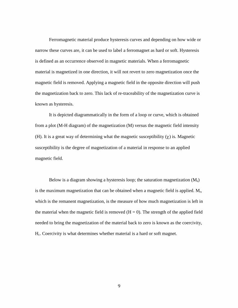
Ferromagnetic material produce hysteresis curves and depending on how wide or
narrow these curves are, it can be used to label a ferromagnet as hard or soft. Hysteresis
is defined as an occurrence observed in magnetic materials. When a ferromagnetic
material is magnetized in one direction, it will not revert to zero magnetization once the
magnetic field is removed. Applying a magnetic field in the opposite direction will push
the magnetization back to zero. This lack of re-traceability of the magnetization curve is
known as hysteresis.
It is depicted diagrammatically in the form of a loop or curve, which is obtained
from a plot (M-H diagram) of the magnetization (M) versus the magnetic field intensity
(H). It is a great way of determining what the magnetic susceptibility (χ) is. Magnetic
susceptibility is the degree of magnetization of a material in response to an applied
magnetic field.
Below is a diagram showing a hysteresis loop; the saturation magnetization (Ms)
is the maximum magnetization that can be obtained when a magnetic field is applied. Mr,
which is the remanent magnetization, is the measure of how much magnetization is left in
the material when the magnetic field is removed (H = 0). The strength of the applied field
needed to bring the magnetization of the material back to zero is known as the coercivity,
Hc. Coercivity is what determines whether material is a hard or soft magnet.

10
3 Figure 1-3 An example of a magnetic hysteresis curve showing magnetization saturation (Ms), remanent magnetization (Mr) and coercivity (Hc) are highlighted. Source - http://www.reddit.com/r/Elements/comments/l1ut9/magnetism (2011).
1.2.3.1 Superparamagnetism
For larger particle sizes, the aligned spin arrangements are divided into regions
called domains. These domain walls exist (multi-domain) throughout the bulk of the
material. For smaller particle sizes, the number of magnetic domains per particle
decreases to a point where it is no longer energetically favorable for domain walls to
exist.
At this critical size range, the particles become single domain due to lack of
boundaries or walls. This makes it easy for the particle to reach magnetic saturation faster
when a magnetic field is applied and this property is called Superparamagnetism.
Nanoparticles with this property have their electron spins align in a preferred orientation
leading to magnetization in that direction.
11
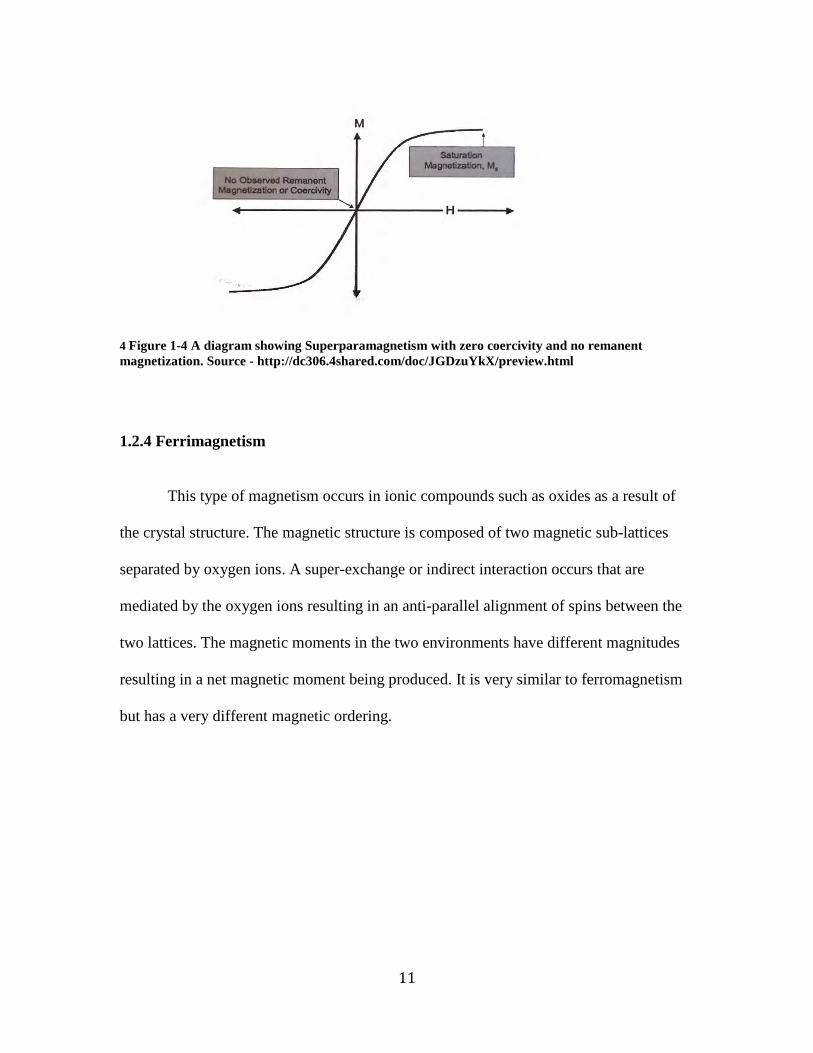
4 Figure 1-4 A diagram showing Superparamagnetism with zero coercivity and no remanent magnetization. Source - http://dc306.4shared.com/doc/JGDzuYkX/preview.html
1.2.4 Ferrimagnetism
This type of magnetism occurs in ionic compounds such as oxides as a result of
the crystal structure. The magnetic structure is composed of two magnetic sub-lattices
separated by oxygen ions. A super-exchange or indirect interaction occurs that are
mediated by the oxygen ions resulting in an anti-parallel alignment of spins between the
two lattices. The magnetic moments in the two environments have different magnitudes
resulting in a net magnetic moment being produced. It is very similar to ferromagnetism
but has a very different magnetic ordering.

12
5 Figure 1-5 A schematic diagram showing spin alignment in a ferrimagnet – magnetic moments are antiparallel and the magnitudes differ. Source – N.A. Spaldin et al (2009).
1.2.5 Antiferromagnetism
For materials with this property, the moments of the two sub-lattices are exactly
equal but opposite so net moment is zero.
6 Figure 1-6 A schematic diagram showing spin alignment in an anti-ferrimagnet – magnetic moments are antiparallel and the magnitudes equal. Source – N.A. Spaldin et al (2009).

13
1.3 Iron Oxide
Maghemite (γ-Fe2O3), magnetite (Fe3O4) and hematite (α-Fe2O3) are some of the
crystalline polymorphs of iron oxides that exist in nature. The iron ions present in the iron
oxides give these structures their magnetic properties. Due to its proven biocompatibility
magnetite and maghemite is seen as the most promising of the three. (Gupta & Gupta,
2005). It is also the most magnetic of all naturally occurring minerals on earth. (Corot,
Robert, Idee et al, 2006)
Since the physical and chemical properties of the nanoparticle are dictated by the
composition of the core, it is imperative to choose a material that will give the
nanoparticle all the required properties.
Magnetite is a ferrimagnetic material although historically it’s been mistaken to
be a ferromagnetic material. It has a cubic inverse spinel structure with oxygen and iron
atoms that exist in two different environments; octahedral sites and tetrahedral sites.
Since the spins in these two sites are opposing and different in magnitude, there is an
overall net magnetization of the material. This gives it the needed magnetic property.
Magnetite nanoparticles were investigated for this application because they are
less sensitive to oxidation than nanoparticles of transition metals and they also display the
desirable magnetic properties.

14
1.4 Methods of synthesis
Iron oxide nanoparticles can be synthesized by a large variety of ways. These
methods of synthesis have been classified into three main groups;
• Physical methods – electron beam lithography, gas-phase deposition
• Wet chemical methods – chemical coprecipitation, hydrothermal
reactions, sonochemical decomposition reaction, etc.
• Microbial methods
For this study the chemical coprecipitation method was selected and used because
it is the simplest and most efficient chemical pathway for MNP synthesis. This method is
also known to produce large quantities of nanoparticles but with a large size distribution.
The nanoparticles also synthesized can be easily functionalized for medical applications.
Since iron oxide nanoparticles are crystalline in nature they are governed by the
principles of crystal formation and growth, which involves two phases – nucleation and
growth. (Jun, Lee & Cheon, 2007). Monodispersed particles are attained if these two
phases are separated as proposed by LaMer and Dinegar. According to this model, there
are three main stages to this synthesis: saturation, nucleation and growth.
Particles must first precipitate out of solution to form small clusters that act as
seeds for further particle growth to occur. Particles only precipitate out when the solution
is saturated; this helps the particles attain the monodispersability condition. (LaMer &
Dinegar, 1950). Ostwald ripening can be used to explain the growth process of the
15
nanoparticle. It shows that, since the formation of larger particles is thermodynamically
favored, in order to decrease the surface energy, larger particles pull material from
smaller ones.
Although this is one of the simplest methods to use it has a few disadvantages that
should be considered. Apart from the large size distribution, it produces nanoparticles,
which aggregate easily and have poor crystallinity resulting in low saturation
magnetization values. The high surface energy of these nanoparticles also makes it hard
to control the particle size on a nano-scale level.
1.5 Stabilization with polymers
Magnetic nanoparticles that display high saturation magnetization as well as high
magnetic susceptibility are of great interest in biotechnology and the medical fields. For
in-vivo applications it is important that well-defined organic coatings surround the nano-
magnetite particles as this prevents aggregation of the nanoparticles in-vivo and may also
enable efficient excretion and protection of the body from toxicity.
Size and surface effects dominate the magnetic behavior of magnetic
nanoparticles. Details of the relationship between shape, surface structure, composition
and the resulting magnetic properties are currently unclear.
Presently, PEI is the industry standard for polymer-mediated transgene delivery
and has been studied comprehensively throughout literature. PEI has become the standard

16
for polymeric transgene delivery due to the excess positive charges on the polymer
backbone resulting in effective condensation of DNA into the nanoparticle, allowing for
sufficient uptake by cells via endocytosis. With toxicity presenting a major limitation to
the use of untargeted PEI in clinical trials, there exists a need to explore other polymers
that will enhance transfection at higher levels.
Polymers provide the advantage of ease of synthesis, ability to be functionalized
and in some cases biodegradability. Poly amido-amines seem to be very comparable to
PEI. These polymers are synthesized using a Michael type addition of various primary or
secondary amines to bisacrylamides. This class of polymers has good water solubility and
biodegradability showing applications in transgene and drug delivery (Guo et al, 2011).
They also have lower toxicity than PEI (Wang et al, 2011). They possess a low surface
charge density that is a contributing factor to the lower cytotoxicity resulting from less
interaction between the polymer and the negatively charged cellular membrane.
1.6 Conclusion
If MNPs are to be used for biological/medical applications, it is crucial that
polymers with low toxicity and high biodegradability be investigated further to make
more strides in the fight against cancer. Hopefully, this research work will help provide
alternatives to PEI, since polyamido-amines seem to be very comparable to it with better
benefits.

17
Chapter 2
COPRECIPITATION SYNTHESIS OF POLYMER TEMPLATED IRON OXIDE NANOPARTICLES
2.1 Introduction
As stated in the previous chapter, the use of magnetic nanoparticles as theranostic
agents is becoming more prevalent in the medical community. Their use for therapeutic
as well as diagnostic undertakings makes them worthwhile to be researched more and
more these days. Magnetic nanoparticles like all other metallic nanoparticles are unstable
without any inorganic or organic coatings on them and so it has become practical for
researchers to further their understanding and knowledge of materials that can be used to
synthesize (nano reactors) as well as stabilize them for theranostic purposes.
There are several methods of synthesizing magnetic nanoparticles (MNPs) but the
coprecipitation method was employed in this research work. Also, there are many
coatings (organic, inorganic) that can be used to stabilize MNPs as mentioned before and
four cationic polymers (chosen from our library) as well as polyethylenimine (PEI) were
used as nano reactors and stabilizers of MNPs synthesized from Ferrous salts. PEI is one
of the preferred standards used in industry as it has been studied thoroughly and is one of
the most promising non-viral vectors that can be used for theranostic purposes. In this
research, PEI was used as the control polymer.
18
The sizes, zeta potential and ICP results of the 5 different polymer templated
MNPs as well as bare MNPs were obtained and compared to help choose potential leads
for magnetofections. SQUID (magnetic measurements) and TEM images were also
collected.
2.2 Materials and Methods
2.2.1 Materials
The ethers used in the polymer synthesis are Neopentyl glycol diglycidyl ether
(NPGDE) and 1,4-cyclohexanedimethanol diglycidyl ether (1,4C), and the amines used
were 3,3’-Diamino-N-methyldipropylamine and 1,4-bis (3-aminopropyl) piperazine
(1,4Bis). All were obtained from Sigma-Aldrich and used with no further modifications.
Branched polyethylenimine (PEI) (MW ∼ 25,000) and Ferrous salts (FeSO4.7H2O) were
also obtained from Sigma Aldrich.
2.2.2 Synthesis of polymers (parallel polymer synthesis)
Four polymers: 1,4C-1,4Bis, 1,4C-3,3’, NPGDE-1,4Bis and NPGDE-3,3’ were
synthesized using the parallel synthesis approach (Barua, et al, 2009) in a ring opening
polymerization. The polymers along with branched PEI were prepared with a
concentration of 40mg/ml (2g of polymer in 50ml of nanopure water) using nanopure
water as a solvent.

19
2.2.3 Synthesis of polymer coated iron oxide nanoparticles.
Iron oxide nanoparticles coated with the above mentioned polymers were
synthesized using the coprecipitation method as reported by Yunfeng Shi et al, 2010 with
slight modifications. The pH of the polymer solutions were measured and kept at 11, to
prevent the introduction of ammonia or alkali hydroxide in the synthesis. Nitrogen gas
was bubbled through the polymer solution to remove any oxidizing agents and to keep
the solution under inert conditions, thus preventing oxidation of the iron ion when
introduced into the polymer solution. 10ml of oxygen-free aqueous FeSO4.7H2O solution
was added to the polymer solution drop-wise over a 20-minute period under vigorous
stirring.
The solution changes from colorless to blue-green after stirring for about 6
hours. After the addition of 0.12ml of peroxide (H2O2) and stirring for 30 minutes, the
solution changes from blue-green to black. The resulting solution is then heated for 3
hours at 800C and allowed to stand overnight. Afterwards the solution is concentrated and
dialyzed for 3 days against water using a cellular membrane with nominal molecular
weight of 2000gmol-1. The solution is then freeze dried to obtain powdered samples.
a b c
7 Figure 2-1 (a) shows the colorless polymer solution under nitrogen atmosphere. (b) is showing the black solution after addition of peroxide (30%wt) and (c) is showing the precipitate during concentrating of the solution by centrifugation.

20
8 Figure 2-2 picture showing freeze-dried samples – 1A (no polymer), 2A (PEI + Fe), 3A (1,4C-1,4Bis + Fe), 4A (1,4C-3,3’+ Fe), 5A (NPGDE-1,4Bis +Fe) and 6A (NPGDE-3,3’ + Fe)
2.2.4 Statistical Analyses. All experiments were carried out in duplicate unless stated otherwise. The
significance between the control and each experimental test condition was determined
using the two-tailed student’s t-test with p < 0.05 being considered significant.
2.3 Results and Discussion
For any biological application, biocompatibility and toxicity must be tested, but
within the scope of this paper, only the physical and magnetic properties of the samples
will be considered.
Magnetic properties of nanoparticles depend on their physical structure and the
chemical phase in which they are found. Biological behavior of the nanoparticles strongly
depends upon the size, shape, charge and nature of the coating used. Data was collected

21
and analyzed for size, surface charge, iron content, magnetic characteristics and TEM
images. Magnetic characteristics will be reviewed in the next chapter.
2.3.1 Particle synthesis
Iron oxide nanoparticles were prepared using the modified chemical
coprecipitation route from Yunfeng Shi et al synthesis. (Shi, Zhou, Wang et al, 2010).
Initial stabilization was conferred to the naked nanoparticles in a basic medium under
inert conditions. Since the pH of the polymer was kept basic (pH =11), there was no need
to add NH4OH (Ammonium Hydroxide) to the reaction solution. This is usually done in
the coprecipitation method to induce a short burst of nucleation. (Beattle, 1989). The
synthesis was carried out under inert conditions by bubbling nitrogen gas through the
polymer solution and aqueous iron sulphate solution to prevent oxidation.
According to literature, nitrogen gas also helps reduce the particle size of the
nanoparticle and this is seen when compared to methods where oxygen is not removed.
The polymer solution was stirred vigorously with a magnetic stirrer when the iron
solution was added to maximize uniformity of nanoparticles formed. (Kim, Zhang, Voit,
Rao et al, 2001).
Ferrous ions are known to bind readily to the amine groups in the polymers to
form ferrous hydroxide. The reaction proceeded from a colorless solution, to a blue-green
intermediate color then to black after the addition of peroxide, which is consistent with
the pathways for the formation of magnetite nanoparticles.

22
2.3.2 Size (hydrodynamic diameter)
In almost all cases, nanoparticles are polydisperse and the heterogeneity in sizes
gives rise to different values when reviewing number or intensity-weighted means. In this
research, dynamic light scattering (DLS) was employed in determining the sizes and size
distribution (using % intensity and % number) of the nanoparticles synthesized. This was
done using the Delsa Nano Submicron Particle Size and Zeta Potential Particle Analyzer
(Malvern) and the hydrodynamic diameter values are reported in nm. The polydispersity
index of each sample was also measured and reported.
Transmission Electron Microscopy (TEM) was also used to determine the size
distribution of the samples. Following synthesis, the nanoparticles were visualized using
a JEOL-JEM-2000FX microscope, operating at 200 kV (Leroy Eyring Center for Solid
State Sciences, Arizona State University).
Specimen samples for TEM were prepared by casting a drop of the synthesized
iron oxide nanoparticles dispersed in ultrapure water onto a carbon film on a 200 mesh
copper wire screen (Global Electron Microscopy Technology Co.) and dried in air. Dried
samples were examined by TEM at 80 kV.
For size and surface charge, all samples with the exception of nanoparticles
coated with PEI, were diluted to a concentration of 20mg/ml for better analysis. The PEI
templated nanoparticles were diluted at 1mg/ml. (This is because when diluted at the

23
same concentration as PEI, the intensity for the other polymer or bare nanoparticles were
too low to be read)
Sample Number I (nm) II (nm) III (nm)
No polymer 1A 373.6 288.2 290.3
1B 244.0 271.8 275.3
PEI + Fe 2A 216.1 212.0 211.7
2B 280.0 270.3 269.7
1,4C-1,4Bis +
Fe
3A 54.2 91.06 274.9
3B 23.4 14.62 10.06
1,4C-3,3’ + Fe 4A 3.0 26.18 4.483
4B 75.48 55.85 45.98
NPGDE-1,4Bis
+ Fe
5A 4.23 3.16 4.11
5B 6.87 6.64 6.43
NPGDE-3,3’ +
Fe
6A 22.85 25.95 25.54
6B 421.8 396.3 422.0
1 Table 2-1 The hydrodynamic sizes measured in triplicate for polymer-templated and bare iron nanoparticles formed by coprecipitation.
24
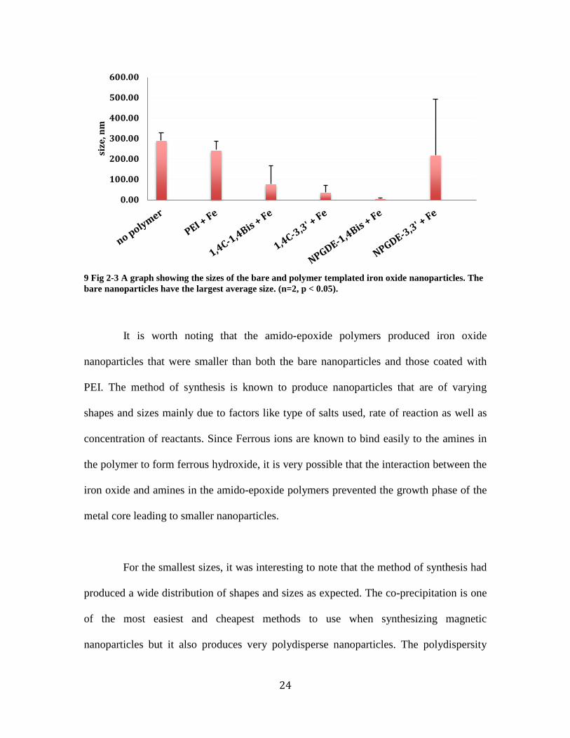
9 Fig 2-3 A graph showing the sizes of the bare and polymer templated iron oxide nanoparticles. The bare nanoparticles have the largest average size. (n=2, p < 0.05).
It is worth noting that the amido-epoxide polymers produced iron oxide
nanoparticles that were smaller than both the bare nanoparticles and those coated with
PEI. The method of synthesis is known to produce nanoparticles that are of varying
shapes and sizes mainly due to factors like type of salts used, rate of reaction as well as
concentration of reactants. Since Ferrous ions are known to bind easily to the amines in
the polymer to form ferrous hydroxide, it is very possible that the interaction between the
iron oxide and amines in the amido-epoxide polymers prevented the growth phase of the
metal core leading to smaller nanoparticles.
For the smallest sizes, it was interesting to note that the method of synthesis had
produced a wide distribution of shapes and sizes as expected. The co-precipitation is one
of the most easiest and cheapest methods to use when synthesizing magnetic
nanoparticles but it also produces very polydisperse nanoparticles. The polydispersity
0.00
100.00
200.00
300.00
400.00
500.00
600.00
size
, nm
25
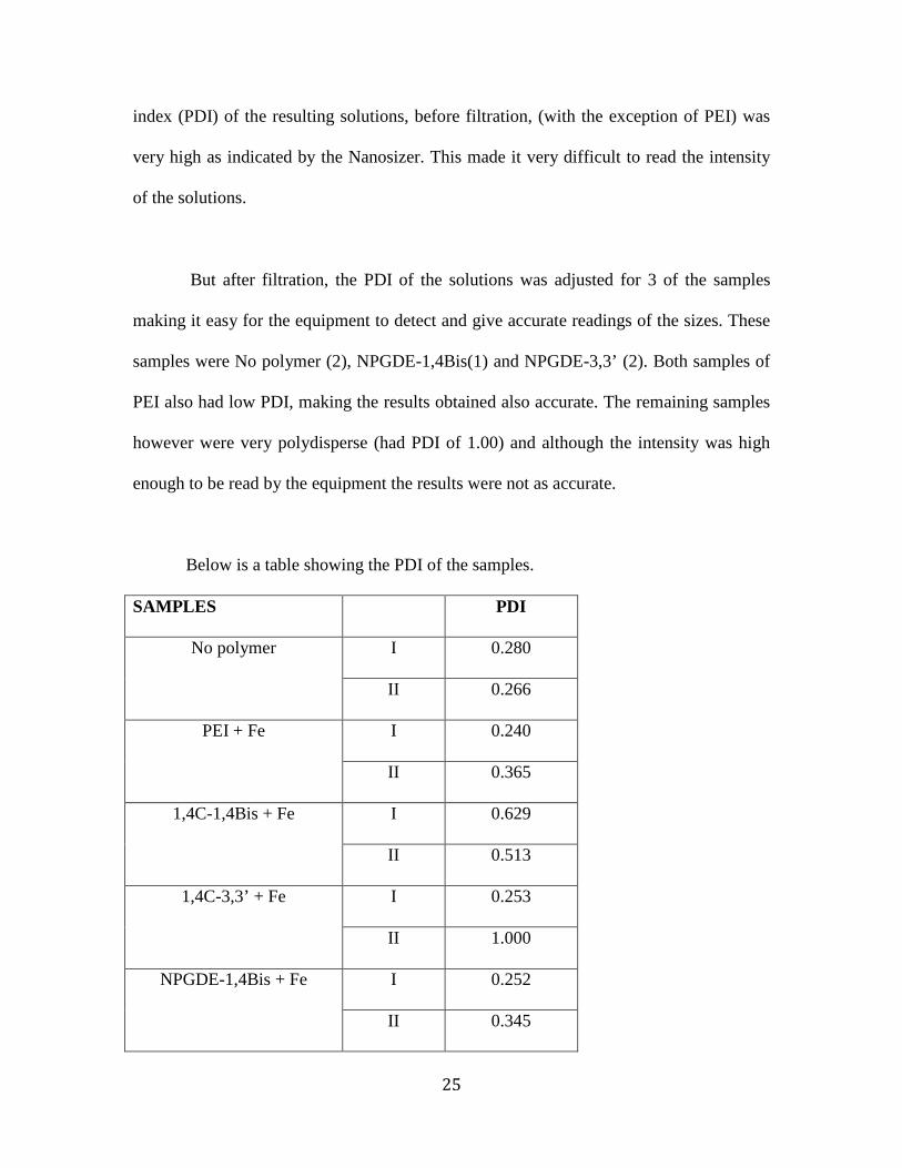
index (PDI) of the resulting solutions, before filtration, (with the exception of PEI) was
very high as indicated by the Nanosizer. This made it very difficult to read the intensity
of the solutions.
But after filtration, the PDI of the solutions was adjusted for 3 of the samples
making it easy for the equipment to detect and give accurate readings of the sizes. These
samples were No polymer (2), NPGDE-1,4Bis(1) and NPGDE-3,3’ (2). Both samples of
PEI also had low PDI, making the results obtained also accurate. The remaining samples
however were very polydisperse (had PDI of 1.00) and although the intensity was high
enough to be read by the equipment the results were not as accurate.
Below is a table showing the PDI of the samples.
SAMPLES PDI
No polymer I 0.280
II 0.266
PEI + Fe I 0.240
II 0.365
1,4C-1,4Bis + Fe I 0.629
II 0.513
1,4C-3,3’ + Fe I 0.253
II 1.000
NPGDE-1,4Bis + Fe I 0.252
II 0.345
26
NPGDE-3,3’ + Fe I 1.000
II 0.452
2 Table 2-2 The table above shows the polydispersity index of the bare and coated iron oxide nanoparticles.
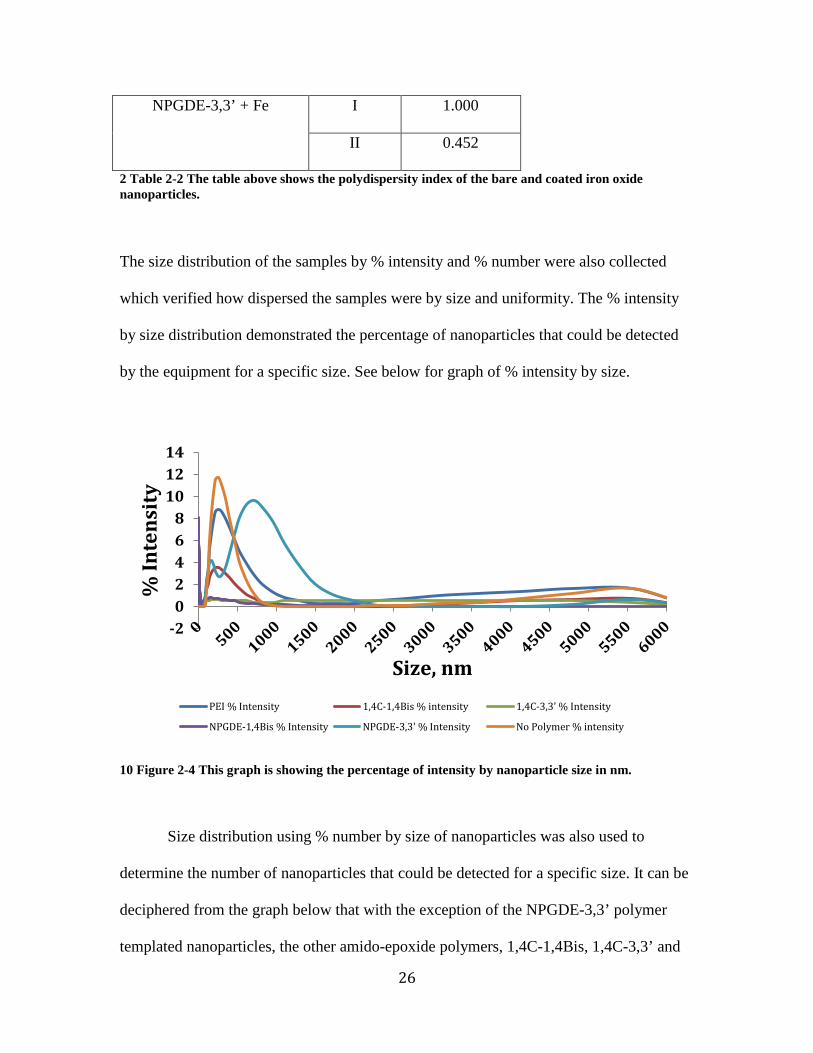
The size distribution of the samples by % intensity and % number were also collected
which verified how dispersed the samples were by size and uniformity. The % intensity
by size distribution demonstrated the percentage of nanoparticles that could be detected
by the equipment for a specific size. See below for graph of % intensity by size.
10 Figure 2-4 This graph is showing the percentage of intensity by nanoparticle size in nm.
Size distribution using % number by size of nanoparticles was also used to
determine the number of nanoparticles that could be detected for a specific size. It can be
deciphered from the graph below that with the exception of the NPGDE-3,3’ polymer
templated nanoparticles, the other amido-epoxide polymers, 1,4C-1,4Bis, 1,4C-3,3’ and
-202468
101214
% In
tens
ity
Size, nm PEI % Intensity 1,4C-1,4Bis % intensity 1,4C-3,3' % Intensity
NPGDE-1,4Bis % Intensity NPGDE-3,3' % Intensity No Polymer % intensity

27
NPGDE-1,4Bis polymer coated nanoparticles had about 10-20% of their number being
less than 50nm. The PEI templated nanoparticles and bare nanoparticles had a higher
number of their quantity in the 50 to 300nm region.
11 Figure 2-5 This graph is showing the size distribution using percentage by number of nanoparticles for specified sizes.
2.3.3 Transmission electron microscopy
As mentioned previously, TEM images were also taken to verify the sizes and
shape of the nanoparticles. From the images taken, there seems to be a confirmation of
how much smaller the amido-epoxide nanoparticles were in comparison to the bare
nanoparticles and the ones coated in PEI.
For the bare nanoparticles, or nanoparticles with no polymer, the images were
fuzzy due to interaction of the beam of the microscope with the sample. It is assumed that
the beam of the microscope affects materials of a magnetic nature.
0
5
10
15
20
25
% N
umbe
r
Size, nm PEI % Number 1,4C-1,4Bis % Number 1,4C-3,3' % Number
NPGDE-1,4Bis % Number NPGDE-3,3' % Number No Polymer % Number
28
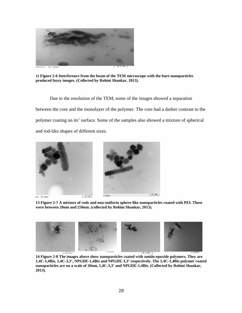
12 Figure 2-6 Interference from the beam of the TEM microscope with the bare nanoparticles produced fuzzy images. (Collected by Rohini Shankar, 2013).
Due to the resolution of the TEM, some of the images showed a separation
between the core and the monolayer of the polymer. The core had a darker contrast to the
polymer coating on its’ surface. Some of the samples also showed a mixture of spherical
and rod-like shapes of different sizes.
13 Figure 2-7 A mixture of rods and non-uniform sphere-like nanoparticles coated with PEI. These were between 20nm and 250nm. (collected by Rohini Shankar, 2013).
14 Figure 2-8 The images above show nanoparticles coated with amido-epoxide polymers. They are 1,4C-1,4Bis, 1,4C-3,3’, NPGDE-1,4Bis and NPGDE-3,3’ respectively. The 1,4C-1,4Bis polymer coated nanoparticles are on a scale of 20nm, 1,4C-3,3’ and NPGDE-1,4Bis. (Collected by Rohini Shankar, 2013).
29
The images from the TEM also show some agglomeration, which may be due to
van der Waal’s forces between the particles.
From the images and hydrodynamic sizes deduced from the DLS machine, it can
be inferred that the amido-epoxide polymer templated nanoparticles had a smaller iron
oxide core as compared to the PEI templated ones and the polymer coatings were much
thinner. As mentioned before, the ferrous ions readily interact with the amines in the
amido-epoxide polymers to form ferrous hydroxide thus curtailing the growth phase of
the nanoparticle in the early stages of the reaction resulting in smaller nanoparticles and
the thin shell may be attributed to the low molecular weight of the polymers. Since the
same concentration of polymers was used, the PEI templated polymers will have a larger
amount of coating or shell on its nanoparticles since it has a larger molecular weight.
2.3.4 Surface charge
Particle aggregation as seen from the images above may be due to van der Waals
forces and/or magnetic dipole-dipole attractive interactions and large surface to volume
ratio, which causes high surface energies. This is very particular to magnetic
nanoparticles. The nanoparticles therefore aggregate to reduce the high surface energy.
The zeta potential, which is a function of the surface charge density and surface structure,
was determined using the Delsa Nano Submicron Particle Size and Zeta Potential Particle
Analyzer (Malvern).

30
Sample Number I (mV) II (mV) III (mV)
No polymer 1A 5.03 7.82 7.74
1B 9.95 9.48 9.95
PEI + Fe 2A 33.70 33.40 34.3
2B 35.60 36.50 37.8
1,4C-1,4Bis +
Fe
3A 2.74 2.53 4.54
3B 6.66 8.07 7.90
1,4C-3,3’ + Fe 4A 10.30 10.50 10.70
4B 12.60 14.20 15.00
NPGDE-1,4Bis
+ Fe
5A 12.40 13.40 10.70
5B 12.70 4.45 7.64
NPGDE-3,3’ +
Fe
6A 12.56 2.24 4.20
6B 18.20 17.5 17.6
3 Table 2-3 The zeta potential of the samples as a function of the surface charge.

31
15 Figure 2-9 A Graph showing the surface charge of the nanoparticles synthesized.
Based on the results obtained, all the polymer coated nanoparticles showed
positive zeta potential, with PEI coated nanoparticles showing the highest zeta potential
(average of about 35.22mV). This confirms that the nanoparticles are cationic in nature
and can therefore be used in magnetofections as well as gene delivery experiments
according to the Proton Sponge Theory, which supports the ability of the cytoplasm in a
cancer cell due to its acidic nature to import protons into the endosome. (Thomas et al,
2005).
1,4C-3,3’ + Fe and both NPGDE polymer based Nanoparticles also show
relatively good zeta potential (between 10-17mv) but with the resulting solution having a
high distribution of differing shapes and sizes it is not surprising that the zeta potential of
the solutions is lower than that of PEI. Further analysis using TEM confirmed this. From
the results it can be deduced that the low surface charge of the amido-epoxide templated
nanoparticles is due to the low amount of polymer coating the nanoparticle. Once again,
0.00 5.00
10.00 15.00 20.00 25.00 30.00 35.00 40.00
Zeta
Pot
enti
al, m
V

32
the molecular weight and concentration of these polymers used may not have been
enough to create a thicker coating hence the lower positive charges.
2.3.5 Iron concentration
The iron concentration of the sample was measured using Inductively Coupled
Plasma Mass Spectrometry (ICPMS) at the Core Research Facilities – Arizona State
University. This was chosen because it can determine the range of metals and non-metals
at low concentrations.
The ICP analysis was performed by digesting 3 different weighted amounts of
each sample with a combination of Nitric acid and Hydrochloric acid (8ml: 2ml) by
volume. The results were presented by percent weight of iron in the previously weighed
samples and they indicate that the iron content of all the polymer-coated samples was
greatly reduced as compared to the uncoated sample.
1,4C-3,3’ + Fe sample had the least amount of iron as confirmed by the size
distribution analysis performed also. Since a higher content of iron in the iron oxide core
can inevitably increase the magnetization values of the nanoparticle, we seek to create
nanoparticles with higher iron content. In comparing all the polymer templated
nanoparticles, the one with PEI had the highest content of iron. This could be due to the
growth of the iron oxide core being more than that of the amido-epoxide templated
nanoparticles. It is possible that the higher molecular weight presented the solution with
more amine-ferrous oxide interaction resulting in larger amounts being created during the

33
growth phase giving us larger particles. This can be seen in the larger iron oxide core in
the TEM images and size above.
16 Figure 2-10 A graph showing percentage of Iron content in the synthesized nanoparticles
2.3.6 Conclusion
Looking at results from the size, TEM images and ICP analysis, the following
conclusions can be made.
- The size of the polymer templated nanoparticles is greatly affected by the
type of polymer used (presence of amines) and the molecular weight and
concentration of polymer as well.
- The presence of amines allows for the interaction to proceed between the
amines in the polymer and the ferrous ions, resulting in a faster rate of
reaction in creating small sized nanoparticles. The interaction between
the reactants determines when the growth phase is terminated as the
polymer coats the stabilized particles formed. PEI had larger particle
sizes due to the large amount of amines present.
0.0010.0020.0030.0040.0050.0060.00
% o
f Iro
n pr
esen
t

34
- Since the PEI polymer was heavier, in terms of molecular weight, a
higher concentration promoted more interaction with its amines and the
ferrous ions resulting in nanoparticles that were larger than those of the
amido-epoxide templated nanoparticles. Therefore it will be prudent to
change the concentration of the amido-epoxide polymers to cancel out
this variation.
The results therefore show that although smaller sizes can be attained with the
amido-epoxide polymers synthesized by the Rege lab, the iron content of the
nanoparticles were found to be very low and this can be altered by altering the amount of
polymer or concentration of polymer used in the synthesis.
35
Chapter 3
MAGNETIC CHARACTERIZATION OF POLYMER TEMPLATED IRON OXIDE NANOPARTICLES
3.1 Introduction
The saturation magnetization of the polymer coated and bare nanoparticles was
determined using a Superconducting Quantum Interference Device (SQUID) at the
LeRoy Eyring Center for Solid State Science. The magnetic moments of the stabilized
nanoparticles was rapidly saturated in the presence of an externally applied magnetic
field, up to about 40000 Oe AT 300K. The following magnetization curves were obtained
as a result of this analysis.
Magnetization values of magnetic nanoparticles are affected by the amount of
iron content found in the iron oxide core, the amount of polymer coating the nanoparticle
as well as the presence of oxygen during synthesis. The oxygen content in the polymer
was greatly reduced by bubbling nitrogen gas and keeping an inert atmosphere during
synthesis to prevent the reduction of the Ferrous ions and causing rust.
The iron content determined by ICP analysis was discussed in the previous
chapter and showed the PEI templated nanoparticles to have a higher iron content than
the amido-epoxide templated ones. It was also inferred from the TEM images that the
polymer coating surrounding these nanoparticles were very thin so the magnetization
values are expected to match those from literature.

36
3.2 Results and discussion
There was a decrease in the saturation magnetization of all the polymer-coated
nanoparticles, which can be attributed to the use of the polymer as an agent of
stabilization.
The polymer on the surface of the iron oxide restricts the crystal growth of the
iron core resulting in a relatively slow process that takes a toll on the magnetization
saturation value. (Shi et al, 2010).
17 Figure 3-1 A plot of the magnetization curves of all samples prepared. Magnetic moments were rapidly saturated in the presence of a magnetic field of about 40000 Oe.
For all six nanoparticle samples there was no hysteresis in the magnetization, with
both remanence and coercivity being zero in each case. Magnetic remanence is the
amount of magnetism remaining in a sample after exposure to a field.
37
All the samples showed paramagnetic properties, which is characteristic of metals
with unpaired electrons, which in this case is Iron. Paramagnetism is a magnetic property
of materials whereby there is a net magnetic moment due to the partial alignment of
magnetic moments in the direction of the applied field. PEI coated iron oxide
nanoparticles and the bare nanoparticle showed as superparamagnetic curves due to the
steep initial slope of magnetization with high magnetic susceptibility. Magnetization of a
paramagnetic sample returns to zero when the applied external field is removed due to the
electron spin moments and orbital moments cancelling each other out.
Looking at the plot above, the highest magnetization when the highest magnetic
field of 40000 Oe was applied was about 32 emu/g for the uncoated iron nanoparticles.
Fe +1,4C-3,3’ nanoparticles had the least magnetization (6.73 emu/g), although this is
low, it is less than that of the uncoated nanoparticles and this will give them the property
of being rapidly and easily separated from any reaction medium. This is very critical for
biomedical applications. According to literature (Shi et al, 2009), when the hyper-
branched PEI (HPEI) of molecular weight 60,000 Da was used the saturation
magnetization was about 46.4 emu per gram and that with the molecular weight of 10,000
Da was 39.8 emu per gram. These showed higher magnetization results because of the
hyper-branching in the polymer allowing for better stabilization of the iron oxide. Since
the PEI used was not hyper-branched, it is understandable to have lower magnetization
values.

38
As has been detected in much research work, the magnetization values of the
nanoparticles are lower than what the bulk materials typically produce and the results
shown are consistent with this.
The low magnetic saturation of the amido-epoxide templated nanoparticles may
be due to the method of synthesis employed. It has been reported that, although the
coprecipitation method is the simplest and most efficient route of synthesis, it has the
disadvantage of producing large particle size distribution, aggregation and poor
crystallinity that results in low saturation magnetization values. (Gupta & Gupta, 2005).
The lower molecular weight of these polymers may also attribute to the low values as
they may not be as good stabilizing agents as PEI but further heating and increasing of
ferrous salts may help improve this.
Also according to some literature, a linear correlation might exist between the
particle size, surface effects and saturation magnetization. Thus, if there are any defects
on the surface of the nanoparticles it may result in disordered crystal orientation on the
surface that can decrease the saturation magnetization of smaller particles. (Varanda,
Jafelicci et al, 2002). The dead layer which is about 1nm thick existing between the core
and the polymer surface may also decrease the magnetization saturation due to the
environmental effects of the surface atoms. (Sato et al, 1987).

39
3.3 Conclusion
Since the amido-epoxide polymer coated nanoparticles had relatively lower
saturation magnetization values in comparison to the PEI coated ones, it is prudent to say
more work needs to be done to increase these values.
This can be done by increasing the quantity of the ferrous salt during precipitation
and heating for a longer time period. Although these polymers produced nanoparticles
with smaller sizes, the amount of iron core being low, significantly affected the magnetic
properties of the nanoparticles.
The saturation magnetization could also be affected by the amount of iron found
in the core of the nanoparticles, as iron is directly responsible for the magnetic nature of
the material.
Working further on varying reaction criteria (pH, reaction rates, quantity and
concentration of reactants) could potentially lead to better magnetic results, but must be
taken not to vary these conditions too much and end up with larger particles.
40
Chapter 4
FUTURE WORK
4.1 Magnetofections
Amido-epoxide polymers are polycations like PEI, they can be used for
transfecting vector DNA into cells and with the influence of a magnetic field these
nanoparticles can be used to perform magnetofections, which may yield better results
than transfections.
4.1.1 Coprecipitation synthesis modification
Since the nanoparticles coated with amido-epoxide polymers yielded lesser
magnetic saturation values, more experiments will be set up to vary conditions of the
synthesis. Effort will be put into increasing the ferrous salts to see if there will be an
increment in the magnetic properties of the nanoparticles.
Also, further heating was suggested to help improve the reaction between the
polymer and ferrous salts, by aiding the growth phase of the iron core. This will also be
tested for different times to see the effect of heat on the magnetic properties.

41
4.2 Magnetic Resonance Imaging (MRI)
Although smaller nanoparticles (< 50 nm) are taken up by the reticuloendothelial
system (RES), the small size is beneficial due to longer blood circulation time. This will
result in greater time for specific localization.
Since three of the amido-epoxide polymer coated nanoparticles had a high
distribution of its size in this range (10nm -50nm), the future plan will be to employ some
of these nanoparticles in an MRI application.

42
REFERENCES
Arruebo, M., Fernandez-Pacheco, R., Ibarra, M. R. & Santamaria, J. (2007). Magnetic nanoparticles for drug delivery. Nanotoday, 2(3), 22- 31.
Barua, S., Rege, K. (2010). The influence of mediators of intracellular trafficking on transgene expression efficacy of polymer-plasmid DNA complexes. Biomaterials, 31(22), 5894- 5902.
Beattle, J. K. (1989). Monodisperse colloids of transition metal and lanthanide compounds. Pure Applied Chemistry, 61, 937- 941.
Corot, C., Robert, P., Idee, M., et al (2006). Recent advances in iron oxide nanocrytal technology for medical imaging. Advanced Drug Delivery Reviews, 58(14), 471- 504.
Dobson, J. (2006). Magnetic nanoparticles for drug delivery. Drug Development Research, 67, 55- 60.
Guo, M., Yan, Y., Zhang, H., et al (2008). Magnetic and pH-responsive nanocarriers with multilayer core-shell architecture for anticancer drug delivery. Journal of Material Chemistry, 18, 5104- 5112.
Gupta, A., Curtis, A., (2004). Lactoferrin and ceruloplasmin derivatized superparamagnetic iron oxide nanoparticles for targeting cell surface receptors. Biomaterials, 25(15), 3029- 3040.
Gupta, K., Ajay & Gupta Mona (2005). Synthesis and surface engineering of iron oxide Nanoparticles for biomedical applications. Biomaterials, 26, 3995– 4021.
Jun, Y., Lee, J., Cheon, J. (2007). Artificially engineered magnetic nanoparticles for ultra-sensitive molecular imaging. Natural Medicine, 13(1), 95- 99.
Kim, D. K., Zhang, Y., Voit, W., Rao, K., et al (2001). Synthesis and characterization of surfactant coated superparamagnetic Monodispersed iron oxide nanoparticles. Journal of Magnetic Materials, 225(1-2), 30- 36.

43
LaMer, V., Dinegar, R. (1950). Theory, production and mechanism of formation of Monodispersed hydrosols. Journal of American Chemistry Society, 72(11), 4847- 4854.
Li, Y., Pei, Y., Zhang, X., et al (2001). PEGylated PLGA nanoparticles as protein carriers: synthesis, preparation and biodistribution in rats. Journal of Control Release, 71, 203– 211.
Lodhia, J., Mandarano, G., Ferris, N. J., et al (2009). Development and use of iron oxide nanoparticles (part 1): Synthesis of iron oxide nanoparticles for MRI. Biomedical Imaging and Intervention Journal.
Sato, T., Lijima, T., Sekin, M., et al (1987). Magnetic properties of ultrafine ferrite particles. Journal of Magnetic materials, 65, 252.
Schweiger, C., Pietzonka, C., Heverhagen, J. and Kissel, T. (2011). Novel magnetic iron oxide nanoparticles coated with poly(ethyleneimine)-g-poly(ethylene glycol) for potential biomedical application: Synthesis, stability cytotoxity and MR imaging. International Journal of Pharmaceutics, 408, 130- 137.
Shi, Y., Zhou, L., Wang, R., Pang, Y., et al (2010). In situ preparation of magnetic nonviral gene vectors and magnetofections in vitro. Nanotechnology, 21, 115- 123.
Sun, C., Lee, J. & Zhang, M. (2008). Magnetic nanoparticles in MR imaging and drug Delivery. Advanced Drug Delivery Reviews 60, 1252– 1265.
Varanda, L., Jafelicci, P., O’Grady, K., et al (2002). Structural and Magnetic transformation of Monodispersed iron oxide particles in a reducing atmosphere. Journal of Applied Physics, 92(4), 2079.
Wang, Y. H., Zheng, M., Meng, F. et al (2011). Branched polyethylenimine derivatives with reductively cleavable periphery for safe and efficient in vitro gene transfer. Biomacromolecules, 12(4), 1032- 1040.

44
Xie, J., Lee, S. & Chen, X. (2010). Nanoparticle-based theranostic agents. Advanced Drug Delivery Reviews 62, 1064-1079.


