Eur Respir J 1992, 5, 997-1003
REVIEW
Steroid-induced myopathy and its significance to respiratory disease: a known disease rediscovered
P.N.R. Dekhuijzen, M. Decramer
Steroid-induced myopathy and its significance to respiratory disease: a known disease rediscovered. P.N.R. Dekhuijzen, M. Decramer. ABSTRACT: Skeletal muscle myopathy is a well-known side-effect of systemically administered corticosteroids. In recent years renewed attention is being paid to the involvement of the respiratory muscles and its consequent significance in pulmonary patients. Two different clinical patterns of steroid-induced muscular changes are known. In acute myopathy and atrophy after short term treatment with high doses of steroids, generalized muscle atrophy and rhabdomyolysis occur, including the respiratory muscles. Chronic steroid myopathy, occurring after prolonged treatment with moderate doses, is characterized by the gradual onset of proximal limb muscle weakness and may be accompanied by reduced respiratory muscle force.
Animal studies demonstrated diaphragmatic myopathy and atrophy similar to the alterations in peripheral skeletal muscles. Fluorinated steroids induced selective type lib (fast-twitch glycolytic) fibre atrophy, resulting in changes in contractile properties of the diaphragm. Non-fluorinated steroids may also induce histological, biochemical and functional alterations in the diaphragm. Observations in patients with collagen vascular disorders and with asthma and chronic obstructive pulmonary disease (COPD) underline the potential hazards of treatment with corticosteroids to respiratory muscle structure and function. Eur Respir J, 1992, 5, 997-1003.
Respiratory Muscle Research Unit, Laboratory for Pneumology and Respiratory Division, University Hospital, Katholieke Universiteit Leuven, B-3000 Leuvcn, Belgium.
Correspondence: M. Decramer, Respiratory Division, University Hospital, Weligerveld I , 3212 Pellenberg, Belgium
Keywords: Atrophy corticosteroids myopathy respiratory muscles
Received: January 20 1992 Accepted for publication May 7 1992
This study was supported by grants from the Dutch Asthma Foundation and the "Nationaal Fonds voor Wetenschappelijk Onder:wek".
Intravenous and oral corticosteroids are often used in the treatment of obstructive pulmonary disease, interstitial lung disease and collagen vascular disorders. Among the many side-effects are myopathy and atrophy, affecting striated muscles of the limbs. This occurs primarily when fluorinated steroids such as triamcinolone and dexamethasone are used [1-3]. In patients, proximal muscle wasting and weakness predominate the clinical picture. Biopsy studies of peripheral muscles in humans and animals show selective atrophy of the type Ub (fast-twitch glycolytic) fibres [3- 7]. Due to their continuous activity the respiratory muscles, especially the diaphragm, were thought to be relatively spared by this process.
function is already affected by hyperinflation, hypoxaemia, hypercapnia and malnutrition. Moreover, a reduced average activity level of these patients will lead to the development of disuse atrophy.
In recent years, however, several studies in animals have shown that diaphragmatic structure and function may be severely compromised by the use of systemic steroids l8-12). Diaphragmatic atrophy and weakness were the main findings in these animal studies.
The extent to which diaphragmatic function may be compromised by steroid administration in patients is not fully understood [13- 15]. The clinical implications of these changes, however, may be potentially serious in patients with chronic obstructive pulmonary disease (COPD) and asthma. In these patients, diaphragmatic
In the present review, literature on corticosteroidinduced pathological and physiological changes in respiratory muscles will be discussed, as well as their potential clinical relevance and implications for therapy.
Clinical picture of steroid-induced myopathy
Two distinct types of steroid-induced myopathy are well-known to the clinician: acute and chronic steroid myopathy.
Acute steroid myopathy
This complication of treatment with systemic corticosteroids is infrequently encountered [16-20] . Generalized muscle weakness, involving the respiratory muscles, occurs 5-7 days after the onset of treatment with high doses of corticosteroids (e.g. hydrocortisone 0.9-4 g·day·1 i.v, dexamethasone 40-80 mg·day-1 i .v.

998 P.N.R. DEKHUIJZEN, M. DECRAMER
[ 19]). In one study [20], there appeared to be a correlation between the occurrence of this complication and the total dose of steroids administered. Acute atrophy was encountered with total doses >5.4 g of hydrocortisone in six days, whereas no signs of myopathy were noted with total doses <4 g [20].
In most cases, high levels of creatine kinase were found (1,000-100,000 U·/·1), associated to myoglobinuria, indicating rhabdomyolysis. Muscle biopsies show focal and diffuse necrosis and atrophy of all fibre types [17, 19], without predilection for type lib fibres. Previous use of systemic corticosteroids was apparently not involved in the development of this rare complication of steroid therapy. Recovery may be prolonged to more than six months [ 19].
Chronic steroid myopathy
This is the classical type of steroid-induced myopathy, occurring after prolonged administration of prednisolone in a dose of about 40-60 mg·day·1
• Proximal muscle weakness is a prominent clinical feature, preventing patients from climbing stairs or elevating their arrns. Fluorinated steroids (such as triamcinolone and dexamethasone) seem to produce weakness and myopathy more often than non-fluorinated corticosteroids such as hydrocortisone and prednisone [1-3].
Serum levels of muscle e11zymes (creatine kinase (CK), serum glutamic oxalo-acetic transaminase (SGOT) and aldolase) are usually normal [2, 3, 21]. Lactate dehydrogenase (LDH), however, was reported to be elevated in one study of patients with systemic lupus erythematosus [22], and in another paper on patients with COPD or asthma [15]. Creatine excretion in the urine is massively increased, from several days before the clinical appearence of myopathy [3]. Myoglobinuria or rhabdomyoJysis are absent. In biopsies of the diseased muscles, the main finding is an atrophy of type Jib fast-twitch glycolytic (FG) fibres [3-7].
Table 1. Steroid-induced myopathy in animal studies
The distribution of the different fibre types, however, was not changed [7, 23, 24). Recovery of weakness associated with chronic use of steroids may take many weeks or months [25].
Corticosteroids preferentially affect less active muscles [26]. Moreover, muscles with predominantly type Jib fibres are especially susceptible to steroid-induced wasting [27, 28]. In this respect the almost continuously active diaphragm, mainly composed of type I and Ha fibres, would not be expected to be affected by corticosteroids. Recent animal studies, however, have cast doubt on this expectation.
Respiratory muscle involvement in animal studies
The effects of steroids on physiological, histochemical and biochemical characteristics of the diaphragm have been described in several animal studies. The results of these studies are summarized in tables I, 2 and 3. MooRE et al. [8] investigated the effects of cortisone on the diaphragm in rats (table 1). After daily intramuscular injection of 100 mg·kg-1 cortisone acetate for ten days the weight of the diaphragm decreased by about 30%. The force generating capacity of the diaphragm decreased in proportion to the reduction in weight, but was not altered when expressed per unit cross-sectional area (table 2). During the forcefrequency curve fatigue (i .e. a reduction in maximal fused tetanic tension) occurred in the control groups, but not in the steroid-treated group. This may be explained by a selective atrophy of the type lib fibres, leaving muscle mainly composed of fatigue-resistant type I and Ila fibres.
The effects of treatment over two weeks with cortisone acetate (10 mg·kg·1 q·d.) on the diaphragm and the m. extensor digitorum longus (EDL), (a limb muscle mainly composed of fast-twitch Ua and lib fibres) of rabbits were studied by FERGUSON et al. (11].
Animal Steroid Body Diaphragm Soleus Gastro- EDL Reference n dose, duration weight weight weight cnemius weight
weight
Rats cortisone acetate i.m. -14% -30% -36% -28% MOORE et al. (8] 11 100 mg·kg·' q.d. 10 days
Hamsters triamcinolone i.m. -27% = -45% WILCOX et al. [9] 4 3 mg·kg·' q.d. 3 weeks
Rats triamcinolone i.m. -30% -30% = -27% VURES et al. [10] 80 1.2 mg·kg·• q.d. 8 days
Rats dexamethasone i.p. -18% -30% SASSON et al. [12] 10 1 mg·kg·• q.d. 2 weeks
Rats dexamethasone i.p. -23% -32% = SASSON et a/. [12] 9 4 mg·kg·• q.d. 2 weeks
Rats dexamethasone i.p. -25% -26% -28% SASSON et al. [12] 7 I mg·kg·1 q.d. 7 weeks
EDL: m. extensor digitorium longus; =: no change; - : not measured.
STEROrD MYOPATHY AND RESPfRATORY DISEASE 999
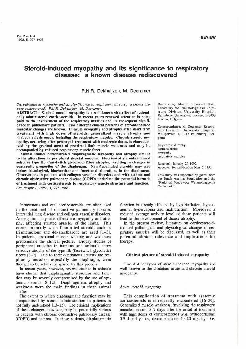
Table 2. - Diaphragmatic contractile properties
Animal Steroid Pt/CSA Po/CSA Pt/Po TPT 1/2 RT Force Fatigue Reference n dose, duration freq.
curve
Rats cortisone acetate i.m. = = = MOORE et al. [8) 11 100 mg·kg-' q.d. 10 days
Hamsters triamcinolone i.m. +20% +31% i at 10, J. Wn.cox et al. [9) 4 3 mg·kg·• q.d. 3 weeks 20, 30Hz
Rats triamcinolone i .m. -62% J. J. at 50 VrrREs et al. [10] 80 1.2 mg·kg·• q.d. 8 days and 100Hz
Rats dexamethasone i.p = = = = = SASSON et al. [12) 10 1 mg·kg·• q.d. 2 weeks
Rats dexamethasone i.p. -22% = J. at all = SASSON et al. (12) 9 4 mg·kg· ' q.d. 2 weeks freq.
Rats dexamethasone i.p. -31% -12% = -24% J. at all = SASSON et al. [12) 7 1 mg·kg·• q.d. 7 weeks freq.
Pt: twitch tension; CSA: cross-sectional area; Po: tetanic tension; TPT: time-to-peak tension; 1/2 RT: half-relaxation time. =: no change; -: not measured.
Table 3. Histological, histochemical and biochemical changes in the diaphragm
Animal Steroid Gross Histochemistry Biochemistry Reference n dose, duration pathology ---
Rabbits cortisone acetate i.m. Vacuolization Glycogen i FERGUSON et al. [11] 9 10 mg·kg·• q.d. 2 weeks Fragmentation
Myonecrosis Lactate i Rabbits cortisone acetate i.m. Vacuolization Fibre types = Glycogen i FERGUSON et al. [29]
13 10 mg·kg·• q.d. 3 weeks Myonecrosis Fibre CSA J. of all fibres Lactate i
Hamsters triamcinolone i.m. More angulated Fibre types = Aggregate areas WILCOX et al. [9] 4 3 mg·kg·' q.d. 3 weeks muscle fibres of SDH staining
in type I and II No myonecrosis lib fibre fibres
size J. CSA: cross-sectional area; SDH: succinate dehydrogenase. =: no change; -: not measured.
Weight loss did not occur in these animals. Histological examination of the diaphragm showed extensive vacuolization, fragmentation of contractile material and loss of fibre cross-sectional area (table 3). Glycogen and lactate levels were significantly elevated in the diaphragm, whereas in the EDL only lactate was increased. No significant pathological changes occurred in the EDL. Maximal transdiaphragmatic pressure (Pdimax) was not altered by the treatment. Respiratory muscle endurance, measured by inspiratory threshold loading, was significantly decreased. These results indicate that even with a moderate dose of cortisone acetate histological and biochemical changes occur, resulting in a decrease in diaphragmatic endurance but not in strength.
Atrophy of type lib fibres of the diaphragm, without changes in type I or Ila fibres, was found in hamsters treated with triamcinolone (3 mg·kg·• q.d.) during two weeks [9]. Triamcinolone induced profound loss of body weight. Maximal force of the
diaphragm per unit cross-sectional area did not change. The force-frequency curve shifted to the left; the fatigability of the diaphragm was diminished. These findings are expected to result from the relative increase of type I and lla fibres, which are less fatigable than the type lib fibres. The overall effect is expected to be detrimental because of the loss of muscle mass.
Contrasting findings in rabbits, however, were obtained in a later study from FERGUSON et al. [29]. In this study 28 rabbits were treated, during three weeks, with cortisone acetate (10 mg·kg·• q.d. i.m.). In the diaphragm a reduction in size of all fibre types was shown, including type I fibres. Atrophy of type lib fibres, but not of type I or IIa, was present in intercostal, sternocleidomastoid and EDL muscles. This type I atrophy in the diaphragm may clearly affect the endurance capacity of this muscle. The discrepancy between these fmdings and the results of WJLCOX et al. [9] may, in part, be related to species differences in fibre composition of the diaphragm and the response

1000 P .N.R. DEKHUUZEN, M. DECRAMER
to corticosteroids. Moreover, these differences may further be explained by the use of a non-fluorinated steroid, cortisone acetate [29], in contrast to triamcinolone [9], a fluorinated steroid. The authors also raise the possibility of a different corticosteroid receptor [30] and/or another type I fibre [31] in the diaphragm compared to peripheral and other respiratory muscles.
In another study [10], 80 rats were treated with a single injection of triamcinolone acetate 12 mg·kg·•, representing a daily dose of 1.2 mg·kg·•. After 8 days, total body weight and mass of the diaphragm and EDL decreased in proportion (about 30%). Transdiaphragmatic pressure during bilateral phrenic nerve stimulation at different frequencies and the force-frequency curve in vitro showed no differences in the low frequency range, but a decrease at higher frequencies (50 and 100 Hz) compared to a control group. Significant, but smaller, changes were also noted in the EDL. Diaphragmatic peak twitch tension was not changed, but peak tetanic force was significantly decreased in the steroid-treated group. In this model, it is likely that atrophy (reflected by the decreased weights of the muscles) as well as myopathy (indicated by the generation of less force per unit cross-sectional area) were induced.
Several treatment regimens in rats were compared in the study of SASSON et al. [12]: dexamethasone in low dose (1 mg·kg-1 q.d.) and high dose (4 mg·kg·• q.d.) during two weeks, and prolonged treatment during seven weeks with low dose dexamethasone (I mg·kg-1
q.d.). Dose-related weight loss was found in all animals. Diaphragm weight loss was noted in the high dose and the prolonged low dose groups. In these two treatment groups, maximal tetanic tension decreased significantly, and the force-frequency relationship was shifted to the right. The recovery from fatigue was not influenced by dexamethasone.
From these animal studies, it is apparent that high doses of fluorinated [9, lO, 12] and non-fluorinated [8, 11] steroids, administered during short periods, may severely affect diaphragmatic structure and function. From the study of FERGUSON et al. [11], it can be concluded that changes may even occur with relatively low doses of cortisone acetate. The effects of longterm treatment with low doses of steroids remain to be investigated, nor has the effect of intermittent therapy consisting of repeated bursts of steroid administration been studied. The latter two questions may be of paramount importance to the treatment of COPD and asthma patients.
Steroid-induced changes in human respiratory muscles
In an early report from BowYER et al. [32], respiratory and peripheral muscle strength were studied in steroid-dependent asthmatic patients. Maximal inspiratory mouth pressure (P1max) correlated with hip flexor strength. The authors also reported a good correlation between P1max and the daily dose of
steroids, but did not provide further details on this statement.
PICADO et al. [33] studied the effects of the use of steroids on the strength of the respiratory muscles and the m. deltoideus in patients with asthma. Steroiddependent patients (n==34), who took an average dose of 12 mg·day· 1 during the last 8 yrs were compared to a group of non-steroid dependent asthmatic patients with the same age and sex distribution. P1max, maximal expiratory mouth pressure (PF.max) and the strength of the m. deltoideus were not significantly different between the two groups. However, steroid myopathy may not necessarily develop with these relatively low doses of corticosteroids. Moreover, the effects of repeated bursts of steroids were not appreciated in this study.
JANSSENS and DECRAMER [13] described two patients with collagen vascular disorders who showed a progressive decrease in Punax and PEmax during treatment with steroids. Gradual steroid tapering resulted in clinical improvement and marked increase in maximal mouth pressures, whilst creatinuria decreased.
In a study of 21 patients with COPD or asthma, significant correlations were found between respiratory and peripheral muscle strength and average daily dose of steroids in the last six months [14]. Eight of the 21 patients suffered from generalized muscle weakness. The average daily dose of methylprednisolone during the last six months exceeded 4 mg in seven out of the eight patients, in contrast to only three of the 13 patients with normal muscle strength.
W ANG et al. l34] compared the effects of 20 mg prednisolone orally for two weeks in eight normal subjects in a placebo-controlled study. They found no ,significant effects on Ptmax, PEmax, Pdimax or inspiratory muscle endurance. It is not excluded, however, that the effects of steroids may be more pronounced in the presence of other factors that affect diaphragmatic function, as in COPD patients. In addition, longer duration of treatment and higher doses may be required to produce effects.
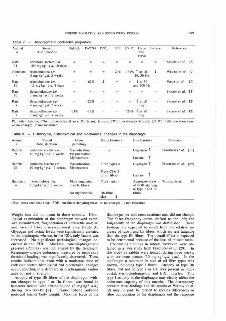
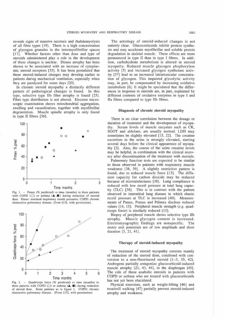
In a recent report, two patients with asthma and one with COPD, who developed myopathy of respiratory and peripheral muscles during treatmen with methylprednisolone, were described [15] (figs. 1 and 2). P1max and PEmax were reduced to mean 38% and 48% predicted, respectively, whilst quadriceps force was reduced to 31% predicted. Tapering of the dose was fo llowed by an increase to 74% and 92% of P1max and PEmax, respectively, after three months. Quadriceps force increased to 62% after six months.
These observations underline the potential hazards of treatment with corticosteroids, and the prolonged duration of recovery after discontinuation of steroid treatment.
Pathogenesis and aetiology of steroid-induced atrophy and myopathy
Pathological examination of muscles of patients treated acutely with high doses of systemic steroids
STEROID MYOPATHY AND RESPIRATORY DISEASE 1001
reveals signs of massive necrosis and rhabdomyolysis of all fibre types [19]. There is a high concentration of glycogen granules in the intermyofibrillar spaces [ 17]. Whether factors other than dose and type of steroids administered play a role in the development of these changes is unclear. Disuse atrophy has been shown to be associated with an increase of cytoplasmic steroid receptors [35]. It has been postulated that these steroid-induced changes may develop earlier in patients during mechanical ventilation, especially when they are paralysed for some days [20].
In chronic steroid myopathy a distinctly different pattern of pathological changes is found. In this type, selective type lib fibre atrophy is found [25]. Fibre type distribution is not altered. Electron microscopic examination shows mitochondrial aggregation, swelling and vacuolization, together with myofibrillar degeneration. Muscle spindle atrophy is only found in type II fibres [36].
100 • D
80 D 0 • "0 ~ • 0. 60 ~ • 0
0 X CIJ E 40 • D "" a: "" •
20
0 0 2 3
Time months Fig. I. - P1max (% predicted) vs time (months) in three patients with COPD (0) or asthma (.&. .•) during reduction of steroid dose. P1max: maximal inspiratory mouth pressures; COPD: chronic obstructive pulmonary disease. (from [15], with permission).
100 . . . . D -80 .
"0 . -~ . --. -0. lilt --~ / -
0 --. -Q) 60 . D · - D ~ . ' --
.E . en li p -' 0. 40 : -Q) (.) . g. -·· 'o ~
. ' - _ ....... ' -CIJ - ~ - -
. ::I 0 20
~ -
0 0 2 3 4 5 6
Time months Fig. 2. - Quadriceps force (% predicted) vs time (months) in three patients with COPD (0) or asthma (.&., •> during reduction of steroid dose. Same patients as in figure I . COPD: chronic obstructive pulmonary disease . (From [ 15], with permission)
The aetiology of steroid-induced changes is not entirely clear. Glucocorticoids inhibit protein synthesis and may accelerate myofibrillar and soluble protein degradation in skeletal muscle. These effects are more pronounced in type II than in type I fibres. In addition, carbohydrate metabolism is altered in steroid myopathy. Reduced muscle glycogen phophorylase activity [5] and increased glycogen synthetase activity [37] lead to an increased intramuscular concentration of glycogen. This impaired glycolytic activity may, in part, be compensated by increasing oxidative metabolism [6]. It might be speculated that the differences in response to steroids are, in part, explained by different contents of oxidative enzymes in type I and Ila fibres compared to type lib fibres.
Diagnosis of chronic steroid myopathy
There is no clear correlation between the dosage or duration of treatment and the development of myopathy. Serum levels of muscle enzymes such as CK, SGOT and aldolase, are usually normal. LDH may sometimes be slightly elevated [15, 22]. The creatine excretion in the urine is strongly elevated, starting several days before the clinical appearence of myopathy [3]. Also, the course of the urine creatine levels may be helpful, in combination with the clinical recovery after discontinuation of the treatment with steroids.
Pulmonary function tests are expected to be similar to those observed in patients with respiratory muscle weakness [38, 39]. A slightly restrictive pattern is found, due to reduced muscle force [13] . The diffusion capacity for carbon dioxide may be reduced because of microatelectases [38]. Lung compliance is reduced with low recoil pressure at total lung capacity (TLC) [38]. This is in contrast with the pattern observed in interstitial lung disease in which elastic recoil pressure at TLC is increased [40] . Measurements of P1max, PEmax and Pdimax disclose reduced values [14, 15]. Peripheral muscle strength (e.g. quadriceps force) is similarly reduced [15].
Biopsy of peripheral muscle shows selective type lib atrophy. Muscle glycogen content is increased . Electromyographic findings are nonspecific. The motor unit potentials are of low amplitude and short duration [3, 21, 41].
Therapy of steroid-induced myopathy
The treatment of steroid myopathy consists mainly of reduction of the steroid dose, combined with conversion to a non-fluorinated steroid [1-3, 35, 42]. Androgens partially antagonize glucocorticoid-induced muscle atrophy [21, 43, 44], in the diaphragm [45]. The role of these anabolic steroids in patients with COPD or asthma who are treated with glucocorticoids has not yet been elucidated.
Physical exercises, such as weight-lifting [46] and treadmill walking [47] partially prevent steroid-induced atrophy and weakness.
1002 P.N.R. DEKHUJJZEN, M. DECRAMER
References
J. Williams RS. - Triamcinolone myopathy. Lancet, 1959; 1: 698-701. 2. Golding DN, Murray SM, Pearch GW, Thompson M. - Corticosteroid myopathy. Ann Phys Med, 1961; 5: 171 - 176. 3. Askari A, Yignos PJ, Moskowitz RW. - Steroid myopathy in connective tissue disease. Am J Med, 1976; 6 I: 485-492. 4. MacLean K, Schurr PH. - Reversible amyotrophy complicating treatment with fluorocortisone. Lancet, 1959; 1: 701-703. 5. Smith B. - Histological and histochemical changes in the muscles of rabbits given the corticosteroid triamcinolone. Neurology. 1964; 14: 857- 863. 6. Yignos PJ, Greene R. - Oxidative respiration of skeletal muscle in experimental corticosteroid myopathy. J Lab Clin Med, 1973; 81: 365-378. 7. Gardiner PF, Edgerton YR. - Contractile responses of rat fast-twitch and slow-twitch muscles to glucocorticoid treatment. Muscle Nerve, 1979; 2: 274-281. 8. Moore BJ, Miller MJ, Feldman HA, Reid MB. - Diaphragm atrophy and weakness in cortisone-treated rats. J Appl Physiol, 1989; 67: 2420- 2426. 9. Wilcox PG, Hards JM, Bockhold K, Bressler B, Pardy RL. - Pathologic changes and contractile properties of the diaphragm in corticosteroid myopathy in hamsters: comparison to peripheral muscle. Am J Respir Cell Mol Bioi, 1989; 1: 191-199. I 0. Viires N, Pavlovic D, Pariente R, Aubier M. - Effects of steroids on diaphragmatic function in rats. Am Rev Respir Dis, 1990; 124: 34-38. ll. Ferguson GT, Jrvin CG, Chemiack RM. - Effects of corticosteroids on diaphragm function and biochemistry in the rabbit. Am Rev Respir Dis, 1990; 141: 156-163. 12. Sasson L, Tarasiuk A, Heimer D, Bark H. - Effect of dexamethasone on diaphragmatic and soleus muscle mor-phology and fatigability. Respir Physiol, 1991; 85: 15-28. 13. Janssens S, Decramer M. - Corticosteroid-induced myopathy and the respiratory muscles. Report of two cases. Chest, 1989; 95: 1160-1162. 14. Rogiers Ph, Lacquet LM, Decramer M. - Steroidinduced myopathy in patients with COPD and asthma. (Abstract). Eur Respir J, 1991 ; 4: 346. 15. Decramer M, Stas KJ. - Corticosteroid-induced myopathy involving respiratory muscles in patients with COPD or asthma. Am Rev Respir Dis, 1992, (In Press). 16. MacFarlane lA, Rosenthal FD. - Severe myopathy after status asthmaticus. Lancet, 1977; ii: 615. 17. van Marle W, Woods KL. - Acute hydrocortisone myopathy. Br Med J, 1980; 281: 271-272. 18. Knox AJ, Mascie-Taylor 8, Muers MF. - Acute hydrocortisone myopathy in acute severe asthma. Thorax, 1986; 41: 411-412. 19. Williams TJ, O'Hehir RE, Czuruy D, Home M, Bowes G. - Acute myopathy in severe acute asthma treated with intravenously administered corticosteroids. Am Rev Respir Dis, 1988; 137: 460-463. 20. Shee CD. - Risk factors for hydrocortisone myopathy in acute severe asthma. Respir Med, 1990; 84: 229- 233. 21. Afifi AK, Bergman RA, Harvcy JC. - Steroid myopathy. Clinical, histologic and cytologic observations. John Hopkins Med J, 1968; 123: 158-174. 22. Kanayama Y, Shiota K, Horiguchi T, Kato N,
Ohi A, Inoue T. - Correlation between steroid myopathy and serum lactate dehydrogenase in systemic lupus erythematosus. Arch Intern Med, 1981; 141: 1176-1179. 23. Clark AF, Vignos PJ. - Experimental corticosteroid myopathy: effect on myofibrillar ATPase activity and protein degeneration. Muscle Nerve, 1979; 2: 265- 273. 24. Ruff RL, Martyn 0 , Gordon AM. - Glucocorticoidinduced atrophy is not due to impaired excitability in rat muscle. Am J Plzysiol, 1982; 243: E512-E52l. 25. Ruff, R.L. - Endocrine myopathies. In: Engel AG, Banker BQ, eds. Myology. McGraw-Hill Book Co., 1986; 1871- 1906. 26. Goldberg AL, Goodman HM. - Relationship between cortisone and muscle work in determining muscle size. J Physio/, 1969; 200: 667-675. 27. Gardiner PF, Botterman BR, Eldred E, Simpson DR, Edgerton YR. - Metabolic and contractile changes in fast and slow muscle of the cat after glucocorticoid-induced atrophy. Exp New·ol, 1978; 62: 241-255. 28. Roy RR, Gardiner PF, Simpson DR, Edgerton YR. -Glucocorticoid-induced atrophy in different fibre types of selected rat jaw and hind-limb muscles. Archs Oral Bioi, 1983; 28: 639-643. 29. Ferguson GT, lrvin CG, Cherniack RM. - Effects of corticosteroids on respiratory muscle histopathology. Am Rev Respir Dis, 1990; 142: 1047- 1052. 30. Carlstedt-Duke J, Gustafsson JA. - Structure and function of the glucocorticoid receptor. J Steroid Biochem, 1987; 27: 99-104. 31. Reid MB, Ericson GC, Feldman HA, Johnson RL. -Fibre types and fibre diameters in canine respiratory muscles. ' App/ Physiol, 1987; 62: 1705-17 I 2. 32. Bowyer SL, La Mothe MP, Hollister JR. - Steroid myopathy: incidence and detection in a population with asthma. J Allergy Cl in lmmunol, I 985; 76: 234-242. 33. Picado C, Fiz AJ, Montserrat JM, Grau JM, FemandezSola J, Luengo MT, Casademont J, Agusti-Yidal A. -Respiratory and skeletal muscle function in steroiddependent bronchial asthma. Am Rev Respir Dis, 1990; 141: 14-20. 34. Wang Y, Zintel T, Yasques A, Gallagher CG. - Corticosteroid therapy and respiratory muscle function in humans. Am Rev Respir Dis, 1991; 144: 108- 112. 35. DuBois DC, Almon RR. - Disuse atrophy of skeletal muscle is associated with an increase in number of glucocorticoid receptors. Endocrinology, 1980; 107: 1649-165 I. 36. Ste rn LZ, Hannapel LK. - The muscle spindle in cortisone-induced muscular atrophy. Exp Neurol. 1973; 40: 484-490. 37. Shoji S, Takagi A, Sugita H, Toyokura Y. - Muscle glycogen metabolism in steroid-induced myopathy of rabbits. Exp Neurol, 1974; 45: l-7. 38. De Troyer A, Botenstein S, Cordier R. - Analysis of lung volume restriction in patients with respiratory muscle weakness. Thorax, 1980; 35: 603-610. 39. Braun NMT, Arora NS, Rochestcr OF. - Respiratory muscle and pulmonary function in polymyositis and other proximal myopathies. Thorax, 1983; 38: 616- 623. 40. Yemault JC, De Jonghe M, De Coster A, Englert M. - Pulmonary mechanics in diffuse fibrosing alveolitis. Bull Eur Physiopathol Respir, 1975; 11: 231 - 234. 41. Yates DAH. - Steroid myopathy. Rheumatol Phys Med, 1971; 11: 28-33.

STEROID MYOPATHY AND RESPIRATORY DISEASE 1003
42. Duvenci J, Chodosh S, Segal MS. - Dexamethasone therapy in bronchial asthma. Ann Allergy, 1959; 17: 695-700. 43. Sakai Y, Kobayashi K, Twata N. - Effects of anabolic steroids and vitamin B complex upon myopathy induced by corticosteroids. Eur J Physiol, 1978; 52: 353-359. 44. Santidrian S, Young YR. - Effect of androgens on the rate of muscle protein breakdown in rats treated with glucocorticoids. Reproduction, 1980; 4: 331- 337.
45. Reid MB, Wang N, Haack KE, Miller MJ. -Anabolic steroids protect skeletal muscles against cortisone-induced atrophy. (Abstract). Faseb J, 1990; 4: 1068. 46. Gardiner PF, Hibl B, Simpson DR, Roy R, Edgerton YR. - Effects of a mild weight-lifting program on the progress of glucocorticoid-induced atrophy in rat hindlimb muscles. Pflugers Arch, 1980; 385: 147-153. 47. Hickson RC, Davis JR. - Partial prevention of glucocorticoid-induced muscle atrophy by endurance training. Am J Physiol, 1981; 241: 226-232.


![[PPT]Steroid Induced Diabetes - Healing, Teaching & … Induced... · Web viewACE/ADA Task Force on Inpatient Diabetes. Diabetes Care. 2006;29(8):1955-1962. Bolus insulin to keep](https://cdn.vdocuments.us/doc/165x107/5ab7ace77f8b9a684c8bcb29/pptsteroid-induced-diabetes-healing-teaching-inducedweb-viewaceada.jpg)

