
SRINIVAS SRIDHARNANOMEDICINE SCIENCE AND TECHNOLOGY CENTERNANOMEDICINE SCIENCE AND TECHNOLOGY CENTER
ELECTRONIC MATERIALS RESEARCH INSTITUTE DEPARTMENT OF PHYSICS, NORTHEASTERN UNIVERSITY,RADIATION ONCOLOGY , HARVARD MEDICAL SCHOOL
www.igert.neu.edu

Director : Srinivas Sridhar, Ph.D.CAS Distinguished Professor of PhysicsDirector : Srinivas Sridhar, Ph.D.CAS Distinguished Professor of Physics
sagar.physics.neu.eduwww igert neu edu
Nanotechnology for Radiation OncologyNanotechnology for Radiation Oncology Magnetic Theranostic NanoplatformsMagnetic Theranostic Nanoplatforms
Visiting Professor of Radiation Oncology, Harvard Medical SchoolVisiting Professor of Radiation Oncology, Harvard Medical School
Funded by NIH, NSF, Hospitals
www.igert.neu.edu
With Harvard Medical School• Smart Implants for BIS‐IGRT• Gold Nanoparticles for Radiotherapy• Green Quantum Dots for Xray Photodynamic
With Harvard Medical School• Smart Implants for BIS‐IGRT• Gold Nanoparticles for Radiotherapy• Green Quantum Dots for Xray Photodynamic
Magnetic Theranostic Nanoplatforms• MR Contrast Enhancement• Magnetic Targeting• Magnetic Hyperthermia
Magnetic Theranostic Nanoplatforms• MR Contrast Enhancement• Magnetic Targeting• Magnetic Hyperthermia
TherapyTherapy
Multi‐modal Imaging Platforms• MR, SPECT, CT, PET• Nanotechnology in the Operating Room of the
Multi‐modal Imaging Platforms• MR, SPECT, CT, PET• Nanotechnology in the Operating Room of the
Dental and Orthopedic NanomedicineWith Harvard School of Dental Medicine•Smart Implants for Bone Regeneration •Slow release agents for cements and
Dental and Orthopedic NanomedicineWith Harvard School of Dental Medicine•Smart Implants for Bone Regeneration •Slow release agents for cements and• Nanotechnology in the Operating Room of the
Future• Nanotechnology in the Operating Room of the Future
•Slow release agents for cements and membranes•Slow release agents for cements and membranes
Nanoparticles in k l
MR Imaging of Theranostic Magnetic
www.igert.neu.edu
skin using multi‐modal imaging
Theranostic Magnetic Nanoiposomes

www.igert.neu.edu

From nano to micro
NNATUR
DNA base DNA turn Protein Virus CellsBacteria
RAL
1nm 10nm 100nm m0.1nm m
Metal MIcelleNanoassembly Liposome
DNA t h
SYNT
16nm4nm
DNA nanotechTHETI
www.igert.neu.edu
IC

Designing Utili k l d fDesigning Nano: The
computation revolution
• Utilize knowledge of interactions to design nanoelements
e o ut o
Making Nano: The
nanofabricatio n revolution
• Top‐down nanolithograpjy• Bottom up Self – assembly
Seeing Nano: The imaging revolution
• Atomic, Electron Microscopies
• Optical microscopies, d
www.igert.neu.edu
quantum dots
NANOMEDICINE A di i di i d th
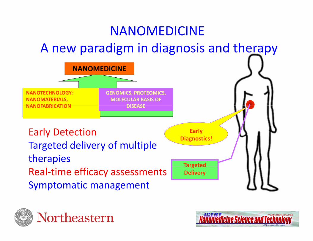
NANOMEDICINE
A new paradigm in diagnosis and therapy
NANOTECHNOLOGY:NANOMATERIALS, NANOFABRICATION
GENOMICS, PROTEOMICS, MOLECULAR BASIS OF
DISEASE
Early Early Detection
NANOFABRICATION DISEASE
Diagnostics!
Targeted
yTargeted delivery of multiple therapies
Targeted DeliveryReal‐time efficacy assessments
Symptomatic management
www.igert.neu.edu

ARTISTS CONCEPTIONS OF AARTISTS CONCEPTIONS OF A FANTASTIC FUTURE FOR NANOMEDICINE
NANOBOTS TO CLEAN ARTERIES
NANOHUMVEES TO
Tim Fonseca
DESTROY TUMOR CELLS
www.igert.neu.edu

NANOMEDICINE
NANOTECHNOLOGY APPLIED TO:
•Cancer•Cardiovascular Diseases•Infectious Diseases•Orthopedics•Genomics•ProteomicsO th l l•Opthalmology
•Neuroscience•
www.igert.neu.edu
• ………..

THERANOSTIC NANOPLATFORMS
EARLY REAL‐TIME MONITORING
TARGETED
DIAGNOSTICS MONITORINGTHERAPY
www.igert.neu.edu
Figure: Opensource Handbook of Nanoscience and Nanotechnology

Cellular andNanoplatforms
•Polymers, lipids, ll lf
Characterization toolkit
•Nanoscale Mi i SEM
Cellular and Phyiological Mechanisms
•Apoptosis
Medical Challenge
• Image guided drug d liorganelles, self‐
assembling ampiphiles,
•Magnetic nanoparticles
Microscopies: SEM, TEM, AFM, STM, NSOM,
• Fluorescence confocal and optical
•Delivery to nucleus, mitochondria or ribosome
•Endocytosis, Cellular uptake
delivery• Localized energy delivery
•Nano‐enhanced implants
•Metallic nanoparticles
•Quantum dots•Nanoporous coatings on implants
microscopy• Spectroscopies: Femtosecond optical spectroscopy, XAFS,
• Zeta Potential,
•Gene Silencing• Organ biodistribution and pharmacokinetics
•Radiation Oncology•Dental and Orthopedic Nanomedicine
www.igert.neu.edu
coatings on implants ,SQUID, Coulter

Multi‐functional Nanoplatforms
PEGSpacerAntibody Endosome
BufferingAgent
HIV TATP tid
NANOCOATED IMPLANTS
TAT
d
Peptide
pDNA orOligonucleotide
pH‐SensitiveFluorescentLabel
Radioactive Label
Magnetic core40 nm 30 nm40 nm40 nm40 nm 30 nm30 nm30 nm
SENSING DRUG ELUTINGSENSING, DRUG ELUTING PLATFORMS, SCAFFOLDS AND
TEMPLATES, NEUROCHIP
Au or Fe‐Au NP1‐100nm
Micelles10‐50nm
Liposomes100‐250 nm
Polymeric NP~ 20nm – 10 m
SENSING TARGETING DELIVERY
www.igert.neu.edu
SENSING, TARGETING, DELIVERY

NANOPARTICLE FORMULATIONS OF DRUGS
www.igert.neu.edu

www.igert.neu.edu
Ferrari, Nature

Table 1. Examples of Nanomaterials in Clinical Use.* Nanomaterial Trade Name Application Target Adverse Effects Manufacturer Current Status
Examples of Nanomedicine in Clinical Use Kim, et. al. NEJM 2010
Nanomaterial Trade Name Application Target Adverse Effects Manufacturer Current Status Metallic Iron oxide Feridex MRI contrast Liver Back pain, vaso- Bayer Schering FDA approved
dilatation Resovist MRI contrast Liver None Bayer Schering FDA approved Combidex MRI contrast Lymph nodes None Advanced Magnetics In phase 3 clin-
ical trials NanoTherm Cancer therapy Various forms Acute urinary MagForce In phase 3 clin-
retention ical trials Gold Verigene In vitro diag- Genetic Not applicable Nanosphere FDA approved
nostics Aurimmune Cancer therapy Various forms Fever CytImmune Sciences In phase 2 clin-
ical trials Nanoshells Auroshell Cancer therapy Head and neck Under investigation Nanospectra In phase 1 clin-
Biosciences ical trials Semiconductor
Quantum dot Qdots, EviTags, Fluorescent con- Tumors, cells, Not applicable Life Technologies, Research
semiconductor trast, in vitro tissues, and eBioscience, use only t l di ti l l Nnanocrystals diagnostics molecular Nanoco,
sensing CrystalPlex, structures Cytodiagnostics
Organic Protein Abraxane Cancer therapy Breast Cytopenia Abraxis Bioscience FDA approved Liposome Doxil/Caelyx Cancer therapy Various forms Hand–foot syndrome, Ortho Biotech FDA approved
stomatitisstomatitis Polymer Oncaspar Cancer therapy Acute lymphoblas- Urticaria, rash Rhône-Poulenc Rorer FDA approved
tic leukemia CALAA-01 Cancer therapy Various forms Mild renal toxicity Calando In phase 2 clin-
ical trials Dendrimer VivaGel Microbicide Cervicovaginal Abdominal pain, Starpharma In phase 2 clin-
dysuria ical trials
www.igert.neu.edu
dysuria ical trials Micelle Genexol-PM Cancer therapy Various forms Peripheral sensory Samyang For phase 4
neuropathy, clinical neutropenia trials

Theranostic Magnetic Nanoplatforms
Other options for targeting:
TARGETING IMAGING GUIDED DRUG & ENERGY DELIVERY
Solid tumor
Apply magnetic field
p g g1 ‐ Direct injection into tumor site2 ‐ Coating NMP with antibodies to target tumorApply magnetic field
to concentrate particles
Inject NMPs IV,NMP ill i l h h
g
Modulate field to release drug from particles
NMP will circulate through the blood stream
particles
From Biophan
www.igert.neu.edu

SuperParamagnetic Iron Oxide Nanoparticles (SPIONS)
M vs B
Nanoparticles (SPIONS)
0.E+001.E-052.E-053.E-054.E-05
mu/
uL)
hexanemicelle
SPION in hexane at 100 Oe
4.E-05
-4.E-05-3.E-05-2.E-05-1.E-05
-0.5 -0.3 -0.1 0.1 0.3 0.5
M (e
3.E-05
3.E-05
4.E-05
4.E 05u/
ul)
ZFCFC
Blocking
temperature (TB) of
SPIONs
B (T)
5 E-06
1.E-05
2.E-05
2.E-05
M (e
muSuperparamagnetism
No hysteresis in M‐H curves
www.igert.neu.edu
0.E+00
5.E 06
0 50 100 150 200 250 300 350T (K)

Magnetic Nanoplatforms @ NU
TAT
Y
TAT
Y
I id ldIron oxide NPs
Iron oxide‐goldCore‐shell NPs MagNP + polyelectrolye MagNP + antibody
Magnetic NanoLiposome SPION‐micellew. Torchilin
Magnetic NanoLiposomew. Campbell
Magnetic NanoEmulsionw. Amiji
For hydrophilic drugs eg
www.igert.neu.edu
For hydrophobic drugs, e.g. taxanesdrugs, eg. Adriamycin

MULTI‐FUNCTIONAL MAGNETIC NANOPLATFORM: MAGNETIC CATIONIC LIPOSOMEMAGNETIC CATIONIC LIPOSOME
Typical size : 100 – 250 nm
PEG (Circulation)
Fluorescent Label
Magnetic guidanceMRI contrast enhancement
With Robert Campbell
MCLformulated andcharacterized inprevious work.
All components are FDA
www.igert.neu.edu
= PEG approvedsiRNA

MULTI‐MODAL IN VIVO IMAGING
Apply Magnet
Inject cancer cells
No magnet
Inject MCL IV or IT
Dr. Zhenghong
Lee, Case W
Bi di t ib ti t d SPECT/CT Fl
Western U
niversity
MR imaging Biodistribution study using radio‐tagged MCL
SPECT/CT Fluorescence imaging
www.igert.neu.edu

STABLE FORMULATIONS OF ML, EXTENSIVELY CHARACTERIZEDDPPC:DOTAP:CHOL and DOPE‐PEG5000 Campbell , et. al.p
Vigorous uptake in HMVEC‐D cellsLiposomes
MagnetoLiposomes
www.igert.neu.edu
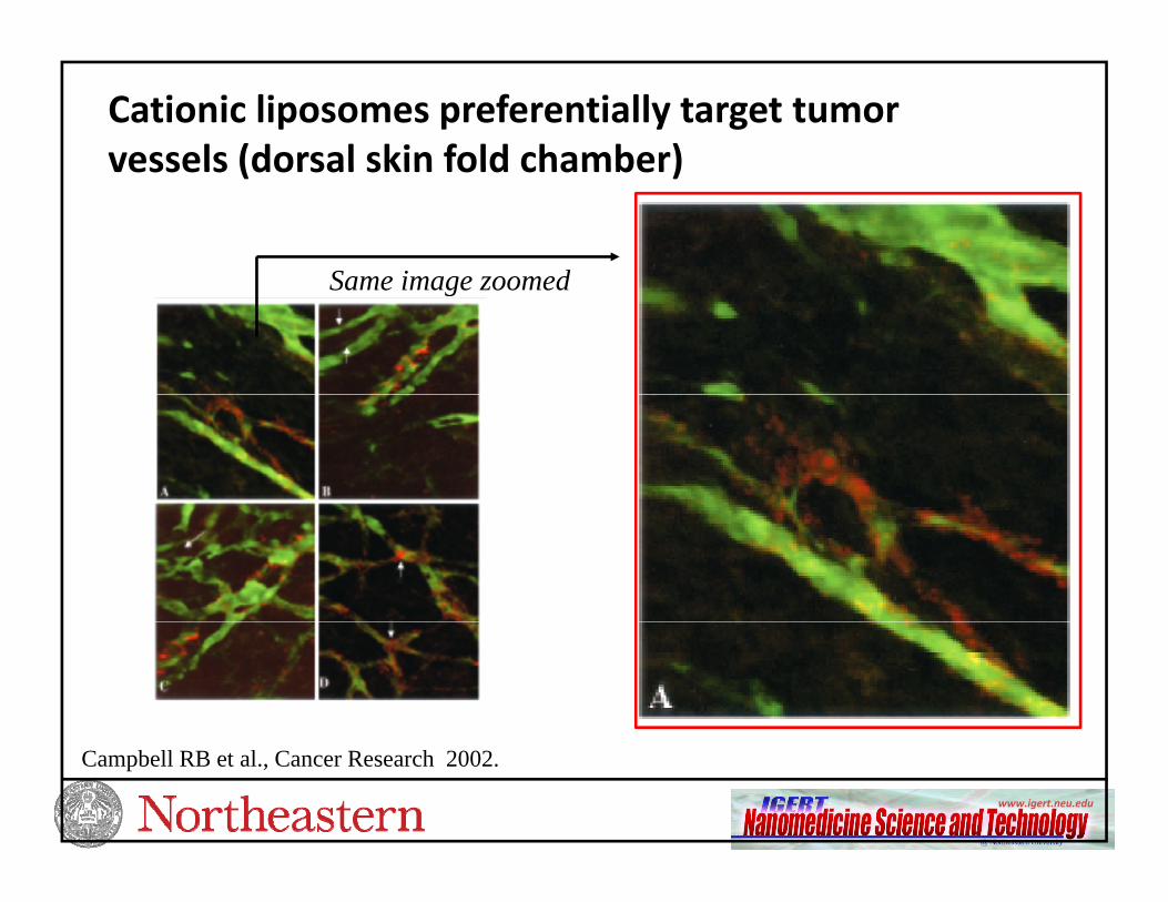
Cationic liposomes preferentially target tumor vessels (dorsal skin fold chamber)vessels (dorsal skin fold chamber)
Same image zoomedSame image zoomed
www.igert.neu.edu
Campbell RB et al., Cancer Research 2002.

Vinblastine‐loaded MCLs improved melanoma treatment: in vivo therapeutic benefit
Mechanism of action:
Campbell , et. al.
Mechanism of action: Vinblastine sulfate- binds to the microtubular proteins of the mitotic spindle, causing mitotic arrest and cell
death
Magnet improvedTreatment outcomeTreatment outcome
www.igert.neu.edu

T1‐scan of healthy animal before and i di l f i j iimmediately after injection.
LiverLiver
Spleen
Melanoma tumor model
www.igert.neu.edu
Melanoma tumor modelMNL ARE LONG CIRCULATING AND CAN BE OBSERVED FOR > 48 HOURS

MAGNETIC LIPOSOMESMAGNETIC GUIDANCE LEADS TO INCREASED ACCUMULATION IN TUMNOR
T2‐scan of intravenous injected mouse with magnet
Magnet placed externally for 1 hour
T2‐scan of intravenous injected mouse with magnet applied for 1hr imaged 24 and 48 hours after injection.
Gultepe, et. al., Nanomedicine (2010)
www.igert.neu.edu
With Robert Campbell, Craig Ferris, Praveen Kulkarni

NANOMAGNETIC THERMAL THERAPY
MAGNETIC NANOPARTICLES ABSORB
THERMAL ABLATION > 45CCOAGULATION, NECROSIS
MAGNETIC NANOPARTICLES ABSORB ENERGY FROM AC MAGNETIC FIELDSRELEASE ENERGY TO TUMOR
NON INVASIVEHYPERTHERMIA 41‐45 CREVERSIBLE, SENSITIZATION
•NON‐INVASIVE •LOCALIZED •CAN BE IMAGED
NANOTECHNOLOGY AND NEWMAGNETIC SOURCES HAVE LED TO A REVIVALNANOTECHNOLOGY, AND NEW MAGNETIC SOURCES, HAVE LED TO A REVIVAL OF INTEREST IN MAGNETIC HYPERTHERMIACLINICAL TRIALS IN EUROPEDARTMOUTH CANCER NANOTECH CENTER ESTABLISHED
www.igert.neu.edu

IN VITRO HEATING USING MAGNETIC NANOLIPOSOMES
Hyperthermia of Mag C - Liposomes
110 0
20 mg/ml10 mg/ml5 mg/ml2 5 mg/ml
80 0
90.0
100.0
110.0
)
2.5 mg/mlBlank liposomes
50.0
60.0
70.0
80.0
pera
ture
(CRAPID TEMPERATURE
THERMAL ABLATION
20.0
30.0
40.0
50.0Te
mINCREASE WITH APPLICATION OF AC MAGNETIC FIELD360 kHz, 70 kA drive
HYPERTHERMIA
10.00 5 10 15 20 25 30
Time (minutes)
www.igert.neu.eduTHERMAL THERAPY FEASIBLE WITH MNL

SPECT /CT Single photon emission computed tomography (SPECT)Xray computed tomography (CT)
M i Li I 111 di l b lMagnetic Liposome + In111 radiolabel
IT‐no magnet‐24hrs post‐i j ti
IT‐magnet‐ 24hrs post‐injection
injection
www.igert.neu.edu

MULTI‐FUNCTIONAL NANOPLATFORMS FOR THE OPERATING ROOM OF THE FUTURE
MRI
Ch th ti
SPECT/CT
ChemotherapeuticsBiologics, siRNA
SPECT/CT
PET
Radio TherapyHardware Integration – MRI, PET, CT, ..In vivo fiducial marker – not on the table, but in the aorta
www.igert.neu.edu
,or tumor!Improved image registration for treatment planning

CLINICAL TRANSLATION OF NANOMEDICINES
Valley of death
SAFETY
I go home today They cured me
SAFETY POLICYETHICS
I go home today. They cured meusing this new miracle drug. Itwill be years before it isapproved for humans.
COMMERCIALIZATION
www.igert.neu.edu

Nanotechnology for Smart Implants
Reasons for implant failures: Scar tissue formation; Inflammatory response; Lack of tissue integration; Infection Nanotechnology based
techniques offer solution
Non‐erodible coatings on TiO2 for cardiovascular stents and other implantsimplants
Smart implants for Image Guided Radio Therapy
http://www.alberox.com/articles/MAC_MedicalImageFINAL.jpg
www.igert.neu.edu30
Gold TiO2
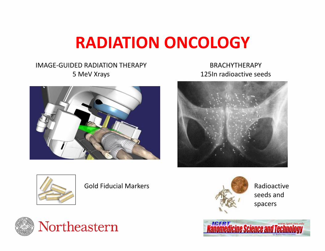
RADIATION ONCOLOGYRADIATION ONCOLOGYIMAGE‐GUIDED RADIATION THERAPY
5 MeV XraysBRACHYTHERAPY
125In radioactive seeds
Gold Fiducial Markers Radioactive seeds and spacers
www.igert.neu.edu
spacers

Nanotechnology for Radiation OncologyBIOLOGICAL IN SITU IMAGE‐GUIDED RADIATION THERAPY (BIS‐IGRT)
Cormack, Makrigiorgos, D’Amico, Nguyen, Kumar, Sridhar
BIOLOGICAL IN SITU IMAGE‐GUIDED RADIATION THERAPY (BIS‐IGRT)
Dual drug release strategies for localized delivery over 8 weeks
Brachytherapy spacers of IGRT coated with radiosensitizer-releasing nanoparticles, lead to a radiosensitized volume resulting in increased efficacy of radiation therapy.
Combined chemo + radiation therapy i ffi i t i ti ll
www.igert.neu.edu
is more efficient synergistically

Join us at
www igert neu edu
Join us atTim Fonseca
www.igert.neu.edusagar.physics.neu.edu


