This is an Open Access document downloaded from ORCA, Cardiff University's institutional
repository: http://orca.cf.ac.uk/102637/
This is the author’s version of a work that was submitted to / accepted for publication.
Citation for final published version:
Butt, Nauman M., Lambert, Jonathan, Ali, Sahra, Beer, Philip A., Cross, Nicholas C. P.,
Duncombe, Andrew, Ewing, Joanne, Harrison, Claire N., Knapper, Steven, McLornan, Donal,
Mead, Adam J., Radia, Deepti and Bain, Barbara J. 2017. Guideline for the investigation and
management of eosinophilia. British Journal of Haematology 176 (4) , pp. 553-572.
10.1111/bjh.14488 file
Publishers page: http://dx.doi.org/10.1111/bjh.14488 <http://dx.doi.org/10.1111/bjh.14488>
Please note:
Changes made as a result of publishing processes such as copy-editing, formatting and page
numbers may not be reflected in this version. For the definitive version of this publication, please
refer to the published source. You are advised to consult the publisher’s version if you wish to cite
this paper.
This version is being made available in accordance with publisher policies. See
http://orca.cf.ac.uk/policies.html for usage policies. Copyright and moral rights for publications
made available in ORCA are retained by the copyright holders.

1
Guideline for the investigation and management of eosinophilia
Nauman M. Butt,1 Jonathan Lambert,2 Sahra Ali,3 Philip A. Beer,4 Nicholas C.P.
Cross,5 Andrew Duncombe,6 Joanne Ewing,7 Claire N. Harrison,8 Steven Knapper,9
Donal McLornan,10 Adam J. Mead,11 Deepti Radia,8 and Barbara J. Bain.12
Writing group: British Committee for Standards in Haematology
1Royal Liverpool and Broadgreen University Teaching Hospitals NHS Trust, Liverpool, 2University College London Hospitals NHS Foundation Trust, London 3Hull and East
Yorkshire Hospitals NHS Trust, 4Wellcome Trust Sanger Institute, Cambridge, 5Faculty
of Medicine, University of Southampton, Southampton, 6Department of Haematology,
University Hospital Southampton, Southampton, 7Heart of England NHS Foundation
Trust, Birmingham, 8Guy’s and St Thomas’ NHS Foundation Trust, London, 9Division of
Cancer & Genetics, School of Medicine, Cardiff University, Cardiff., 10King's College
Hospital NHS Foundation Trust, London, 11MRC Molecular Haematology Unit,
Weatherall Institute of Molecular Medicine, University of Oxford and BRC Blood Theme,
NIHR Oxford Biomedical Centre, Oxford, UK and 12Imperial College London, St
Mary’s Hospital, London, UK
Keywords: eosinophilia, hypereosinophilia, eosinophilic leukaemia, HES
2
Introduction
The guideline group was selected to be representative of UK-based medical experts with
an interest in myeloproliferative neoplasms and eosinophilia. PubMed and EMBASE
were searched systematically for publications in English until August 2015 using the key
words eosinophilia, hypereosinophilia, eosinophilic leukaemia and HES. The writing
group produced the draft guideline which was subsequently revised by consensus by
members of the General Haematology and Haemato-oncology Task Forces of the British
Committee for Standards in Haematology. The guideline was then reviewed by a
sounding board of UK haematologists and representatives from the Nordic MPN Study
Group, the British Committee for Standards in Haematology (BCSH) and the British
Society for Haematology (BSH) Committee and comments were incorporated where
appropriate. The ‘GRADE’ system was used to quote levels and grades of evidence,
details of which can be found in Table I. The objective of this guideline is to provide
healthcare professionals with clear guidance on the investigation and management of
eosinophilia.
Guideline Update
There is no previous BCSH guideline for this topic.
Aim

3
The purpose of this guideline is to provide a practical approach to the investigation and
management of eosinophilia.
Key recommendations
The underlying cause of eosinophilia should be sought and possible
eosinophil-associated end organ damage should be evaluated (Grade 1B)
Assessment of underlying cause
A detailed medical history should be taken and a thorough physical
examination should be performed (Grade 1C).
The history should include:
o assessment for allergic disorders, skin rashes and cardiorespiratory,
gastrointestinal and constitutional symptoms.
o a detailed travel history, particularly for tropical travel; travel even in
the remote past may be relevant.
o a detailed drug history.
All patients should have a full blood count, blood film examination and
routine tests of renal and liver function, a bone profile, lactate
dehydrogenase and erythrocyte sedimentation rate and/or C-reactive protein
(Grade 1C)
Patients who are otherwise well with mild to moderate eosinophilia between
0.5 and 1.5 × 10⁹/l may not require further testing. Patients with systemic
symptoms and those with persistent eosinophilia (at least 1.5 × 10⁹/l),
4
irrespective of suspected organ damage, should be considered for additional
testing for an underlying cause
Specific causes of reactive eosinophilia, based on clinical suspicion, should be
confirmed or excluded at an early stage by appropriate testing (Grade 1C).
Patients with an eosinophil count of at least 1.5 × 10⁹/l with no obvious cause
should be investigated for a possible haematological neoplasm with clonal
eosinophilia, initially by peripheral blood analysis for FIP1L1-PDGFRA by
fluorescence in situ hybridisation (FISH) or nested reverse transcriptase
polymerase chain reaction (RT-PCR) (Grade 1C).
Serum tryptase estimation should be performed if the differential diagnosis
includes chronic eosinophilic leukaemia or systemic mastocytosis (Grade 1B).
In the absence of an identifiable cause and with negative peripheral blood
analysis for FIP1L1-PDGFRA by FISH or nested RT-PCR, a bone marrow
aspirate, trephine biopsy and cytogenetic analysis should be performed; the
possibility of an underlying lymphoma or of the lymphocytic variant of
hypereosinophilic syndrome should be evaluated, including consideration of
immunophenotyping of peripheral blood and bone marrow lymphocytes and
analysis for T-cell receptor gene rearrangement (Grade 1C). The possibility
of systemic mastocytosis or other myeloid neoplasm should be considered.
Assessment for possible eosinophil-associated end organ damage

5
End organ damage should be assessed using chest radiography and/or
computed tomography (CT) of the thorax, echocardiography, serum
troponin T and pulmonary function testing. (Grade 1C).
An unprovoked thromboembolic event should be recognised as a possible
manifestation of eosinophil-associated tissue damage (Grade 2C).
In patients with end organ damage, the frequency of further serial
evaluations of organ function should be determined by the severity and
extent of organ compromise and/or by worsening of the eosinophilia (Grade
2C).
Emergency treatment
Patients requiring emergency treatment for severe or life-threatening
eosinophilia should receive high-dose corticosteroids (Grade 1B).
Patients receiving corticosteroids, in whom there is a risk of strongyloides
infection, should receive concomitant ivermectin to prevent potentially fatal
hyperinfection (Grade 1B)
Treatment of clonal eosinophilia
Patients with clonal eosinophilia with FIP1L1-PDGFRA (including patients
presenting with acute leukaemia), should be treated with low dose imatinib
(Grade 1B).
Patients with clonal eosinophilia with PDGFRB rearrangement or ETV6-
ABL1 fusion should receive standard dose imatinib (Grade 1B).
6
Patients with clonal eosinophilia with ETV6-FLT3 fusion should be
considered for sunitinib or sorafenib therapy (Grade 2B)
Patients with clonal eosinophilia with JAK2 rearrangement should be
considered for ruxolitinib therapy (Grade 2B)
Patients with acute myeloid leukaemia (AML) with clonal eosinophilia and
no molecular or cytogenetic abnormality suggesting likely response to a
tyrosine kinase inhibitor should be offered standard AML induction therapy
(Grade 1A).
Patient with other haematological neoplasms with clonal eosinophilia should
have treatment directed at management of the neoplasm. If there is organ
damage or dysfunction relating to the eosinophilia, treatment with
corticosteroids should also be given (Grade 1C).
Treatment of lymphocytic variant of hypereosinophilic syndrome
Patients with the lymphocytic variant of hypereosinophilic syndrome (HES)
can be managed in the same manner as idiopathic HES (grade 2B)
Treatment of idiopathic hypereosinophilic syndrome
Patients with idiopathic HES should be treated in the first instance with
corticosteroids (see emergency treatment above).
7
Patients with idiopathic HES who do not respond adequately to
corticosteroids, or who require prolonged corticosteroid therapy, or who are
intolerant of corticosteroids, should be considered for a short trial (4‒6 weeks)
of imatinib, immunomodulatory agents (interferon alpha, ciclosporin or
azathioprine), myelosuppressive therapy (hydroxycarbamide) or monoclonal
antibody therapy with mepolizumab (anti-interleukin 5), the latter
preferably as part of a clinical trial (Grade 2B).
Alemtuzumab, an anti-CD52 monoclonal antibody, should be considered for
patients with severe idiopathic HES unresponsive to other therapies, and
may be useful in patients with idiopathic HES-associated cardiac and
cerebral dysfunction. (Grade 2B)
Role of haemopoietic stem cell transplantation (HSCT)
HSCT should be considered for cases with clonal eosinophilia with FGFR1
rearrangement, patients with chronic eosinophilic leukaemia, not otherwise
specified and those HES patients refractory to or intolerant of both
conventional tyrosine kinase inhibitor (TKI) therapy and experimental
medical therapy, where available, or who display progressive end organ
damage. (Grade 2C)
8
Definitions
Eosinophilia is defined as an elevation of the eosinophil count above levels observed in
healthy subjects, usually taken as above 0.5 × 10⁹/l. Eosinophil counts are higher in
neonates than in adults and the values gradually fall in the elderly. There is no sex or
ethnic variation in the eosinophil count. Definitions of hypereosinophilia (HE) and the
hypereosinophilic syndrome (HES) are based on the proposal by Chusid et al (Chusid et
al, 1975) that required eosinophils to be 1.5 × 10⁹/l or greater in the hypereosinophilic
syndrome with evidence of eosinophil-mediated organ damage or dysfunction after
exclusion of the other potential causes for the organ damage. This criterion was
subsequently accepted in the World Health Organization (WHO) classification of chronic
eosinophilic leukaemia, not otherwise specified (CEL, NOS) (Bain et al, 2008).
Biology
The normal bone marrow contains between 1% and 6% eosinophils and these produce an
eosinophil count in the peripheral blood of 0.05–0.5 × 10⁹/l (Valent et al, 2012).
Eosinophil production in the marrow is tightly controlled by a network of transcription
factors (McNagny & Graf 2002) and is driven by various cytokines, principally
interleukin (IL)-5, IL-3 and granulocyte-macrophage colony-stimulating factor (GM-CSF)
produced by activated T lymphocytes, stromal cells and mast cells, triggering
differentiation and activation (Ackerman & Bochner 2007). Such cytokines are also the
main drivers in reactive eosinophilia in contrast to clonal eosinophilia where tyrosine
kinase fusions are common, typically involving the genes coding for platelet-derived

9
growth factor receptor alpha (PDGFRA), platelet-derived growth factor receptor beta
(PDGFRB) or fibroblast growth factor receptor 1 (FGFR1) (Gotlib et al, 2006).
Under normal conditions, eosinophils may also be found in lymphoid organs and the
mucosa of the gastrointestinal tract and uterus but very rarely in other tissues. However,
prolonged or marked activation of eosinophils may cause migration into non-native
tissues such as the skin, heart and lung where they may cause end-organ damage
principally through the induction of thrombosis and fibrosis (Gleich 2000).
Epidemiology
Primary and idiopathic eosinophil disorders are rare and probably under-diagnosed
conditions. A large population based study in a general practice setting from Copenhagen
demonstrated an incidence of eosinophilia (defined as a count of at least 0.5 × 10⁹/l) of
4% (Andersen et al, 2015).
Causes of Eosinophilia
The causes of eosinophilia are numerous and are conventionally divided into three main
categories – secondary (reactive), primary and idiopathic – as indicated in Table II. These
are discussed below. HES predominantly affects males, with an estimated male-to-female
ratio ranging up to 9 : 1 (Weller & Bubley 1994). This is partly explained by the fact that
the FIP1L1-PDGFRA fusion gene occurs almost exclusively in males.
Secondary (reactive) eosinophilia

10
These form the majority of cases of eosinophilia.
Allergic Disorders
Allergic disorders such as atopic dermatitis, asthma and seasonal allergic disorders
(rhinitis/hayfever) can result in a cytokine-driven non-clonal eosinophilia which is
usually mild (less than 1.5 × 10⁹/l) with the degree of blood eosinophilia and tissue
infiltration generally correlating with the severity of the disease (Horn et al, 1975). The
eosinophilia usually resolves with control of the underlying condition.
Dermatological causes (non-allergic)
Wells syndrome (eosinophilic cellulitis) is a recurring granulomatous dermatitis with
eosinophilia (Wells 1971) characterised by (i) sudden onset annular or circinate
erythematous-oedematous patches that rapidly evolve to morphoea-like slate-blue
plaques, (ii) a histological appearance characterized by the presence of 'flame figures' and
(iii) an inconstant blood eosinophilia. Similar histological appearances can be seen in
other dermatological conditions but these can often be discriminated clinically. Wells
variants have been described (El-Khalawany et al, 2013) as well as associations with
other disorders including connective tissue disease (Yin & Xie 2012).
Drug-induced eosinophilia
Drug hypersensitivity should always be considered as a cause for unexplained
eosinophilia. The list of agents is extensive and includes dietary supplements and herbal
11
remedies (Klion 2009). The clinical manifestations associated with drug-induced
eosinophilia range from asymptomatic to life-threatening (Klion 2009). Rarely a drug
reaction with eosinophilia and systemic symptoms (DRESS syndrome) occurs 3‒6 weeks
after the introduction of a new drug. This syndrome is characterised by a triad of a skin
eruption, fever and internal organ involvement (lung, liver, kidneys, lymph nodes or heart)
(Dong et al, 2014; Sultan et al, 2015). Drug-induced vasculitis and eosinophilia is also
reported, manifesting with purpura, arthralgia and myalgia with possible kidney and lung
involvement (Roujeau et al, 2014).
Infectious diseases
A detailed review of infectious causes of eosinophilia has recently been published
(O'Connell & Nutman 2015). Important infective agents and their diagnostic tests are
outlined in Table III. The British Infection Society and the Hospital for Tropical Diseases
have published UK recommendations for the investigation of eosinophilia in patients
with a tropical travel history (Checkley et al, 2010).
Gastrointestinal disorders
Primary gastrointestinal disorders are rarely associated with gastrointestinal tissue
eosinophilia with or without peripheral blood eosinophilia. Table IV highlights the main
disorders that need to be considered in the differential diagnosis.
12
Vasculitides
Polyarteritis nodosa (PAN) results in inflammation and injury to medium-sized and small
arteries leading to ischaemia or haemorrhage in a variety of tissues and organs, though
the kidneys are usually spared. Renal sparing and negative antineutrophil cytoplasmic
antibodies (ANCA) are useful in differentiating PAN from other systemic necrotizing
vasculitides (Hernandez-Rodriguez et al, 2014). Eosinophilia is occasionally seen but,
when present, eosinophilic granulomatosis with polyangiitis must be considered (Watts
et al, 2007).
Eosinophilic granulomatosis with polyangiitis (EGPA/Churg-Strauss syndrome): EGPA
can present with a variety of clinicopathological features depending on the organs
involved: allergic rhinitis, acute hepatitis, diarrhoea (mesenteric ischaemia), restrictive
cardiomyopathy, peripheral neuropathy, skin lesions and, rarely, renal disease. ANCA is
useful to support the diagnosis and is positive in approximately two-thirds of patients –
mostly perinuclear ANCA (p-ANCA) against myeloperoxidase (MPO). The diagnosis is
confirmed by tissue biopsy.
Rheumatological Diseases
Systemic lupus erythematosus (SLE): eosinophilia is not uncommonly described in
individual case reports but there are no systematic studies on the incidence and severity
of eosinophilia in SLE and eosinophilia does not form one of the diagnostic criteria for
the condition (Petri et al, 2012).
13
Eosinophilic fasciitis (Shulman disease): this is a rare scleroderma-like syndrome of
unknown cause, thought to be immune mediated with resultant painful swelling and
progressive induration and thickening of skin/soft tissues of limbs and trunk. Laboratory
findings are those of a peripheral blood eosinophilia, hypergammaglobulinaemia and
elevated erythrocyte sedimentation rate (ESR). There are no universally accepted
diagnostic criteria but in a recent review (Pinal-Fernandez et al, 2014) peripheral blood
eosinophilia formed one of four minor criteria for the diagnosis. There is an association
with aplastic anaemia (De Masson et al, 2013).
Rheumatoid arthritis (RA): a recent prospective French study showed that approximately
3% of patients with RA have an eosinophilia, which is usually mild and transient
(Guellec et al, 2015). Although this prevalence does not differ from that of the Danish
population study, defined in the same manner (Andersen et al, 2015), eosinophilia did
appear to predict a poorer response to disease-modifying antirheumatic drug use after 3
years (Guellec et al, 2015). A smaller Argentinian study demonstrated a 7% prevalence
of eosinophilia amongst RA patients, but in all these cases a parasitic cause for the
eosinophilia was subsequently demonstrated (Chiardola et al, 2008).
Respiratory disease
Löffler disease: Löffler first described this transient pulmonary reaction with
reticulonodular shadowing on chest radiology associated with a peripheral blood
eosinophilia in 1932. Patients present with a low grade fever and a cough for 7‒10 days,
14
which is usually due to an allergic reaction in the alveoli as a result of a parasitic
infection (see Infectious disease) or medications (See Drug-Induced). Occasionally
patients present with skin lesions. The onset of symptoms is typically 2‒3 weeks
following exposure to parasites and 3‒4 days following ingestion of medication. This is
usually a self limiting disease with symptoms subsiding within 3‒4 weeks of eliminating
the causal agent. The differential diagnosis is allergic bronchopulmonary aspergillosis
and asthma.
Allergic bronchopulmonary aspergillosis is caused by hypersensitivity to Aspergillus
fumigatus and results in uncontrolled asthma and recurrent pulmonary infiltrates which
can progress to bronchiectasis and pulmonary fibrosis (Agarwal et al, 2013). Diagnostic
criteria include a history of asthma or cystic fibrosis, elevated aspergillus-specific
immunoglobulin (Ig) E and IgG, elevated serum IgE (1000 ng/ml or > 417 iu/ml), wheal-
and-flare skin reaction to aspergillus antigen and an eosinophil count greater than 1.0 ×
109/l.
Sarcoidosis: there is no diagnostic test for sarcoidosis though a mild peripheral blood
eosinophilia, raised serum angiotensin converting enzyme (ACE) level and a tissue
biopsy demonstrating the presence of non-caseating granulomas in affected organs
suggests the diagnosis.
Neoplastic disorders with secondary non-clonal eosinophilia

15
Solid tumours: a range of non-haematological neoplasms have been reported to cause
reactive eosinophilia, with a prevalence of 0.5% to 7% (Montgomery et al, 2013). The
presence of eosinophilia is often associated with more advanced metastatic disease. In
cases of unexplained eosinophilia, careful clinical evaluation and radiological studies
should be carried out to exclude underlying occult malignancy (Klion 2009).
Lymphoproliferative disorders: reactive eosinophilia occurs in a broad spectrum of B-
and T-cell lymphoproliferative disorders. In Hodgkin lymphoma the prevalence of
eosinophilia is 15% (Vaughan Hudson et al, 1987) and in non-Hodgkin lymphoma
prevalence ranges from 2% to 20% (Montgomery et al, 2013) with a higher prevalence in
T-cell than B-cell lymphomas. Reactive eosinophilia can occur in acute lymphoblastic
leukaemia/lymphoma (Grimaldi & Meeker 1989). In the case of B-lineage acute
lymphoblastic leukaemia (ALL) with t(5;14)(q31.1;q32.1), it results from dysregulation
of the IL3 gene by proximity to the IGH locus. When eosinophils are part of the
neoplastic clone the eosinophilia is primary rather than reactive and the case falls into the
group of haematological neoplasms with clonal eosinophilia.
Lymphocytic variant HES (L-HES)
L-HES is caused by an expansion of demonstrably clonal or phenotypically aberrant T-
lymphoid cells in the peripheral blood with a secondary, reactive eosinophilia (Simon et
al, 1999), without overt lymphoproliferative disease. Such abnormal T-cell populations
have been reported to be present in 17% to 27% of otherwise unexplained HES (Ogbogu
et al, 2009; Simon et al, 1999). There are no current consensus diagnostic criteria and

16
diagnosis rests on demonstration of an abnormal T-cell population by flow cytometry,
with a broad range of phenotypes reported including CD3-CD4+, CD3+CD4-CD8- and
CD3+CD4+CD7- (Roufosse 2009). Some cases of L-HES harbour T-cell receptor gene
rearrangement (Simon et al, 1999), but this alone is insufficient to make the diagnosis of
L-HES in the absence of phenotypically abnormal T cells (Roufosse 2009). Clinical
manifestations of disease in L-HES are typically cutaneous (Simon et al, 1999).
Miscellaneous Causes
Atheroembolic disease: cholesterol atheroembolic disease develops as a consequence of
cholesterol microembolisation following rupture of atheromatous aortic plaques after
arterial catheterisation procedures, vascular surgery or following anticoagulant or
thrombolytic therapy. A transient eosinophilia has been reported in the acute phase of the
illness (Kasinath & Lewis 1987; Scolari et al, 2007), thought to be driven by increased
IL-5 production by activated T cells at the surface of emboli (Cogan et al, 1995). The
diagnosis is usually confirmed by biopsy of an affected organ.
Graft-Versus-Host Disease: eosinophilia can be a feature of both acute and chronic graft-
versus-host disease (GVHD) following allogeneic haematopoietic stem cell
transplantation (HSCT) (Ahmad et al, 2011; Imahashi et al, 2010; Jacobsohn et al, 2004)
although the mechanisms underlying this are unclear. Conflicting data exist regarding the
prognostic significance of the eosinophilia (Ahmad et al, 2011; Imahashi et al, 2010).

17
Gleich's syndrome: this rare disease was first described in 1984 (Gleich et al, 1984) and
is characterised by episodic angio-oedema with eosinophilia. It is associated with raised
IgM and C1 esterase levels are normal. There is no evidence that this syndrome leads to
end organ damage. Steroids may reduce the severity of attacks.
Clonal eosinophilia
A number of haematological neoplasms may be accompanied by an eosinophilia in which
the eosinophils are part of the neoplastic clone, and the eosinophilia is thus primary.
These are listed in Table II.
Clonal eosinophilia should be suspected in patients who present with unexplained
isolated eosinophilia, possibly representing chronic eosinophilic leukaemia (CEL), or
with haematological features consistent with chronic myelomonocytic leukaemia with
eosinophilia (CMML-Eo) or atypical chronic myeloid leukaemia with eosinophilia
(aCML-Eo). The neoplastic disorders to be considered in these patients include those
with JAK2V617, KITD816V or rearrangement of PDGFRA, PDGFRB or FGFR1 (Bain
2008), PCM1-JAK2 (Bain & Ahmad 2014) and, rarely, BCR-JAK2 (Bain & Ahmad
2014), ETV6-JAK2 (Bain & Ahmad 2014), ETV6-ABL1 (Nand et al, 2009) or ETV6-
FLT3 (Walz et al, 2011). Patients with BCR-ABL1-positive chronic myelogenous
leukaemia in chronic phase do not have disproportionate eosinophilia, although the
eosinophils are clonal; however predominant eosinophilia can occur at the time of disease
acceleration or blast transformation.

18
Rarely patients with acute myeloid leukaemia associated with t(8;21)(q22;q22.1) or
inv(16)(p13.1q22) present with prominent peripheral blood eosinophilia (AML-Eo).
Apparent AML-Eo can also represent transformation of a myeloproliferative neoplasm
(MPN) with rearrangement of PDGFRA, PDGFRB or FGFR1.
Patients who present with ALL with eosinophilia may have either reactive eosinophilia or
a leukaemia arising in a pluripotent lymphoid-myeloid stem cell in which the eosinophils
are clonal. T-ALL/T lymphoblastic lymphoma with associated clonal eosinophilia can
occur with PDGFRA and FGFR1 rearrangements and with ETV6-FLT3; a small number
of cases of T lymphoblastic lymphoma (Chmielecki et al, 2012; Ondrejka et al, 2014) or
unspecified lymphoid blast phase (Metzgeroth et al, 2013) have also been reported in
MPNs with PDGFRB rearrangement. Rarely B-ALL/B lymphoblastic lymphoma
transformation is seen in association with PDGFRA rearrangement (Trempat et al, 2003).
B-ALL/B lymphoblastic lymphoma occurs more often in association with FGFR1
rearrangement and mixed phenotype acute monoblastic/precursor-B ALL (Yamamoto et
al, 2006) has also been reported in this context. Cases with PCM1-JAK2 can suffer a B
lymphoblastic transformation.
Eosinophilia can be a feature of systemic mastocytosis when it can be postulated that the
eosinophilia may be either cytokine driven, clonal or a combination of the two.
Chronic eosinophilic leukaemia, not otherwise specified is, by definition, BCR-ABL1-
negative without rearrangement of PDGFRA, PDGFRB or FGFR1 (Bain et al, 2008).
Cases with PCM1-JAK2, ETV6-JAK2 or BCR-JAK2 should also be excluded. The
eosinophil count must be at least 1.5 × 10⁹/l and, to make a distinction from AML-Eo,

19
blast cells must be less that 20% in both the peripheral blood and bone marrow and t(8;21)
and inv(16) must be absent. There may be some increase in neutrophils and monocytes or,
occasionally, basophils. For the condition to be recognised as leukaemic in nature there
must be an increase of blast cells in the blood or marrow or cytogenetic or other evidence
of clonality. Cytogenetic abnormalities observed have included trisomy 8 and i(17q).
CEL, not otherwise specified (CEL, NOS) is a rare and aggressive disorder associated
with a median survival of 20 months and a high rate of acute transformation (Helbig et al,
2012; Klion 2011).
Idiopathic hypereosinophilia and the idiopathic hypereosinophilic syndrome
Idiopathic hypereosinophilia (idiopathic HE) and the idiopathic hypereosinophilic
syndrome (idiopathic HES) are diagnoses of exclusion in patients who have been
appropriately assessed with a detailed history, physical examination and thorough
investigation without any cause being found. Both are defined by an eosinophil count of
1.5 × 10⁹/l or more (Chusid et al, 1975) with, in the case of idiopathic HES, there also
being tissue damage. Organ systems involved include the heart, lungs, skin, peripheral
and central nervous systems and gastrointestinal tract. Thromboembolic complications
are common. Some cases are likely to represent a reactive condition consequent on an
unrecognised underlying cause. Others cases may represent eosinophilic leukaemia which
is sometimes confirmed on follow up when blast transformation occurs.

20
Evaluation of patients presenting with eosinophilia
The evaluation of eosinophilia is centred on investigating for a possible underlying cause
and assessing possible eosinophil-associated end organ damage or dysfunction. These
investigations are usually performed in parallel.
The diagnostic process begins with a detailed medical history involving the assessment
for allergic disorders such as asthma, eczema, urticaria and hay fever. A history of skin
rashes or lymphadenopathy should be sought. Cardiorespiratory and gastrointestinal
symptoms should be evaluated. Constitutional symptoms should be noted including fever,
drenching night sweats, weight loss, pruritus and alcohol-induced pain. A detailed travel
history, particularly of tropical travel, should be taken; even travel in the remote past may
be relevant. A detailed drug history should be taken.
A thorough physical examination should be performed.
Initially, all patients should have a full blood count performed and a blood film
examined. This is to verify the eosinophil count as hypogranular eosinophils may not be
counted accurately by automated counters. The arbitrary eosinophil count accepted for a
diagnosis of CEL, NOS (Bain et al, 2008) and idiopathic HES (Chusid et al, 1975) is a
count of at least 1.5 × 10⁹/l (but molecular analysis permits certain entities to be
diagnosed with a lower count). The blood film may indicate an alternative cause for the
eosinophilia, such as parasitic disease, or may show morphological evidence of an
underlying haematological neoplasm including blast cells, neutrophilia and left shift,
monocytosis, basophilia, dysplastic features or circulating lymphoma cells or mast cells.

21
The cytological features of eosinophils are not helpful in the differential diagnosis since
striking abnormalities can occur in reactive eosinophilia and sometimes clonal
eosinophils are cytologically fairly normal.
Routine tests should be performed for renal, liver and bone profile, lactate dehydrogenase
and ESR and/or C-reactive protein (CRP).
Further testing is dependent on the suspected diagnosis based on the history, examination
and the results of these initial investigations, and on the degree of clinical urgency.
Suggested investigations are outlined in Table V. In patients who are otherwise well with
mild to moderate eosinophilia between 0.5 and 1.5 × 10⁹/l, further testing may not be
indicated. Patients with systemic symptoms or those with persistent eosinophilia (at least
1.5 × 10⁹/l), with or without suspected organ damage, should be considered for additional
testing for primary and secondary causes of eosinophilia and for evaluation of organ
damage.
Secondary (reactive) causes of eosinophilia should be confirmed or excluded at an early
stage. In patients with an eosinophilia of at least 1.5 × 10⁹/l with no obvious secondary
cause, a haematological neoplasm with clonal eosinophilia should be considered. In a
non-urgent situation, it is prudent to do the least invasive tests first with peripheral blood
analysis for FIP1L1-PDGFRA by fluorescence in situ hybridisation (FISH) or nested
reverse transcriptase polymerase chain reaction (RT-PCR). Serum tryptase estimation is
indicated if the differential diagnosis includes CEL or systemic mastocytosis. Otherwise,
a bone marrow aspirate, trephine biopsy and cytogenetic analysis should be performed.

22
Morphological assessment of the marrow is vital for exclusion of haematological and
non-haematological malignancy. If serum tryptase is elevated, a diagnosis of systemic
mastocytosis should be considered and molecular analysis for KIT on a bone marrow
aspirate should be performed. However it should be noted that serum tryptase is also
often elevated in patients with CEL with FIP1L1-PDGFRA who may also have increased
bone marrow mast cells; because of their sensitivity to imatinib, it is very important that
such cases are not misdiagnosed as systemic mastocytosis. In addition, occasional cases
of unexplained eosinophilia may test positive for JAK2V617F (Schwaab et al, 2015).
Abnormalities that are particularly sought on cytogenetic and molecular analysis and
their clinical significance are summarised in Table VI. It is important to stress that
almost all tyrosine kinase fusions apart from FIP1L1-PDGFRA are associated with
visible cytogenetic rearrangements and therefore we recommend bone marrow
cytogenetic analysis for cases with a suspected underling MPN. Break-apart FISH
analysis for specific loci may be used as an alternative but may be a relatively expensive
approach. Any suspected fusion should be confirmed by RT-PCR and sequencing to
ensure that targeted therapy is used appropriately and to facilitate subsequent molecular
monitoring.
Key recommendations
The underlying cause of eosinophilia should be sought and possible
eosinophil-associated end organ damage should be evaluated (Grade 1B)
Assessment of underlying cause
23
A detailed medical history should be taken and a thorough physical
examination should be performed (Grade 1C).
The history should include:
o assessment for allergic disorders, skin rashes and cardiorespiratory,
gastrointestinal and constitutional symptoms.
o a detailed travel history, particularly for tropical travel; travel even in
the remote past may be relevant.
o a detailed drug history.
All patients should have a full blood count, blood film examination and
routine tests of renal and liver function, a bone profile, lactate
dehydrogenase and erythrocyte sedimentation rate and/or C-reactive protein
(Grade 1C)
Patients who are otherwise well with mild to moderate eosinophilia between
0.5 and 1.5 × 10⁹/l may not require further testing. Patients with systemic
symptoms and those with persistent eosinophilia (at least 1.5 × 10⁹/l),
irrespective of suspected organ damage, should be considered for additional
testing for an underlying cause
Specific causes of reactive eosinophilia, based on clinical suspicion, should be
confirmed or excluded at an early stage by appropriate testing (Grade 1C).
Patients with an eosinophil count of at least 1.5 × 10⁹/l with no obvious cause
should be investigated for a possible haematological neoplasm with clonal
eosinophilia, initially by peripheral blood analysis for FIP1L1-PDGFRA by

24
fluorescence in situ hybridisation (FISH) or nested reverse transcriptase
polymerase chain reaction (RT-PCR) (Grade 1C).
Serum tryptase estimation should be performed if the differential diagnosis
includes chronic eosinophilic leukaemia or systemic mastocytosis (Grade 1B).
In the absence of an identifiable cause and with negative peripheral blood
analysis for FIP1L1-PDGFRA by FISH or nested RT-PCR, a bone marrow
aspirate, trephine biopsy and cytogenetic analysis should be performed; the
possibility of an underlying lymphoma or of the lymphocytic variant of
hypereosinophilic syndrome should be evaluated, including consideration of
immunophenotyping of peripheral blood and bone marrow lymphocytes and
analysis for T-cell receptor gene rearrangement (Grade 1C). The possibility
of systemic mastocytosis or other myeloid neoplasm should be considered.
Assessment of tissue damage
In patients with suspected tissue damage as a consequence of eosinophilia, investigations
are directed at assessment of organ involvement including chest radiography and/or
computed tomography (CT) of the thorax, echocardiography, serum troponin T and
pulmonary function testing (see Table VII). An unprovoked thromboembolic event is a
recognised consequence of hypereosinophilia and is a manifestation of tissue damage.
25
In patients found to have tissue damage, the frequency of further serial evaluations of
organ function is determined by the severity and extent of organ compromise and/or by
worsening of the eosinophilia
Key recommendations
Assessment for possible eosinophil-associated end organ damage
End organ damage should be assessed using chest radiography and/or
computed tomography (CT) of the thorax, echocardiography, serum
troponin T and pulmonary function testing. (Grade 1C).
An unprovoked thromboembolic event should be recognised as a possible
manifestation of eosinophil-associated tissue damage (Grade 2C).
In patients with end organ damage, the frequency of further serial
evaluations of organ function should be determined by the severity and
extent of organ compromise and/or by worsening of the eosinophilia (Grade
2C).
Treatment of patients with eosinophilia
The treatment of eosinophilia should be directed at the underlying cause. Specific
treatment of secondary (reactive) eosinophilia is outside of the scope of this guideline and
specialist referral should be made where indicated. Emergency treatment, treatment of
clonal eosinophilia and treatment of idiopathic HES are dealt with below.
Emergency treatment
26
There is no consensus on the absolute eosinophil level in the peripheral blood at which
treatment is deemed necessary in completely asymptomatic patients (Gotlib 2014). The
absolute eosinophil count does not correlate well with the degree or risk of organ damage
(Brito-Babapulle 2003; Flaum et al, 1981; Schooley et al, 1981). There is some evidence
for urgent treatment in cases with a high count of degranulated eosinophils since cardiac
damage has been found to correlate with a degranulated eosinophil count of 1 × 10⁹/l or
more (Spry et al, 1983). In the absence of identified organ damage, there is no evidence
to indicate when or if treatment should be initiated. However in cases with significant
organ dysfunction, particularly cardiac or pulmonary, emergency treatment is required.
The aim of therapy is to reduce the absolute eosinophil count and reduce tissue
infiltration and eosinophil-mediated tissue damage (Klion 2009). A response assessment
has been proposed by the Nordic study group based on (i) normalisation of eosinophil
count, other haematological parameters and biochemical indicators such as IgE and
serum tryptase; (ii) no evidence of organ involvement or symptoms; (iii) quality of life
assessment (Bjerrum et al, 2012). This has yet to be validated.
Corticosteroids
High dose corticosteroids are the mainstay of emergency treatment and may be indicated
whilst awaiting the results of initial investigations. The evidence for their use is limited
and largely restricted to numerous case reports and small case series, many of which were
published prior to the understanding of the molecular characterisation of
hypereosinophilic syndromes (Klion et al, 2006; Roufosse & Weller 2010; Simon &
Klion 2012; Weller & Bubley 1994). Although there is no evidence for the use of
27
corticosteroids in combination with other immunosuppressive or myelosuppressive
agents as first line therapy, this may be prudent to lessen eosinophil-mediated tissue
damage.
Where there is evidence of life-threatening organ involvement treatment should start with
the equivalent of 1 mg/kg/day of methylprednisolone intravenously. Otherwise, oral
prednisolone is generally used at a dose of 0.5–1 mg/kg/day for 1‒2 weeks. In extreme
eosinophilia, consideration could be given to the concomitant administration of
allopurinol for a short period. Corticosteroids can be slowly tapered over a period of 2‒3
months to the lowest possible maintenance dose to retain response. Complete and partial
response rates vary, typically between 64 and 85% (Helbig et al, 2013; Helbig et al, 2014;
Ogbogu et al, 2009) with reported maintenance doses of prednisone (or equivalent)
ranging widely between 1 mg and 60 mg daily for periods between 2 months and 20
years. The toxicity of long-term corticosteroids needs to be considered, and measures
should be taken to limit the risk. In patients with a strong possibility of strongyloides
exposure (see Table III), concomitant empirical ivermectin therapy should be given (200
μg/kg/day for 2 days) to prevent potentially fatal hyperinfection (Ramanathan & Nutman
2008).
Steroid-unresponsive cases may require alternative therapeutic approaches and it has
been proposed that in cases where the eosinophil count remains greater than 1.5 × 10⁹/l
after one month of therapy or if a patient requires a maintenance dose of prednisolone (or
equivalent) of greater than 10 mg daily a second-line agent should be considered (see
Treatment of idiopathic hypereosinophilic syndrome).
28
Key recommendations
Emergency treatment
Patients requiring emergency treatment for severe or life-threatening
eosinophilia should receive high-dose corticosteroids (Grade 1B).
Patients receiving corticosteroids, in whom there is a risk of strongyloides
infection, should receive concomitant ivermectin to prevent potentially fatal
hyperinfection (Grade 1B)
Treatment of primary (clonal) eosinophilic disorders
Chronic leukaemias with clonal eosinophilia and a specific molecular target
In clonal eosinophilia the highest priorities are to provide emergency treatment when
required and to recognise entities in which specific therapy with a tyrosine kinase
inhibitor (TKI) is indicated as highlighted in Table VI. Patients with significant organ
dysfunction, particularly cardiac or pulmonary, require emergency corticosteroid
treatment alongside specific TKI therapy when appropriate.
Cases associated with FIP1L1-PDGFRA are highly sensitive to imatinib and a starting
dose of 100 mg daily should be commenced (Baccarani et al, 2007). Dose titration, up to
400 mg daily, is dependent on eosinophil count and molecular response. Imatinib should
also be commenced in patients presenting in acute leukaemic transformation as they may
enter remission with imatinib even in the absence of chemotherapy (Barraco et al, 2014).

29
Acquired imatinib resistance is uncommon but a T674I mutation, and less commonly a
D842V mutation, leading to multi-TKI resistance has been observed in some cases.
Cases associated with PDGFRB rearrangement or an ETV6-ABL1 fusion gene are
responsive to imatinib at a dose of 400 mg daily. Molecular monitoring is indicated.
Neoplasms associated with ETV6-FLT3 may be responsive to sunitinib or sorafenib
(Walz et al, 2011).
Ruxolitinib has demonstrated activity in cases with PCM1-JAK2 or other JAK2
rearrangement. Doses are adapted to platelet counts in line with the summary of product
characteristics for ruxolitinib. Although a complete remission may be achieved, this is
often of limited duration (Schwaab et al, 2014).
Chronic leukaemias with clonal eosinophilia without a specific molecular target
Clonal disorders without a specific molecular targeted therapy can be treated as for
idiopathic HES as described below. Occasional patients have responded to imatinib and
in view of the good safety profile of this agent, a short trial (4‒6 weeks) is justified.
Patients with at least four features of an MPN are more likely to respond to imatinib
(Khoury et al, 2016). Cases associated with FGFR1 rearrangement have a poor prognosis
and intensive AML-type induction treatment followed by haematopoietic stem cell
transplantation (HSCT) may be the best option. Because of the poor prognosis of CEL,
NOS (Helbig 2012) this approach could also be justified in these cases. Response is
assessed by monitoring a clonal marker when possible and by the eosinophil count.

30
Other haematological malignancies with an associated clonal eosinophilia
Patients with AML with clonal eosinophilia and no identifiable molecular or cytogenetic
abnormality should be offered standard AML induction therapy. In patients with other
haematological neoplasms with an associated clonal eosinophilia, treatment should be
directed towards management of the underlying cause. If there is organ damage or
dysfunction relating to the eosinophilia, addition of corticosteroids is indicated.
Key recommendations
Treatment of clonal eosinophilia
Patients with clonal eosinophilia with FIP1L1-PDGFRA (including patients
presenting with acute leukaemia), should be treated with low dose imatinib
(Grade 1B).
Patients with clonal eosinophilia with PDGFRB rearrangement or ETV6-
ABL1 fusion should receive standard dose imatinib (Grade 1B).
Patients with clonal eosinophilia with ETV6-FLT3 fusion should be
considered for sunitinib or sorafenib therapy (Grade 2B)
Patients with clonal eosinophilia with JAK2 rearrangement should be
considered for ruxolitinib therapy (Grade 2B)
Patients with acute myeloid leukaemia (AML) with clonal eosinophilia and
no molecular or cytogenetic abnormality suggesting likely response to a

31
tyrosine kinase inhibitor should be offered standard AML induction therapy
(Grade 1A).
Patient with other haematological neoplasms with clonal eosinophilia should
have treatment directed at management of the neoplasm. If there is organ
damage or dysfunction relating to the eosinophilia, treatment with
corticosteroids should also be given (Grade 1C).
Treatment of Lymphocyte variant HES
Appropriate management is similar to that of idiopathic HES (see under relevant heading).
Corticosteroids are indicated for primary management. Ciclosporin may be useful as a
steroid-sparing agent and mepolizumab has shown efficacy in this setting (Ogbogu et al,
2009; Rothenberg et al, 2008).
Key recommendations
Treatment of lymphocytic variant of hypereosinophilic syndrome
Patients with the lymphocytic variant of HES can be managed in the same
manner as idiopathic HES (grade 2B)
Treatment of idiopathic hypereosinophilic syndrome
Corticosteroids
32
In general, corticosteroids are the first-line therapy for idiopathic HES, and
immunomodulatory and myelosuppressive agents are reserved for steroid-unresponsive
disease or are used as adjuvant steroid-sparing therapy.
Imatinib
As in cases of clonal eosinophilia without a specific molecular target, patients with
idiopathic HES failing first line corticosteroids should be considered for a short trial (4‒6
weeks) of low dose imatinib given the good safety profile of this agent.
Immunomodulatory agents
Interferon-alpha: Interferon-alpha targets both eosinophils and T cells making it a
rational therapy for many hypereosinophilic disorders. Its mechanism of action and role
in the treatment of hypereosinophilic syndromes including idiopathic HES have been
extensively reviewed (Butterfield 2005).
Improvement in the eosinophil count is associated with improvement in organ
dysfunction including clinical symptoms and organomegaly (hepatomegaly,
splenomegaly or both) (Fruehauf et al, 1993; Murphy et al, 1990; Zielinski & Lawrence
1990), cardiopulmonary effects (Yamada et al, 1998), mucosal ulcers (Barouky et al,
2003) and cutaneous manifestations (Mohr et al, 1995). It may take several weeks to
achieve a response.

33
The optimal starting dose of interferon-alpha in hypereosinophilic disorders has yet to be
defined. A wide variety of effective doses have been reported between 1 and 5 million
units/m2/day (Butterfield 2005). The side effects are usually dose dependent and
frequently dose limiting (Ogbogu et al, 2009). Maintenance doses may be lower than
initiation doses (Busch, et al, 1991; Canonica et al, 1995). There are limited data on the
efficacy of once weekly pegylated-interferon as an alternative to conventional interferon-
alpha (Butterfield & Weiler 2012).
Ciclosporin: ciclosporin is a calcineurin inhibitor, is used primarily in HES as a steroid-
sparing immunosuppressive agent despite a relative paucity of published information,
largely limited to case reports. Ciclosporin impairs T-cell activation hence its value in
lymphocyte-variant HES. There are also reports of sustained clinical responses when
ciclosporin is added to prednisolone in previously steroid-resistant idiopathic HES
(Akiyama et al, 1997; Fukuta et al, 2001, Zabel & Schlaak 1991), when used as a steroid-
sparing agent in idiopathic paediatric HES (Hosoki et al, 2011; Nadarajah et al, 1997)
(and also in cases of eosinophilic cellulitis and fasciitis (Kim et al, 2013, Tahara et al,
2008)).
A variety of effective ciclosporin doses have been reported, generally with gradual
tapering following clinical response. The largest published experience in HES is that of
11 patients (within a 188-patient retrospective case series) who received ciclosporin at
doses of 150‒500 mg/24 hrs (median 200 mg); of the 5 patients who received ciclosporin
monotherapy, 1 patient achieved a complete response with 2 partial responders although,

34
notably, ciclosporin was discontinued early in 9 of the 11 patients, due to either lack of
efficacy or poor tolerance (Ogbogu et al, 2009).
Azathioprine: azathioprine is a purine analogue used commonly in combination with
corticosteroids as a steroid-sparing agent. There are case reports of its use in
hypereosinophilic syndromes particularly in those presenting with cardiological
complications including endomyocardial fibrosis (Pineton de Chambrun et al, 2015) and
eosinophilic myositis (Aggarwal et al, 2001; Fozing et al, 2014). The recommended
starting dose is 1‒3 mg/kg/day and this should be adjusted, within these limits, depending
on the clinical and haematological response. This may not be evident for weeks or
months. Lower starting doses should be considered in patients with renal and/or hepatic
insufficiency or those receiving concomitant allopurinol.
Myelosuppressive therapy
Hydroxycarbamide: hydroxycarbamide is a non-alkylating ribonucleotide reductase
inhibitor, which has been used as a myelosuppressive agent at dose of 0.5 to 3 g daily to
lower the eosinophil count as a corticosteroid-sparing agent either alone (Ogbogu et al,
2009) or in combination with interferon (Butterfield 2005).
Other myelosuppressive therapy: haematological benefit has been observed with other
agents such as vincristine, cyclophosphamide, etoposide, cladribine and cytarabine but
the evidence for their use is limited (Gotlib 2014).
35
Monoclonal antibodies
Anti-interleukin 5 monoclonal antibodies: interleukin 5 is the primary cytokine involved
in eosinophil development and is frequently elevated in patients with HES (Owen et al,
1989). Two monoclonal anti-IL5 antibodies have shown promising efficacy:
mepolizumab, a fully humanised murine antibody, and reslizumab, a humanised rat
antibody.
Mepolizumab has shown efficacy in steroid-refractory (Plotz et al, 2003) and steroid-
dependent HES (Ogbugu et al, 2009; Rothenberg et al, 2008). Roufosse et al (Roufosse
et al, 2010; Roufosse et al, 2013) reported that patients receiving the highest doses of
prednisolone at the outset responded better to mepolizumab than those on lower doses.
However patients with the greatest fall in eosinophil counts did not experience fewer
HES-related symptoms. The drug was well tolerated. There is currently no evidence on
its effectiveness in improving end-organ damage in HES.
There are fewer data on reslizumab. Klion et al (Klion et al, 2004) found that two of four
HES patients responded to monthly reslizumab infusions but relapsed following cessation
of therapy. Response was not predicted by FIP1L1-PDGFRA status or baseline IL5 levels.
Alemtuzumab: two initial reports indicate that alemtuzumab may induce clinical and
haematological remissions in patients with HES unresponsive to steroids,
hydroxycarbamide, interferon and imatinib (Pitini et al, 2004; Sefcick et al, 2004; Strati,
et al, 2013). Patients relapsing after therapy may achieve durable responses following re-
36
treatment with alemtuzumab. The principal complications were infections (including
cytomegalovirus reactivation) and infusion reactions. Two case reports suggest that
alemtuzumab can reverse established cardiac and cerebral dysfunction in patients with
FIP1L1-PDGFRA-negative HES (Perini et al, 2009; Sye et al, 2012).
Key recommendations
Treatment of idiopathic hypereosinophilic syndrome
Patients with idiopathic hypereosinophilic syndrome (idiopathic HES) should
be treated in the first instance with corticosteroids (see emergency treatment
above).
Patients with idiopathic HES who do not respond adequately to
corticosteroids, or who require prolonged corticosteroid therapy, or who are
intolerant of corticosteroids, should be considered for a short trial (4‒6 weeks)
of imatinib, immunomodulatory agents (interferon alpha, ciclosporin or
azathioprine), myelosuppressive therapy (hydroxycarbamide) or monoclonal
antibody therapy with mepolizumab (anti-interleukin 5), the latter
preferably as part of a clinical trial (Grade 2B).
Alemtuzumab, an anti-CD52 monoclonal antibody, should be considered for
patients with severe idiopathic HES unresponsive to other therapies, and
may be useful in patients with idiopathic HES-associated cardiac and
cerebral dysfunction. (Grade 2B)

37
Role of haematopoietic stem cell transplantation (HSCT):
Allogeneic HSCT has been performed in a small number of patients with refractory or
debilitating HES that was idiopathic or ill-defined and prolonged remissions have been
reported (Cooper et al, 2005; Ueno et al, 2002). A lack of clinical trials, small numbers
reported and clinical heterogeneity make it impossible to offer definitive
recommendations. Although both myeloablative and reduced intensity conditioning
regimens have been used, there remains insufficient evidence to give recommendations
on conditioning regimen or intensity (Cooper et al, 2005; Halaburda et al, 2006; Juvonen
et al, 2002; Ueno et al, 2002).
Cases of eosinophilia associated with FGFR1 rearrangement have a poor prognosis and
intensive AML-type induction treatment followed by HSCT may be the best option.
Because of the poor prognosis of CEL, NOS (Helbig 2012) this approach could also be
justified in these cases.
HSCT should also be considered for those HES patients refractory to or intolerant of both
conventional TKI therapy and experimental medical therapy, where available, or who
display progressive end organ damage (Fathi et al, 2014).
Key recommendations
38
Role of haemopoietic stem cell transplantation (HSCT)
HSCT should be considered for cases with clonal eosinophilia with FGFR1
rearrangement, patients with chronic eosinophilic leukaemia, not otherwise
specified and those HES patients refractory to or intolerant of both
conventional tyrosine kinase inhibitor (TKI) therapy and experimental
medical therapy, where available, or who display progressive end organ
damage. (Grade 2C)

39
Declarations of conflicts of interest
None of the authors have any competing financial interest or conflict of interest
associated with these guidelines.
Acknowledgements
Dr Mike Brown, Consultant in Infectious Diseases and Tropical Medicine, The Hospital
for Tropical Diseases, London, UK.
Dr Ole Weis Bjerrum, Department of Hematology, Rigshospitalet, University Hospital of
Copenhagen, Denmark. Nordic MPN Study Group.
Dr Elizabeth Soilleux, Consultant Histopathologist, Oxford University Hospitals NHS
Trust, Oxford, UK.

40
Table I. Evidence statements and grades of recommendations.
GRADE nomenclature
STRENGTH OF RECOMMENDATIONS:
Strong (grade 1): Strong recommendations (grade 1) are made when there is
confidence that the benefits do or do not outweigh harm and burden. Grade 1
recommendations can be applied uniformly to most patients. Regard as
'recommend'.
Weak (grade 2): Where the magnitude of benefit or not is less certain a weaker
grade 2 recommendation is made. Grade 2 recommendations require judicious
application to individual patients. Regard as ‘suggest’.
QUALITY OF EVIDENCE
The quality of evidence is graded as high (A), moderate (B) or low (C). To put this in
context it is useful to consider the uncertainty of knowledge and whether further
research could change what we know or our certainty.
(A) High Further research is very unlikely to change confidence in the estimate of
effect. Current evidence derived from randomised clinical trials without important
limitations.
(B) Moderate Further research may well have an important impact on confidence in
the estimate of effect and may change the estimate. Current evidence derived from
randomised clinical trials with important limitations (e.g. inconsistent results, imprecision
wide confidence intervals or methodological flaws e.g. lack of blinding,
large losses to follow up, failure to adhere to intention to treat analysis), or very
strong evidence from observational studies or case series (e.g. large or very large
and consistent estimates of the magnitude of a treatment effect or demonstration of
a dose-response gradient).
(C) Low Further research is likely to have an important impact on confidence in the
estimate of effect and is likely to change the estimate. Current evidence from
observational studies, case series or just opinion.

41
Table II Causes of eosinophilia
A. Secondary (reactive) eosinophilia
Allergic disorders
- Asthma
- Atopic dermatitis/eczema
- Seasonal allergic disorders (rhinitis syndromes/hayfever)
Dermatological disorders (non-allergic)
- Wells syndrome
Drugs
- Including antibiotics, anticonvulsants
Infectious diseases
- Parasitic infections*
- Fungal infections*
Gastrointestinal disorders
- Primary gastrointestinal eosinophilic disorders including eosinophilic
oesophagitis
- Chronic pancreatitis
- Inflammatory bowel disease
- Coeliac disease
Vasculitides
- Polyarteritis nodosa
- Eosinophilic granulomatosis with polyangiitis (Churg-Strauss syndrome)
Rheumatological disease
- Systemic lupus erythematosus
- Eosinophilic fasciitis (Shulman disease)
- Rheumatoid Arthritis
Respiratory disease
- Löffler syndrome
- Allergic bronchopulmonary aspergillosis
42
- Sarcoidosis
Neoplasms (non-haematological and haematological in which the eosinophils are
not part of the neoplastic clone)
- Solid tumours
- Lymphomas and acute lymphoblastic leukaemia (the majority of cases in
which the eosinophils are non-clonal)
- Systemic mastocytosis†
Lymphocytic variant hypereosinophilic syndrome
Miscellaneous causes
- Atheroembolic disease
- Chronic graft-versus-host disease
- Gleich's syndrome (episodic angio-oedema with eosinophilia)
B Primary (clonal) eosinophilia
Haematological neoplasms with clonal eosinophilia (i.e. neoplasms where the
eosinophils form part of the neoplastic clone)
- Myeloid and lymphoid neoplasms with rearrangement of PDGFRA,
PDGFRB or FGFR1 or with PCM1-JAK2, ETV6-JAK2 or BCR-JAK2
- Chronic eosinophilic leukaemia, not otherwise specified (CEL, NOS)
including cases with ETV6-ABL1, ETV6-FLT3 or other tyrosine kinase
fusion genes
- Atypical chronic myeloid leukaemia with eosinophilia (aCML-Eo)
- Chronic myelomonocytic leukaemia with eosinophilia (CMML-Eo)
- Chronic myelogenous leukaemia in accelerated phase or transformation
(occasional cases)
- Other myeloproliferative neoplasm in transformation (occasional cases)
- Acute myeloid leukaemia with eosinophilia, particularly with
t(8;21)(q22;q22.1) or inv(16)(p13.1q22) (occasional cases only) (AML-
Eo)
- Acute lymphoblastic leukaemia, only if eosinophils demonstrated to be
part of the neoplastic clone
- Systemic mastocytosis†
C Idiopathic eosinophilia
No detectable primary or secondary causes for eosinophilia

43
* In the presence of opportunistic or unusual infections, concomitant HIV infection
should be considered.
† Systemic mastocytosis – the eosinophilia may be clonal, cytokine driven or a
combination of both
44
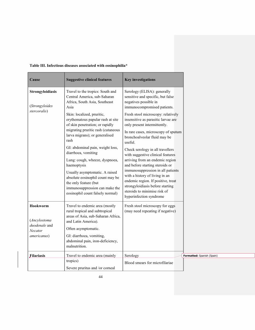
Table III. Infectious diseases associated with eosinophilia*
Cause Suggestive clinical features Key investigations
Strongyloidiasis
(Strongyloides
stercoralis)
Travel to the tropics: South and
Central America, sub-Saharan
Africa, South Asia, Southeast
Asia
Skin: localized, pruritic,
erythematous papular rash at site
of skin penetration; or rapidly
migrating pruritic rash (cutaneous
larva migrans); or generalised
rash
GI: abdominal pain, weight loss,
diarrhoea, vomiting
Lung: cough, wheeze, dyspnoea,
haemoptysis
Usually asymptomatic. A raised
absolute eosinophil count may be
the only feature (but
immunosuppression can make the
eosinophil count falsely normal)
Serology (ELISA): generally
sensitive and specific, but false
negatives possible in
immunocompromised patients.
Fresh stool microscopy: relatively
insensitive as parasitic larvae are
only present intermittently.
In rare cases, microscopy of sputum
bronchoalveolar fluid may be
useful.
Check serology in all travellers
with suggestive clinical features
arriving from an endemic region
and before starting steroids or
immunosuppression in all patients
with a history of living in an
endemic region. If positive, treat
strongyloidiasis before starting
steroids to minimise risk of
hyperinfection syndrome
Hookworm
(Ancylostoma
duodenale and
Necator
americanus)
Travel to endemic area (mostly
rural tropical and subtropical
areas of Asia, sub-Saharan Africa,
and Latin America).
Often asymptomatic.
GI: diarrhoea, vomiting,
abdominal pain, iron-deficiency,
malnutrition.
Fresh stool microscopy for eggs
(may need repeating if negative)
Filariasis Travel to endemic area (mainly
tropics)
Severe pruritus and /or corneal
Serology
Blood smears for microfilariae
Formatted: Spanish (Spain)

45
(Loa loa,
Wuchereria
bancrofti,
Mansonella
perstans, Brugia
malayi, onchocerca
spp.)
precipitates (onchocerca spp.)
Transient subcutaneous swellings
(Loa loa)
Lymphoedema (W. bancrofti or B.
malayi)
Eosinophilia may be only feature
Skin snips and slit lamp
examination (Onchocerca volvulus)
Ascariasis
(Ascaris
lumbricoides)
Travel to endemic area (mainly
tropics), more common in
children
GI: ascending cholangitis,
obstructive jaundice, bile duct
perforation, bowel obstruction (all
rare)
Lung: cough, wheeze, dyspnoea,
haemoptysis
Fresh stool microscopy for eggs
and larvae
Abdominal radiograph showing
dilated bowel; worm aggregates
may be visible as a ‘whirlpool’ shadow.
Ultrasonography may demonstrate
hepatobiliary or pancreatic
ascariasis
Toxocariasis
(Toxocara canis or,
less commonly,
Toxocara cati)
Contact with domestic dogs or
cats.
Usually asymptomatic; may cause
fever and anorexia.
Rarely causes visceral larva
migrans (hepatitis and
pneumonitis), or ocular,
neurological and cardiac
symptoms
Serology (ELISA)
Trichinellosis
(trichinella spp.)
Ingestion of undercooked pork (or
rarely other meats) several days
before onset of symptoms.
May be asymptomatic, or cause
fever, headache, vomiting and
diarrhoea.
Musculoskeletal (due to invasion
of muscle by parasite): myositis,
Serology (ELISA, latex
agglutination) – may be false
negative in first 3-4 weeks of
infection.
Occasionally Western blot on blood
or muscle biopsy may be needed.
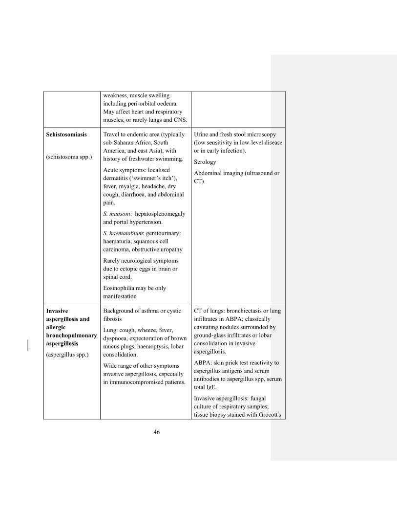
46
weakness, muscle swelling
including peri-orbital oedema.
May affect heart and respiratory
muscles, or rarely lungs and CNS.
Schistosomiasis
(schistosoma spp.)
Travel to endemic area (typically
sub-Saharan Africa, South
America, and east Asia), with
history of freshwater swimming.
Acute symptoms: localised
dermatitis (‘swimmer’s itch’), fever, myalgia, headache, dry
cough, diarrhoea, and abdominal
pain.
S. mansoni: hepatosplenomegaly
and portal hypertension.
S. haematobium: genitourinary:
haematuria, squamous cell
carcinoma, obstructive uropathy
Rarely neurological symptoms
due to ectopic eggs in brain or
spinal cord.
Eosinophilia may be only
manifestation
Urine and fresh stool microscopy
(low sensitivity in low-level disease
or in early infection).
Serology
Abdominal imaging (ultrasound or
CT)
Invasive
aspergillosis and
allergic
bronchopulmonary
aspergillosis
(aspergillus spp.)
Background of asthma or cystic
fibrosis
Lung: cough, wheeze, fever,
dyspnoea, expectoration of brown
mucus plugs, haemoptysis, lobar
consolidation.
Wide range of other symptoms
invasive aspergillosis, especially
in immunocompromised patients.
CT of lungs: bronchiectasis or lung
infiltrates in ABPA; classically
cavitating nodules surrounded by
ground-glass infiltrates or lobar
consolidation in invasive
aspergillosis.
ABPA: skin prick test reactivity to
aspergillus antigens and serum
antibodies to aspergillus spp, serum
total IgE.
Invasive aspergillosis: fungal
culture of respiratory samples;
tissue biopsy stained with Grocott's
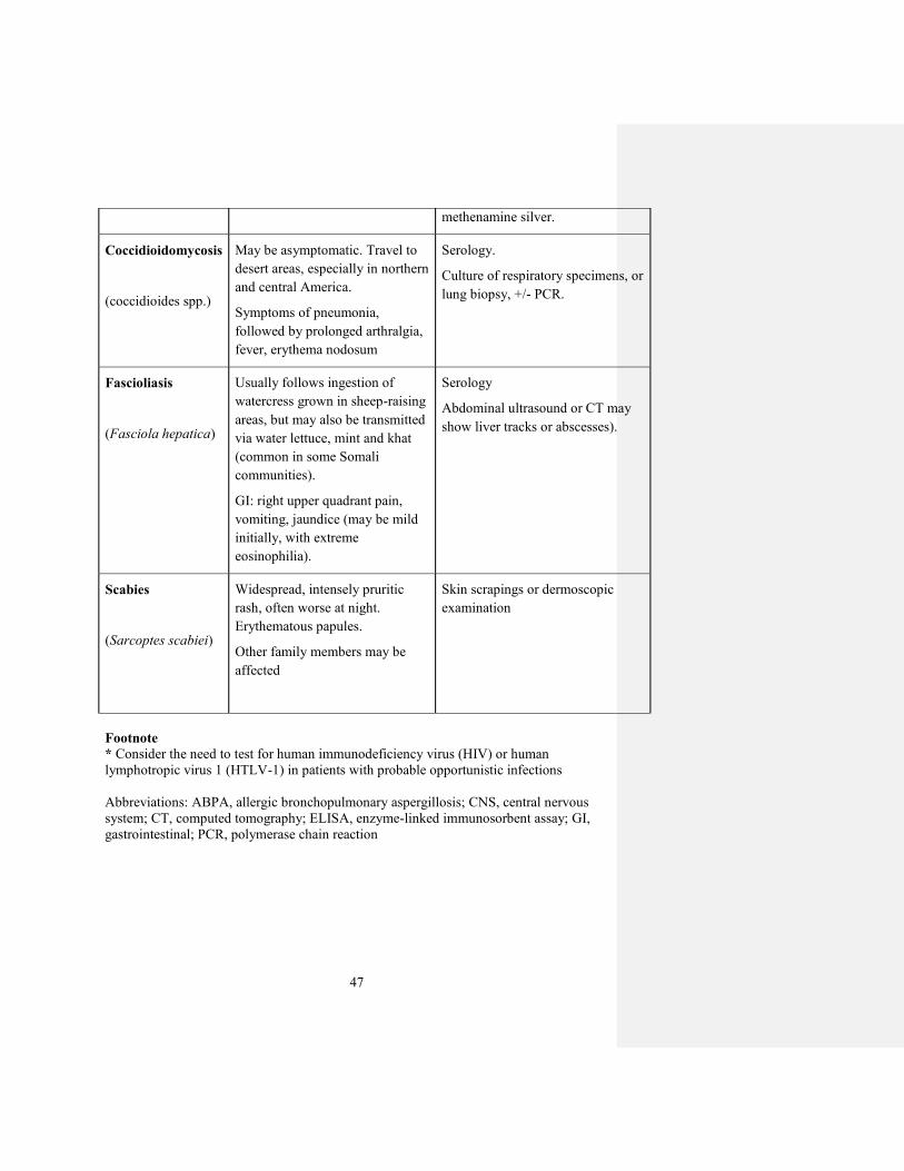
47
methenamine silver.
Coccidioidomycosis
(coccidioides spp.)
May be asymptomatic. Travel to
desert areas, especially in northern
and central America.
Symptoms of pneumonia,
followed by prolonged arthralgia,
fever, erythema nodosum
Serology.
Culture of respiratory specimens, or
lung biopsy, +/- PCR.
Fascioliasis
(Fasciola hepatica)
Usually follows ingestion of
watercress grown in sheep-raising
areas, but may also be transmitted
via water lettuce, mint and khat
(common in some Somali
communities).
GI: right upper quadrant pain,
vomiting, jaundice (may be mild
initially, with extreme
eosinophilia).
Serology
Abdominal ultrasound or CT may
show liver tracks or abscesses).
Scabies
(Sarcoptes scabiei)
Widespread, intensely pruritic
rash, often worse at night.
Erythematous papules.
Other family members may be
affected
Skin scrapings or dermoscopic
examination
Footnote
* Consider the need to test for human immunodeficiency virus (HIV) or human
lymphotropic virus 1 (HTLV-1) in patients with probable opportunistic infections
Abbreviations: ABPA, allergic bronchopulmonary aspergillosis; CNS, central nervous
system; CT, computed tomography; ELISA, enzyme-linked immunosorbent assay; GI,
gastrointestinal; PCR, polymerase chain reaction

48
Table IV. Primary gastrointestinal disorders associated with gastrointestinal tissue
eosinophilia with or without peripheral blood eosinophilia
Disorder General Comments
Primary gastrointestinal eosinophilic
disorders
1) Primary eosinophilic oesophagitis
2) Primary eosinophilic gastritis
3) Primary eosinophilic colitis
A myriad of clinical symptoms may be present
depending on which segment of the gastrointestinal
tract is involved. These may include fatigue,
dysphagia, weight loss, vomiting, gastric dysmotility
or diarrhoea. The peripheral blood eosinophil count
is not always abnormal. Diagnosis involves
comprehensive history and examination, evaluation
of secondary causes of gastrointestinal eosinophilia
and biopsy findings (Prussin 2014; Zuo &
Rothenburg 2007).
Chronic Pancreatitis Can be associated with peripheral blood eosinophilia.
Incidence is higher in autoimmune pancreatitis than
in non-autoimmune causes (Wang et al, 2009)
Inflammatory Bowel Disease Both ulcerative colitis and Crohn disease can be
associated with peripheral blood eosinophilia.
Ulcerative colitis with eosinophilia may be
associated with a more severe clinical phenotype,
including primary sclerosing cholangitis (Barrie et al,
2013; Suttor et al, 2009).
Coeliac Disease Can be associated with eosinophilic oesophagitis
(Thompson et al, 2012)
49
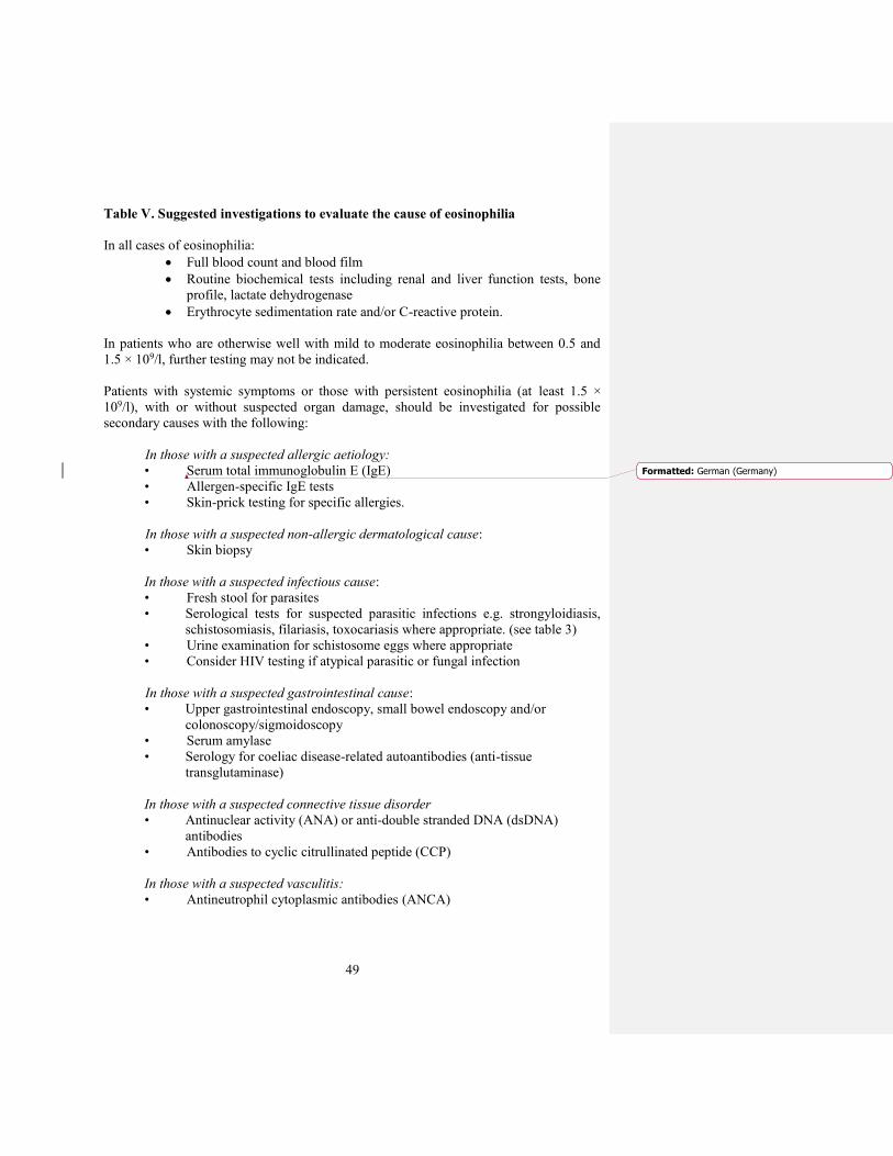
Table V. Suggested investigations to evaluate the cause of eosinophilia
In all cases of eosinophilia: Full blood count and blood film Routine biochemical tests including renal and liver function tests, bone
profile, lactate dehydrogenase Erythrocyte sedimentation rate and/or C-reactive protein.
In patients who are otherwise well with mild to moderate eosinophilia between 0.5 and
1.5 × 10⁹/l, further testing may not be indicated.
Patients with systemic symptoms or those with persistent eosinophilia (at least 1.5 ×
109/l), with or without suspected organ damage, should be investigated for possible
secondary causes with the following:
In those with a suspected allergic aetiology:
• Serum total immunoglobulin E (IgE)
• Allergen-specific IgE tests
• Skin-prick testing for specific allergies.
In those with a suspected non-allergic dermatological cause:
• Skin biopsy
In those with a suspected infectious cause:
• Fresh stool for parasites
• Serological tests for suspected parasitic infections e.g. strongyloidiasis,
schistosomiasis, filariasis, toxocariasis where appropriate. (see table 3)
• Urine examination for schistosome eggs where appropriate
• Consider HIV testing if atypical parasitic or fungal infection
In those with a suspected gastrointestinal cause:
• Upper gastrointestinal endoscopy, small bowel endoscopy and/or
colonoscopy/sigmoidoscopy
• Serum amylase
• Serology for coeliac disease-related autoantibodies (anti-tissue
transglutaminase)
In those with a suspected connective tissue disorder
• Antinuclear activity (ANA) or anti-double stranded DNA (dsDNA)
antibodies
• Antibodies to cyclic citrullinated peptide (CCP)
In those with a suspected vasculitis:
• Antineutrophil cytoplasmic antibodies (ANCA)
Formatted: German (Germany)

50
• Serology for hepatitis B virus (HBV), hepatitis C virus (HCV), human
immunodeficiency virus (HIV), cytomegalovirus (CMV) and parvovirus
B19
In those with suspected respiratory disease:
• Appropriate imaging
• Bronchoscopy with bronchoalveolar lavage/endobronchial
ultrasonography
In those with a suspected lymphoma, non-haematological malignancy or T-cell
driven eosinophilia:
• Appropriate imaging and tissue biopsy
• Peripheral blood T-cell immunophenotyping and T-cell receptor gene
rearrangement studies
Miscellaneous:
• Tests for atheroembolic disease.
• Immunoglobulins and C1 esterase levels*
* If the differential diagnosis includes Gleich’s syndrome
51
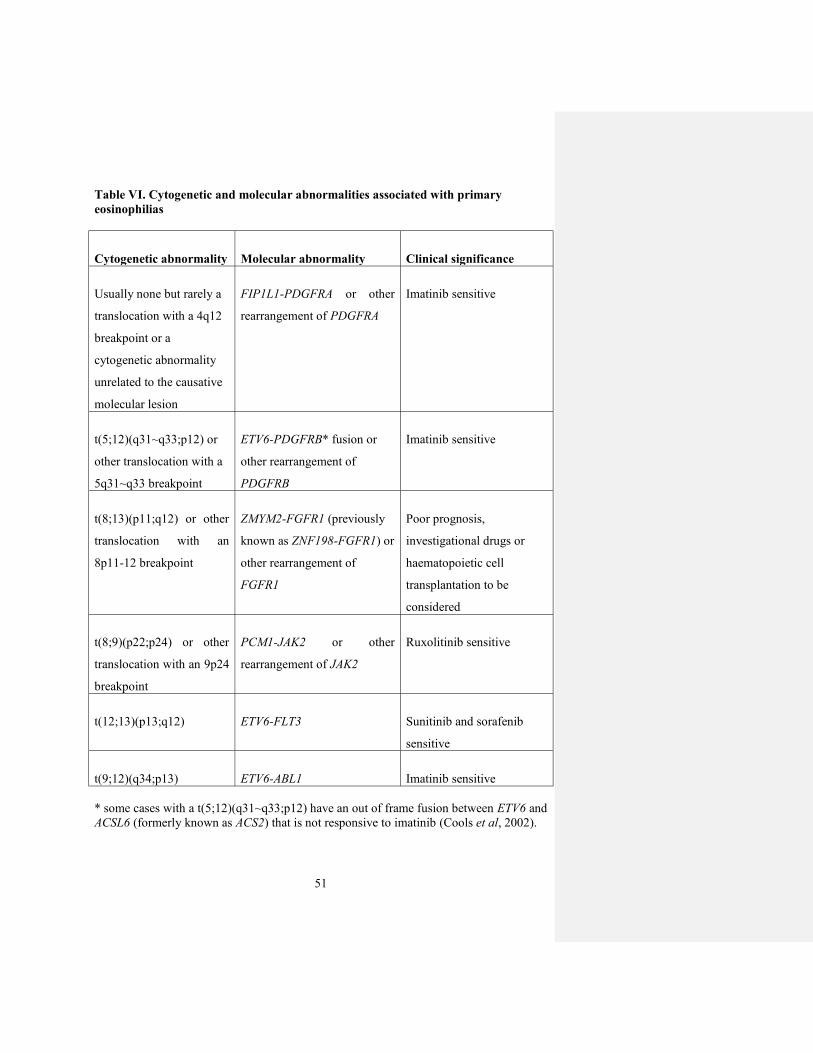
Table VI. Cytogenetic and molecular abnormalities associated with primary
eosinophilias
Cytogenetic abnormality Molecular abnormality Clinical significance
Usually none but rarely a
translocation with a 4q12
breakpoint or a
cytogenetic abnormality
unrelated to the causative
molecular lesion
FIP1L1-PDGFRA or other
rearrangement of PDGFRA
Imatinib sensitive
t(5;12)(q31~q33;p12) or
other translocation with a
5q31~q33 breakpoint
ETV6-PDGFRB* fusion or
other rearrangement of
PDGFRB
Imatinib sensitive
t(8;13)(p11;q12) or other
translocation with an
8p11-12 breakpoint
ZMYM2-FGFR1 (previously
known as ZNF198-FGFR1) or
other rearrangement of
FGFR1
Poor prognosis,
investigational drugs or
haematopoietic cell
transplantation to be
considered
t(8;9)(p22;p24) or other
translocation with an 9p24
breakpoint
PCM1-JAK2 or other
rearrangement of JAK2
Ruxolitinib sensitive
t(12;13)(p13;q12) ETV6-FLT3 Sunitinib and sorafenib
sensitive
t(9;12)(q34;p13) ETV6-ABL1 Imatinib sensitive
* some cases with a t(5;12)(q31~q33;p12) have an out of frame fusion between ETV6 and
ACSL6 (formerly known as ACS2) that is not responsive to imatinib (Cools et al, 2002).

52
Table VII. Assessment of End-Organ Damage
Cardiac assessment: chest radiography, electrocardiogram, echocardiogram, serum
troponin T
Pulmonary assessment: pulmonary function tests including spirometry, O2 saturation
and transfer factor of the lung for carbon monoxide (TLCO)
Footnote:
If there is a strong suspicion of cardiac dysfunction in the absence of obvious
echocardiographic abnormalities, a specialist cardiology review should be sought.

53
Figure 1. Testing algorithm for possible haematological neoplasms with clonal
eosinophilia
Eosinophilia (eosinophil count at least 1.5 ×
109/l) with or without suspected organ damage No obvious underlying cause
Presence of reciprocal
translocations involving 4q12
(PDGFRA), 5q31-33 (PDGFRB),
8p11-12 (FGFR1), 9p24 (JAK2),
13q12 (FLT3) or, potentially, loci
of other tyrosine kinase genes
T cell
immunophenotyping
(+/- TCR
rearrangement
studies)
• Peripheral blood analysis for FIP1L1-PDGFRA by FISH or
nested RT-PCR
• Serum mast cell tryptase
• Bone marrow aspirate, trephine biopsy and cytogenetic
analysis
Myeloid and lymphoid
neoplasms with eosinophilia and
abnormalities of PDGFRA,
PDGFRB, FGFR1 or JAK2
or
Chronic Eosinophilic Leukaemia
(NOS)
Lymphocytic variant
hypereosinophilia
Abnormal
RT-PCR and sequence
confirmation of any fusion
suspected by cytogenetic
analysis
Yes
No Evidence of other
WHO-defined
Myeloid Neoplasm
with associated
Eosinophilia
Yes
MDS, MPN
including SM,
MDS/MPN or
Chronic
Eosinophilic
Leukaemia
(NOS)
No
Normal
Idiopathic
hypereosinophilic
syndrome or
idiopathic
hypereosinophilia
54
REFERENCES
Ackerman, S.J. & Bochner, B.S. (2007) Mechanisms of eosinophilia in the pathogenesis
of hypereosinophilic disorders. Immunol Allergy Clin North Am, 27, 357-375.
Agarwal, R., Chakrabarti, A., Shah, A., Gupta, D., Meis, J.F., Guleria, R., Moss, R.,
Denning, D.W. & group, A.c.a.I.w. (2013) Allergic bronchopulmonary
aspergillosis: review of literature and proposal of new diagnostic and
classification criteria. Clin Exp Allergy, 43, 850-873.
Aggarwal, A., Bergin, P., Jessup, P., Kaye, D. Hypersensitivity myocarditis presenting as
cardiogenic shock. J Heart Lung Transplant, 20, 1241–1244.
Ahmad, I., Labbe, A.C., Chagnon, M., Busque, L., Cohen, S., Kiss, T., Lachance, S., Roy,
D.C., Sauvageau, G. & Roy, J. (2011) Incidence and prognostic value of
eosinophilia in chronic graft-versus-host disease after nonmyeloablative
hematopoietic cell transplantation. Biol Blood Marrow Transplant, 17, 1673-1678.
Akiyama, Y., Shimizu, A., Kimura, A., Onodera, R., Minatani, M., Horikoshi, K., Jingu,
K., Fukakusa, M., Kotajima, F. & Sato, T. (1997) [Successful treatment of
hypereosinophilic syndrome with cyclosporin and steroids]. Nihon Kyobu Shikkan
Gakkai Zasshi, 35, 541-545.
Andersen, C.L., Siersma, V.D., Hasselbalch, H.C., Vestergaard, H., Mesa, R., Felding, P.,
Olivarius, N.D., Bjerrum, O.W. (2015). Association of the blood eosinophil count
with hematological malignancies and mortality. Am J Hematol., 90, 225-9.
Baccarani, M., Cilloni, D., Rondoni, M., Ottaviani, E., Messa, F., Merante, S., Tiribelli,
M., Buccisano, F., Testoni, N., Gottardi, E., de Vivo, A., Giugliano, E., Iacobucci,
I., Paolini, S., Soverini, S., Rosti, G., Rancati, F., Astolfi, C., Pane, F., Saglio, G.
& Martinelli, G. (2007) The efficacy of imatinib mesylate in patients with
FIP1L1-PDGFRalpha-positive hypereosinophilic syndrome. Results of a
multicenter prospective study. Haematologica, 92, 1173-1179.
Bain, B.J. & Ahmad, S. (2014) Should myeloid and lymphoid neoplasms with PCM1-
JAK2 and other rearrangements of JAK2 be recognized as specific entities? Br J
Haematol, 166, 809-817.
Bain BJ, G.D., Vardiman JW and Horny H-P (2008) Chronic eosinophilic leukaemia, not
otherwise specified. In Swerdlow SH, Campo E, Harris NL, Jaffe ES, Pileri SA,
Stein H, Thiele J and Vardiman JW, World Health Organization Classification of
Tumours of Haematopoietic and Lymphoid Tissue, 4th Edn, IARC Press, Lyon. pp
51–53., 51–53.
Bain, B.J.G., D.G.; Vardiman, J. W.; Horny, H.P. (2008) Chronic eosinophilic leukaemia ,
not otherwise specified,. In: World Health Organisation Classification of
Tumours of Haematopoietic and Lymphoid Tissues, pp. 68-73. IARC Press, Lyon.
Barouky, R., Bencharif, L., Badet, F., Salles, G., Vital Durand, D. & Rousset, H. (2003)
Mucosal ulcerations revealing primitive hypereosinophilic syndrome. Eur J
Dermatol, 13, 207-208.
Barraco, D., Carobolante, F., Candoni, A., Simeone, E., Piccaluga, P., Tabanelli, V. &
Fanin, R. (2014) Complete and long-lasting cytologic and molecular remission of
FIP1L1-PDGFRA-positive acute eosinophil myeloid leukaemia, treated with low-
dose imatinib monotherapy. Eur J Haematol, 92, 541-545.
55
Bjerrum, O.W. (2011) Interferon treatment in patients with hypereosinophilia. Curr Drug
Targets, 12, 429-432.
Bjerrum O.W., Pelliniemi, T-T,, Wadenvik, H. (2012) Nordic MPD Study Group -
Guidelines for the diagnosis and treatment of eosinophilia. (homepage:
www.nmpn.org/guidelines)
Brito-Babapulle, F. (2003) The eosinophilias, including the idiopathic hypereosinophilic
syndrome. Br J Haematol, 121, 203-223.
Busch, F.W., Schmidt, H. & Steinke, B. (1991) Alpha-interferon for the
hypereosinophilic syndrome. Ann Intern Med, 114, 338-339.
Butterfield, J.H. (2005) Interferon treatment for hypereosinophilic syndromes and
systemic mastocytosis. Acta Haematol, 114, 26-40.
Butterfield, J.H. & Weiler, C.R. (2012) Use of pegylated interferon in hypereosinophilic
syndrome. Leuk Res, 36, 192-197.
Canonica, G.W., Passalacqua, G., Pronzato, C., Corbetta, L. & Bagnasco, M. (1995)
Effective long-term alpha-interferon treatment for hypereosinophilic syndrome. J
Allergy Clin Immunol, 96, 131-133.
Checkley, A.M., Chiodini, P.L., Dockrell, D.H., Bates, I., Thwaites, G.E., Booth, H.L.,
Brown, M., Wright, S.G., Grant, A.D., Mabey, D.C., Whitty, C.J., Sanderson, F.,
British Infection, S. & Hospital for Tropical, D. (2010) Eosinophilia in returning
travellers and migrants from the tropics: UK recommendations for investigation
and initial management. J Infect, 60, 1-20.
Chmielecki, J., Peifer, M., Viale, A., Hutchinson, K., Giltnane, J., Socci, N.D., Hollis,
C.J., Dean, R.S., Yenamandra, A., Jagasia, M., Kim, A.S., Dave, U.P., Thomas,
R.K. & Pao, W. (2012) Systematic screen for tyrosine kinase rearrangements
identifies a novel C6orf204-PDGFRB fusion in a patient with recurrent T-ALL
and an associated myeloproliferative neoplasm. Genes Chromosomes Cancer, 51,
54-65.
Chiardola, F., Schneeberger, E., Citera, G., Rosemffet, G., Kuo, L., Santillan, G.,
Maldonado-Cocco, J.. (2008) Prevalence and clinical significance of eosinophilia
in patients with rheumatoid arthritis in Argentina. J Clin Rheumatol, 14, 211-13.
Chusid, M.J., Dale, D.C., West, B.C. & Wolff, S.M. (1975) The hypereosinophilic
syndrome: analysis of fourteen cases with review of the literature. Medicine
(Baltimore), 54, 1-27.
Cogan, E., Schandene, L., Papadopoulos, T., Crusiaux, A. & Goldman, M. (1995)
Interleukin-5 production by T lymphocytes in atheroembolic disease with
hypereosinophilia. J Allergy Clin Immunol, 96, 427-429.
Cools J, Mentens N, Odero MD, Peeters P, Wlodarska I, Delforge M, Hagemeijer A,
Marynen P (2002). Evidence for position effects as a variant ETV6-mediated
leukemogenic mechanism in myeloid leukemias with a t(4;12)(q11-q12;p13) or
t(5;12)(q31;p13). Blood, 99, 1776-84.
Cooper, M.A., Akard, L.P., Thompson, J.M., Dugan, M.J. & Jansen, J. (2005)
Hypereosinophilic syndrome: long-term remission following allogeneic stem cell
transplant in spite of transient eosinophilia post-transplant. Am J Hematol, 78, 33-
36.
56
Dong, A., Wang, Y., Gao, L. & Zuo, C. (2014) Increased FDG uptake of the bone
marrow mimicking malignancy in a patient of eosinophilia secondary to
Sparganum mansoni infection. Clin Nucl Med, 39, 640-642.
De Masson, A., Bouaziz, J-D., de Latour, R.P., et al. Severe Aplastic Anemia Associated
With Eosinophilic Fasciitis: Report of 4 Cases and Review of the Literature.
Medicine. 2013;92:69-81
El-Khalawany, M., Al-Mutairi, N., Sultan, M. & Shaaban, D. (2013) Eosinophilic
annular erythema is a peculiar subtype in the spectrum of Wells syndrome: a
multicentre long-term follow-up study. J Eur Acad Dermatol Venereol, 27, 973-
979.
Fathi, A.T., Dec, G.W. Jr, Richter, J.M., Chen, Y.B., Schwartzenberg, S.S., Holmvang,
G., Hasserjian, R.P. (2014). Case records of the Massachusetts General Hospital.
Case 7-2014. A 27-year-old man with diarrhea, fatigue, and eosinophilia. N. Engl.
J. Med., 370, 861-72.
Flaum, M.A., Schooley, R.T., Fauci, A.S. & Gralnick, H.R. (1981) A clinicopathologic
correlation of the idiopathic hypereosinophilic syndrome. I. Hematologic
manifestations. Blood, 58, 1012-1020.
Fozing, T., Zouri, N., Tost,A., Breit, R., Seeck, G., Koch, C., Oezbek, C. (2014)
Management of a patient with eosinophilic myocarditis and normal peripheral
eosinophil count: case report and literature review. Circ Heart Fail, 7, :692-4.
Fruehauf, S., Fiehn, C., Haas, R., Doehner, H. & Hunstein, W. (1993) Sustained
remission of idiopathic hypereosinophilic syndrome following alpha-interferon
therapy. Acta Haematol, 89, 91-93.
Fukuta, A., Hara, T., Tsurumi, H. & Moriwaki, H. (2001) [Hypereosinophilic syndrome
with DIC treated successfully with a combination of high-
dosemethylprednisolone and cyclosporin A]. Rinsho Ketsueki, 42, 1145-1147.
Gleich, G.J., Schroeter, A.L., Marcoux, J.P., Sachs, M.I., O'Connell, E.J., Kohler, P.F.
(1984). Episodic angioedema associated with eosinophilia. N. Engl. J. Med., 310,
1621–6.
Gleich, G.J. (2000) Mechanisms of eosinophil-associated inflammation. J Allergy Clin
Immunol, 105, 651-663.
Gotlib, J. (2014) World Health Organization-defined eosinophilic disorders: 2014 update
on diagnosis, risk stratification, and management. Am J Hematol, 89, 325-337.
Gotlib, J., Cross, N.C. & Gilliland, D.G. (2006) Eosinophilic disorders: molecular
pathogenesis, new classification, and modern therapy. Best Pract Res Clin
Haematol, 19, 535-569.
Grimaldi, J.C. & Meeker, T.C. (1989) The t(5;14) chromosomal translocation in a case of
acute lymphocytic leukemia joins the interleukin-3 gene to the immunoglobulin
heavy chain gene. Blood, 73, 2081-2085.
Guellec, D., Milin, M., Cornec, D., et al. (2015). Eosinophilia predicts poor clinical
outcomes in recent-onset arthritis: results from the ESPOIR cohort. RMD Open, 1,
e000070.
Halaburda, K., Prejzner, W., Szatkowski, D., Limon, J. & Hellmann, A. (2006)
Allogeneic bone marrow transplantation for hypereosinophilic syndrome: long-
term follow-up with eradication of FIP1L1-PDGFRA fusion transcript. Bone
Marrow Transplant, 38, 319-320.
Formatted: French (France)
Deleted:
57
Helbig, G., Hus, M., Francuz, T., Dziaczkowska-Suszek, J., Soja, A. & Kyrcz-Krzemien,
S. (2014) Characteristics and clinical outcome of patients with hypereosinophilia
of undetermined significance. Med Oncol, 31, 815.
Helbig, G., Soja, A., Bartkowska-Chrobok, A. & Kyrcz-Krzemien, S. (2012) Chronic
eosinophilic leukemia-not otherwise specified has a poor prognosis with
unresponsiveness to conventional treatment and high risk of acute transformation.
Am J Hematol, 87, 643-645.
Helbig, G., Wisniewska-Piaty, K., Francuz, T., Dziaczkowska-Suszek, J. & Kyrcz-
Krzemien, S. (2013) Diversity of clinical manifestations and response to
corticosteroids for idiopathic hypereosinophilic syndrome: retrospective study in
33 patients. Leuk Lymphoma, 54, 807-811.
Hernandez-Rodriguez, J., Alba, M.A., Prieto-Gonzalez, S. & Cid, M.C. (2014) Diagnosis
and classification of polyarteritis nodosa. J Autoimmun, 48-49, 84-89.
Horn, B.R., Robin, E.D., Theodore, J. & Van Kessel, A. (1975) Total eosinophil counts
in the management of bronchial asthma. N Engl J Med, 292, 1152-1155.
Hosoki, K., Nagao, M., Iguchi, K., Ihara, T., Yamada, Y., Higashigawa, M., Kephart,
G.M., Kita, H. & Fujisawa, T. (2011) An 8-year-old boy with hypereosinophilic
syndrome. Int Arch Allergy Immunol, 155 Suppl 1, 117-122.
Imahashi, N., Miyamura, K., Seto, A., Watanabe, K., Yanagisawa, M., Nishiwaki, S.,
Shinba, M., Yasuda, T., Kuwatsuka, Y., Terakura, S. & Kodera, Y. (2010)
Eosinophilia predicts better overall survival after acute graft-versus-host-disease.
Bone Marrow Transplant, 45, 371-377.
Jacobsohn, D.A., Schechter, T., Seshadri, R., Thormann, K., Duerst, R. & Kletzel, M.
(2004) Eosinophilia correlates with the presence or development of chronic graft-
versus-host disease in children. Transplantation, 77, 1096-1100.
Juvonen, E., Volin, L., Koponen, A. & Ruutu, T. (2002) Allogeneic blood stem cell
transplantation following non-myeloablative conditioning for hypereosinophilic
syndrome. Bone Marrow Transplant, 29, 457-458.
Kasinath, B.S. & Lewis, E.J. (1987) Eosinophilia as a clue to the diagnosis of
atheroembolic renal disease. Arch Intern Med, 147, 1384-1385.
Kim, S.H., Kwon, J.E. & Kim, H.B. (2013) Successful treatment of steroid-dependent
eosinophilic cellulitis with cyclosporine. Allergy Asthma Immunol Res, 5, 62-64.
Klion, A.D. (2009) How I treat hypereosinophilic syndromes. Blood, 114, 3736-3741.
Klion, A.D. (2011) Eosinophilic myeloproliferative disorders. Hematology Am Soc
Hematol Educ Program, 2011, 257-263.
Klion, A.D., Bochner, B.S., Gleich, G.J., Nutman, T.B., Rothenberg, M.E., Simon, H.U.,
Wechsler, M.E., Weller, P.F. & The Hypereosinophilic Syndromes Working, G.
(2006) Approaches to the treatment of hypereosinophilic syndromes: a workshop
summary report. J Allergy Clin Immunol, 117, 1292-1302.
Klion, A.D., Law, M.A., Noel, P., Kim, Y.J., Haverty, T.P. & Nutman, T.B. (2004)
Safety and efficacy of the monoclonal anti-interleukin-5 antibody SCH55700 in
the treatment of patients with hypereosinophilic syndrome. Blood, 103, 2939-
2941.
Khoury P, Desmond R, Pabon A, Holland-Thomas N, Ware JM, Arthur DC, Kurlander R,
Fay MP, Maric I, Klion AD. (2016). Clinical features predict responsiveness to
imatinib in platelet derived growth factor receptor alpha-negative

58
hypereosinophilic syndrome. Allergy. 2016 Jan 21.[Epub ahead of
print]McNagny, K. & Graf, T. (2002) Making eosinophils through subtle shifts in
transcription factor expression. J Exp Med, 195, F43-47.
Metzgeroth, G., Schwaab, J., Gosenca, D., Fabarius, A., Haferlach, C., Hochhaus, A.,
Cross, N.C., Hofmann, W.K. & Reiter, A. (2013) Long-term follow-up of
treatment with imatinib in eosinophilia-associated myeloid/lymphoid neoplasms
with PDGFR rearrangements in blast phase. Leukemia, 27, 2254-2256.
Mohr, C., Schutte, B., Hildebrand, A., Luger, T.A. & Kolde, G. (1995) Eosinophilic
pustular folliculitis: successful treatment with interferon-alpha. Dermatology, 191,
257-259.
Montgomery, N.D., Dunphy, C.H., Mooberry, M., Laramore, A., Foster, M.C., Park, S.I.
& Fedoriw, Y.D. (2013) Diagnostic complexities of eosinophilia. Arch Pathol
Lab Med, 137, 259-269.
Murphy, P.T., Fennelly, D.F., Stuart, M. & O'Donnell, J.R. (1990) Alfa-interferon in a
case of hypereosinophilic syndrome. Br J Haematol, 75, 619-620.
Nadarajah, S., Krafchik, B., Roifman, C. & Horgan-Bell, C. (1997) Treatment of
hypereosinophilic syndrome in a child using cyclosporine: implication for a
primary T-cell abnormality. Pediatrics, 99, 630-633.
Nand, R., Bryke, C., Kroft, S.H., Divgi, A., Bredeson, C. & Atallah, E. (2009)
Myeloproliferative disorder with eosinophilia and ETV6-ABL gene
rearrangement: efficacy of second-generation tyrosine kinase inhibitors. Leuk Res,
33, 1144-1146.
O'Connell, E.M. & Nutman, T.B. (2015) Eosinophilia in Infectious Diseases. Immunol
Allergy Clin North Am, 35, 493-522.
Ogbogu, P.U., Bochner, B.S., Butterfield, J.H., Gleich, G.J., Huss-Marp, J., Kahn, J.E.,
Leiferman, K.M., Nutman, T.B., Pfab, F., Ring, J., Rothenberg, M.E., Roufosse,
F., Sajous, M.H., Sheikh, J., Simon, D., Simon, H.U., Stein, M.L., Wardlaw, A.,
Weller, P.F. & Klion, A.D. (2009) Hypereosinophilic syndrome: a multicenter,
retrospective analysis of clinical characteristics and response to therapy. J Allergy
Clin Immunol, 124, 1319-1325 e1313.
Ondrejka, S.L., Jegalian, A.G., Kim, A.S., Chabot-Richards, D.S., Giltnane, J.,
Czuchlewski, D.R., Shetty, S., Sekeres, M.A., Yenamandra, A., Head, D., Jagasia,
M. & Hsi, E.D. (2014) PDGFRB-rearranged T-lymphoblastic
leukemia/lymphoma occurring with myeloid neoplasms: the missing link
supporting a stem cell origin. Haematologica, 99, e148-151.
Owen, W.F., Rothenberg, M.E., Petersen, J., Weller, P.F., Silberstein, D., Sheffer, A.L.,
Stevens, R.L., Soberman, R.J. & Austen, K.F. (1989) Interleukin 5 and
phenotypically altered eosinophils in the blood of patients with the idiopathic
hypereosinophilic syndrome. J Exp Med, 170, 343-348.
Perini, G.F., Santos, F.P. & Hamerschlak, N. (2009) Alemtuzumab in the reversal of
encephalopathy associated with hypereosinophilic syndrome. Br J Haematol, 147,
592.
Petri, M., Orbai, A.M., Alarcon, G.S., Gordon, C., Merrill, J.T., Fortin, P.R., Bruce, I.N.,
Isenberg, D., Wallace, D.J., Nived, O., Sturfelt, G., Ramsey-Goldman, R., Bae,
S.C., Hanly, J.G., Sanchez-Guerrero, J., Clarke, A., Aranow, C., Manzi, S.,
Urowitz, M., Gladman, D., Kalunian, K., Costner, M., Werth, V.P., Zoma, A.,
59
Bernatsky, S., Ruiz-Irastorza, G., Khamashta, M.A., Jacobsen, S., Buyon, J.P.,
Maddison, P., Dooley, M.A., van Vollenhoven, R.F., Ginzler, E., Stoll, T.,
Peschken, C., Jorizzo, J.L., Callen, J.P., Lim, S.S., Fessler, B.J., Inanc, M.,
Kamen, D.L., Rahman, A., Steinsson, K., Franks, A.G., Jr., Sigler, L., Hameed, S.,
Fang, H., Pham, N., Brey, R., Weisman, M.H., McGwin, G., Jr. & Magder, L.S.
(2012) Derivation and validation of the Systemic Lupus International
Collaborating Clinics classification criteria for systemic lupus erythematosus.
Arthritis Rheum, 64, 2677-2686.
Pinal-Fernandez, I., Selva-O' Callaghan, A. & Grau, J.M. (2014) Diagnosis and
classification of eosinophilic fasciitis. Autoimmun Rev, 13, 379-382.
Pineton de Chambrun, M., Charron, P., Vauthier-Brouzes, D., Cluzel, P., Haroche, J.,
Kahn, J.E., Amoura, Z., Aubart, F.C. (2015). Reversible Severe Eosinophilic
Endomyocardial Fibrosis During Pregnancy: A Case Report. Medicine
(Baltimore), 94(32), e1307
Pitini, V., Teti, D., Arrigo, C. & Righi, M. (2004) Alemtuzumab therapy for refractory
idiopathic hypereosinophilic syndrome with abnormal T cells: a case report. Br J
Haematol, 127, 477.
Plotz, S.G., Simon, H.U., Darsow, U., Simon, D., Vassina, E., Yousefi, S., Hein, R.,
Smith, T., Behrendt, H. & Ring, J. (2003) Use of an anti-interleukin-5 antibody in
the hypereosinophilic syndrome with eosinophilic dermatitis. N Engl J Med, 349,
2334-2339.
Ramanathan, R. & Nutman, T. (2008) Strongyloides stercoralis infection in the
immunocompromised host. Curr Infect Dis Rep, 10, 105-110.
Rothenberg, M.E., Klion, A.D., Roufosse, F.E., Kahn, J.E., Weller, P.F., Simon, H.U.,
Schwartz, L.B., Rosenwasser, L.J., Ring, J., Griffin, E.F., Haig, A.E., Frewer, P.I.,
Parkin, J.M., Gleich, G.J. & Mepolizumab, H.E.S.S.G. (2008) Treatment of
patients with the hypereosinophilic syndrome with mepolizumab. N Engl J Med,
358, 1215-1228.
Roufosse, F. (2009) Hypereosinophilic syndrome variants: diagnostic and therapeutic
considerations. Haematologica, 94, 1188-1193.
Roufosse, F., de Lavareille, A., Schandene, L., Cogan, E., Georgelas, A., Wagner, L., Xi,
L., Raffeld, M., Goldman, M., Gleich, G.J. & Klion, A. (2010) Mepolizumab as a
corticosteroid-sparing agent in lymphocytic variant hypereosinophilic syndrome.
J Allergy Clin Immunol, 126, 828-835 e823.
Roufosse, F. & Weller, P.F. (2010) Practical approach to the patient with
hypereosinophilia. J Allergy Clin Immunol, 126, 39-44.
Roufosse, F.E., Kahn, J.E., Gleich, G.J., Schwartz, L.B., Singh, A.D., Rosenwasser, L.J.,
Denburg, J.A., Ring, J., Rothenberg, M.E., Sheikh, J., Haig, A.E., Mallett, S.A.,
Templeton, D.N., Ortega, H.G. & Klion, A.D. (2013) Long-term safety of
mepolizumab for the treatment of hypereosinophilic syndromes. J Allergy Clin
Immunol, 131, 461-467 e461-465.
Roujeau, J.C., Haddad, C., Paulmann, M. & Mockenhaupt, M. (2014) Management of
nonimmediate hypersensitivity reactions to drugs. Immunol Allergy Clin North
Am, 34, 473-487, vii.

60
Schooley, R.T., Flaum, M.A., Gralnick, H.R. & Fauci, A.S. (1981) A clinicopathologic
correlation of the idiopathic hypereosinophilic syndrome. II. Clinical
manifestations. Blood, 58, 1021-1026
Schwaab J, Umbach R, Metzgeroth G, Naumann N, Jawhar M, Sotlar K, Horny HP,
Gaiser T, Hofmann WK, Schnittger S, Cross NC, Fabarius A, Reiter A.(2015)
KIT D816V and JAK2 V617F mutations are seen recurrently in hypereosinophilia
of unknown significance. Am J Hematol, 90, 774-7.
Schwaab, J., Knut, M., Haferlach, C., Metzgeroth, G., Horny, H.P., Chase, A., Tapper,
W., Score, J., Waghorn, K., Naumann, N., Jawhar, M., Fabarius, A., Hofmann,
W.K., Cross, N.C. & Reiter, A. (2015) Limited duration of complete remission on
ruxolitinib in myeloid neoplasms with PCM1-JAK2 and BCR-JAK2 fusion genes.
Ann Hematol, 94, 233-238.
Scolari, F., Ravani, P., Gaggi, R., Santostefano, M., Rollino, C., Stabellini, N., Colla, L.,
Viola, B.F., Maiorca, P., Venturelli, C., Bonardelli, S., Faggiano, P. & Barrett, B.J.
(2007) The challenge of diagnosing atheroembolic renal disease: clinical features
and prognostic factors. Circulation, 116, 298-304.
Sefcick, A., Sowter, D., DasGupta, E., Russell, N.H. & Byrne, J.L. (2004) Alemtuzumab
therapy for refractory idiopathic hypereosinophilic syndrome. Br J Haematol, 124,
558-559.
Simon, H.U. & Klion, A. (2012) Therapeutic approaches to patients with
hypereosinophilic syndromes. Semin Hematol, 49, 160-170.
Simon, H.U., Plotz, S.G., Dummer, R. & Blaser, K. (1999) Abnormal clones of T cells
producing interleukin-5 in idiopathic eosinophilia. N Engl J Med, 341, 1112-1120.
Spry, C.J., Tai, P.C. & Davies, J. (1983) The cardiotoxicity of eosinophils. Postgrad Med
J, 59, 147-153.
Strati, P., Cortes, J., Faderl, S., Kantarjian, H. & Verstovsek, S. (2013) Long-term follow-
up of patients with hypereosinophilic syndrome treated with Alemtuzumab, an
anti-CD52 antibody. Clin Lymphoma Myeloma Leuk, 13, 287-291.
Sultan, S.J., Sameem, F. & Ashraf, M. (2015) Drug reaction with eosinophilia and
systemic symptoms: manifestations, treatment, and outcome in 17 patients. Int J
Dermatol, 54, 537-542.
Syed, F.F., Bleeker, J.S., Glockner, J., Pardanani, A. & Cooper, L.T., Jr. (2012) Response
to alemtuzumab in FIP1L1/PDGFRA-negative hypereosinophilic myocarditis on
serial cardiac magnetic resonance imaging. J Am Coll Cardiol, 59, 430.
Tahara, K., Yukawa, S., Shoji, A., Hayashi, H. & Tsuboi, N. (2008) Long-term remission
by cyclosporine in a patient with eosinophilic fasciitis associated with primary
biliary cirrhosis. Clin Rheumatol, 27, 1199-1201.
Trempat, P., Villalva, C., Laurent, G., Armstrong, F., Delsol, G., Dastugue, N. &
Brousset, P. (2003) Chronic myeloproliferative disorders with rearrangement of
the platelet-derived growth factor alpha receptor: a new clinical target for
STI571/Glivec. Oncogene, 22, 5702-5706.
Ueno, N.T., Anagnostopoulos, A., Rondon, G., Champlin, R.E., Mikhailova, N.,
Pankratova, O.S., Zoubarovskaya, L.S., Semenova, E.V., Afanasyev, B.V.,
O'Brien, S., Andreeff, M. & Zaritskey, A.Y. (2002) Successful non-myeloablative
allogeneic transplantation for treatment of idiopathic hypereosinophilic syndrome.
Br J Haematol, 119, 131-134.

61
Valent, P., Klion, A.D., Horny, H.P., Roufosse, F., Gotlib, J., Weller, P.F., Hellmann, A.,
Metzgeroth, G., Leiferman, K.M., Arock, M., Butterfield, J.H., Sperr, W.R.,
Sotlar, K., Vandenberghe, P., Haferlach, T., Simon, H.U., Reiter, A. & Gleich,
G.J. (2012) Contemporary consensus proposal on criteria and classification of
eosinophilic disorders and related syndromes. J Allergy Clin Immunol, 130, 607-
612 e609.
Vaughan Hudson, B., Linch, D.C., Macintyre, E.A., Bennett, M.H., MacLennan, K.A.,
Vaughan Hudson, G. & Jelliffe, A.M. (1987) Selective peripheral blood
eosinophilia associated with survival advantage in Hodgkin's disease (BNLI
Report No 31). British National Lymphoma Investigation. J Clin Pathol, 40, 247-
250.
Walz, C., Erben, P., Ritter, M., Bloor, A., Metzgeroth, G., Telford, N., Haferlach, C.,
Haferlach, T., Gesk, S., Score, J., Hofmann, W.K., Hochhaus, A., Cross, N.C. &
Reiter, A. (2011) Response of ETV6-FLT3-positive myeloid/lymphoid neoplasm
with eosinophilia to inhibitors of FMS-like tyrosine kinase 3. Blood, 118, 2239-
2242.
Watts, R., Lane, S., Hanslik, T., Hauser, T., Hellmich, B., Koldingsnes, W., Mahr, A.,
Segelmark, M., Cohen-Tervaert, J.W. & Scott, D. (2007) Development and
validation of a consensus methodology for the classification of the ANCA-
associated vasculitides and polyarteritis nodosa for epidemiological studies. Ann
Rheum Dis, 66, 222-227.
Weller, P.F. & Bubley, G.J. (1994) The idiopathic hypereosinophilic syndrome. Blood,
83, 2759-2779.
Wells, G.C. (1971) Recurrent granulomatous dermatitis with eosinophilia. Trans St Johns
Hosp Dermatol Soc, 57, 46-56.
Yamada, O., Kitahara, K., Imamura, K., Ozasa, H., Okada, M. & Mizoguchi, H. (1998)
Clinical and cytogenetic remission induced by interferon-alpha in a patient with
chronic eosinophilic leukemia associated with a unique t(3;9;5) translocation. Am
J Hematol, 58, 137-141.
Yamamoto, K., Kawano, H., Nishikawa, S., Yakushijin, K., Okamura, A. & Matsui, T.
(2006) A biphenotypic transformation of 8p11 myeloproliferative syndrome with
CEP1/FGFR1 fusion gene. Eur J Haematol, 77, 349-354.
Yin, G. & Xie, Q. (2012) Systemic lupus erythematosus associated with Wells' syndrome.
Rheumatol Int, 32, 1087-1089.
Zabel, P. & Schlaak, M. (1991) Cyclosporin for hypereosinophilic syndrome. Ann
Hematol, 62, 230-231.
Zielinski, R.M. & Lawrence, W.D. (1990) Interferon-alpha for the hypereosinophilic
syndrome. Ann Intern Med, 113, 716-718.


