Article
Somatic Primary piRNA Bi
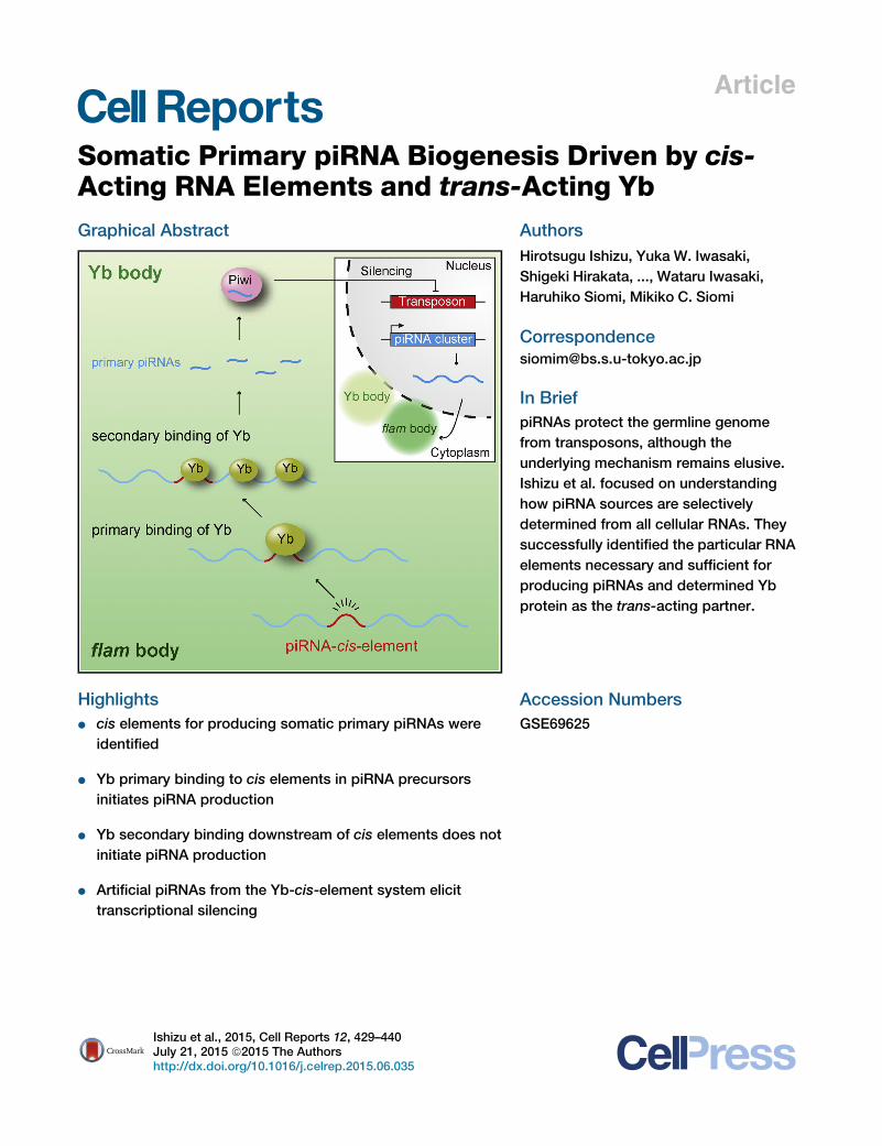
ogenesis Driven by cis-Acting RNA Elements and trans-Acting YbGraphical Abstract
Highlights
d cis elements for producing somatic primary piRNAs were
identified
d Yb primary binding to cis elements in piRNA precursors
initiates piRNA production
d Yb secondary binding downstream of cis elements does not
initiate piRNA production
d Artificial piRNAs from the Yb-cis-element system elicit
transcriptional silencing
Ishizu et al., 2015, Cell Reports 12, 429–440July 21, 2015 ª2015 The Authorshttp://dx.doi.org/10.1016/j.celrep.2015.06.035
Authors
Hirotsugu Ishizu, Yuka W. Iwasaki,
Shigeki Hirakata, ..., Wataru Iwasaki,
Haruhiko Siomi, Mikiko C. Siomi
In Brief
piRNAs protect the germline genome
from transposons, although the
underlying mechanism remains elusive.
Ishizu et al. focused on understanding
how piRNA sources are selectively
determined from all cellular RNAs. They
successfully identified the particular RNA
elements necessary and sufficient for
producing piRNAs and determined Yb
protein as the trans-acting partner.
Accession Numbers
GSE69625
Cell Reports
Article
Somatic Primary piRNA Biogenesis Drivenby cis-Acting RNA Elements and trans-Acting YbHirotsugu Ishizu,1 Yuka W. Iwasaki,2 Shigeki Hirakata,1 Haruka Ozaki,3 Wataru Iwasaki,1,3 Haruhiko Siomi,2
and Mikiko C. Siomi1,*1Department of Biological Sciences, Graduate School of Science, The University of Tokyo, Tokyo 113-0032, Japan2Department of Molecular Biology, Keio University School of Medicine, Tokyo 160-8582, Japan3Department of Computational Biology, Graduate School of Frontier Sciences, The University of Tokyo, Tokyo 113-0032, Japan
*Correspondence: [email protected]://dx.doi.org/10.1016/j.celrep.2015.06.035
This is an open access article under the CC BY-NC-ND license (http://creativecommons.org/licenses/by-nc-nd/4.0/).
SUMMARY
Primary piRNAs in Drosophila ovarian somatic cellsarise from piRNA cluster transcripts and the 30
UTRs of a subset of mRNAs, including Traffic jam(Tj) mRNA. However, it is unclear how these RNAsare determined as primary piRNA sources. Here, weidentify a cis-acting 100-nt fragment in the Tj 30
UTR that is sufficient for producing artificial piRNAsfrom unintegrated DNA. These artificial piRNAswere effective in endogenous gene transcriptionalsilencing. Yb, a core component of primary piRNAbiogenesis center Yb bodies, directly bound the Tj-cis element. Disruption of this interaction markedlyreduced piRNA production. Thus, Yb is the trans-acting partner of the Tj-cis element. Yb-CLIP re-vealed that Yb binding correlated with somaticpiRNA production but Tj-cis element downstreamsequences produced few artificial piRNAs. We thuspropose that Yb determines primary piRNA sourcesthrough two modes of action: primary binding to ciselements to specify substrates and secondary bind-ing to downstream regions to increase diversity inpiRNA populations.
INTRODUCTION
PIWI-interacting RNAs (piRNAs) interact with PIWI proteins
to form piRNA-induced silencing complexes (piRISCs), which
repress target genes, mostly transposons, either transcription-
ally or at the post-transcriptional level by cleaving transcripts in
the cytoplasm (Aravin et al., 2007; Brennecke et al., 2007; Ghil-
diyal and Zamore, 2009; Ishizu et al., 2012; Juliano et al., 2011;
Khurana and Theurkauf, 2010; Siomi et al., 2011). Interestingly,
not all cells in the gonads use both mechanisms. Follicle cells
in Drosophila ovaries use transcriptional silencing but lack
piRISC-mediated post-transcriptional silencing, while germ cells
possess both transcriptional and post-transcriptional piRISC
machineries (Ishizu et al., 2012). In Bombyx ovaries, only post-
transcriptional silencing occurs (Kawaoka et al., 2009). This vari-
ation largely depends on which PIWI proteins are expressed in a
given cell type; transcriptional silencing requires nuclear PIWI
proteins while post-transcriptional silencing requires cyto-
plasmic PIWI proteins (Huang et al., 2013; Malone et al., 2009).
Primary piRNAs are produced from single-stranded long non-
coding RNAs transcribed from piRNA clusters in a Dicer-inde-
pendent manner (Houwing et al., 2007; Vagin et al., 2006). The
Drosophila genome contains 142 piRNA clusters (Brennecke
et al., 2007), whose expression is regulated differently in different
cell types. flamenco (flam), a representative of unidirectional
piRNA clusters, is expressed only in follicle cells, whereas the
bidirectional cluster 42AB is expressed specifically in nurse cells
(Brennecke et al., 2007). The types of transposon fragments in-
serted in individual piRNA clusters also vary; therefore, piRNA
populations differ among cell types. piRNAs in nurse cells are
rather complex because primary piRNAs are amplified through
the amplification loop, yielding secondary piRNAs (Ishizu et al.,
2012). Recent studies showed that secondary piRNAs further
produce phased trailer piRNAs (Han et al., 2015; Mohn et al.,
2015). Follicle cells do not use this amplification system and
thus only contain primary piRNAs.
The biogenesis of somatic primary piRNAs has been studied
using ovaries and an ovarian somatic cell (OSC) line (Olivieri
et al., 2010; Saito et al., 2009). A current model suggests that
upon transcription flam-piRNA precursors are localized to peri-
nuclear Flam bodies (Murota et al., 2014) and processed at adja-
cent Yb bodies (Olivieri et al., 2010; Saito et al., 2010). Yb bodies
contain many piRNA factors besides Yb (Haase et al., 2010;
Olivieri et al., 2012; Qi et al., 2011; Saito et al., 2009; Saito
et al., 2010; Zamparini et al., 2011). Zucchini (Zuc), an endonu-
clease required for processing piRNA intermediates into mature
piRNAs, is localized on the surface of mitochondria (Choi et al.,
2006; Han et al., 2015; Ipsaro et al., 2012; Mohn et al., 2015;
Nishimasu et al., 2012; Olivieri et al., 2012). Yb bodies tend to
be observed in inter-mitochondrial regions (Murota et al.,
2014; Nishimasu et al., 2012; Szakmary et al., 2009). This
arrangement of organelles appears crucial for accelerating
piRNA processing because it centralizes all the necessary fac-
tors in the cytoplasm. Upon maturation, piRNAs associate
with Piwi, a Drosophila PIWI protein, to form piRISCs, which
are then translocated to the nucleus to implement nuclear trans-
poson silencing through chromatin modifications on target
transposon loci with support from co-factors such as GTSF1/
Asterix and Maelstrom (Donertas et al., 2013; Ishizu et al.,
Cell Reports 12, 429–440, July 21, 2015 ª2015 The Authors 429

Figure 1. Artificial piRNA Production from an Inserted Fragment into
the 30 UTR of Tj
(A) Top: schematic drawings of EGFP-tj WT and EGFP-tj MT constructs con-
sisting of EGFP CDS (green) and the Tj-30 UTR (orange). The ‘‘hotspot’’
element (24 nt) in the Tj 30 UTR of EGFP-tj WT was substituted with a 24-nt
arbitrary sequence (shown by a red box) in EGFP-tj MT. Center: schematic
drawing of TjmRNA. Tj-piRNAs (green bars) (Saito et al., 2009) are mapped. Tj
30 UTR (415–490) is shown by a black bar. Bottom: alignments of Tj-piRNAs
derived from Tj 30 UTR (415–490). The hotspot sequence is shown in orange.
The 24-nt arbitrary sequence inserted in EGFP-tj MT is shown in red.
(B) Left: scheme of the experiments for detecting piRNAs derived from EGFP-tj
WT and EGFP-tj MT in OSCs. Upper right: total RNAs and piRNAs in the Piwi
complex immunopurified from OSCs were subjected to northern blotting
analysis. WT and MT probes detect natural Tj-piRNAs originating from the first
hotspot in the Tj 30 UTR and artificial piRNAs produced from the 24-nt random
sequence embedded in EGFP-tj MT, respectively. Artificial MT-piRNAs
associate with Piwi in OSCs expressing EGFP-tj MT. Lower right: the amounts
of Piwi immunoprecipitated from OSCs expressing EGFP-tj WT and EGFP-tj
MT were examined by western blotting using an anti-Piwi antibody.
2012; Muerdter et al., 2013; Ohtani et al., 2013; Olivieri et al.,
2010; Saito et al., 2010)
flam is themajor source of primary piRNAs in OSCs and follicle
cells in the ovaries (Brennecke et al., 2007; Malone et al., 2009;
Saito et al., 2009). flam is largely occupied by transposon rem-
nants, whose orientation predominantly opposes that of the
parental transposons; thus, most primary piRNAs arising from
430 Cell Reports 12, 429–440, July 21, 2015 ª2015 The Authors
the piRNA cluster act as antisense oligos to repress parental
transposons (Brennecke et al., 2007; Malone et al., 2009; Saito
et al., 2009). Some protein-coding genes such as Traffic jam
(Tj) also act as primary piRNA sources (Robine et al., 2009; Saito
et al., 2009), and genic piRNA sources express proteins in OSCs
and follicle cells. The TJ protein, encoded by Tj, is a large Maf
transcriptional factor necessary for controlling gonad morpho-
genesis (Li et al., 2003). Loss of Tj function abolishes Piwi
expression in follicle cells. However, Piwi expression in nurse
cells is not influenced by TJ loss. Thus, the dependence of
Piwi expression on TJ differs between follicle cells and germ cells
(Saito et al., 2009).
Only a limited number of transcripts serve as somatic primary
piRNA precursors. However, the mechanism underlying the
recognition and selection of these transcripts as piRNA precur-
sors is poorly understood. To better understand the mechanism,
we used the Tj 30 UTR as representative of somatic primary
piRNA sources to identify a cis element and its trans-acting part-
ner necessary for producing primary piRNAs in OSCs.
RESULTS
Production of Artificial Primary piRNAs from a RandomSequence Inserted into the Tj 30 UTRFirst, we constructed a plasmid, EGFP-tj WT, consisting of
the EGFP coding sequence (CDS; 717 nt) and the Tj 30 UTR(1,467 nt) (Figure 1A). We previously showed that piRNAs are
not produced uniformly along the Tj 30 UTR; rather, it containstwo hotspots that generate substantial amounts of Tj-piRNAs
(Saito et al., 2009). In the current study, the first hotspot (24 nt)
in EGFP-tj WT was replaced with a 24-nt arbitrary sequence,
yielding a mutant construct, EGFP-tj MT (Figure 1A). The 24-nt
sequence did not match any region in the Drosophila genome
(data not shown).
OSCs were transfected individually with EGFP-tj WT and
EGFP-tj MT constructs. After transfection, northern blotting
was performed with two specific probes, WT and MT, designed
to detect natural Tj-piRNAs originating from the first hotspot in
the Tj 30 UTR and artificial piRNAs produced from the 24-nt
random sequence embedded in EGFP-tj MT, respectively. The
MT probe detected small RNAs of 23–29 nt in length among total
RNAs isolated from OSCs after transfection with EGFP-tj MT
(Figure 1B). Interestingly, the signals were highly enriched in
the Piwi-piRISC fraction immunopurified from OSCs, demon-
strating that the artificial piRNAs produced from the 24-nt
random sequence in EGFP-tj MT were loaded onto Piwi (Fig-
ure 1B). These artificial piRNAs (MT-piRNAs) were undetected
in OSCs transfected with EGFP-tj WT, confirming MT probe
specificity (Figure 1B). The WT probe detected Tj-piRNAs in
both cells (Figure 1B). However, the signal intensity appeared
slightly higher in OSCs transfected with EGFP-tj WT, likely
because of exogenous expression of Tj-piRNAs from the
construct.
Identification of a cis-Acting Element in the Tj 30 UTRTo narrow down the region that acts as a cis element in the
expression of MT-piRNAs from EGFP-tj MT, three deletion mu-
tants, MT-1 to MT-3, were produced (Figure 2A). A control

Figure 2. Identification of a cis-Regulatory Element in the Tj 30 UTR(A) Schematic drawings of EGFP-tj MT and its mutant constructs. MT-4 con-
tains theActin42A 30 UTR (360 nt) (light blue) instead of the Tj 30 UTR. The 24-ntartificial sequence (red) exists also in MT-4.
(B) WT-piRNA and MT-piRNA production was monitored by northern blotting
analysis using WT and MT probes as in Figure 1B.
(C) Schematic drawings ofMT-2 and itsmutant constructs. The 100-nt random
sequence inserted in MT-2-1 and MT-2-2 is shown (purple). A stable stem-
loop structure was inserted into the 50 UTR of MT-2-3 to inhibit EGFP
translation.
(D) WT-piRNA and MT-piRNA production was monitored by northern blotting
analysis using WT and MT probes as in Figure 2B.
(E) Western blotting shows the expression levels of EGFP protein in OSCs after
transfection. b-tubulin was detected as a loading control.
construct, MT-4, in which the Tj 30 UTR was substituted with the
Actin42A 30 UTR, was also produced (Figure 2A). The 24-nt inser-
tion was maintained in the Actin42A 30 UTR of MT-4.
Total RNAs isolated from OSCs and RNAs from the Piwi-
piRISC fraction immunopurified from cells were subjected to
northern blotting. MT-piRNAs were produced to a similar extent
from MT-1 and MT-2 as from EGFP-tj MT (Figure 2B). In sharp
contrast, MT-3 and MT-4 failed to produce MT-piRNAs (Fig-
ure 2B). MT-3 and MT-4 mRNA expression levels were nearly
equal to those of MT-1 and MT-2 (Figure S1A), suggesting that
the 100-nt fragment present in MT-2 but missing from MT-3
serves as the cis element for producing MT-piRNAs from down-
stream regions.
To confirm that the 100-nt fragment acts as a bona fide cis-
acting element for generating MT-piRNAs, the element was
replaced with a 100-nt random sequence, yielding MT-2-1 (Fig-
ure 2C). Another mutant, MT-2-2, was produced by exchanging
the second 100-nt region of the 30 UTR in MT-2 with the random
sequence inserted into MT-2-1 (Figure 2C). Northern blotting re-
vealed that MT-2-2, but not MT-2-1, expressed MT-piRNAs
similarly to MT-2 (Figure 2D).
Tj encodes Tj-piRNAs and TJ protein, both of which are de-
tected in OSCs and ovaries (Brennecke et al., 2007; Li et al.,
2003; Malone et al., 2009; Saito et al., 2009). We next assessed
if MT-2 mRNA translation is required for primary piRNA produc-
tion. A hairpin structure that would presumably interfere with
translation was inserted into the 50 UTR of MT-2, yielding
MT-2-3 (Figure 2C). Western blotting using an anti-EGFP anti-
body confirmed that no EGFP was expressed from MT-2-3 (Fig-
ure 2E). MT-2-3 mRNAs were expressed in OSCs (Figure S1B).
Likewise, MT-piRNAs were expressed from MT-2-3 to a similar
extent as from MT-2 and MT-2-2 (Figure 2D). Thus, translation
of piRNA precursors appears to be dispensable for primary
piRNA production.
Deletion of the Tj-cis Element from the Drosophila
Genome using the CRISPR/Cas9 SystemTo examine whether deletion of the Tj-cis element from the
Drosophila genome would cause defective Tj-piRNA biogen-
esis, we used the CRISPR/Cas9 system (Hsu et al., 2014). We
constructed three plasmids expressing short-guide RNAs
(sgRNAs) that target specific sites in the Tj locus: targets 1–3
(Figure 3A). sgRNAs were expressed in OSCs for targeting
either target 1 + target 2 or target 1 + target 3. We then per-
formed PCR to examine if genomic deletion occurred as
expected. DNA fragments of the expected sizes, 599 nt
(target 1 + target 2) and 530 nt (target 1 + target 3), were de-
tected only when sgRNAs had been expressed (Figure S2A).
Single PCR bands were produced after cloning of each cell
line (Figure 3A). Sequencing of the PCR fragments determined
the purity of the clones and the deletion sites (Figure S2B), con-
firming the generation of two deletion mutant cell lines: OSC-
Dtj-cis 1 and OSC-Dtj-cis 2.
We conducted northern blotting using a WT probe (Figure 1B)
to detect endogenous Tj-piRNAs in both wild-type and mutant
OSC lines. This revealed that the piRNAs arising from the first
Tj 30 UTR hotspot (Figure 1A) in normal OSCs were barely de-
tected in OSC-Dtj-cis 1 and OSC-Dtj-cis 2 cells (Figure 3B).
The production of Idefix-piRNAs arising from other genomic
loci was barely affected by Tj-cis-element deletion (Fig-
ure 3B). Deep sequencing and genome mapping of piRNAs
co-immunoprecipitated with Piwi showed that the numbers of
Tj-piRNAs, particularly from the 200-nt region neighboring the
Tj-cis element, were severely decreased in OSC-Dtj-cis 1 and
Cell Reports 12, 429–440, July 21, 2015 ª2015 The Authors 431
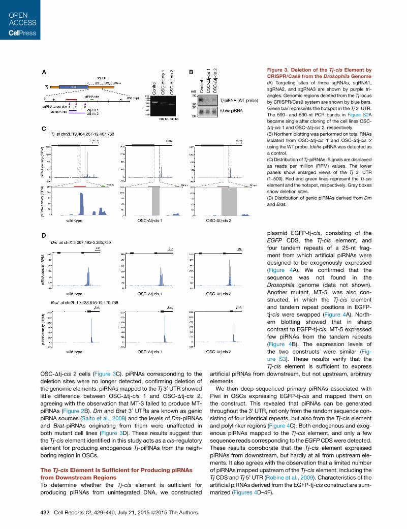
Figure 3. Deletion of the Tj-cis Element by
CRISPR/Cas9 from the Drosophila Genome
(A) Targeting sites of three sgRNAs, sgRNA1,
sgRNA2, and sgRNA3 are shown by purple tri-
angles. Genomic regions deleted from the Tj locus
by CRISPR/Cas9 system are shown by blue bars.
Green bar represents the hotspot in the Tj 30 UTR.The 599- and 530-nt PCR bands in Figure S2A
became single after cloning of the cell lines OSC-
Dtj-cis 1 and OSC-Dtj-cis 2, respectively.
(B) Northern blotting was performed on total RNAs
isolated from OSC-Dtj-cis 1 and OSC-Dtj-cis 2
using theWT probe. Idefix-piRNAwas detected as
a control.
(C) Distribution of Tj-piRNAs. Signals are displayed
as reads per million (RPM) values. The lower
panels show enlarged views of the Tj 30 UTR
(1–500). Red and green lines represent the Tj-cis
element and the hotspot, respectively. Gray boxes
show deletion sites.
(D) Distribution of genic piRNAs derived from Dm
and Brat.
OSC-Dtj-cis 2 cells (Figure 3C). piRNAs corresponding to the
deletion sites were no longer detected, confirming deletion of
the genomic elements. piRNAs mapped to the Tj 30 UTR showed
little difference between OSC-Dtj-cis 1 and OSC-Dtj-cis 2,
agreeing with the observation that MT-3 failed to produce MT-
piRNAs (Figure 2B). Dm and Brat 30 UTRs are known as genic
piRNA sources (Saito et al., 2009) and the levels of Dm-piRNAs
and Brat-piRNAs originating from them were unaffected in
both mutant cell lines (Figure 3D). These results suggest that
the Tj-cis element identified in this study acts as a cis-regulatory
element for producing endogenous Tj-piRNAs from the neigh-
boring region in OSCs.
The Tj-cis Element Is Sufficient for Producing piRNAsfrom Downstream RegionsTo determine whether the Tj-cis element is sufficient for
producing piRNAs from unintegrated DNA, we constructed
432 Cell Reports 12, 429–440, July 21, 2015 ª2015 The Authors
plasmid EGFP-tj-cis, consisting of the
EGFP CDS, the Tj-cis element, and
four tandem repeats of a 25-nt frag-
ment from which artificial piRNAs were
designed to be exogenously expressed
(Figure 4A). We confirmed that the
sequence was not found in the
Drosophila genome (data not shown).
Another mutant, MT-5, was also con-
structed, in which the Tj-cis element
and tandem repeat positions in EGFP-
tj-cis were swapped (Figure 4A). North-
ern blotting showed that in sharp
contrast to EGFP-tj-cis, MT-5 expressed
few piRNAs from the tandem repeats
(Figure 4B). The expression levels of
the two constructs were similar (Fig-
ure S3). These results verify that the
Tj-cis element is sufficient to express
artificial piRNAs from downstream, but not upstream, arbitrary
elements.
We then deep-sequenced primary piRNAs associated with
Piwi in OSCs expressing EGFP-tj-cis and mapped them on
the construct. This revealed that piRNAs can be generated
throughout the 30 UTR, not only from the random sequence con-
sisting of four identical repeats, but also from the Tj-cis element
and polylinker regions (Figure 4C). Both endogenous and exog-
enous piRNAs mapped to the Tj-cis element, and only a few
sequence reads corresponding to theEGFPCDSwere detected.
These results corroborate that the Tj-cis element expressed
piRNAs from downstream, but hardly at all from upstream ele-
ments. It also agrees with the observation that a limited number
of piRNAs mapped upstream of the Tj-cis element, including the
Tj CDS and Tj 50 UTR (Robine et al., 2009). Characteristics of the
artificial piRNAs derived from the EGFP-tj-cis construct are sum-
marized (Figures 4D–4F).

Figure 4. The Tj-cis Element Is Sufficient to
Produce Genic piRNAs from Its Down-
stream Region
(A) Schematic drawings of EGFP-tj-cis and its
mutant constructs. The Tj-cis element (100 nt)
is shown in orange. Blue boxes indicate 25-nt
random sequences, which are repeated four times
in each construct.
(B) Artificial piRNAs derived from the random se-
quences [blue in (A)] were detected by northern
blotting using a 50-nt probe complementary to two
tandem repeats. The WT probe detected endog-
enous Tj-piRNAs.
(C) piRNA sequencing reads mapping to the
EGFP-tj-cis construct. Signals are displayed as
RPM values.
(D) The size distribution of piRNAs derived from the
repeat region in EGFP-tj-cis.
(E) Bar diagrams indicating nucleotide of 50 end in
piRNAs mapped to the repeat region.
(F) piRNA 50 end coverage over a 25-nt random
sequence (minimal unit of the repeat sequence).
Primary piRNA Production Largely Depends onYb Binding to the cis elementWepreviously performedHITS crosslinking immunoprecipitation
(CLIP) experiments in OSCs using an anti-Yb antibody (Murota
et al., 2014). Here, to expand the read numbers, we constructed
two new Yb-CLIP libraries (Figure S4A), which showed a high
correlation for tag sequences (Figure S4B). Bioinformatic anal-
ysis revealed that Yb bound strongly and persistently with
the Tj 30 UTR, but not the Tj CDS or 50 UTR (Figure 5A). The
Tj-cis element resided in one of the strong, if not the strongest,
Yb association sites in the 30 UTR (Figure 5A). Comparison of
Cell Reports 12, 429–
Yb-CLIP mapping data with piRNA map-
ping data obtained in this study (Fig-
ure 3C; wild-type) showed that they
greatly overlap (Figure S4C), suggesting
the direct involvement of Yb in deter-
mining substrates in somatic primary
piRNA biogenesis.
Unlike MT-2, MT-3 failed to produce
artificial MT-piRNAs in OSCs (Figures 2A
and 2B). However, Yb-binding marks
were observed on regions shared with
MT-2 and MT-3 (Figures 2A and 5A). Tj-
R1, a 121-nt sequence adjacent to the
Tj-cis element, was within the shared re-
gions, so we next investigated whether
Tj-R1 could also produce artificial
piRNAs. The Tj-cis element in EGFP-tj-
cis (Figure 4A) was replaced with Tj-R1
to yield construct MT-6 (Figure 5B).
Northern blotting revealed that limited
artificial piRNAs were produced from
MT-6 in sharp contrast to EGFP-Tj-cis
(Figures 5B and S4D). RNA immunopre-
cipitation (RIP) experiments followed by
qRT-PCR showed that MT-6 transcripts
only weakly bound Yb (Figure 5C). This suggested that despite
the close proximity of Tj-R1 to the Tj-cis element and Yb-binding
marks determined by CLIP, it was ineffective in driving piRNA
production. We also constructed MT-7, in which Tj-R1 was re-
placed with Tj-R2, encompassing another 100-nt sequence
following Tj-R1. MT-7 transcripts showed little Yb-binding ca-
pacity and produced only a small amount of artificial piRNAs
(Figures 5B, 5C, and S4D). Yb-RNA binding determined by
CLIP may therefore reflect primary binding to cis elements that
provokes piRNA production, and secondary binding that does
not provoke piRNA production but determines the domains to
440, July 21, 2015 ª2015 The Authors 433
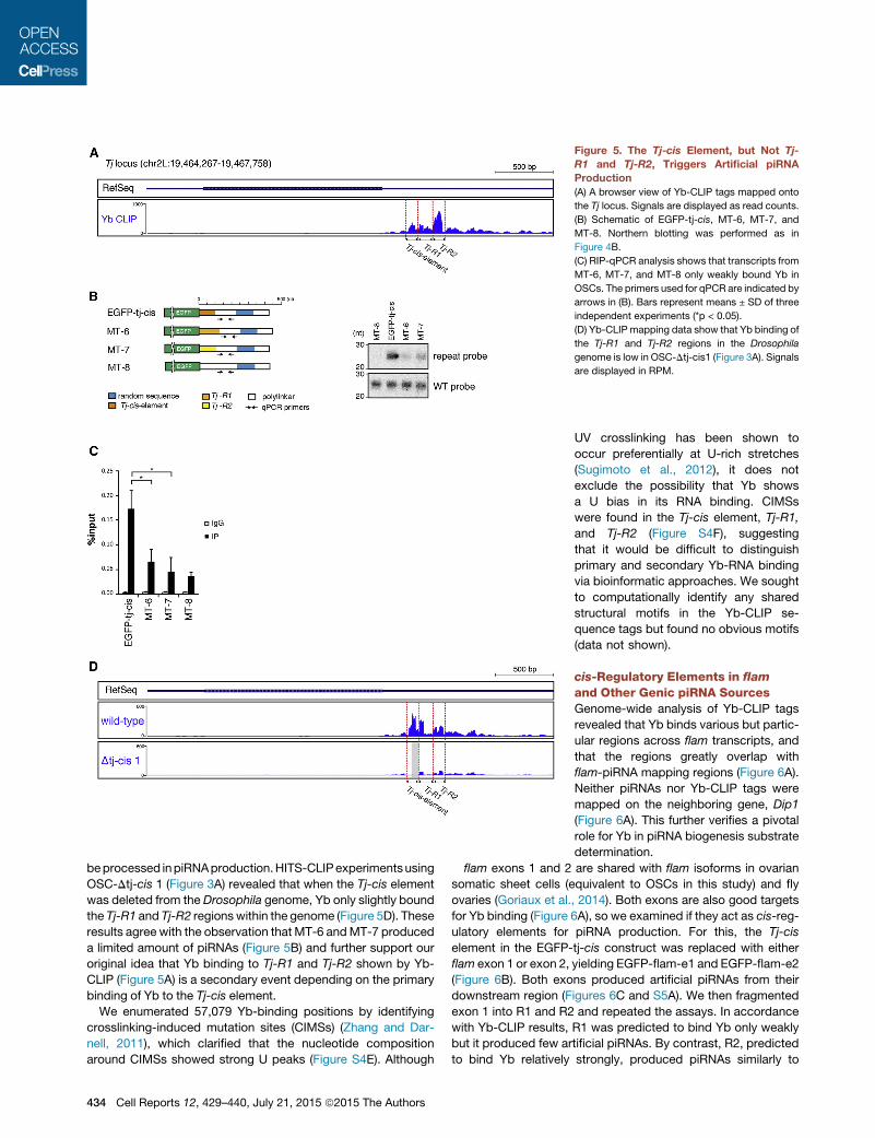
Figure 5. The Tj-cis Element, but Not Tj-
R1 and Tj-R2, Triggers Artificial piRNA
Production
(A) A browser view of Yb-CLIP tags mapped onto
the Tj locus. Signals are displayed as read counts.
(B) Schematic of EGFP-tj-cis, MT-6, MT-7, and
MT-8. Northern blotting was performed as in
Figure 4B.
(C) RIP-qPCR analysis shows that transcripts from
MT-6, MT-7, and MT-8 only weakly bound Yb in
OSCs. The primers used for qPCR are indicated by
arrows in (B). Bars represent means ± SD of three
independent experiments (*p < 0.05).
(D) Yb-CLIP mapping data show that Yb binding of
the Tj-R1 and Tj-R2 regions in the Drosophila
genome is low in OSC-Dtj-cis1 (Figure 3A). Signals
are displayed in RPM.
beprocessed inpiRNAproduction.HITS-CLIPexperimentsusing
OSC-Dtj-cis 1 (Figure 3A) revealed that when the Tj-cis element
was deleted from the Drosophila genome, Yb only slightly bound
the Tj-R1 and Tj-R2 regions within the genome (Figure 5D). These
results agree with the observation that MT-6 andMT-7 produced
a limited amount of piRNAs (Figure 5B) and further support our
original idea that Yb binding to Tj-R1 and Tj-R2 shown by Yb-
CLIP (Figure 5A) is a secondary event depending on the primary
binding of Yb to the Tj-cis element.
We enumerated 57,079 Yb-binding positions by identifying
crosslinking-induced mutation sites (CIMSs) (Zhang and Dar-
nell, 2011), which clarified that the nucleotide composition
around CIMSs showed strong U peaks (Figure S4E). Although
434 Cell Reports 12, 429–440, July 21, 2015 ª2015 The Authors
UV crosslinking has been shown to
occur preferentially at U-rich stretches
(Sugimoto et al., 2012), it does not
exclude the possibility that Yb shows
a U bias in its RNA binding. CIMSs
were found in the Tj-cis element, Tj-R1,
and Tj-R2 (Figure S4F), suggesting
that it would be difficult to distinguish
primary and secondary Yb-RNA binding
via bioinformatic approaches. We sought
to computationally identify any shared
structural motifs in the Yb-CLIP se-
quence tags but found no obvious motifs
(data not shown).
cis-Regulatory Elements in flam
and Other Genic piRNA SourcesGenome-wide analysis of Yb-CLIP tags
revealed that Yb binds various but partic-
ular regions across flam transcripts, and
that the regions greatly overlap with
flam-piRNA mapping regions (Figure 6A).
Neither piRNAs nor Yb-CLIP tags were
mapped on the neighboring gene, Dip1
(Figure 6A). This further verifies a pivotal
role for Yb in piRNA biogenesis substrate
determination.
flam exons 1 and 2 are shared with flam isoforms in ovarian
somatic sheet cells (equivalent to OSCs in this study) and fly
ovaries (Goriaux et al., 2014). Both exons are also good targets
for Yb binding (Figure 6A), so we examined if they act as cis-reg-
ulatory elements for piRNA production. For this, the Tj-cis
element in the EGFP-tj-cis construct was replaced with either
flam exon 1 or exon 2, yielding EGFP-flam-e1 and EGFP-flam-e2
(Figure 6B). Both exons produced artificial piRNAs from their
downstream region (Figures 6C and S5A). We then fragmented
exon 1 into R1 and R2 and repeated the assays. In accordance
with Yb-CLIP results, R1 was predicted to bind Yb only weakly
but it produced few artificial piRNAs. By contrast, R2, predicted
to bind Yb relatively strongly, produced piRNAs similarly to

Figure 6. Analysis of cis Elements in flam
and CG9257 Transcripts
(A) A browser view of Yb-CLIP tags and piRNA
sequences in wild-type OSCs (Figure 3C) mapped
onto the flam locus. The bottom two rows show
zoom-in mapping data of the top row. Signals are
displayed as read counts.
(B) Schematic of EGFP-tj-cis, EGFP-flam-e1, and
EGFP-flam-e2.
(C) Northern blotting was performed as in Fig-
ure 4B.
(D) A browser view of Yb-CLIP tags and piRNA
sequences in wild-type OSCs (Figure 3C) mapped
onto the flam exon 1. Two deletion mutants, R1
and R2, are indicated. Signals are displayed as
read counts. Schematic of EGFP-flam-e1, EGFP-
flam-e1-R1, and EGFP-flam-e1-R2.
(E) Northern blotting was performed as in Fig-
ure 4B.
(F) A browser view of Yb-CLIP tags and piRNA
sequences in wild-type OSCs (Figure 3C) mapped
onto the CG9257 locus. Three fragments, R1, R2,
and R3, used to produce the constructs are indi-
cated. Signals are displayed as read counts.
Schematic of EGFP-CG9257-R1, EGFP-CG9257-
R2, and EGFP-CG9257-R3 constructs.
(G) Northern blotting was performed as in Fig-
ure 4B.
full-length exon 1 (Figures 6D, 6E, and S5B). Yb-RNA binding
therefore plays a crucial role in driving piRNA production.
We further extended our experiments to the genic piRNA
gene CG9257 (Saito et al., 2009). Yb-CLIP showed that Yb
bound almost the entire 30 UTR of the transcripts (Figure 6F),
and regions R1 and R2 in the CG9257 30 UTR acted as cis-
regulatory elements similar to the Tj-cis element (Figures 6F
and 6G). However, a deletion mutant of R2, R3, produced few
piRNAs, despite its moderate binding with Yb. R3 may there-
fore represent a secondary binding site of Yb, as well as Tj-R1
and Tj-R2.
Exogenously Expressed piRNAs Are SufficientlyAbundant to Elicit the Silencing of Endogenous GenesNext, we assessed whether artificial piRNAs expressed under
the control of the Tj-cis element were able to repress endoge-
Cell Reports 12, 429–
nous genes in OSCs. We chose Krimper
(Krimp) as an endogenous gene target
because its knockdown previously had
no effect on piRNA biogenesis or func-
tion (Olivieri et al., 2012). We constructed
four plasmids (Krimp-50, Krimp-CDS-1,
Krimp-CDS-2, and Krimp-30) expressingpiRNAs targeting the 50 UTR, CDS-1
(1–50), CDS-2 (591–640), and the 30
UTR of Krimp mRNA, respectively (Fig-
ures 7A and 7B). All plasmids were
based on the EGFP-tj-cis construct by
inserting three identical copies of Krimp
targeting sequences (50 nt each; except
for the sequence targeting the 50 UTR,
which was 72 nt) in front of the tandem repeats. The insertion
was made in an antisense orientation; thus, piRNAs arising
from the insertions should act as antisense to Krimp mRNAs.
Northern blotting confirmed the expression of Krimp-piRNAs
(Figure 7C).
To determine the efficiency of Krimp-piRNAs in Krimp
silencing, OSCs were transfected with Krimp-50, Krimp-CDS-1,
Krimp-CDS-2, and Krimp-30, together with another plasmid con-
taining a blasticidin-resistance gene. On day 3 after transfection,
blasticidin selection was applied to remove untransfected cells
(Figure S6A). Western blotting with an anti-Krimp antibody (Na-
gao et al., 2011) showed that Krimp was significantly repressed
in OSCs when Krimp-piRNAs targeting the Krimp CDS (1–50)
and 30 UTR were expressed (Figure S6B). Conversely, Krimp-
piRNAs targeting the 50 UTR and CDS (591–640) repressed
Krimp to a lesser extent (Figure S6B).
440, July 21, 2015 ª2015 The Authors 435

Figure 7. Artificial piRNAs Are Driven by the
Tj-cis Element Silence Endogenous Genes
in OSCs
(A) Schematic of constructs for expressing artificial
Krimp-piRNAs. Genomic DNA fragments corre-
sponding to Krimp 50 UTR, CDS-1, CDS-2, and the
30 UTR (see in B) are inserted three times into
EGFP-tj-cis.
(B) Four regions in the Krimp gene were selected
as Krimp-piRNA generation sites: Krimp-50 (1–72),Krimp-CDS-1 (1—50), Krimp-CDS-2 (591–640),
and Krimp-30 (1–50).(C) Production of exogenous Krimp-piRNAs was
monitored by northern blotting. Artificial piRNAs
expressed from the random sequence repeats
(blue) were also detected.
(D) Immunofluorescence using anti-EGFP and
anti-Krimp antibodies was performed on OSCs
expressing exogenous Krimp-piRNAs. Krimp
forms cytoplasmic foci called Krimp body (arrow-
heads), which are not observed in EGFP-positive
OSCs. Scale bars represent 20 mm.
(E) Two regions in the Tj gene were selected as Tj-
piRNA generation sites: Tj-50 and Tj-CDS.
(F) The production of exogenous Tj-piRNAs was
monitored by northern blotting using probes cor-
responding to the particular regions. Artificial
piRNAs expressed from the random sequence
repeats (blue) were also detected.
(G) Immunofluorescence using anti-EGFP and
anti-TJ antibodies was performed on OSCs
expressing exogenous Tj-piRNAs. Scale bars
represent 20 mm.
Considering that blasticidin treatment might not fully eliminate
untransfected cells, leading to an apparent weakness in the
silencing effect, we performed immunofluorescence using an
anti-Krimp antibody. This determined the silencing effect only
in cells in which Krimp-piRNAs had been generated, because
cells considered EGFP-positive showed high EGFP mRNA
expression, suggesting a high level of Krimp-piRNA expression.
As expected, Krimp bodies (Nagao et al., 2011; Olivieri et al.,
2012), cytoplasmic granules in which Krimp strongly accumu-
lates, disappeared in EGFP-positive cells regardless of the
constructs used for transfection (Figures 7D and S6C). This indi-
cated that Krimp silencing occurred regardless of where Krimp-
piRNAs targeted Krimp mRNA.
Krimp-piRNAs exogenously expressed from the constructs
were loaded onto Piwi to form piRISCs (Figure 7C). To examine
if Krimp silencing occurred Piwi-dependently, the efficiency of
Krimp silencing was assessed in Piwi-depleted OSCs. When
Piwi was present in OSCs, Krimp expression was strongly down-
regulated by Krimp-piRNAs (Figure S6D). However, when Piwi
was depleted, Krimp expression was restored (Figure S6D).
436 Cell Reports 12, 429–440, July 21, 2015 ª2015 The Authors
Thus, Krimp silencing mediated by
exogenous piRNAs appears to be Piwi
dependent.
To assess whether Krimp silencing
mediated by exogenous Krimp-piRNAs
is transcriptional, we performed chro-
matin-immunoprecipitation (ChIP). The association of RNA
pol II with Krimp was significantly reduced by Krimp-piRNAs
targeting the Krimp CDS (1–50) and 30 UTR (Figure S6E). Under
these conditions, the accumulation of H3K9me3 at Krimp
increased (Figure S6E). The expression of Krimp-piRNAs in
the sense orientation only slightly affected RNA pol lI and
H3K9me3 binding to Krimp (Figure S6F). These results suggest
that exogenously expressed piRNAs trigger transcriptional
silencing in OSCs.
We also set out to determine the silencing effect of artificial
piRNAs against Tj. Primary piRNAs targeting Tj mRNAs (Fig-
ure 7E) were expressed in OSCs. Two plasmids were produced
based on the EGFP-tj-cis construct, as in Figure 7A, by inserting
three identical Tj targeting sequences (Tj-50 and Tj-CDS; 50 nt
each) in front of the tandem repeats. Northern blotting confirmed
the expression of corresponding piRNAs (Figure 7F). Immunoflu-
orescence using anti-Tj antibodies showed that EGFP-positive
cells expressing artificial Tj-50-piRNAs and Tj-CDS-piRNAs
lacked TJ signals in the nucleus (Figure 7G) (note that these arti-
ficial piRNAs were ‘‘antisense’’ to Tj mRNA while endogenous

Tj-piRNAs are ‘‘sense’’ to Tj mRNA). Thus, artificial piRNAs
produced under control of the Tj-cis element appear capable
of silencing endogenous genes.
DISCUSSION
Yb bodies and Flam bodies in OSCs are considered to be the
centers for primary piRNA maturation/piRISC formation and
piRNA intermediate storage, respectively, and exist in close
proximity (Murota et al., 2014; Olivieri et al., 2010; Saito et al.,
2010). The formation of both bodies depends on the Yb protein,
particularly its RNA-binding activity (Murota et al., 2014). In the
absence of this, piRNA processing fails, resulting in piRNA
loss, although piRNA intermediates and processing factors are
present in the cytosol. Thus, Yb binding to piRNA sources cen-
tralizes all necessary ingredients for piRNA biogenesis, which
is crucial for primary piRNA production (Murota et al., 2014). In
this study, we continued the study on Yb and discovered that
the direct association of Yb with a specific �100-nt element
(i.e., cis element) within the piRNA precursors provokes somatic
primary piRNA biogenesis fromdownstream regions. Insertion of
the Yb-binding element within RNA molecules that do not other-
wise serve as piRNA precursors converts the RNA transcripts
into piRNA sources. Artificial primary piRNAs were mapped
only downstream, but not upstream, of regions of the Yb-binding
element. Previous studies demonstrated that natural genic
piRNAs mostly arise from 30 UTRs rather than mRNA CDS or 50
UTRs (Robine et al., 2009; Saito et al., 2009). The present study
also showed that few Tj-piRNAs mapped to the Tj CDS (Fig-
ure 3C), and that few Yb-CLIP tags were also found in the Tj
CDS (Figure 5A). Thus, Yb determines not only substrate speci-
ficity but also processing directionality in the somatic primary
piRNA biogenesis pathway. This may occur through the Yb-
controlled recruitment of other piRNA factors, such as another
putative RNA helicase Armi and endonuclease Zuc, only to
downstream sequences. We are currently investigating this
possibility.
Yb-CLIP tags greatly overlap with primary piRNA-producing
loci in the genome. This strongly supports the idea that Yb
is the central player in determining substrates in the piRNA
pathway. An unexpected but intriguing observation in our study
is that Tj-R1 and Tj-R2 in the Tj 30 UTR show strong Yb-binding
marks, as does the Tj-cis element, but provoked very little artifi-
cial piRNA production in contrast to the Tj-cis element (Fig-
ure 5B). Yb-CLIP experiments showed that Yb binding to Tj-R1
and Tj-R2 within the Drosophila genome largely depends on
Yb binding to its upstream Tj-cis element (Figure 5D). We there-
fore propose a model in which Yb determines primary piRNA
sources by two sequential modes of action: primary binding to
cis elements that represents selection of piRNA precursors
among cellular RNAs, then secondary binding to downstream re-
gions, representing the defining domains to be processed by
precursors (Figure S7). This complexity in determining piRNA
precursors could ensure the high diversity in piRNA populations,
which is a unique feature of piRNAs (Aravin et al., 2007; Bren-
necke et al., 2007; Lau et al., 2009).
The RNA-binding activity of Yb is required for primary piRNA
production in OSC. Yb mutants carrying a point mutation within
the DEAD box showed little RNA binding activity (Murota et al.,
2014). When these Yb mutants were expressed individually in
OSC lacking endogenous Yb, piRNA precursors were not accu-
mulated in Flam bodies, and few piRNAs were produced. As a
consequence, transposons were de-silenced. Therefore, there
is little doubt that the RNA-binding activity of Yb through the
DEAD-box is indispensable for primary piRNA production.
HITS-CLIP experiments clarified direct interaction of Yb with
piRNA sources, including Tj mRNA. Insertion of a particular
Yb-bound RNA element within Tj mRNA, i.e., the Tj-cis element,
upstream of any given RNA molecule enables the arbitrary
sequences to produce artificial piRNAs. Deletion of the Tj-cis
element from the Drosophila genome significantly abolished
piRNA production from its downstream region spanning at least
�200 nt. These observations strongly support our model (Fig-
ure S7), in which Yb is the trans-acting factor that recognizes
and binds cis elements within piRNA precursors to provoke
primary piRNA biogenesis in ovarian somatic cells. However, it
does not exclude the possibility that Yb collaboratively achieves
this taskwith unknown factors.Moreover, we are not certain if Yb
is the uppermost factor in the cytoplasmic phase of the biogen-
esis pathway upon nuclear transport of piRNA precursors. We
are currently engaged in addressing these challenging questions
in the laboratory.
EXPERIMENTAL PROCEDURES
Cell Culture and Transfection
OSCs were cultured as described previously (Saito et al., 2009; Niki et al.,
2006). OSC transfection was carried out using Xfect Transfection Reagent
(Clontech) as described previously (Murota et al., 2014). Blasticidin (Life Tech-
nologies) was added to the media at 50 mg/ml upon co-transfection using
a plasmid carrying a blasticidin-resistant gene. RNAi was performed as
described previously (Murota et al., 2014).
Plasmid Construction
To generate the EGFP-tj WT construct, the full-length Tj 30 UTR was first PCR-
amplified with the primers tj-30UTR F/R (Table S1, all oligonucleotides were
purchased from Invitrogen) from OSC cDNA samples using KOD plus DNA po-
lymerase (Toyobo) and then cloned between XhoI and BamHI sites of pEGFP-
C1 (Clontech). The polylinker region between EGFP and Tj 30 UTRwas removed
by inverse PCR using the primers delta-mcs F/R. The EGFP-tj 30UTR insert was
subcloned into the NheI and BamHI sites of pAcM (Saito et al., 2009). The Cas9
plasmid, phsp70-Cas9, and the sgRNA plasmid, pU6-BbsI-chiRNA, were pur-
chased fromAddgene. Target sequenceswere synthesized asoligomers and in-
serted into the BbsI site of pU6-BbsI-chiRNA. Detailedmethods for constructing
other plasmids are described in the Supplemental Information.
Derivation of the Tj-cis Element Deletion Mutant OSCs
OSCswere transfectedwith 4mgofCas9plasmid, 4mgof sgRNAplasmids, and
400 ng of the blasticidin resistance gene plasmid as described above. Cells
were incubated for 48 hr post-transfection prior to blasticidin selection. Three
days after addition of blasticidin, 5.03 103 cells were passaged in 6-cm dishes
and allowed to grow in blasticidin containingmedium. After 6–7 days of culture,
colonies were picked and passaged in single wells of 24-well plates and
allowed to grow to confluence in blasticidin free medium. Genomic DNA was
extracted using QuickExtract DNA Extraction Solution (Epicenter) following
the manufacturer’s protocol. The genomic region flanking the CRISPR target
site was PCR amplified and analyzed by electrophoresis on 1% agarose gels.
Immunoprecipitation
Immunoprecipitation from OSCs was performed as previously described
(Saito et al., 2009). Anti-Piwi antibody (Saito et al., 2006) was immobilized on
Cell Reports 12, 429–440, July 21, 2015 ª2015 The Authors 437

Dynabeads protein G (Invitrogen). Total RNAs were isolated from the immuno-
precipitates with phenol-chloroform and precipitated with ethanol.
Northern Blotting
Total RNA was isolated from OSCs using RNAzol RT (Molecular Research
Center). Any contaminating DNA was digested by TURBO DNase (Ambion).
For small RNA detection, northern blotting analysis was carried out using
5 mg of total RNAs as described previously (Saito et al., 2006). The DNA oligo-
nucleotides used are summarized in Table S1. mRNAs were purified from
20 mg of total RNA using Oligotex-dT30 (Takara) according to the manu-
facturer’s instructions, and northern blotting analysis was carried out as
described previously (Murota et al., 2014). DNA probes were synthesized
using a Random Primer DNA Labeling Kit Ver. 2 (Takara) in the presence of32P-dCTP. Templates of random-primed probes were generated by PCR.
For the EGFP probe, the EGFP ORF was amplified using the primers EGFP
F/R with EGFP-C1 vector as template. For the GAPDH probe, the GAPDH
ORFwas amplified using the primers GAPDHF/Rwith OSC cDNA as template.
Western Blotting
Western blotting was performed as described previously (Saito et al., 2006).
Anti-Piwi (Saito et al., 2006) (1:1,000 dilution), anti-EGFP (MBL) (1:1,000 dilu-
tion), anti-Yb (Murota et al., 2014) (1:1,000 dilution), and anti-tubulin (DSHB)
(1:2,000 dilution) antibodies were used.
Small RNA Cloning and Sequencing
For piRNA cloning, coimmunoprecipitated piRNA was extracted from Piwi im-
munoprecipitates using phenol and chloroform, and gel-purified piRNAs were
cloned using the NEBNext Small RNA Library Sample Prep Set (NEB). piRNA
libraries were analyzed on a HiSeq2000 (Illumina).
Small RNA Sequencing Data Analysis
Sequencing of small RNA libraries generated from Piwi immunoprecipitates
were performed usingHiSeq2000. Adaptor sequenceswere removed fromob-
tained reads andmapped to the Release 5 assembly of theDrosophila genome
using Bowtie (Langmead et al., 2009), allowing zero mismatch and extracting
the readsmapped uniquely to the genome. Reads originating from EGFP-tj-cis
construct were mapped against construct sequence using Bowtie, allowing
zero mismatch and default parameters. The reads were normalized to RPM
(read per million) by the number of Drosophila genome-mapped reads.
HITS-CLIP
CLIP was performed basically as described previously (Jaskiewicz et al.,
2012). OSCs (1–2 3 108) were UV crosslinked by irradiating uncovered
with 200 mJ/cm2 of 254 nm UV, followed by lysis with lysis buffer (20 mM
HEPES-KOH [pH 7.3], 1 mM EDTA, 1 mM DTT, 150 mM sodium chloride,
2 mg/ml pepstatin, 2 mg/ml leupeptin, 0.5% aprotinin, 0.5% NP40), and cell
debris was removed by centrifugation. Immunoprecipitation was performed
using 20 mg of anti-Yb antibody (Murota et al., 2014), followed by the treatment
of 10 U/ml RNaseT1 (Roche). Dephosphorylation of RNA segments crosslinked
to immunoprecipitated proteins was performed by calf intestinal alkaline
phosphase (CIP form NEB), and the samples were 50 end labeled, followed
by NuPAGE (Life Technologies) and transfer to the membrane. Protein-bound
RNA signal was cut out from the membrane, and RNA was isolated by phenol/
chloroform extraction. For library construction, preadenylated 30 adaptor was
ligated to the RNA samples using T4 RNA Ligase 2, truncated KQ (NEB), fol-
lowed by 50 adaptor ligation using T4 RNA Ligase 1 (NEB). Adaptor-ligated
RNA samples were reversed transcribed using SuperScript III (Life Technolo-
gies) and PCR amplified using Q5 High-Fidelity DNA Polymerase (NEB).
Bioinformatic Analysis
CLIP tags were mapped to Release 5 assembly of the Drosophila genome as
previously described (Murota et al., 2014). CIMSs were detected according to
Zhang and Darnell (2011). For each genomic position where at least one dele-
tion was observed on themapped CLIP tags, the number of mapped CLIP tags
(k) and that of tags with deletions (m) were counted. The null distribution of m
for given k was obtained by permuting positions of the deletions. The permu-
tation was conducted by preserving the distribution of the CLIP tags on the
438 Cell Reports 12, 429–440, July 21, 2015 ª2015 The Authors
genome and the positional distribution of deletions relative to the 50 ends of
the CLIP tags. The false discovery rate (FDR) was estimated using the cumu-
lative distributions of m for a given k, and the positions with FDR <0.01 were
detected as CIMSs.
Assessing Reproducibility of HITS-CLIP Data
Given two biological replicate data sets of HITS-CLIP, their reproducibility was
assessed as follows. For each CIMS, in one data set (query) and in the other
(target), we collected reads that were mapped to the CIMS position and to
the same strand using BEDTools (version 2.18.1) and an in-house Perl script.
The boundaries of each read cluster were defined as the left-most and right-
most positions of the collected reads. For each cluster in query, the cluster
in target that overlapped with it by at least 1 bp and whose CIMS position
was closest to the query CIMS position was identified. Then, Pearson and
Spearman’s rank correlation coefficients between their read numbers were
calculated. If multiple clusters in target satisfied the above criterion (i.e., with
the same CIMS distances), the averaged read number was used.
RIP and qPCR
OSCs (4–5 3 107) were homogenized in lysis buffer (20 mM HEPES-KOH
[pH 7.3], 150 mM sodium chloride, 1 mM DTT, 1 mM EDTA, 2 mg/ml pepstatin,
2 mg/ml leupeptin, 0.5% aprotinin, 0.5% NP40, and 40 U/ml RNasin [Prom-
ega]), and cell debris was removed by centrifugation. One-tenth of the lysate
was saved as RNA input, and total RNAs were isolated using ISOGEN-LS
reagent (Nippongene). Anti-Yb antibody (Murota et al., 2014) (2 mg) was immo-
bilized on Dynabeads protein G. Lysates were incubated with beads for 2 hr at
4�C before washing three times with IP wash buffer (20 mM HEPES-KOH
[pH 7.3], 300 mM sodium chloride, 1 mM DTT, 2 mg/ml pepstatin, 2 mg/ml leu-
peptin, 0.5% aprotinin, 0.05% NP40) and three times with high-salt wash
buffer (20 mM HEPES-KOH [pH 7.3], 500 mM sodium chloride, 1 mM DTT,
2 mg/ml pepstatin, 2 mg/ml leupeptin, 0.5% aprotinin, and 0.05% NP40). Total
RNAs were isolated from the immunoprecipitates with phenol-chloroform and
were precipitated with ethanol. For qPCR analysis following RNA immunopre-
cipitation, a fixed volume of RNA isolated from input and immunoprecipitates
was used for reverse transcription. Reverse transcription was performed using
ReverTra Ace qPCR RT Master Mix (Toyobo). qPCRs were performed using
the specific qPCR primers mcs F/R (Table S1) in the StepOnePlus Real-
Time PCR System (Life Technologies). THUNDERBIRD SYBR qPCR Mix
(Toyobo) was used as described in the instruction manual. The specific enrich-
ment was analyzed based on a percent input calculation.
Immunofluorescence
Immunofluorescence of OSCs was performed using anti-EGFP IgG2b (MBL)
(1:500 dilution), anti-Krimp IgG1 (Nagao et al., 2011) (1:250 dilution), and
anti-TJ IgG1 (1:250 dilution) antibodies as described previously (Murota
et al., 2014). Alexa-Fluor-488-conjugated anti-mouse IgG2b (Molecular
Probes) and Alexa-Fluor-594-conjugated anti-mouse IgG1 (Molecular Probes)
antibodies were used as secondary antibodies (1:1,000 dilution). Anti-TJ
monoclonal antibody was produced by immunizing mice with GST-TJ protein
as previously described (Saito et al., 2009).
ChIP and qPCR
OSCs (1–2 3 108) were crosslinked with 1% formaldehyde for 5 min at room
temperature, and crosslinking was then quenched with 125 mM glycine for
5 min at room temperature. Fixed cells were washed twice with PBS, har-
vested by scraping, lysed with swelling buffer (25 mM HEPES-KOH [pH 7.3],
1.5 mM magnesium chloride, 10 mM potassium chloride, 0.1% NP40, 1 mM
DTT, 1 3 Halt Protease Inhibitor Cocktail [Thermo Scientific]), and pelleted.
Nuclear pellets were extracted by sonication buffer (50 mM HEPES-KOH
[pH 7.3], 140mMsodium chloride, 1mMEDTA, 1%Triton X-100, 0.1% sodium
deoxycholate, 0.1% SDS, 13 Halt Protease Inhibitor Cocktail), sonicated in a
Covaris S220 focused ultrasonicator, and diluted. After removing input, the
DNA-protein complexes were incubated with 2 mg of nonimmune IgG anti-
body, anti-Pol II antibody 4H8 (CST), or anti-H3K9me3 antibody (Active motif)
at 4�C overnight. DNA-protein complexes were precipitated using Dynabeads
Protein G for 1 hr. The beads were consequently washed with low-salt wash
buffer (20 mM Tris-HCl [pH 8.0], 150 mM sodium chloride, 2 mM EDTA,
0.1% SDS, 0.1% Triton X-100, 1 mM PMSF), high-salt wash buffer (20 mM
Tris-HCl [pH 8.0], 500 mM sodium chloride, 2 mM EDTA, 0.1% SDS, 0.1%
Triton X-100, 1 mM PMSF), LiCl wash buffer (10 mM Tris-HCl [pH 8.0], 1 mM
EDTA, 250 mM lithium chloride, 1% NP40, 1% sodium deoxycholate,
1 mM PMSF), and twice with TE (10 mM Tris-HCl [pH 8.0], 1 mM EDTA,
1 mM PMSF). After washing, immunoprecipitated chromatin was eluted by
elution buffer (50 mM Tris-HCl [pH 8.0], 10 mM EDTA, 1% SDS). Eluted
chromatin and input samples were reverse crosslinked for 12–16 hr at 65�C.ChIP DNA was treated with RNase A for 30 min at 37�C, and then treated
with proteinase K for 60 min at 55�C. DNA was purified by extraction with
phenol/chloroform/isoamyl alcohol and ethanol precipitation. Pellet Paint NF
Co-Precipitant (Merck Millipore) was added to the DNA prior to precipitation.
qPCRs were performed using ChIP DNA with specific qPCR primers Krimper
F/R (Table S1) in StepOnePlus Real-Time PCR Systems. THUNDERBIRD
SYBR qPCRMix was used as described in the instructionmanual. The specific
enrichment was analyzed by percentage input calculation.
ACCESSION NUMBERS
Deep sequencing data sets have been deposited in the NCBI GEO and are
available under accession number GEO: GSE69625.
SUPPLEMENTAL INFORMATION
Supplemental Information includes Supplemental Experimental Procedures,
seven figures, and one table and can be found with this article online at
http://dx.doi.org/10.1016/j.celrep.2015.06.035.
ACKNOWLEDGMENTS
We thank the other members of the M.C.S. laboratories for their discussions
and comments on the manuscript. We also thank T. Fukunaga for bioinformat-
ics assistance. This work was supported by a Grant-in-Aid for Scientific
Research from the Ministry of Education, Culture, Sports, Science and Tech-
nology (MEXT) of Japan to H.I., Y.W.I., W.I., H.S., and M.C.S.
Received: May 19, 2015
Revised: June 8, 2015
Accepted: June 10, 2015
Published: July 9, 2015
REFERENCES
Aravin, A.A., Hannon, G.J., and Brennecke, J. (2007). The Piwi-piRNA pathway
provides an adaptive defense in the transposon arms race. Science 318,
761–764.
Brennecke, J., Aravin, A.A., Stark, A., Dus, M., Kellis, M., Sachidanan-
dam, R., and Hannon, G.J. (2007). Discrete small RNA-generating loci
as master regulators of transposon activity in Drosophila. Cell 128,
1089–1103.
Choi, S.Y., Huang, P., Jenkins, G.M., Chan, D.C., Schiller, J., and Frohman,
M.A. (2006). A common lipid links Mfn-mediated mitochondrial fusion and
SNARE-regulated exocytosis. Nat. Cell Biol. 8, 1255–1262.
Donertas, D., Sienski, G., and Brennecke, J. (2013). Drosophila Gtsf1 is an
essential component of the Piwi-mediated transcriptional silencing complex.
Genes Dev. 27, 1693–1705.
Ghildiyal, M., and Zamore, P.D. (2009). Small silencing RNAs: an expanding
universe. Nat. Rev. Genet. 10, 94–108.
Goriaux, C., Desset, S., Renaud, Y., Vaury, C., and Brasset, E. (2014). Tran-
scriptional properties and splicing of the flamenco piRNA cluster. EMBO
Rep. 15, 411–418.
Haase, A.D., Fenoglio, S., Muerdter, F., Guzzardo, P.M., Czech, B., Pappin,
D.J., Chen, C., Gordon, A., and Hannon, G.J. (2010). Probing the initiation
and effector phases of the somatic piRNA pathway in Drosophila. Genes
Dev. 24, 2499–2504.
Han, B.W., Wang, W., Li, C., Weng, Z., and Zamore, P.D. (2015). Noncoding
RNA. piRNA-guided transposon cleavage initiates Zucchini-dependent,
phased piRNA production. Science 348, 817–821.
Houwing, S., Kamminga, L.M., Berezikov, E., Cronembold, D., Girard, A., van
den Elst, H., Filippov, D.V., Blaser, H., Raz, E., Moens, C.B., et al. (2007). A role
for Piwi and piRNAs in germ cell maintenance and transposon silencing in
Zebrafish. Cell 129, 69–82.
Hsu, P.D., Lander, E.S., and Zhang, F. (2014). Development and applications
of CRISPR-Cas9 for genome engineering. Cell 157, 1262–1278.
Huang, X.A., Yin, H., Sweeney, S., Raha, D., Snyder, M., and Lin, H. (2013). A
major epigenetic programming mechanism guided by piRNAs. Dev. Cell 24,
502–516.
Ipsaro, J.J., Haase, A.D., Knott, S.R., Joshua-Tor, L., and Hannon, G.J. (2012).
The structural biochemistry of Zucchini implicates it as a nuclease in piRNA
biogenesis. Nature 491, 279–283.
Ishizu, H., Siomi, H., and Siomi, M.C. (2012). Biology of PIWI-interacting RNAs:
new insights into biogenesis and function inside and outside of germlines.
Genes Dev. 26, 2361–2373.
Jaskiewicz, L., Bilen, B., Hausser, J., and Zavolan, M. (2012). Argonaute
CLIP—a method to identify in vivo targets of miRNAs. Methods 58, 106–112.
Juliano, C., Wang, J., and Lin, H. (2011). Uniting germline and stem cells: the
function of Piwi proteins and the piRNA pathway in diverse organisms.
Annu. Rev. Genet. 45, 447–469.
Kawaoka, S., Hayashi, N., Suzuki, Y., Abe, H., Sugano, S., Tomari, Y.,
Shimada, T., and Katsuma, S. (2009). The Bombyx ovary-derived cell line
endogenously expresses PIWI/PIWI-interacting RNA complexes. RNA 15,
1258–1264.
Khurana, J.S., and Theurkauf, W. (2010). piRNAs, transposon silencing, and
Drosophila germline development. J. Cell Biol. 191, 905–913.
Langmead, B., Trapnell, C., Pop, M., and Salzberg, S.L. (2009). Ultrafast and
memory-efficient alignment of short DNA sequences to the human genome.
Genome Biol. 10, R25.
Lau, N.C., Robine, N., Martin, R., Chung, W.J., Niki, Y., Berezikov, E., and Lai,
E.C. (2009). Abundant primary piRNAs, endo-siRNAs, and microRNAs in a
Drosophila ovary cell line. Genome Res. 19, 1776–1785.
Li, M.A., Alls, J.D., Avancini, R.M., Koo, K., and Godt, D. (2003). The large Maf
factor Traffic Jam controls gonad morphogenesis in Drosophila. Nat. Cell Biol.
5, 994–1000.
Malone, C.D., Brennecke, J., Dus, M., Stark, A., McCombie, W.R., Sachida-
nandam, R., and Hannon, G.J. (2009). Specialized piRNA pathways act in
germline and somatic tissues of the Drosophila ovary. Cell 137, 522–535.
Mohn, F., Handler, D., and Brennecke, J. (2015). Noncoding RNA. piRNA-
guided slicing specifies transcripts for Zucchini-dependent, phased piRNA
biogenesis. Science 348, 812–817.
Muerdter, F., Guzzardo, P.M., Gillis, J., Luo, Y., Yu, Y., Chen, C., Fekete, R.,
and Hannon, G.J. (2013). A genome-wide RNAi screen draws a genetic frame-
work for transposon control and primary piRNA biogenesis in Drosophila. Mol.
Cell 50, 736–748.
Murota, Y., Ishizu, H., Nakagawa, S., Iwasaki, Y.W., Shibata, S., Kamatani,
M.K., Saito, K., Okano, H., Siomi, H., and Siomi, M.C. (2014). Yb integrates
piRNA intermediates and processing factors into perinuclear bodies to
enhance piRISC assembly. Cell Rep. 8, 103–113.
Nagao, A., Sato, K., Nishida, K.M., Siomi, H., and Siomi, M.C. (2011). Gender-
specific hierarchy in nuage localization of PIWI-interacting RNA factors in
Drosophila. Front. Genet. 2, 55.
Niki, Y., Yamaguchi, T., and Mahowald, A.P. (2006). Establishment of stable
cell lines of Drosophila germ-line stem cells. Proc. Natl. Acad. Sci. USA 103,
16325–16330.
Nishimasu, H., Ishizu, H., Saito, K., Fukuhara, S., Kamatani, M.K., Bonnefond,
L., Matsumoto, N., Nishizawa, T., Nakanaga, K., Aoki, J., et al. (2012). Struc-
ture and function of Zucchini endoribonuclease in piRNA biogenesis. Nature
491, 284–287.
Cell Reports 12, 429–440, July 21, 2015 ª2015 The Authors 439
Ohtani, H., Iwasaki, Y.W., Shibuya, A., Siomi, H., Siomi, M.C., and Saito, K.
(2013). DmGTSF1 is necessary for Piwi-piRISC-mediated transcriptional
transposon silencing in the Drosophila ovary. Genes Dev. 27, 1656–1661.
Olivieri, D., Sykora, M.M., Sachidanandam, R., Mechtler, K., and Brennecke, J.
(2010). An in vivoRNAi assay identifiesmajor genetic and cellular requirements
for primary piRNA biogenesis in Drosophila. EMBO J. 29, 3301–3317.
Olivieri, D., Senti, K.A., Subramanian, S., Sachidanandam, R., and Brennecke,
J. (2012). The cochaperone shutdown defines a group of biogenesis factors
essential for all piRNA populations in Drosophila. Mol. Cell 47, 954–969.
Qi, H., Watanabe, T., Ku, H.Y., Liu, N., Zhong, M., and Lin, H. (2011). The Yb
body, a major site for Piwi-associated RNA biogenesis and a gateway for
Piwi expression and transport to the nucleus in somatic cells. J. Biol. Chem.
286, 3789–3797.
Robine, N., Lau, N.C., Balla, S., Jin, Z., Okamura, K., Kuramochi-Miyagawa,
S., Blower, M.D., and Lai, E.C. (2009). A broadly conserved pathway generates
3’UTR-directed primary piRNAs. Curr. Biol. 19, 2066–2076.
Saito, K., Nishida, K.M., Mori, T., Kawamura, Y., Miyoshi, K., Nagami, T.,
Siomi, H., and Siomi, M.C. (2006). Specific association of Piwi with rasiRNAs
derived from retrotransposon and heterochromatic regions in the Drosophila
genome. Genes Dev. 20, 2214–2222.
Saito, K., Inagaki, S., Mituyama, T., Kawamura, Y., Ono, Y., Sakota, E., Kotani,
H., Asai, K., Siomi, H., and Siomi, M.C. (2009). A regulatory circuit for piwi by
the large Maf gene traffic jam in Drosophila. Nature 461, 1296–1299.
440 Cell Reports 12, 429–440, July 21, 2015 ª2015 The Authors
Saito, K., Ishizu, H., Komai, M., Kotani, H., Kawamura, Y., Nishida, K.M.,
Siomi, H., and Siomi, M.C. (2010). Roles for the Yb body components Ar-
mitage and Yb in primary piRNA biogenesis in Drosophila. Genes Dev. 24,
2493–2498.
Siomi, M.C., Sato, K., Pezic, D., and Aravin, A.A. (2011). PIWI-interacting
small RNAs: the vanguard of genome defence. Nat. Rev. Mol. Cell Biol.
12, 246–258.
Sugimoto, Y., Konig, J., Hussain, S., Zupan, B., Curk, T., Frye, M., and Ule, J.
(2012). Analysis of CLIP and iCLIPmethods for nucleotide-resolution studies of
protein-RNA interactions. Genome Biol. 13, R67.
Szakmary, A., Reedy, M., Qi, H., and Lin, H. (2009). The Yb protein defines a
novel organelle and regulates male germline stem cell self-renewal in
Drosophila melanogaster. J. Cell Biol. 185, 613–627.
Vagin, V.V., Sigova, A., Li, C., Seitz, H., Gvozdev, V., and Zamore, P.D. (2006).
A distinct small RNA pathway silences selfish genetic elements in the germline.
Science 313, 320–324.
Zamparini, A.L., Davis, M.Y., Malone, C.D., Vieira, E., Zavadil, J., Sachidanan-
dam, R., Hannon, G.J., and Lehmann, R. (2011). Vreteno, a gonad-specific
protein, is essential for germline development and primary piRNA biogenesis
in Drosophila. Development 138, 4039–4050.
Zhang, C., and Darnell, R.B. (2011). Mapping in vivo protein-RNA interactions
at single-nucleotide resolution from HITS-CLIP data. Nat. Biotechnol. 29,
607–614.


