SLET in Bilateral Limbal Stem Cell Deficiency from HLA matched sibling
Dr Harshavardhan GhorpadeMS, DNB, FRCS(Glasgow), FICO, FCOS
Fellowship Cornea and Ocular surface, Nottingham, UK and Ghent, Belgium.
Director, DOVS, Fortis Hospital, Vashi, Navi Mumbai
Consultant Cornea and Refractive /Director, Cornea transplant services ,
Director ,Saroj Specialty Eye Clinic, Vashi, Navi Mumbai
Prof. Dr Sunil Moreker, MS
Consultant, Oculoplasty and Glaucoma.
DOVS, Fortis Hospital, Vashi, Navi Mumbai
CPD /CME committee member, International Council of Ophthalmology
No Financial Disclosures
Vashi, Navi Mumbai

Introduction:-
• Simple Limbal Epithelial Transplant (SLET) was first described by Dr Virender Sangwan et al as a
procedure in which we quote.. “A 2×2 mm strip of donor limbal tissue was obtained from the healthy eye
and divided into eight to ten small pieces… After surgical preparation of the recipient ocular surface, these
tiny limbal transplants were distributed evenly over an amniotic membrane placed on the cornea.”
• Wills Eye Hospital group and Amescua et al described a modified technique for SLET
• But both procedures were described in Unilateral cases
Reference:-
1.Simple limbal epithelial transplantation (SLET): a novel surgical technique for the treatment of unilateral limbal stem
cell deficiency . Br J Ophthalmol bjophthalmol-2011-301164. doi:10.1136/bjophthalmol-2011-301164
2. Amescua G, Atallah M, Nikpoor N, et al. Modified simple limbal epithelial transplantation using cryopreserved
amniotic membrane for unilateral limbal stem cell deficiency. Am J Ophthalmol. 2014;158:4694-4675.
Vashi, Navi Mumbai

Our case :- Bilateral stem cell Deficiency
• 28 year old male had a history of BM transplantation from his HLA matched sister
for thalassaemia.
• It was followed by a severe graft vs host (GVHD) reaction leading to bilateral
limbal stem cell failure.
• This led to his vision being 6/12 in right eye and HMCF worsening to almost PL
in his left eye, severe photophobia and congestion in left eye with a dense pannus
on the cornea with positive fluorescein stain.
• He was not on any medications and IOP was digitally normal.
• Right fundus was normal but the left fundus could not be visualised.
• Bscan was normal.
Vashi, Navi Mumbai

Material and method:-Amescua modification & Sangwan SLET
• Patient was taken under General anaesthesia. His pannus was excised. There were very deep stromal vascularisation as well which had to be left behind. An amniotic membrane (AM) was placed on the bare surface epithelial side up and sutured to the excised peripheral conjunctiva.
• Next a 2mm strip of corneal limbal stem cells was harvested from the superior limbus of his sister and cut into 10 to 12 small bits and spread over the AM on the cornea. It was secured by tisseal glue and covered by a BCL.
• Next another layer of AM with epithelial side down was placed over the stem cells and sutured to the peripheral conjunctiva. This is the sandwich technique of SLET described by Amescua et al . SLET has been described by Dr Virender Sangwanfrom LVPEI, India.
• Systemic Immunosuppression was monitored by Dr Mayur Moreker from Bombay Hospital
Reference:-Amescua G, Atallah M, Nikpoor N, et al. Modified simple limbal epithelial transplantation using cryopreserved amniotic membrane for unilateral limbal stem cell deficiency. Am J Ophthalmol. 2014;158:4694-4675.
Vashi, Navi Mumbai
Stem cell under the amniotic membrane withamniotic membranecovering it
Results:-• One month:- the cornea began to clear up and his vision began to improve. His photophobia had decreased to a
great extent and he was comfortable. The Bandage Contact Lens( BCL) was replaced and he was kept on low
dose steroids and antibiotic eye drops. Vision was CF 5m.
• 2 months post op :- his BCL was discontinued and all his sutures were removed. Post removal his irritation and
congestion became much less and he was very comfortable and got back to work. Lubricants, low potency
steroid, preservative free lubricants and Tacrolimus 0.1% eye ointment at night continued . Vision was 6/36.
• 3 months post op :- Unaided vision 6/24. Cornea clearing well. Vessels very few.
• 6 months post op:- cornea shows minimal staining, few deep vessels, pupil and fundus visible, normal and vision
6/9(p). Patient is only on lubricants and local Tacrolimus at night, IOP normal, Fundus normal.
Vashi, Navi Mumbai
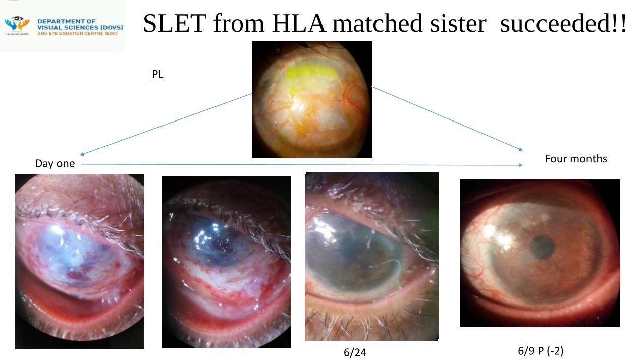
SLET from HLA matched sister succeeded!!
Four monthsDay one
6/24 6/9 P (-2)
PL
Vashi, Navi Mumbai
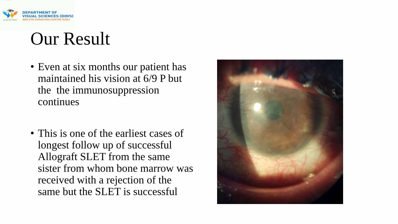
Our Result
• Even at six months our patient has maintained his vision at 6/9 P but the the immunosuppression continues
• This is one of the earliest cases of longest follow up of successful Allograft SLET from the same sister from whom bone marrow was received with a rejection of the same but the SLET is successful
Vashi, Navi Mumbai
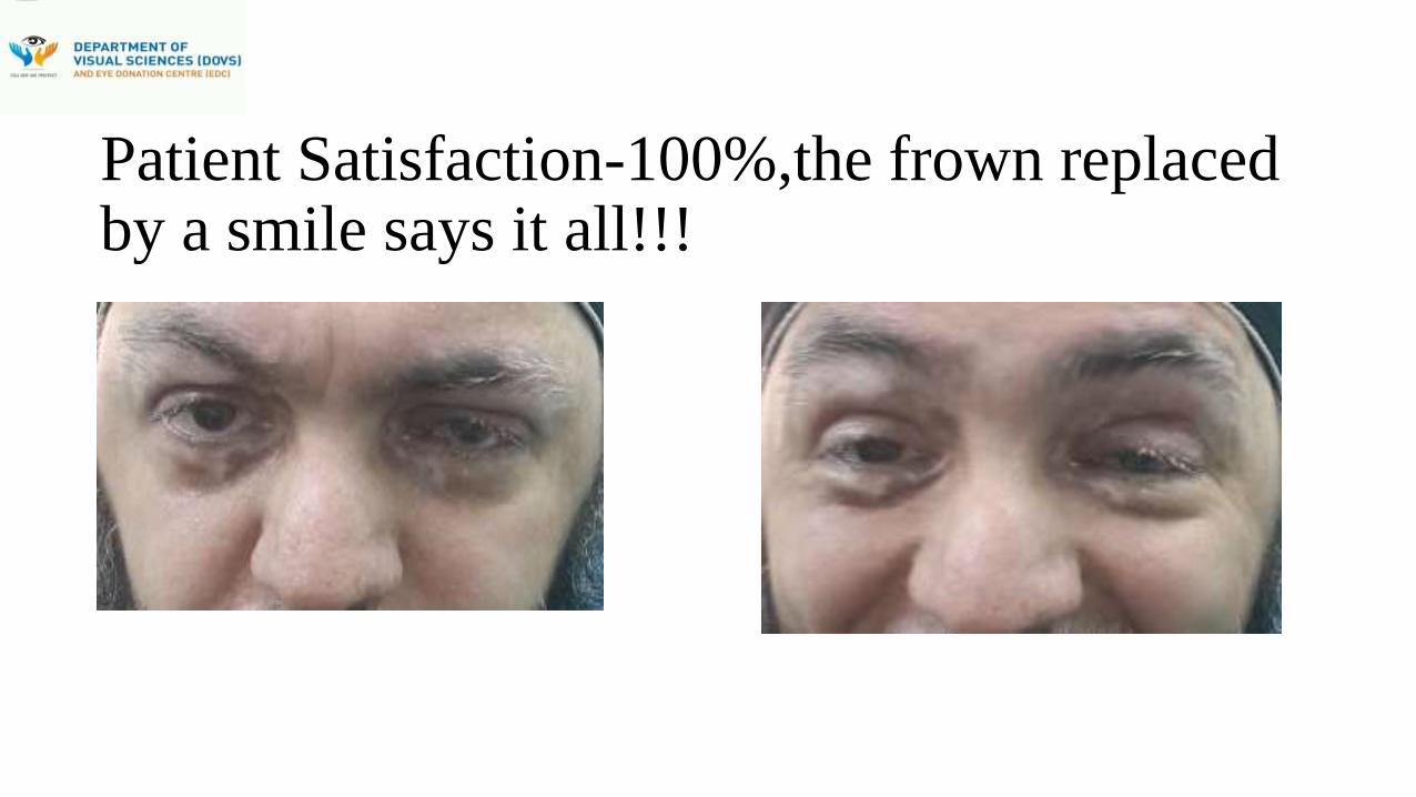
Patient Satisfaction-100%,the frown replaced by a smile says it all!!!
Vashi, Navi Mumbai

Discussion:-
• Living-related limbal allograft transplantation has been reported to be limited by
the amount of limbal stem cells that can be harvested
• There are risks reported of donor site stem cell deficiency and so more than one
donor may be required but the surgical technique used in our case helped us
distribute the small amount of stem cells harvested and they grew in situ. The
donor did not have any complication and only one donor was used
References:-
1. Tsubota K, Shimmura S, Shinozaki N, Holland EJ, Shimazaki J. Clinical application of living-related conjunctival-limbal allograft. Am J Ophthalmol 2002;133:134-5
2.Sangwan VS, Jain R, Basu S, Bagadi AB, Sureka S, Mariappan I, MacNeil S. Transforming ocular surface stem cell research into successful clinical practice. Indian J Ophthalmol 2014;62:29-40
Vashi, Navi Mumbai
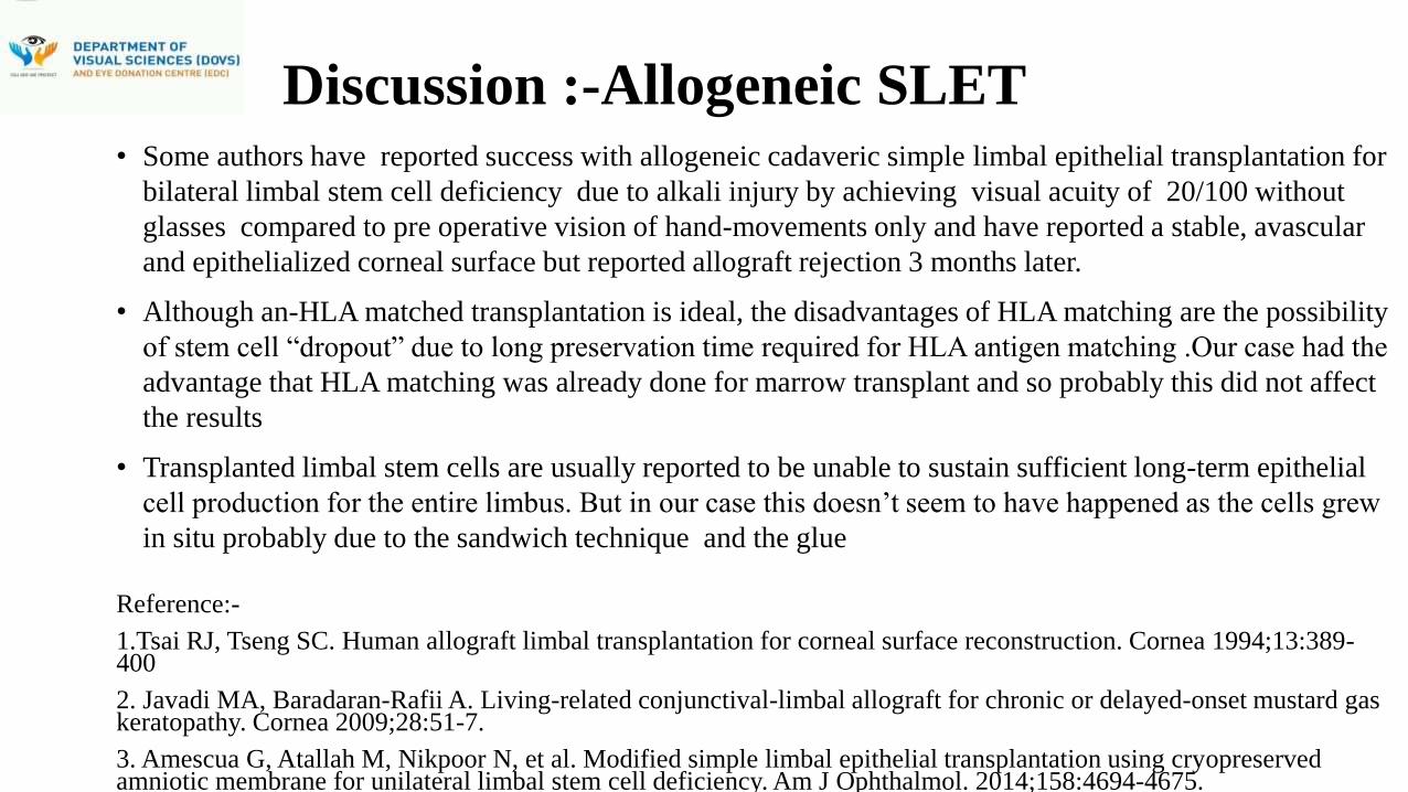
Discussion :-Allogeneic SLET• Some authors have reported success with allogeneic cadaveric simple limbal epithelial transplantation for
bilateral limbal stem cell deficiency due to alkali injury by achieving visual acuity of 20/100 without
glasses compared to pre operative vision of hand-movements only and have reported a stable, avascular
and epithelialized corneal surface but reported allograft rejection 3 months later.
• Although an-HLA matched transplantation is ideal, the disadvantages of HLA matching are the possibility
of stem cell “dropout” due to long preservation time required for HLA antigen matching .Our case had the
advantage that HLA matching was already done for marrow transplant and so probably this did not affect
the results
• Transplanted limbal stem cells are usually reported to be unable to sustain sufficient long-term epithelial
cell production for the entire limbus. But in our case this doesn’t seem to have happened as the cells grew
in situ probably due to the sandwich technique and the glue
Reference:-
1.Tsai RJ, Tseng SC. Human allograft limbal transplantation for corneal surface reconstruction. Cornea 1994;13:389-400
2. Javadi MA, Baradaran-Rafii A. Living-related conjunctival-limbal allograft for chronic or delayed-onset mustard gas keratopathy. Cornea 2009;28:51-7.
3. Amescua G, Atallah M, Nikpoor N, et al. Modified simple limbal epithelial transplantation using cryopreserved amniotic membrane for unilateral limbal stem cell deficiency. Am J Ophthalmol. 2014;158:4694-4675.
Vashi, Navi Mumbai

Discussion :- Choices as discussed in Literature
• “Limbal stem cell transplantation has been reported to be , currently the only
approved human stem cell therapy in India besides mesenchymal stem cells for blood
cancers
• If both eyes have total limbal stem cell deficiency then one needs to chose between
allogeneic-limbal transplantation which requires long term immunosuppression or the
autologous Cultivated Oral Mucous Epithelial transplantation ( COMET) which is
reported by experts to give not so satisfactory visual outcomes”
Reference:-
1. Sangwan VS, Jain R, Basu S, Bagadi AB, Sureka S, Mariappan I, MacNeil S. Transforming ocular surface stem cell research into successful clinical practice. Indian J Ophthalmol 2014;62:29-40
2. Nishida K, Yamato M, Hayashida Y, Watanabe K, Yamamoto K, Adachi E, et al. Corneal reconstruction with tissue engineered cell sheets composed of autologous oral mucosal epithelium. N Engl J Med 2004;16:1187-96
Vashi, Navi Mumbai
Conclusion:-
• Our case provides probably one answer ( subject to longer follow up) in
such complex bilateral situations and if we manage to maintain his vision
for a long time ,more than the present duration of six months, we can
probably conclude that SLET as proposed by Sangwan et al and modified
by Amescua et al and others from Wills Eye, makes it possible even for
corneal surgeons in low resource settings, in two tier non metro townships
to provide long lasting solutions in such complex bilateral cases without
the need for advanced institutes with sophisticated cell biology laboratories
as has been also concluded by innovators of this particular procedure in
India, Dr Virender Sangwan and his group
Vashi, Navi Mumbai


