
SEARCH FOR ANTIFUNGAL COMPOUNDS FROM EXTRACTS OF
BASIDIOMYCETES AGAINST PHYTOPATHOGEN Fusarium oxysporium f. sp.
lycopersici
NJUE ALICE WANJIKU
A research thesis submitted to the Graduate School in partial fulfilment for the
requirements of the Master of Science Degree in Chemistry of Egerton University
EGERTON UNIVERSITY
OCTOBER 2009

ii
DECLARATION AND RECOMMENDATION
Declaration
I, Njue Alice Wanjiku, hereby declare that this research thesis is my original work
and has not been submitted for an award in any other institution of learning to the best of my
knowledge.
Signed:…………………………. Date:……………………….
SM11/1535/05
Recommendation
We wish to confirm that this research thesis was done under our supervision and has
our approval to be presented for examination as per the Egerton University regulations.
Signed:……………………………. Date:………………………
Dr. J. Ouma Omolo
Egerton University
Signed:……………………………. Date:………………………
Dr. P. K. Cheplogoi
Egerton University
Signed:……………………………. Date:………………………
Dr. D. O. Otaye
Egerton University
iii
COPY RIGHT
All rights reserved. No part of this work may be reproduced, stored in a retrieval
system, or transmitted by any means, mechanical, photocopying, electronic process,
recording, or otherwise copied for public or private use without the prior written permission
from Egerton University.
© Njue Alice Wanjiku
2009

iv
DEDICATION
This work is dedicated to my husband David and my children Wambui, Njue and Njoki for
their love and patience during my studies.
v
ACKNOWLEDGEMENT
I acknowledge and appreciate the following institutions, Departments and
individuals.
1. Egerton University for allowing me to pursue the degree of Master of
Science in Chemistry on part time programme.
2. Research and Extension Division of Egerton University for funding this
research project (CTEE/DVC/RE/023) and for the permission to use the
Integrated Biotechnology Research Laboratory (IBRL).
3. Chemistry Department for accommodating me while pursuing my Msc
degree. Special thanks go to all the members of staff for their guidance,
support and encouragement during my studies.
4. Crops, Horticulture and Soil Department for the permission to use their
green houses to collect the infected tomato plants.
5. Prof. D. A. Mulholland and Mr. M. K. Langat of Surrey University, UK
analysing the compounds using Nuclear Magnetic Resonance (NMR).
6. My supervisors, Dr. J. O. Omolo, Dr. P. K. Cheplogoi and Dr. D. O. Otaye
for their guidance, advice, encouragement and their useful ideas, which
were quite useful for this work.
7. Dr. Andy Foster of Institute for Biotechnology and Drug Research
(IBWF) Kaisseislautern Germany for performing 18s RNA technique for
taxonomic characterisation of the strain JO5125.
8. My family for their love, inspiration and encouragement, throughout my
education endeavours.
9. To God who has been my source of strength throughout my studies and
for enabling me to reach this far.
vi
ABSTRACT
Tomato is one of the most widely cultivated horticutural crops but the production is
affected by the pest problems like Fusarial wilt. Fusarium oxysporium f. sp. Lycopersicit is
a destructive disease of the tomato and one of the main causes of the crop decline
worldwide. The disease is currently managed using synthetic fungicides but there is a
growing concern about the traces of the pesticide residues on the product and resistance to
the current fungicides. Fungicides are applied at various stages of the tomato to protect the
plant from fungal diseases but there is great concern due to environmental pollution and
health problems. Therefore, alternative fungicides from naturally occurring compounds that
are biodegradable offer a great potential for the control of crop fungal pathogens. Fungi are
known to be prolific producers of secondary metabolites that have a wide range of beneficial
biological activities, which can be researched on for crop protection. In this work, the fungi
investigated were collected from different ecological niches in Kenya and kept on agar
slants in the IBRL at Egerton University. For each strain collected, crude extracts were
prepared from sterile submerged liquid nutrient media where growth conditions were
manipulated to trigger production of secondary metabolites. In activity guided screening,
Fusarium oxysporium isolate from a tomato plant, was used as the target organism in agar
diffusion assay. From the ongoing screening of 400 crude extracts about 5% showed
significant and interesting activity. One species, which showed significant and reproducible
activity, was purified for natural antifungal compounds from its submerged cultures. The
selected strain JO5125 was cultivated in constituted liquid nutrient media and crude extracts
prepared from both the broth and mycelia at the end of the fermentation. The crude extracts
were fractionated using both silica gel and reverse phase liquid chromatography to
eventually lead to purification of the responsible compounds. The structure elucidation was
determined using 1- and 2-D NMR spectroscopic techniques. The purified compounds were
zearalenone and 3,4-dimethoxyphenol, which gave a minimum inhibitory concentration
against the pathogen of about 550 and 500ppm, respectively. They are naturally occurring
compounds which qualify as control agents of F. oxysporium with minimum impact in
environment.
vii
TABLE OF CONTENTS
DECLARATION AND RECOMMENDATION ....................................................................... ii
COPY RIGHT .............................................................................................................................. iii
DEDICATION ............................................................................................................................. iv
ACKNOWLEDGEMENT ............................................................................................................ v
ABSTRACT.................................................................................................................................. vi
TABLE OF CONTENTS ........................................................................................................... vii
LIST OF FIGURES ..................................................................................................................... xi
LIST OF TABLES ...................................................................................................................... xii
LIST OF SYMBOLS ABBREVIATIONS AND ACRONYMS ............................................ xiii
CHAPTER ONE ............................................................................................................................ 1
INTRODUCTION ......................................................................................................................... 1
1.1 Background Information ........................................................................................................ 1
1.2 Fusarial wilt causative agent: Fusarium oxysporium ............................................................ 2
1.3 Secondary metabolism in fungi ............................................................................................. 3
1.4 Controlling F. oxysporium using antifungal compounds of fungi ......................................... 4
1.5 Statement of the problem ....................................................................................................... 5
1.6 Objectives .............................................................................................................................. 5
1.6.1 General objective ................................................................................................................... 5
1.6.2 Specific objectives ................................................................................................................. 6
1.7 Justification of the study ........................................................................................................ 6
1.8 Hypothesis ............................................................................................................................. 6
CHAPTER TWO ........................................................................................................................... 7
LITERATURE REVIEW ............................................................................................................. 7
2.1 Biological control of Fusarial wilt ........................................................................................ 7
2.2 Chemical control of Fusarial wilt ......................................................................................... 8
2.3 Synthesised fungicides ......................................................................................................... 10
2.4 Natural antifungal compounds from plants ......................................................................... 11
2.5 Antifungal compounds from fungi ...................................................................................... 12
2.6 Strobilurins - a new class of active substances .................................................................... 16
2.5 Application of research findings from the screened fungi ................................................... 19
viii
CHAPTER THREE ..................................................................................................................... 20
MATERIALS AND METHODS ................................................................................................ 20
3.1 Apparatus and materials ...................................................................................................... 20
3.2 Preparation of liquid media ................................................................................................. 20
3.3 Preparation of test plates ...................................................................................................... 20
3.3.1 Preparation of potato dextrose agar (PDA) media ............................................................... 20
3.3.2 Isolation and culturing of the test organism - F. oxysporium .............................................. 21
3.3.3 Preparation of the test plates for antifungal activity ............................................................ 21
3.4 Cultivation of the selected strains in 250 ml submerged cultures ...................................... 21
3.5 Preparation of crude extracts from mycelium and culture filtrate ...................................... 22
3.5.1 Crude extracts from 250 ml scale initial cultivation ............................................................ 22
3.5.2 Cultivation of the selected strain JO5125 in 1 litre scale replicates .................................... 22
3.5.3 Liquid-solid adsorption resin extraction of the culture filtrate ............................................ 23
3.6 Testing of the crude extracts for anti-fungal activities ........................................................ 23
3.7 Fractionation of the crude extract based on polarity using chromatography ....................... 24
3.7.1 Determination of the dry weight of the crude sample .......................................................... 24
3.7.2 Column chromatography ...................................................................................................... 24
3.7.3 Thin layer chromatography(TLC) ........................................................................................ 25
3.8 Determination of minimum inhibitory concentration (MIC) ............................................... 26
3.9 NMR spectroscopy .............................................................................................................. 26
CHAPTER FOUR ....................................................................................................................... 27
RESULTS AND DISCUSSION .................................................................................................. 27
4.1 Isolation of the test organism - F. oxysporium from diseased tomato plant ........................ 27
4.2 Taxonomic identification of the strain JO5125 ................................................................... 27
4.3 Screening of the crude extracts against the pathogen .......................................................... 27
4.4 Yields of crude extracts from the replicated 1.0L scale ...................................................... 30
4.5 Test cysteine-adduct formation ............................................................................................ 30
4.6 Determination of minimum inhibitory concentration (MIC) for crude extracts .................. 31
4.7 Chromatographic fractionation and purification results ...................................................... 32
4.7.1 Fractionation and purification of mycelium crude extract ................................................... 32
4.7.2 Fractionation and purification of culture filtrate (acetone) crude extract ............................ 33

ix
4.7.3 Fractionation and purification of culture filtrate (methanol) crude extract.......................... 35
4.8 Structure elucidation of the pure compounds ...................................................................... 37
4.8.1 Structure elucidation of NA2, NA6 and NA7 ...................................................................... 37
4.8.2 Structure elucidation of NA3 ............................................................................................... 42
4.9 DISCUSSION ...................................................................................................................... 44
CHAPTER FIVE ......................................................................................................................... 47
5.0 CONCLUSION AND RECOMENDATIONS .............................................................. 47
5.1 Conclusion ........................................................................................................................... 47
5.2 Recommendations ................................................................................................................ 48
6.0 REFERENCES ................................................................................................................ 49
APPENDICES .............................................................................................................................. 57
APPENDIX 1: 1H NMR Spectrum for NA1 .............................................................................. 57
APPENDIX 2: 13
C NMR Spectrum for NA1 ............................................................................. 58
APPENDIX 3: 1H NMR Spectrum for NA2 .............................................................................. 59
APPENDIX 4: 13
C Spectrum NMR for NA2 ............................................................................. 60
APPENDIX 5: 1H NMR Spectrum for NA3 ............................................................................. 61
APPENDIX 6: 13
C NMR Spectrum for NA3 ............................................................................. 62
APPENDIX 7: 1H/
1H COSY NMR Spectrum for NA3 ............................................................ 63
APPENDIX 8: DEPT Spectrum for NA3 .................................................................................. 64
APPENDIX 9: 1H/
13C HMBC Spectrun for NA3 ..................................................................... 65
APPENDIX10: 1H/
13C HSQC-DEPTSpectrum for NA3 ......................................................... 66
APPENDIX 11: 1H/
1H NOESY 2D Spectrum for NA 3 ........................................................... 67
APPENDIX 12: 1H NMR Spectrum for NA6 ............................................................................ 68
APPENDIX 13: 13
C NMR Spectrum for NA6 ........................................................................... 69
APPENDIX 14: 1H NMR Spectrum for NA7 ............................................................................ 70
APPENDIX 15: 13
C NMR spectrum for NA7 ........................................................................... 71
APPENDIX 16: 1H/
1H COSY Spectrum for NA7 ..................................................................... 72
APPENDIX 18: 1H/
13C HMBC Spectrum for NA7 .................................................................. 74
APPENDIX 19: 1H/
13C HMBC Spectum for NA7 .................................................................... 75
APPENDIX 20: 1H/
1H NOESY Spectrum for NA7 .................................................................. 76
APPENDIX 21: 1H/
1H COSY Spectrum for NA 7 .................................................................... 77

x
APPENDIX 22: 1H/
13C HMBC Spectrum for NA7 .................................................................. 78
APPENDIX 23: 1H/
13C HSQC-DEPT Spectrum for NA7 ....................................................... 79
APPENDIX 24: 1H/
1H NOESY Spectrum for NA7 .................................................................. 80
xi
LIST OF FIGURES
Figure 4.1: Summary of preparation of culture and mycelium crude extracts .................. 30
Figure 4.2: Flow scheme illustrating the fraction of mycelium crude extract and subsequent
purification of compounds ............................................................................... 32
Figure 4.3: Flow scheme illustrating the fraction of culture filtrate (acetone) crude extract
and subsequent purification of compounds ..................................................... 34
Figure 4.4: Flow scheme illustrating the fraction of culture filtrate (methanol) crude extract
and subsequent purification of compounds ..................................................... 35
Figure 4.5: Biosynthetic pathway of Zearalerone .............................................................. 42
xii
LIST OF TABLES
Table 4.1: Antifungal activity for crude extracts from the 20 strains of fungal cultures .. 29
Table 4.2: Bioassay results of JO5125 with cysteine against the F. oxysporium .............. 31
Table 4.3: The average MIC and standard deviation of the above crude extracts ............. 31
Table 4.4: The antifungal activity of the intermediate fractions and the purified compounds
against the F. oxysporium ................................................................................ 36
Table 4.5: 1H NMR and
13C NMR data for NA2, NA6, and NA7 .................................... 38
Table 4.6: 1H-NMR,
13C-NMR’ DEPT and HMBC data for compound NA7 .................. 40
Table 4.7: 1H-NMR,
13C-NMR, DEPT and HMBC data for NA3 .................................... 43

xiii
LIST OF SYMBOLS ABBREVIATIONS AND ACRONYMS
13C NMR Carbon-13 Nuclear Magnetic Resonance Spectroscopy
1H NMR Proton Nuclear Magnetic Resonance Spectroscopy
Bc1 Cytochrome bc1
CDCL3 Deuterated chloroform
COSY Correlated Spectroscopy
DEPT Distortionless Enhancement by Polarization Transfer
GC Gas chromatography
GC-MS Gas chromatography-Mass Spectrometry
HMBC Heteronuclear Multiple Bond Coherence
HMQC Heteronuclear Multiple Quantum Coherence
HPLC High performance liquid chromatography
HREIMS High Resolution Electron Impact Mass Spectrometry
HSQC Heteronuclear Multiple Quantum Coherence
IBRL Integrated Biotechnology Research Laboratory
KEX Culture extract
MEX Mycelium extract
MIC Minimum inhibitory concentration
MS Mass Spectrometry
NMR Nuclear Magnetic Resonance Spectroscopy
NOESY Nuclear Overhauser Effect Spectroscopy
PDA Potato Dextrose Agar
PPM Parts per million
TLC Thin layer chromatography
UV Ultra-Violet radiations

1
CHAPTER ONE
INTRODUCTION
1.1 Background Information
In the last one hundred years, control of plant diseases and pests has depended
increasingly on the extensive use of synthetic chemicals. In fact, these pesticides have
become established as an essential input in growing of horticultural crops and without their
use crop yields would be severely reduced to uneconomic levels. Many soil borne noxious
organisms are microscopic making monitoring intrinsically difficult and thus costly. In
general, soil-borne diseases are limiting factors in the production of many crops and
accounts for 10 - 20% annul yield losses. Losses in individual fields can be as high as 100%
(Fravel et al., 2005). Management of soil-borne pathogens is necessary for a secure and
stable supply of food as well as for maintaining agricultural exports and profit maximisation.
The structural, physical and biological complexity of the soil environment in which
soil borne pathogens interact with plant roots inherently limits the options available for
disease control. The already proven measures include conventional breeding and genetic
engineering of disease-resistant plants, application of disease-suppressing cultural practices,
and to some extent, use of antagonistic biological agents against the microorganisms that
cause plant disease. Control through crop rotation or other cultural practices is not very
feasible in most countries anymore and not always profitable for intensive use (Copping and
Menn, 2000). Resistance in widely used agricultural fungicides has emerged as a significant
problem. The situation is worsened further by lack of arsenal of fungicides that attack soil
borne pathogens. Many chemical pesticides have been or are being phased out (like
organochlorine insecticides, methyl bromide) either because of potential human risk,
environmental pollution, effects on non-target organisms or the development of pest
resistance. African agricultural products are still restricted in overseas markets just because
the commodities or products are treated with synthetic fungicides and do no meet the basic
requirement (Gullino et al., 2000).
However, none of the fungicides used have been successful since there are
unintended consequences highlighted above. The use of broad-spectrum fungicides further
results in imbalances within the microbial community creating unfavourable conditions for

2
the activity of beneficial organisms leading to suppression of soil fertility (Ristaino and
Thomas, 2000). Risk associated with implementation of alternative technologies is
inherently higher due to climatic and pest crop variability. The global consensus to reduce
inputs of chemical pesticides which are perceived as being hazardous by some consumers
has provided challenges for the development of novel, benign, sustainable crop protection
strategies. Therefore, development of alternatives to these synthetic fungicides can be sought
from higher fungi like ascomyectes and basidiomycetes to discover selective and
environmentally safe fungicides for use in agriculture.
1.2 Fusarial wilt causative agent: Fusarium oxysporium
The genus Fusarium is one of the most injurious and causes diseases that are found
in a number of plants: cereals, grasses, legumes and horticultural crops. F. oxysporium is a
species-complex that includes many host-specific plant pathogens. A common characteristic
of F. oxysporium strains is the ability to parasitize plant roots, usually without inducing
symptoms. The pathogen penetrates plants through root tips or wounds on roots and reaches
the xylem. Vascular wilt diseases caused by the strains of these fungi are usually highly
destructive whether they occur in cultivated crops or indigenous species. Under warm and
conducive conditions the losses could reach 95% of production in some fields (Bailey and
Lazarovits, 2003). The interaction with the host involves a specialized colonization of the
vascular system setting in motion a series of biochemical and morphological reactions that
culminate into the characteristic symptoms of these diseases - the wilting of the host plant.
The economic damage caused by Fusarium wilt diseases has inspired considerable
research on those species affecting important crop species. Management of the disease is
complicated by the speed at which it spreads. In corn, Fusarium wilts may reduce yield by
50% (Agrios, 1998). It has been reported that virtually any kernel of dried corn, anywhere,
can be shown to contain Fusarium, if proper isolation techniques are used (Cavigelli et al.,
2005). Pathogenic strains of F. oxysporium are placed into formae speciales according to
their host range. There about 150 host specific formae speciales described for this vascular
wilt pathogen. They can also live as saprotrophs in the soils, so crop rotation is usually not
sufficient for control. Fusarial wilts are most severe under warm soil conditions and in
greenhouses.
3
Fusarium wilts are caused by pathogens that reside in the soil and survive on
infected debris, in alternative host or as resistant propagules of which several can be
produced in the life cycle of a species. The epidemiology of these pathogens is also
extremely complex, with propagules being splash borne over short distances or carried on
tools in the course of normal cultivation practices. The use of contaminated soil can also
spread these pathogens to nurseries and shade houses whilst long distance spread is achieved
through use of infected plant material such as seeds or vegetatively propagated material.
Insect vectors can also be involved given their complex life cycle and epidemiology, the
potential for variation within populations and their soil-borne nature, the management of
diseases caused by these pathogens has always been a challenge to plant pathologists and is
likely to continue to be so in the future (Flood, 2003). Fusarium is one of the most widely
distributed pathogen and difficult to control because once an area becomes infected with it,
it remains so indefinitely.
F. oxysporium f. sp. lycopersici is a highly destructive pathogen of both greenhouse
and field grown tomatoes in warm vegetable production areas. The disease causes great
losses, especially on susceptible varieties and under favourable weather conditions. Infected
plants show pronounced vascular discoloration, severe defoliation and stunting which leads
to the death of the plant. Occasionally entire field of tomatoes can be severely damaged
before the crop is harvested. The disease, which is prevalent in Kenya’s tomato growing
zones, causes serious losses because soil and air temperatures are rather high during most of
the season. Heavy damage to tomatoes often occurs in areas of continuous cultivation
because monoculture practices could lead to pathogen build up and subsequent yield losses
(Suleman et al., 2003).
1.3 Secondary metabolism in fungi
Interactions of fungi with other microbes and non-microbial systems often include
the production of biologically active metabolites that affect the potential competitors or
predators. Such ecological phenomena have served as valuable lead to the discovery of
secondary metabolites that have proven to be unlimited source of biologically active
molecules. Using appropriate screening methods, molecules with almost any defined activity
can be obtained, thus, secondary metabolite screening for compounds has been a main stay

4
of the pharmaceutical industry for many years (Demain, 1999) and has increased in
importance in agricultural industry.
The starting point from primary metabolism is the basis of classification of
secondary metabolites according to the biosynthetic precursors (Turner, 1971; Turner and
Aldridge, 1983). The secondary metabolites are, however, low molecular weight, chemically
and taxonomically extremely diverse compounds with obscure function, characteristic
mainly to some specific, distinct types of organisms. The presently known secondary
metabolites of microorganisms display a great number of diverse and versatile biological
activities as well as chemical structures. Hence with application of appropriate fungal
biotechnology, there is a great potential to find novel biologically active compounds and
greatly improve the rate of exploration of uncommon and unreported groups of tropical
fungi. Unique ecosystems may result in unique organisms with unique metabolic pathways
(Lasure, 2000).
1.4 Controlling F. oxysporium using antifungal compounds of fungi
Tropical forest are endowed with enormous and a rich diversity of fungal genetic
resources that can be exploited for new antifungal compounds (Berdy, 2003). Natural
product resources including the microbial world are mainly unexplored both in dimension
and in respect of geographic, ecological and environmental point of view. In some estimates
the worldwide numbers of fungal species were 1.5 million (Hawksworth, 2001). Of these
only 10% have been discovered and described as yet, and barely 1% examined for their
spectrum of secondary metabolites (Arnold et al., 2000). Fungi in their natural habitats
interact competitively and depending on the environment such a microorganism adapts
appropriate survival mechanisms that confer selective advantage over their competitors.
Mechanisms of fungal antagonism and defence often include the production of biologically
active secondary metabolites by species that affects the potential competitors and/or
predators. Laboratory studies clearly indicate that such ecological phenomena can serve as
valuable lead to the discovery of novel natural products many of them potential targets for
agrochemicals and biomedical development (Hostettman and Martson, 2000). Among the
sources of bioactive metabolites less intensively investigated organisms like the higher fungi
seem to hold an excellent promise for new structures with interesting biological activities.
5
Fungi inhabit most climatic zones from the arctic to the tropical rain forest. For more than 5
decades, interesting compounds of different biogenetic origin with antibacterial, antifungal,
phytotoxic, nematocidal, cytostatic, antiviral and other pharmacological activities have been
isolated from basidiomycetes (Thines et al., 2004)
From the early 1990s the number of bioactive compounds isolated from various
filamentous and higher fungal species had continuously increased up to more than 50% by
the year 2000. The need for less toxic, more potent antifungal as well as non-antinfectives,
the evolving resistance to existing fungicides and emergence of new fungal diseases, has
posed a challenge to the researchers of the 1990s (Knight et al., 2003). Especially, the
resistance of pathogenic fungi to fungicides is a serious problem. Therefore, it is quite
probable that highly differentiated fungi such as basidiomycetes will produce a rich array of
secondary metabolites with a great potential to produce novel biologically active compounds
against F. oxysporium. The potential chemical diversity of this vast untapped resource is
surely one of the great driving forces behind today’s search for new antifungal compounds.
1.5 Statement of the problem
Use of synthetic soil fumigants has been widely recommended in the management of
this fungus. With the ban of methyl bromide, the commonly used soil fumigant there is a
problem. Given the moist and warm climatic conditions of tropical soils, F. oxysporium the
causative agent of tomato fusarial wilt, is bound to cause more crop loss. This scenario is
impossible given the serious food and nutritional insecurity in most poor resource
households. Naturally occurring fungicides from fungi, can be a possible alternative to these
synthetic soil fumigants in controlling the fusarial wilt causative agent in tomato crop
production.
1.6 Objectives
1.6.1 General objective
To search for antifungal compounds from submerged cultures of basidiomycetes
against F. oxysporium.
6
1.6.2 Specific objectives
i. To isolate the pathogen from the infected plant.
ii. To screen crude extracts prepared from 400 strains of basidiomycetes against F.
oxysporium using agar diffusion assays.
iii. To cultivate the selected basidiomycete in liquid submerged cultures.
iv. To determine the taxonomy of the selected basidiomycete.
v. To prepare crude extracts from the mycelium and culture filtrate of the selected
basidiomycete using adsorption resin, Mitsubishi HP21 (DIAION).
vi. To carry out bioactivity-guided chromatographic fractionation of the mycelial and
culture filtrate crude extracts, to purify the targeted antifungal compounds.
vii. To carry out structure elucidation of the purified compounds.
viii. To determine minimum inhibitory concentrations of the purified compounds.
1.7 Justification of the study
Control of fungal diseases, fusarial wilts included, has been almost exclusively
controlled by synthetic chemicals. Despite the huge reduction on crops losses occasioned by
these chemicals in the last decade, there are attendant problems especially the non targeted
effects. Researches tailored to discovering environmentally benign compounds are as needed
given that non-targeted effects are responsible for withdrawals of the hitherto effective
synthetic chemicals. Given the unique tropical habitats and the high incidences of fusarial
wilts, it’s a worthwhile to investigate the natural arsenal of antifungal for the control of F.
oxysporium in tomato.
1.8 Hypothesis
From the screening of the crude extracts of the 400 strains of basidiomycetes, a
maximum of 5% will show significant and reproducible activity against F. oxysporium. Out
of the 5% active lot, one basidiomycete will be selected, grown in submerged cultures and
will be expected to produce the desired antifungal compounds.
7
CHAPTER TWO
LITERATURE REVIEW
2.1 Biological control of Fusarial wilt
Biological control of soil-borne plant pathogens is a potential alternative to the use of
soil fumigation, which has been proved to be harmful to the environment (Elizabeth and
Handelsman, 1999). Root of crops can be protected from pathogens to a significant degree
by non-pathogenic root colonizing microorganism applied on the seed. For example, strains
of the fungus Trichoderma have been found to be effective biocontrol agents of various soil-
borne fungal plant pathogens under green house and field conditions (Herman, 2000). This
approach has indeed led to the isolation of several strains of T. harzianum antagonistic to
and effective in controlling plant pathogenic fungi, such as F. oxysporium under such
conditions. In a study, tomato crop survival and yield were increased and disease decreased
following application of T. harzianum either as a seed coating or as an amendment to the
soil in the field (Sivan et al., 1987). Also isolates of T. asperellum from a suppressive mix
prepared from sewage sludge compost showed high ability to suppress Fusarium wilt
(Alabouvette et al., 2002).
Soil borne diseases have been controlled more recently by means of certain
beneficial bacteria that are indigenous to the rhizosphere of plants. They rapidly colonize
and suppress F. oxysporium at the root surface (Ahmed et al., 2007). Certain soils are
naturally suppressive to Fusarium wilt a property due to the role of non-pathogenic F.
oxysporium in the indigenous microbiota of these soils. One of these non-pathogenic F.
oxysporium is Fo47, a strain isolated from a suppressive soil in Chateurenard, France has
been shown to effectively suppress Fusarium wilt of the tomato (Fuchs et al., 1999).
Another biocontrol fungus F. oxysporium strain CS-20 demonstrated potential for reducing
the incidence of Fusarium wilt of the tomato both in the field and green houses. Strain CS-
20 superficially colonizes the interior of the tomato roots, but does not penetrate the vascular
system. It provides biocontrol of Fusarium wilt through a host-mediated mechanism (Fravel
et al., 2005).
Root dip applications of Bacillus subtilis, Pseudomonas fluorescens, Aspergillus
awamori, and Penicillium digitum resulted in significant decline in the rhizosphere

8
population of F. oxysporium. Root dip treatment with the phosphate-solubilising
microorganism tested resulted in significant increase in the yield of tomato being greatest
with A. awamori and P. digitum in pathogen inoculated 36 and 33% (Mujeebur and
Shahana, 2002). Compost obtained from heterogeneous vegetable wastes showed important
suppressive effects against Fusarium species. Fusarium wilt reductions on tomato plant
were generally between 30-65% (Larkin and Fravel, 2002). Crop damage due to disease in
the field is rarely the result of the activity of one pathogen, but as a combination. Because of
the complexity of such a situation, biocontrol studies are difficult to implement and thus a
combination of both chemical and biological agents increase the protection of the crop.
Elmer and McGovern (2004) combined commercial biological products with fungicides and
found that the combination provided a higher degree of suppression of Fusarium wilt of
tomato. The set back of these combinations is lack of knowledge of compatibility of
biocontrol agents with pesticides, which may contribute to failure of biocontrol to perform
as expected. Also fungal biological control agents have a narrow host range and that they act
slowly therefore, giving limited protection. Another major setback in biological control
strategy stems from the fact that these biological control agents have their activities dictated
by various soil conditions such as soil moisture, pH, and even temperature (Copping and
Menn, 2000).
2.2 Chemical control of Fusarial wilt
Chemical control of fungal diseases has a long history, starting with inorganic
compounds such as copper salts and lime-sulfur in the 19th
century and ending with
sophisticated organic compounds such as imidazoles and triazoles in the 1970s and 1980s.
Fungicides remain an important part of disease control strategy in plants. Fungicidal
treatment can keep tomato crops healthy or suppress the infection sufficiently (Merterrhazy
and Bartok, 1996). Various chemicals have been used to control the F. oxysporium disease
such as imidazoles (prochlorax) and the trizoles (Strauss and Labuschagne, 1993) but Song
et al. (2004) found prochlorax and carbendazim to be most effective fungicides in inhibiting
mycelial growth of F. oxysporium. However, the effectiveness of fungicides is threatened by
development of prochlorax and carbendazim resistance in the pathogen population. Allen et
al. (2004) found that benomyl at all concentrations completely inhibited fungal growth of

9
Fusarium species. Calcium cyanide, a nitrogen fertilizer has also been found to reduce the
incidence of the fusarial wilt disease to a certain percentage (Klasse, 2002). Common
inorganic salts such as sodium and potassium bicarbonate have a potential use as an
antifungal agent for controlling the mycelial growth of Fusarium species in crops but are not
very effective (Hang and Woodams, 2003).
Fumigation of soil with Vorlex®
, a commercial mixture of methyl isothiocyanate,
dichloropropenes and dichloropropanes has been reported to reduce populations of
Fusarium in soil (Chew and Hall, 1984). Fumigation of soil with chloropicrin or dazomet
dramatically reduces the population of Fusarium species in fumigated plots but have
environmental implications (Hansen et al., 1996).
Several agricultural systems involving intensive production of high value crops have
become dependent on the use of methyl bromide. The broad-spectrum activity and ease of
application of these materials have led to their use as treatment of choice in a number of
situations. However, the soil fumigation with synthetic organic pesticides has various
hazardous environmental implications (Santos et al., 2006). Although methyl bromide is a
most useful tool in specific instances, there are a number of technical and legislative
limitations that have led to restrictions on its use. It has a substantial phytotoxicity that may
accumulate in excessive levels in commodities that are fumigated several times and have
been a cause of concern in ground water in some European countries. Of most concern is its
ozone depleting potential. Soil fumigation with methyl bromide proved to be effective only
as a short-term control measure in South Africa, due to the successful re-invasion of the
pathogen back to soil after a period of approximately 2 years (Herbert and Marx, 1990).
Both the resistance of the varieties and virulence of the fungal isolates also affect the
fungicidal efficacy. There is also the fact that the regions capability and resources to sustain
methyl bromide phase out beyond 2015 deadline are limited and is dependent on countries
being able to unilaterally address and solve their own pest control problems. The Montreal
protocol schedule required that developed countries phase out methyl bromide by 2005 and
developing countries freeze consumption by 2002, achieve a 20% reduction by 2005 and a
complete phase out by 2015. At least 50% of the countries in Africa have not met this
challenge (U.S. Environmental Protection Agency).
10
Although farmers reported that tomatoes planted with agrochemicals were healthier,
drought tolerant, resistant to diseases and gave better yields. There is need to for effective,
sustainable and environmental friendly fungicides, which will be appropriate to local
farmers. Currently no effective control measure is available for the control of Fusarium wilt
except only prevention strategies (Nel et al., 2006). This concern has provided the impetus
to investigate alternative control measures, like naturally occurring compounds from fungi.
2.3 Synthesised fungicides
Natural compounds have been a major source of bioactive derivatives and lead for
development of synthetic drugs (Butler, 2005). Many naturally occurring compounds are
synthesized so as to improve their physiochemical characteristics and bioactivities. One of
these naturally occurring compounds is chitosan, which has limited activity due to its poor
solubility. To improve its solubility, many derivatives of chitosan have been synthesised and
carboxymethyl chitosan (CMCTS) is the most important one. Chitosan can reduce the
growth of phytopathogenic fungi, which are harmful to field crops, fruit and vegetables.
Antifungal activity of chitosan, CMCTS and the Schiff bases of CMCTS against F.
oxysporium at 500ppm are as follows; chitosan gave inhibitory index of 14.3%, CMCTS
gave 9.1%, 2-(2-Hydroxy-5-nitrobenzyllideneamino)-6-carboxyemthylchitosan
(HNCMCTS) (1) gave 31.2% and 2-(5-chloro-2-hydroxybenzylideneamino)-6-
carboxymethylchitosan (HCCMCTS) (2) gave 43.0%. Schiff bases have activity more than
those of chitosan and CMCTS (Zhanyong et al., 2006). Synthesis of 2-cyano-3-
phenylacrylic acetate (JS399-19) (3), which belongs to the cyanoacrylate fungicide group
exhibited strong fungicidal activity against the plant pathogen Fusarium species in vitro
(Hengkui et al., 2008). Synthesis of 4,4’-bis(2’’-aryl-5’’-methyl/unsubstituted-4’’-oxo-
thiazolidin-3’’-yl) bibenzyl from benzil (4) gave a compound with significant fungitoxicity
at 1000ppm against F. oxysporium (Ibadur et al., 2003). Large numbers of 1,5-
benzothiazepine exhibit a variety of pharmacological activities. In the synthesis of new
fluorinated spiro [1,5]-benzothiazepin-2, 3’ [3’H] -indol] -2' (1 'H)–ones (5) increased the
antifungal activity against F. oxysporium at 1000ppm by 98% (Anshu et al., 1998). The
limiting factor of the synthesized fungicides is lack of combination or alternating application
with other fungicides as a tool to limit fungicide resistance development.

11
CO2Et
CNH2N
N
CH2OCH2CO2H
O
C
H
R
HO
CN
H
HS
O
H
H3C
C6H5
H
(3) JS 339-19
(1) HNCMCTS: R=NO2
(2) HCCMCTS: R=Cl
2
(4) 4,4'-Bis(2"-Arly-5"-methly-4"-oxo-thiazolidin-3'-yl) bibenzyl
(5 )Spiro [1,5]-benzothiazepin-2,3' [3' H] - indol] - 2' (1' H) - ones
NH
S
HN
HH
H3C
n
n
2.4 Natural antifungal compounds from plants
Natural products have been shown to be ideal agrochemicals in a number of ways,
especially given their high potency and specificity. Ever since the introduction of the large
scale screening for natural compounds, they have been playing essentials roles as lead
structures for the development of drugs, pesticides and as characterisation of new selective
targets. In the last decade, the pharmaceutical industry has greatly sped up screening
capacities with the use of Ultra High Throughput Screening (U-HTS) robotic systems, which
can routinely screen thousands of molecules a day for biological activities. In this context,
natural compounds are more and more revisited as rich source of promising model lead
molecules (Piggutt and Karuso, 2004).
The use of botanicals in disease management is as old as humankind and transcends
all stages of civilization. A great variety of plants have broad-spectrum bioactive organic
compounds such as isocyanate, glucocynolate, glycosides, lipids and phenol with
fungicidical and fungistatic properties (Chitwood, 2002). Higher plants such as Warburgia
ugandensis have been explored as potential sources of antimicrobial agents. The aqueous
methanolic extract of the barks of W. ugandensis displayed activity against F. oxysporium.
Muzigadial (6) was isolated with minimum inhibitory concentration of 50ppm (Rugutt et al.,
2006). Many natural diterpenoids are very much functionalised and show good antifungal
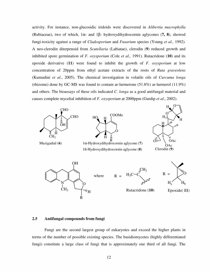
12
activity. For instance, non-glucosidic iridoids were discovered in Alibertia macrophylla
(Rubiaceae), two of which, 1α- and 1β- hydroxydihydrocornin aglycones (7, 8), showed
fungi-toxicity against a range of Cladosporium and Fusarium species (Young et al., 1992).
A neo-clerodin diterpenoid from Scutellaria (Labiatae), clerodin (9) reduced growth and
inhibited spore germination of F. oxysporium (Cole et al., 1991). Rutacridone (10) and its
epoxide derivative (11) were found to inhibit the growth of F. oxysporium at low
concentration of 20ppm from ethyl acetate extracts of the roots of Ruta graveolens
(Kumudini et al., 2005). The chemical investigation in volatile oils of Curcuma longa
(rhizome) done by GC-MS was found to contain ar-lurmerone (51.8%) ar-lurmerol (11.9%)
and others. The bioassays of these oils indicated C. longa as a good antifungal material and
causes complete mycelial inhibition of F. oxysporium at 2000ppm (Gurdip et al., 2002).
CHO
CHO
OH
HCH2
O
O
O
H
H
O
R
COOMeH
H
HO
OAc
OAc Muzigadial (6) 1α-Hydroxydihydrocornin aglycone (7)
1ß-Hydroxydihydrocornin aglycone (8) Clerodin (9)
H
H
N
OH
CH3
O
H
R
H3C C
CH2
Hb
O
Epoxide( 11)Rutacridone (10)
R =R =
Ha
where
2.5 Antifungal compounds from fungi
Fungi are the second largest group of eukaryotes and exceed the higher plants in
terms of the number of possible existing species. The basidiomycetes (highly differentiated
fungi) constitute a large class of fungi that is approximately one third of all fungi. The
13
extraordinary diversity of fungal species suggests virtually limitless potential for secondary
metabolic variation. They are one of largest reservoir for isolating further bioactive
metabolites. Despite their potential for development of biologically active compounds, few
bioactive metabolites have been reported from fungi as compared with higher plants. The
number of bioactive compounds isolated from various fungal species had continuously
increased up to more than 50% by the turn of new millennium (Weber et al., 2007).
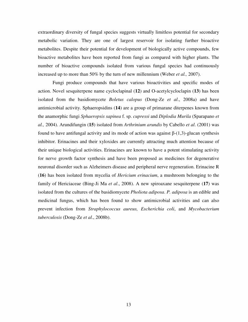
Fungi produce compounds that have various bioactivities and specific modes of
action. Novel sesquiterpene name cycloclapinal (12) and O-acetylcycloclapin (13) has been
isolated from the basidiomycete Boletus calopus (Dong-Ze et al., 2008a) and have
antimicrobial activity. Sphaeropsidins (14) are a group of primarane diterpenes known from
the anamorphic fungi Sphaeropsis sapinea f. sp. cupressi and Diplodia Murila (Sparapano et
al., 2004). Arundifungin (15) isolated from Arthrinium arundis by Cabello et al. (2001) was
found to have antifungal activity and its mode of action was against β-(1,3)-glucan synthesis
inhibitor. Erinacines and their xyloxides are currently attracting much attention because of
their unique biological activities. Erinacines are known to have a potent stimulating activity
for nerve growth factor synthesis and have been proposed as medicines for degenerative
neuronal disorder such as Alzheimers disease and peripheral nerve regeneration. Erinacine R
(16) has been isolated from mycelia of Hericium erinacium, a mushroom belonging to the
family of Hericiaceae (Bing-Ji Ma et al., 2008). A new spiroaxane sesquiterpene (17) was
isolated from the cultures of the basidiomycete Pholiota adiposa. P. adiposa is an edible and
medicinal fungus, which has been found to show antimicrobial activities and can also
prevent infection from Straphylococcus aureus, Escherichia coli, and Mycobacterium
tuberculosis (Dong-Ze et al., 2008b).
14
O
O
O
H
H
R2
OH
R1 O
O
O
OH
H
OH
NH2
O O
O
O O
H
HOOH
AcO
H
CHO OH
O HO
H
R1=R2=OH Cycloclapinal (12)
R1=OH R2=OAC O-Acetylcycloclapin (13)
Sphaeropsidins (14) Arundifungin (15)
Erinacine R (16)
Spiroaxane (17)
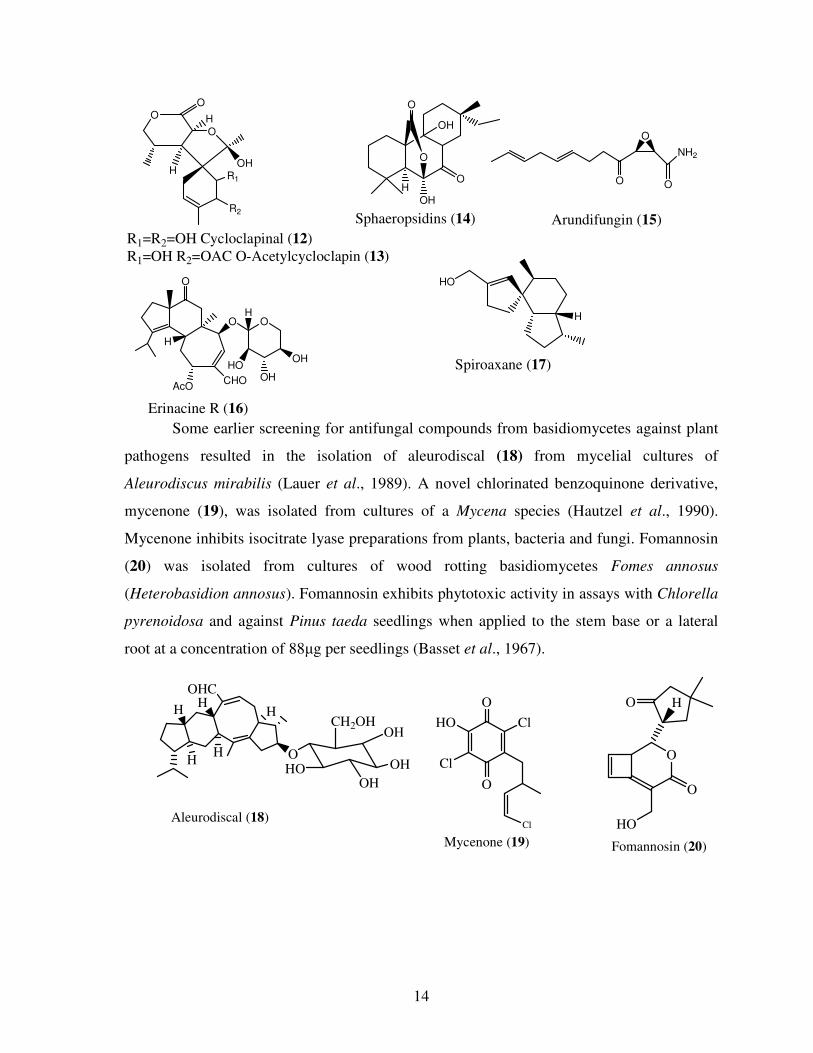
Some earlier screening for antifungal compounds from basidiomycetes against plant
pathogens resulted in the isolation of aleurodiscal (18) from mycelial cultures of
Aleurodiscus mirabilis (Lauer et al., 1989). A novel chlorinated benzoquinone derivative,
mycenone (19), was isolated from cultures of a Mycena species (Hautzel et al., 1990).
Mycenone inhibits isocitrate lyase preparations from plants, bacteria and fungi. Fomannosin
(20) was isolated from cultures of wood rotting basidiomycetes Fomes annosus
(Heterobasidion annosus). Fomannosin exhibits phytotoxic activity in assays with Chlorella
pyrenoidosa and against Pinus taeda seedlings when applied to the stem base or a lateral
root at a concentration of 88µg per seedlings (Basset et al., 1967).
O
O
HO
O
HO
Cl
Cl
O
O
OHO
OH
OH
H
HH
H
OHC
Fomannosin (20)Mycenone (19)
Aleurodiscal (18)
H
Cl
H
OHCH2OH
15
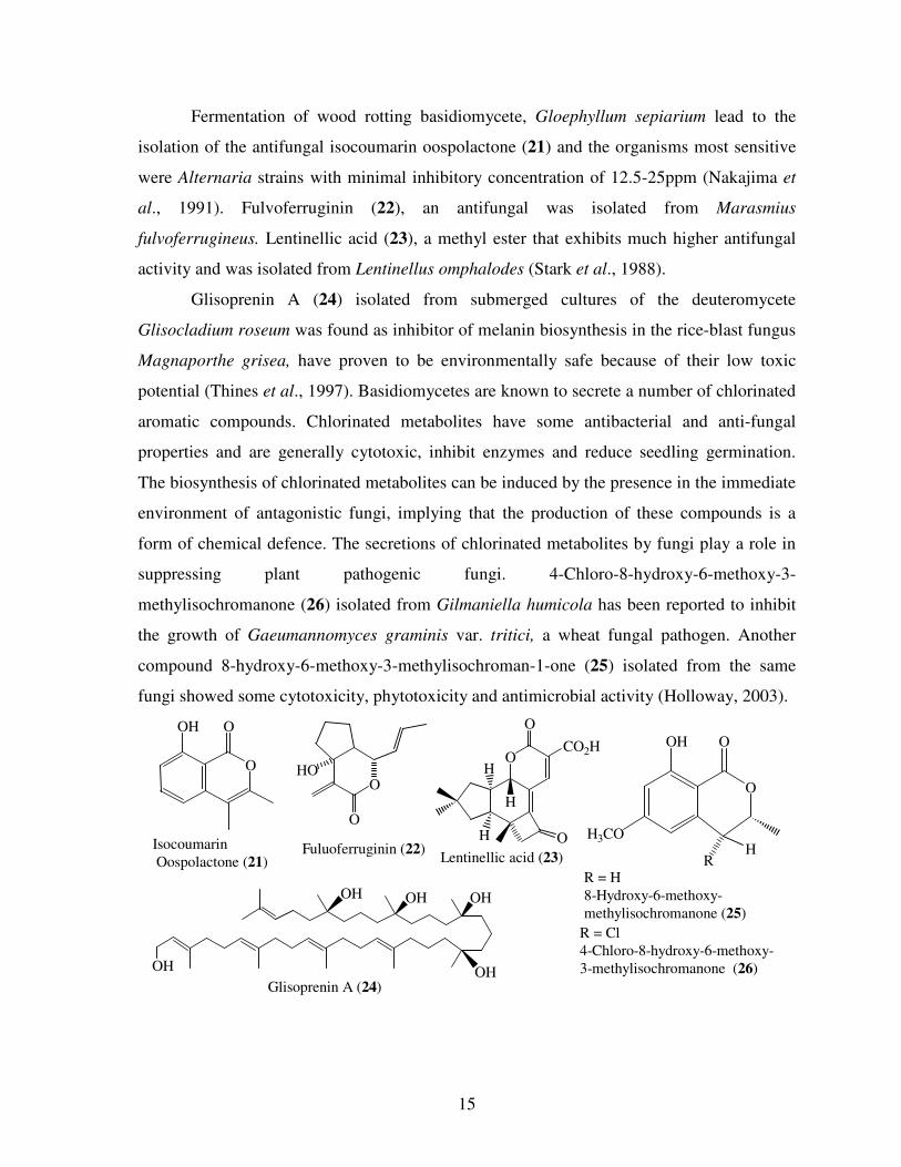
Fermentation of wood rotting basidiomycete, Gloephyllum sepiarium lead to the
isolation of the antifungal isocoumarin oospolactone (21) and the organisms most sensitive
were Alternaria strains with minimal inhibitory concentration of 12.5-25ppm (Nakajima et
al., 1991). Fulvoferruginin (22), an antifungal was isolated from Marasmius
fulvoferrugineus. Lentinellic acid (23), a methyl ester that exhibits much higher antifungal
activity and was isolated from Lentinellus omphalodes (Stark et al., 1988).
Glisoprenin A (24) isolated from submerged cultures of the deuteromycete
Glisocladium roseum was found as inhibitor of melanin biosynthesis in the rice-blast fungus
Magnaporthe grisea, have proven to be environmentally safe because of their low toxic
potential (Thines et al., 1997). Basidiomycetes are known to secrete a number of chlorinated
aromatic compounds. Chlorinated metabolites have some antibacterial and anti-fungal
properties and are generally cytotoxic, inhibit enzymes and reduce seedling germination.
The biosynthesis of chlorinated metabolites can be induced by the presence in the immediate
environment of antagonistic fungi, implying that the production of these compounds is a
form of chemical defence. The secretions of chlorinated metabolites by fungi play a role in
suppressing plant pathogenic fungi. 4-Chloro-8-hydroxy-6-methoxy-3-
methylisochromanone (26) isolated from Gilmaniella humicola has been reported to inhibit
the growth of Gaeumannomyces graminis var. tritici, a wheat fungal pathogen. Another
compound 8-hydroxy-6-methoxy-3-methylisochroman-1-one (25) isolated from the same
fungi showed some cytotoxicity, phytotoxicity and antimicrobial activity (Holloway, 2003).
O
H
O
CO2H
O
H
H
O
OOH
H3COH
R
O
O
HOO
OOH
OH OH
OHGlisoprenin A (24)
R = Cl
4-Chloro-8-hydroxy-6-methoxy-
3-methylisochromanone (26)
Lentinellic acid (23)Fuluoferruginin (22)Isocoumarin
Oospolactone (21)R = H
8-Hydroxy-6-methoxy-
methylisochromanone (25)OH
OH

16
In search for natural products with novel uses particularly related to pest
management, extracts of three endemic species of Flourensia spp. were found to have
antifungal activity on Fusarium oxysporium. The ethanol extracts of F. microphylla, F.
cernua, and F. retniphylla inhibited the mycelial development of F. oxysporium. Inhibition
effect was observed from 10ppm although total inhibition was found at 1000ppm. The
compounds reported to be present in these extracts are glycones of flavanoids 5, 7, 3’-
trihydroxy-3-isobutyroylflavanonol; 5, 7, 3’-trihydroxyflavanone and 5, 7-dihydroxy-3’-
methoxyflavanone (Rodriguez et al., 2007).
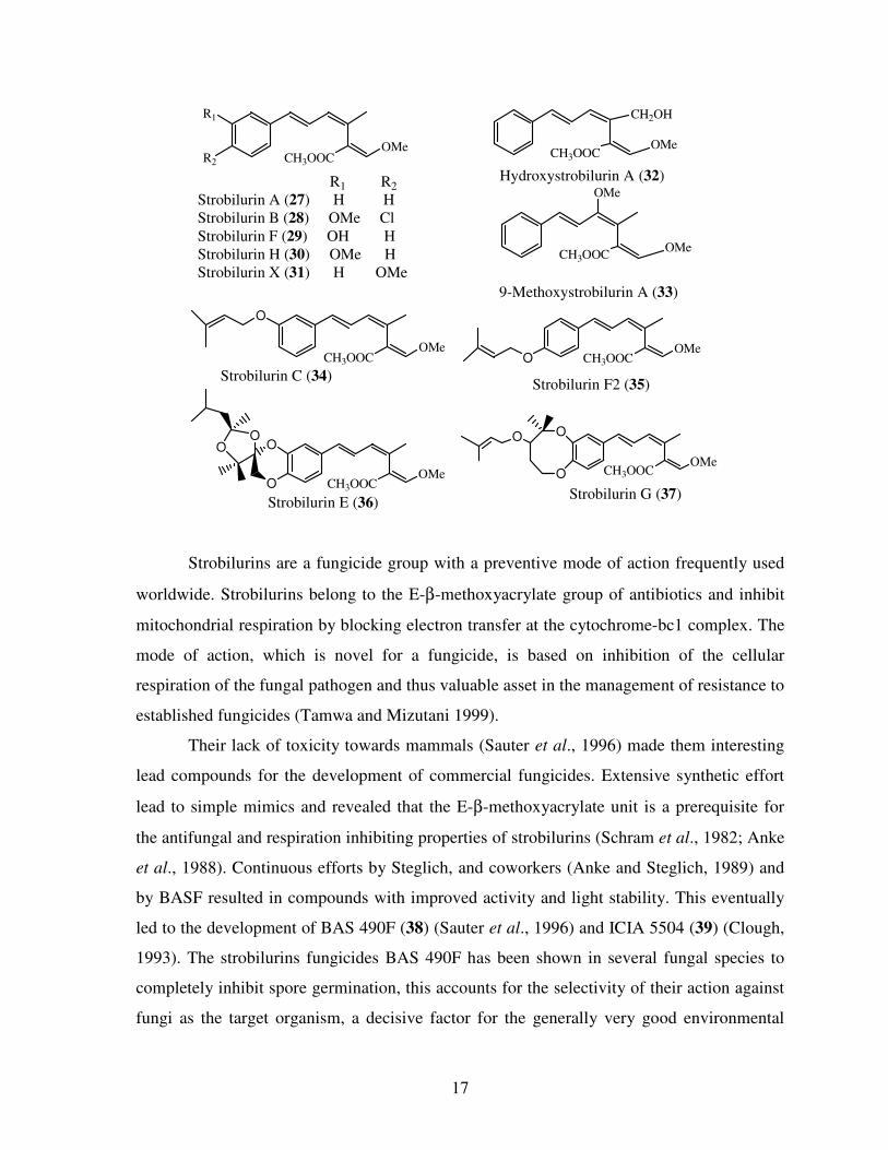
2.6 Strobilurins - a new class of active substances
Strobilurin fungicides have become effective against several different plant
pathogenic fungi. This group is unique in that these fungicides are the first natural lead
synthetic, site-specific compounds to provide significant control of plant diseases caused by
pathogens from all three major groups of fungi: Oomycota, Ascomycota, Basidiomycota.
Fungi that produce strobilurins are found all over the world in all climate zones (Anke and
Steglich, 2000). The discovery of strobilurins (27-37) led to intensive investigation of
basidiomycetes in fermentation for different kinds of biologically active ingredients, chiefly
in Europe, N. America and to some appreciable extent in the Orient (Kaeokamnerd, 1998).
Strobilurins are strong antifungal compounds produced by many basidomycete genera
(Agaricus, Crepidotus, Cyphellopsis, Favolaschia, Filoboletus Hydropus, Mycena,
Oudemansiella, Strobilorus and Xerula).
17
CH3OOCOMe
R1
R2CH3OOC
OMe
CH3OOCOMe
OMe
9-Methoxystrobilurin A (33)
Hydroxystrobilurin A (32) R1 R2
Strobilurin A (27) H H
Strobilurin B (28) OMe Cl
Strobilurin F (29) OH H
Strobilurin H (30) OMe H
Strobilurin X (31) H OMe
CH2OH
O
CH3OOCOMe
O CH3OOCOMe
Strobilurin C (34)Strobilurin F2 (35)
O
O
CH3OOCOMe
OO
CH3OOCOMe
O
O
O
Strobilurin E (36)Strobilurin G (37)
Strobilurins are a fungicide group with a preventive mode of action frequently used
worldwide. Strobilurins belong to the E-β-methoxyacrylate group of antibiotics and inhibit
mitochondrial respiration by blocking electron transfer at the cytochrome-bc1 complex. The
mode of action, which is novel for a fungicide, is based on inhibition of the cellular
respiration of the fungal pathogen and thus valuable asset in the management of resistance to
established fungicides (Tamwa and Mizutani 1999).
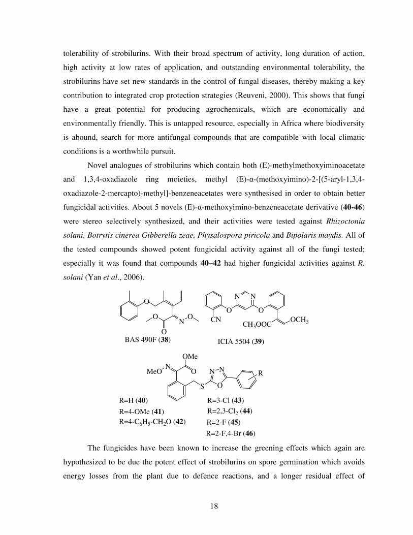
Their lack of toxicity towards mammals (Sauter et al., 1996) made them interesting
lead compounds for the development of commercial fungicides. Extensive synthetic effort
lead to simple mimics and revealed that the E-β-methoxyacrylate unit is a prerequisite for
the antifungal and respiration inhibiting properties of strobilurins (Schram et al., 1982; Anke
et al., 1988). Continuous efforts by Steglich, and coworkers (Anke and Steglich, 1989) and
by BASF resulted in compounds with improved activity and light stability. This eventually
led to the development of BAS 490F (38) (Sauter et al., 1996) and ICIA 5504 (39) (Clough,
1993). The strobilurins fungicides BAS 490F has been shown in several fungal species to
completely inhibit spore germination, this accounts for the selectivity of their action against
fungi as the target organism, a decisive factor for the generally very good environmental
18
tolerability of strobilurins. With their broad spectrum of activity, long duration of action,
high activity at low rates of application, and outstanding environmental tolerability, the
strobilurins have set new standards in the control of fungal diseases, thereby making a key
contribution to integrated crop protection strategies (Reuveni, 2000). This shows that fungi
have a great potential for producing agrochemicals, which are economically and
environmentally friendly. This is untapped resource, especially in Africa where biodiversity
is abound, search for more antifungal compounds that are compatible with local climatic
conditions is a worthwhile pursuit.
Novel analogues of strobilurins which contain both (E)-methylmethoxyiminoacetate
and 1,3,4-oxadiazole ring moieties, methyl (E)-α-(methoxyimino)-2-[(5-aryl-1,3,4-
oxadiazole-2-mercapto)-methyl]-benzeneacetates were synthesised in order to obtain better
fungicidal activities. About 5 novels (E)-α-methoxyimino-benzeneacetate derivative (40-46)
were stereo selectively synthesized, and their activities were tested against Rhizoctonia
solani, Botrytis cinerea Gibberella zeae, Physalospora piricola and Bipolaris maydis. All of
the tested compounds showed potent fungicidal activity against all of the fungi tested;
especially it was found that compounds 40–42 had higher fungicidal activities against R.
solani (Yan et al., 2006).
O
NOO
O
CN
O
NN
O
CH3OOCOCH3
RNN
OS
OMe
NMeO O
BAS 490F (38) ICIA 5504 (39)
R=H (40)
R=4-OMe (41)
R=4-C6H5-CH2O (42)
R=3-Cl (43)
R=2,3-Cl2 (44)
R=2-F (45)
R=2-F,4-Br (46)
The fungicides have been known to increase the greening effects which again are
hypothesized to be due the potent effect of strobilurins on spore germination which avoids
energy losses from the plant due to defence reactions, and a longer residual effect of
19
strobilurins (Jorgensen et al., 1999). Apart from their fungicidal effect strobilurins can
induce physiological and developmental alterations. It has been proposed that, this is due to
inhibition of ethylene biosynthesis, increase of endogeneous cytokinins and reduction in the
carbon dioxide compensation point (Grossmann and Retzlaff, 1997). These effects may
contribute to delayed senescence and thus delayed ripening of the plant. They are site-
specific compounds, which have often indicated a high resistance risk; however, they have a
new mode of action that was difficult for fungi to overcome (Sudisha et al., 2005).
2.5 Application of research findings from the screened fungi
Pathogens continue to be a major problem despite the fact that the world is rich in
under exploited natural products. Although the use of synthetic fungicides in disease
management has long been part of the practices, there is a growing concern due to their
environment and health effects. Some farmers in Kenya currently practice organic farming
aimed at producing healthy crops. The study was based on producing a high yield of healthy
tomato crops using fungicides from naturally occurring compounds, which are
environmentally friendly.
Effective environmental friendly means to prevent or reduce the damage caused by
Fusarium wilt is valuable. The current trend to near zero market tolerance for pesticides
residues in tomatoes provides an additional motivation to search for naturally occurring
chemicals to control the disease. Although strobilurins have proved to be effective
fungicides because of their widespread use on agriculture and originate from natural
compounds, they are commonly used in developed countries. Their efficacies and great
impact of the strobilurin fungicides on agriculture is reflected to very significant
advancements in yield and quality in cereal production (McCartney et al., 2007). Although
they have served as lead compounds for the development of a new generation of industrial
fungicides for crop protection, little evaluation has been done on horticultural crops such as
tomatoes. With application of appropriate fungal biotechnology, there is a great potential to
find novel biologically active compounds from Kenyan natural ecosystem which can be
exploited for the development of safe and effective agrochemicals.
20
CHAPTER THREE
MATERIALS AND METHODS
3.1 Apparatus and materials
Standard laboratory procedures were followed to sterilise media, glassware and
benches. The working bench was continuously maintained sterile with 70% ethanol and a
hot flame, which was also used to sterilise the inoculating needle, blades and wire loops as
well as opening and closing corked sterile flasks.
Liquid and solid media as well as glassware were heat sterilized using an autoclave
(Danfoss 59407-3 No. 375). The apparatus and materials were sterilized twice at a
temperature of 121°C and a pressure of 1.5 bars for 15 minutes. Double sterilization was
used to destroy used and contaminated old plates and other materials used before
incineration. Plates containing F. oxysporium were used and kept in containment for a
maximum of two weeks and later destroyed.
3.2 Preparation of liquid media
Liquid nutrient media was prepared by dissolving 10.0g of molasses, 4.0g glucose,
and 4. g of yeast extract in 1.0L of tap water. The pH of the media was determined using a
pH meters (Fishers Accument Model 610A) and adjusted to 5.5 using 1.0M sodium
hydroxide and 1.0M hydrochloric acid solutions. The media was sterilized immediately by
autoclaving twice at a temperature of 121°C and pressure of 1.5 bars for 15 minutes and left
to cool on the sterile working bench.
3.3 Preparation of test plates
3.3.1 Preparation of potato dextrose agar (PDA) media
The PDA solid media were prepared by autoclaving 39g of the manufacturer’s potato
dextrose agar suspended in 1.0L of distilled water. This was then cooled to 40°C, before
dispensing 15 ml per sterile Petri dish under sterile conditions in a lamina flow hood. PDA
plates were used to grow the strains before being cultured in liquid submerged cultures,
small sliced pieces of the infected tomato plant and culturing of spores of the Fusarium
pathogen.

21
3.3.2 Isolation and culturing of the test organism - F. oxysporium
Infected tomato plants were collected in the green houses of Crops, Horticulture and
Soil (CHS) Department, Egerton University. The infected parts of the plant (roots and stem)
were washed using distilled de-ionised water and then sliced in small pieces. The pieces
from the inner portions were sterilized with 10% of sodium hypochlorite and rinsed with
sterilized distilled de-ionised water. The sterilisation of these pieces was repeated three
times before the pieces were placed on freshly prepared PDA plates and left to grow at room
temperature until the hyphal strands emerged. The cultures were periodically checked for
purity and successively sub-cultured until pure cultures were obtained. The taxonomic
identification of F. oxysporium was performed based on morphological and conidial
characteristics. The mycelium was colourless at first, but at day 7-10 daysit becomes cream-
coloured, pale yellow, pale pink or somewhat purplish. The spores are three to five celled,
septate and with gradually pointed and curved ends.
3.3.3 Preparation of the test plates for antifungal activity
The sterilized PDA media was cooled to a temperature of 40°C in a water bath.
About 10 ml of sterilized distilled water was poured into the plates containing the pure fully-
grown culture of the test organism (F. oxysporium) so as to pick the spores and the
suspension was thoroughly mixed. About 20ml of the spore suspension was added into
250ml of cooled media in the conical flask. The mixture was swirled thoroughly to ensure
uniformity and later dispensed into sterile Petri dishes. This led to approximately 1.0 – 3.0 x
106 spores/ml per plate. Hemocytometer was used to determine concentration of the spores
where the spore density was kept at 106
spores/ml. The antifungal test was carried out
immediately as described later in section 3.6.
3.4 Cultivation of the selected strains in 250 ml submerged cultures
New cultures were prepared from the strains which gave positive results against the
F. oxysporium from their corresponding agar slants, to follow up the reproducibility and
selection of fungal strains for scale-up cultivation. Agar plugs from a well-grown culture of
the fungi were used to inoculate the sterilized liquid malt media. The growth rate was

22
monitored by checking glucose levels using glucose testing strips (Diabur-test ® 5000
(Roche). The growth of the culture was monitored and evaluated daily for any
contamination and biomass build-up.This indicated the progression of growth in the main
culture and once glucose levels were depleted after 21 days, mycelium was separated from
the culture broth by filtration.
3.5 Preparation of crude extracts from mycelium and culture filtrate
3.5.1 Crude extracts from 250 ml scale initial cultivation
Immediately the growth was stopped, mycelium was separated from culture filtrate
by filtration. The crude extract from the culture filtrate were prepared using solvent-solvent
extraction method. It was extracted twice with ethyl acetate in the volume ratio of 1:1. The
combined ethyl acetate extract was dried using anhydrous sodium sulphate to remove
residual water; then filtered and the organic filtrate concentrated under reduced pressure
using a vacuum rotary evaporator at about 40-50oC. The concentrate was transferred into
screw-capped vials and then kept at 4°C awaiting further analysis.
The mycelium was immediately suspended in acetone for 4 hours under constant
agitation with a magnetic stirrer (Gallenkamp). The acetone filtrate was concentrated under
reduced pressure using rotary evaporator to remove acetone and this left an aqueous
solution. The aqueous solution was extracted thrice with equal volume of ethyl acetate. The
combined ethyl acetate solution was dried using anhydrous sodium sulphate, then filtered
and concentrated under reduced pressure using a rotary evaporator. The concentrate was
transferred into screw-capped vials, and kept at 4°C awaiting further analysis.
The crude extracts prepared above were tested and checked for reproducibility of
activity. The strain JO5125 whose extract showed the best reproducibility was selected for
further cultivation on large-scale cultures in 1.0L scale replicates.
3.5.2 Cultivation of the selected strain JO5125 in 1 litre scale replicates
The media was prepared as described in section 3.2. From a well-grown plate of the
selected strain JO5125, pieces of agar plugs (1cm x 1cm) were cut and used to inoculate the
sterilized replicates of 250ml scale of the liquid media. Growth was monitored by
23
accumulation of mycelium and depletion of glucose levels. After evidence of large
accumulation of mycelium the submerged cultures were aseptically transferred into
sterilized 1L scale of the liquid media. The growth profile as a function of time was
monitored by aseptically withdrawing aliquots of samples from the main culture broth and
testing for glucose as previously described in section 3.4. Build-up of the biomass was used
to monitor the rate of growth and as the rate asymptotically approached a constant, the
growth was stopped. Once growth was stopped the mycelium was separated from the culture
filtrate by filtration. The culture filtrates were combined while at the same time mycelia
were also combined. From each of the crude extracts were separately prepared, the
mycelium was processed by organic solvent extraction (refer section 3.5.1) and the culture
filtrate using resin (section 3.5.3).
3.5.3 Liquid-solid adsorption resin extraction of the culture filtrate
A manufacturer’s reverse phase (RP) resin (Mitsubishi HP21 DIAION) was pre-
equilibrated in 1.0M HCl for 72 hours and thoroughly washed with distilled de-ionised
water to neutral pH. The combined culture filtrate was passed thrice through the resin. The
extract-laden column was rinsed with distilled-de-ionised water before elution with organic
solvents. The column was eluted with 300ml of acetone, followed by 300ml of methanol and
the eluents collected. The eluents were concentrated under reduced pressure using rotary
evaporator to remove acetone and methanol, respectively. The aqueous remaining from the
acetone extract was extracted thrice with ethyl acetate to give a dried crude extract, which
was transferred into screw-capped vials and stored at 4°C awaiting further analysis.
3.6 Testing of the crude extracts for anti-fungal activities
Exactly 20µl of the crude extract was applied to a filter paper disc (Rundfilter, ∅6
mm, Schleicher & Schuell) and the solvent left to evaporate. The amount was delivered
using an adjustable (analogue) Eppendorf micropipette. The dry paper disc was carefully
placed onto the surface of the freshly prepared test plate as described in section 3.3.3 and
incubated for duration of 96 hours at room temperature (Rugutt et al., 2006). The results
were evaluated by scoring the inhibition zones around the paper discs and the diameter of
the zones measured in millimetres (mm) and recorded. The larger the diameter of the zone

24
formed the better the activity of the extract. The same procedure was used to test for the
activity of the fractions and pure compounds obtained from the chromatographic separation
of the crude extracts (refer section 3.7.2) and to test for cysteine-adduct formation. This was
done by adding 10µl of 1% cysteine onto a dried paper disk, which was already impregnated
with 20µl of the crude extract. For each there was parallel screen of untreated crude extracts
against F. oxysporium.
3.7 Fractionation of the crude extract based on polarity using chromatography
3.7.1 Determination of the dry weight of the crude sample
The crude extract was dissolved in minimal amount of methanol. About 2.0g of silica
gel was weighed using analytical balance (Precision 310M Swiss Quality) in different
beakers of known weight. To this, small amounts of acetone and methanol extract solution
were slowly added and this was repeated until all the extract was adsorbed onto the silica gel
particles. The solvent was left to evaporate in a running fume hood in a beaker of which was
covered with a perforated aluminium foil to avoid spillage. The silica gel adsorbed extract
was allowed to dry to constant weight and the mass of the crude extract determined.
3.7.2 Column chromatography
This technique was used to fractionate the crude extract into enriched fractions and
eventually to purify the actual active compounds. A glass chromatographic column was
mounted vertically on a fixed support. Fifty grams of silica gel was suspended in 150ml
cyclohexane, and was swirled vigorously until homogenous slurry was obtained. The
column was slurry packed with the silica gel suspension, ensuring that no air is trapped
within the packed slurry. The dry sample adsorbed on to the silica gel was ground to fine
particles, (refer to section 3.7.1) before it was loaded onto the column uniformly as a disc
above the slurry. The incipient air bubbles introduced by the loaded sample were released by
gently tapping the column with air filled rubber. The formed sample disc was anchored in
place with acid-washed sand, which was introduced as a suspension in cyclohexane. This
was done to control turbulence so as to avoid interfering with the formed and anchored
25
sample disc while adding the mobile phase. The column was then eluted with a mobile
phase introduced as discrete solvent gradient system with increasing polarity.
The eluents from the column were collected into test tubes as sequenced fractions
determined by volume and interval time of the collection. Each of the fractions were
correspondingly spotted onto a silica gel pre-coated aluminium TLC plate and developed in
a saturated TLC chamber with an optimal solvent system. To obtain the optimal solvent
system different mixtures of different ratios of cyclohexane/ethyl acetate/methanol were
prepared and evaluated. Once allowed to dry, the developed TLC plate was visualised by
spraying with a freshly prepared p-anisaldehyde solution before heating at 115°C for 10
minutes and by UV lamp preset at fixed wavelengths, λ=254 and 365nm. The visualisation
enabled observation of colour characteristics under the employed technique and
determination of the retardation factor (Rf) values, which were used to pool the collected
fractions into main fractions.
The pooled main fractions were concentrated by removing the organic solvents under
reduced pressure using rotary evaporator. Each fraction was separately transferred to a
screw-capped vial and labelled. Each of these fractions was re-dissolved in 2ml of methanol.
Exactly 20µl of each of the methanol solution was tested against test organism according to
the procedure outlined in section 3.6. The main fractions were further re-chromatographed
but separately, on a smaller column using silica gel in the same solvent system as descried
earlier in this section.
3.7.3 Thin layer chromatography(TLC)
The TLC plate used was silica gel pre-coated aluminium plate (20x20cm, Macherey-
Nagel). The base line and solvent front were marked 1cm from the top and bottom of the
TLC plate, respectively. The point of application of the spots were also marked 0.5cm apart.
The extracts were spotted on the TLC and placed inside the chromatography tank that
contained the saturated solvent systems. In the tank, the TLC plate was developed until the
solvent front was reached. The plates were sprayed with p-anisaldehyde solution, heated to
115oC to visualize the separation. The solvent system that showed good separation on the
TLC plates were noted and used as eluting solvents in the column chromatography. This

26
technique was used to combine the fractions that had the same retardation factor (Rf) values
and to determine the purity of compounds.
3.8 Determination of minimum inhibitory concentration (MIC)
To be able to determine the extent of the observed antifungal activity accurately and
precisely, the crude extracts and pure compounds were tested for minimum inhibitory
concentration (MIC) in a serial dilution assay using modified published methods (Rugutt et
al., 2006). This was done by setting up an array of sterile test tubes, in which 2ml of a
suspension having 1 x 10⁶ spores/ml in 2% (w/v) malt extract were added in each tube. A
blank was prepared by omitting the test substances, while the negative ones contained the
medium only (methanol) the solvent used to dissolve the crude extract and pure compounds.
The extracts were delivered using an analogue micropipette at different concentrations in
sterile tubes under sterile conditions and left to grow for 96 hours at room temperature. The
concentration ranged from 50 to 1500ppm. The lowest concentration whose optical density
corresponds to the blank was taken as the MIC.
3.9 NMR spectroscopy
The pure compounds which showed potent antifungal activity the pathogen were run
to obtain the spectra data using Bruker ARX300 spectrometer. One and two dimension
NMR experiments were carried out on the purified compounds but two dimensions NMR
was only carried on purified compounds that reasonable quantity. The samples were either
dissolved in deuterated chloroform (CDCl3) and the solvent signals (δH 7.26 and δC 77.0)
were used as reference or in deuterated methanol with solvent signals at (δH 4.90, δH 3.40 for
the methyl hydrogen atoms of methanol and δC 49.0). Samples were prepared in 5ml of the
solvent in a 5mm NMR tube and the data was processed using TOPSIN software where they
were attached onto a PNG file. The chemical shifts were reported in ppm relative to the
solvent peaks. From the spectroscopy experiments, structures were proposed based on the
interpretation of the spectra and compared with known compounds in NMR spectroscopic
data reported in literature.

27
CHAPTER FOUR
RESULTS AND DISCUSSION
4.1 Isolation of the test organism - F. oxysporium from diseased tomato plant
The symptoms of the diseased tomato plant was the plant drooping or wilting with
brown discolouration of the vascular region visible in the cross section of infected stems or
roots in the sections tangential to the xylem. The fresh diseased materials were cut in small
piece, sliced off the outer layer and the piece placed on freshly prepared PDA plate. After 96
hours at 25oC the white mycelium grew sufficiently from the cut ends. The white mycelium
was isolated and subcultured in PDA media to get pure cultures using monosporic
techniques (Anguiz, 1989). The mycelium was colourless at first, but with age it became
cream coloured, pale yellow, pale pink, or somewhat purplish.
4.2 Taxonomic identification of the strain JO5125
The fruiting bodies of strains used in this research were collected from indigenous
forest in Kenya (Mt. Kenya and Londiani forests). They were placed on paper bags
serialised using standard methods which included the collector initials, date and sequence in
collection. They were different in morphological features, which indicated that they were
different strains and were collected in the same year (2005). They were kept as herbarium
materials and as pure cultures, which are stored as agar slants in the IBRL Egerton
University. The selected strain JO5125 was collected from Mt. Kenya forest. Further
mycological investigation was done on it using 18 SRNA technology and was identified as
Fusarium species but the genus could not be unequivocally assigned. The mycelium gave a
pink colouration when grown on a PDA plate. This suggested that a Fusarium species was
producing compounds that can control F. oyxporium. This observation had been observed
before at biological level (Fushs et. al., 1999).
4.3 Screening of the crude extracts against the pathogen
About 400 crude extracts prepared from the culture filtrate (Kex) and mycelium
extracts (Mex) were screened against F. oxysporium using agar diffusion technique. This
was done in triplicates to ascertain the results. The clear zone of inhibition around the disc
28
was measured and expressed as inhibition diameter. Twenty of these extracts were found to
give significant activity against the pathogen with zone of inhibition of diameters between
10-40mm shown in table 4.1 below, which was about 5% of the crude extracts screened. The
results significant were as expected since about 5% of the crude extracts screened would
show significant activity against the F. oxysporium which is accepted in screening research
(Rosa, 2005).
The first column in table 4.1 below gives the serial numbers of the basidiomycete,
arranged in ascending order according to their strain code. The letters in the code are the
initials of the collector, the first number gives the year of collection while the remaining
numbers are the sequential position numbers by collection. The third table column gives the
place of collection while the fifth shows the average zone diameter. The extracts that gave
significant activity were recultivated from agar slants and the procedure produced flesh
extracts to check for reproducibility of activity. The outcomes of activity of the crude
extracts are shown in column six which was less than the first activity in column five. This
may be due to difference in secondary metabolites produced by the fungi after recultivation.
Changes in ambient and metabolic conditions cause different enzymatic reaction, which
produces different concentrations of the secondary metabolites. The strain JO50125 was
chosen because it was much more viable than the other strains and both the mycelium and
culture extract were active. It was noted that there was much stronger antifungal activity in
the crude extracts prepared from culture filtrate as compared to those prepared from the
mycelium for strains of fungi that had both types of crude extracts.

29
Table 4.1: Antifungal activity for crude extracts from the 20 strains of fungal cultures
SNo Strain
(code)
Collection
location
Crude
extract
Screening
results Reproducibility
1 JO5057 Mt. Kenya Kex 26±1.7 <10
Mex 20±0.6 <10
2 JO5064 Mt. Kenya Kex 14.3±1.2 Not observed
JO5106 Mt. Kenya Kex 20±1.3 12±2.6
3 JO5115 Mt. Kenya Kex 10±1.2 Not observed
4 JO5125 Mt. Kenya Kex 32±1.4 20±1.7
Mex 20±1.5 10±1.8
5 JO5185 Mt. Kenya Mex 20±1.4 15±1.4
6 JO5301 Mt. Kenya Kex 10±1.6 Not observed
7 JO5304B Mt. Kenya Kex 33±1.6 20±0.8 diffuse
8 JO5315B Mt. Kenya Kex 39±1.2 14±1.2
9 JO5319 Mt. Kenya Kex 19±0.8 22±1.6
10 JO5447A Londiani Kex 12±1.6 <10
11 JO5447B Londiani Kex 23±1.0 Not observed
12 JO5460A Londiani Kex 32±0.6 Not observed
13 JO5460B Londiani Kex 32±1.4 Not observed
14 JO5469 Londiani Mex 15±1.6 <10
15 JO5505A Londiani Kex 30±1.2 Not observed
16 JO5506 Londiani Kex 13.5±1.3 Not observed
17 JO5511A Londiani Kex 30±0.6 <10
Kex: crude extract prepared from extracellular secondary metabolites in the culture filtrate
and Mex: crude extract prepared from intracellular secondary metabolites from mycelia.

30
4.4 Yields of crude extracts from the replicated 1.0L scale
The yields of the crude extracts prepared from the combined culture filtrate and the
mycelium of the cultivated replicates on 1.0L scale of basidiomycete JO5125 are
summarised as shown in figure 4.1. From Mitsubishi (DIAION) resin elution, it was noted
that the crude extract from the methanol eluent (5.46g) had the highest yield in comparison
to the acetone eluted crude extract (2.36g). This is expected since methanol being polar,
extracts up the abundant compounds in the cultures.
31.0 Litres of culture broth
Filtration usingBuchner filtration system
953.25 grammes ofcombined mycelium
30.0 Litres of combinedculture filtrate
2.36 grammes 5.46 grammes
adsorption on Mitsubishi(DIAION) resin
Elution with 300ml of100% acetone
Elution with 300ml of 100% MeOH
2.09 grammes
Figure 4.1: Summary of preparation of culture and mycelium crude extracts
4.5 Test cysteine-adduct formation
Nature of activity of the crude extracts was verified using cysteine to check for easy
adduct forming functional groups (like polyacetylene). The results showed that the zone
diameter of extracts with cysteine was the same as the diameter of extracts without cysteine.
This shows that there could be absence of adduct forming functional groups in the
compounds present in the crude extracts since the zone diameter remained the same after
addition of cysteine. The following results were obtained as shown in Table 4.2:

31
Table 4.2: Bioassay results of JO5125 with cysteine against the F. oxysporium
Strain Zone diameter of extracts
with Cysteine (mm)
Zone diameter of extracts
without Cysteine (mm)
JO5125 Kex (Acetone) 15±1.6 15±1.6
JO5125 Kex (Methanol) 10±1.2 10±1.2
JO5125 Mex 14±1.0 14±1.0
4.6 Determination of minimum inhibitory concentration (MIC) for crude extracts
Evaluation of MIC was carried out for the crude extracts in a serial dilution assay. Stock
solutions of the crude extracts were prepared by dissolving the extracts in methanol. The
different amounts of the crude extracts dissolved in 1 ml of methanol were 54.1mg of the
strain JO5125 Mex, 31.8mg of JO5125 Kex methanol and 137.6mg of JO5125 Kex acetone.
For each of the stock solutions prepared, a concentration range was tested from 50 to 1000
ppm. This was done at intervals leading to 7 concentrations prepared namely; 50, 100, 200,
300, 400, 500 and 1000ppm. This series were set up in 9 replicates, average MIC and
standard deviation were calculated and the results are shown in the table 4.3 below:
Table 4.3: The average MIC and standard deviation of the above crude extracts
Crude extract Mean±Std (ppm)
JO5125 Mex 433.3±50.0
JO5125 Kex acetone 222.2±44.1
JO5125 Kex methanol 455.6±52.7
From the Mitsubishi resin liquid-adsorption extraction process, the crude extract for
acetone eluent was 222.2±44.1ppm, methanol eluent was 455.56±52.7ppm and for the
mycelium prepared (Mex) extract activity was 433.3±50ppm. From the table the crude
extract (JO5125 Kex) coming with the acetone eluent was found be more active than the
mycelium crude extract. This indicated that the active compounds were relatively less polar.
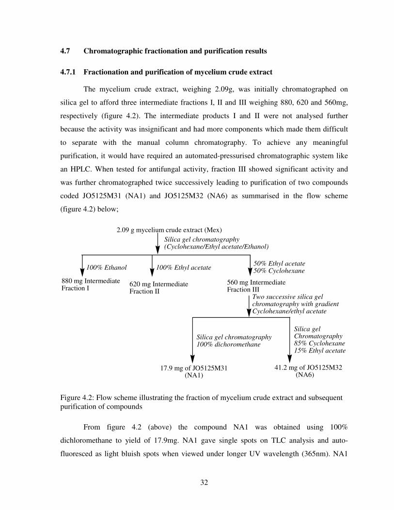
32
4.7 Chromatographic fractionation and purification results
4.7.1 Fractionation and purification of mycelium crude extract
The mycelium crude extract, weighing 2.09g, was initially chromatographed on
silica gel to afford three intermediate fractions I, II and III weighing 880, 620 and 560mg,
respectively (figure 4.2). The intermediate products I and II were not analysed further
because the activity was insignificant and had more components which made them difficult
to separate with the manual column chromatography. To achieve any meaningful
purification, it would have required an automated-pressurised chromatographic system like
an HPLC. When tested for antifungal activity, fraction III showed significant activity and
was further chromatographed twice successively leading to purification of two compounds
coded JO5125M31 (NA1) and JO5125M32 (NA6) as summarised in the flow scheme
(figure 4.2) below;
2.09 g mycelium crude extract (Mex)
880 mg IntermediateFraction I
620 mg IntermediateFraction II
560 mg IntermediateFraction III
17.9 mg of JO5125M31 (NA1)
41.2 mg of JO5125M32 (NA6)
Two successive silica gel chromatography with gradientCyclohexane/ethyl acetate
Silica gel chromatography(Cyclohexane/Ethyl acetate/Ethanol)
100% Ethanol 100% Ethyl acetate50% Ethyl acetate50% Cyclohexane
Silica gel chromatography100% dichoromethane
Silica gelChromatography85% Cyclohexane15% Ethyl acetate
Figure 4.2: Flow scheme illustrating the fraction of mycelium crude extract and subsequent
purification of compounds
From figure 4.2 (above) the compound NA1 was obtained using 100%
dichloromethane to yield of 17.9mg. NA1 gave single spots on TLC analysis and auto-
fluoresced as light bluish spots when viewed under longer UV wavelength (365nm). NA1
33
was further subjected to NMR experiments and the spectra are attached in appendices 1 and
2. From the spectral data it was easily discerned that the compound was an unsaturated fatty
acid. The 13
C spectrum had peak at 179.29ppm, which is a typical carbonyl carbon in
carboxylic acid, and two olefinic carbons that gave carbon signals at 129.9 and 130.2ppm
confirming mono unsaturated fatty acid. The other carbon signals were appearing in the
aliphatic carbon range between 10-40ppm. The results of structure elucidation corroborated
the fact that this compound did not show any significant activity.
From intermediate fraction III, another compound NA6 (41.2mg) was found to have
single spot when analysed on TLC. It gave a yellow colour when sprayed with p-
anisaldehyde. NA6 was found to have significant antifungal activity and was tested in serial
dilution assay in 9 replicates and it gave a minimum inhibitory concentration of
550±35.4ppm. The detailed discussion of the NMR spectra for NA6 will be presented and
discussed in section 4.8.1.
4.7.2 Fractionation and purification of culture filtrate (acetone) crude extract
The 2.36g crude extract, prepared from acetone elution of Mitsubishi (DIAION)
resin (refer figure 4.1), was chromatographed to give three intermediate fractions IV, V and
VI weighing 680, 820 and 420mg, respectively. The intermediate fractions IV and VI were
further chromatographed twice successively because they both had good separation profiles
on TLC plated and had significant activity. Intermediate product IV gave two pure
compounds JO5125K41 (NA2) and JO5125K42 (NA3) weighing 12.5 and 11.5mg
respectively (see figure 4.3 below). Similarly intermediate fraction VI gave three pure
compounds (see figure 4.3) JO5125K61 (NA7), JO5125K62 (NA8) and JO5125K63 (NA9)
weighing 15.0, 6.5 and 2.9mg, respectively.
One of the compounds purified from intermediate fraction IV, NA2, gave a single
spot and yellow colour on TLC analysis. The compound was found to show significant
activity and was tested in a serial dilution assay in 9 replicates to give a minimum inhibitory
concentration (MIC) value of 550±35.4ppm. This compound (NA2) was further analysed in
NMR experiments and the spectra obtained are attached as appendices 3 and 4. The detailed
discussion of the NMR spectra for NA2 will be presented and discussed in section 4.8.1.

34
From the intermediate fraction IV, another compounds NA3 was purified and gave a
single spot on TLC analysis. This spot gave a red colour when spayed with p-anisaldehyde.
The 17.8mg was divided into two portions 11.8mg for NMR experiments and 6.0mg was
used for antifungal activity testing. Given that the amount left for antifungal testing in serial
dilution assay was low, the experiment was conducted in duplicate that gave MIC value of
500ppm. With only two data generated for the MIC value, only the mean is reported and
standard deviation was not calculated.
2.36g culture filtrate (acetone) crude extract (Kex)
680mg Intermediate
Fraction IV820mg Intermediate
Fraction V
420mg Intermediate
Fraction VI
15.0mg of
JO5125K61
(NA7)
2.9mg of
JO5125K63
(NA9)
Two successive silica gel
chromatography
90% Ethyl acetate/10% Methanol
Silica gel chromatography
(Cyclohexane/Ethyl acetate/Ethanol)
100% Ethanol100% Ethyl acetate50% Ethyl acetate
50% Cyclohexane
Silica gel chromatography
100% Ethyl acetate
12.5mg of
JO5125K41
(NA2)
17.8mg of
JO5125K42
(NA3)
3.5mg of
JO5125K62
(NA8)
Silica gel chromatography with
gradient Cyclohexane-Ethyl acetate
Silica gel chromatography
80% Cyclohexane
20% Ethyl acetate
Figure 4.3: Flow scheme illustrating the fraction of culture filtrate (acetone) crude extract
and subsequent purification of compounds
The intermediate fraction V (820mg), though obtained high yields, was not
processed further because the TLC profiles showed that there were compounds that were not
easily separable. The fraction also showed very insignificant antifungal activity and further
purification of the active compounds was not prioritized.
Intermediate fraction VI was further chromatographed using 100% ethyl acetate on
silica gel to afford 3 pure compounds NA7, NA8 and NA9 as shown in the flow scheme in
figure 4.3. Whereas NA7 was obtained in substantial amounts sufficient for both MIC

35
determination and NMR analysis, the yield for NA8 and NA9 were quite low. All the
amounts that were obtained for each of the latter two were sent for NMR analysis and only
gave 1H-NMR spectra. The detailed discussion of the NMR spectra for NA7 will be
presented and discussed in section 4.8.1. In addition to low yield for NA8, there were also
impurities that could not allow further experiments. NA9 was not enough for further
experiments to be performed.
4.7.3 Fractionation and purification of culture filtrate (methanol) crude extract
The methanol 5.46g crude extract was chromatographed as shown in figure 4.4 to
afford three intermediate fraction VII, VIII and IX weighing 1020, 1360 and 1450mg,
respectively. Fractions VII and IX showed weaker activity with the former having better
separation profile on TLC, hence prioritised for further purification. This was motivated by
the fact that the yield was high, there were lots of non-active compounds whose presence
were masking the activity of minor components when definite concentrations were prepared.
This led to fraction VII giving two pure compounds JO5125K71 (NA4) and JO5125K72
(NA5) was weighing 6.2 and 7.5mg, respectively.
5.46g culture filtrate (methanol) crude extract (Kex)
1020mg IntermediateFraction VII
1360mg IntermediateFraction VIII
1450mg IntermediateFraction IX
Silica gel chromatography(Cyclohexane/Ethyl acetate/Ethanol)
100% Ethanol100% Ethyl acetate50% Cyclohexane50% Ethyl acetate
6.2mg ofJO5125K71 (NA4)
7.5mg ofJO5125K72 (NA5)
Silica gel chromatography withisocratic 100% Ethyl acetate
Silica gel chromatography100% Ethyl acetate
Figure 4.4: Flow scheme illustrating the fraction of culture filtrate (methanol) crude extract
and subsequent purification of compounds

36
NA4 and NA5 were obtained in low yields and were only analysed using 1H NMR
experiments. The yields were not sufficient enough for further experiments to be conducted
given the power of the NMR machine. The intermediate fractions VIII and IX were not
chromatographed further when the activity and the separation profiles were considered.
Table 4.4: The antifungal activity of the intermediate fractions and the purified compounds
against the F. oxysporium
Intermediate Fraction Antifungal activity
(mm)
Corresponding purified compound
I 10±1.3 None
II 9±0.9 None
III 15±1.6 JO5125M31 (NA1)
JO5125M32 (NA6)
IV 12±0.6 JO5125K41 (NA2)
JO5125K42 (NA3)
V <10 None
VI 15±1.2 JO5125K61 (NA7)
JO5125K62 (NA8)
JO5125K63 (NA9)
VII 10±2.1 JO5125K71 (NA4)
Each of the intermediate factions (I – IX) was tested for activity using agar diffusion
assay with the results presented in the second column in table 7 against the corresponding
fraction in the first column. From each fraction the corresponding purified compounds are
listed in the third column. When the NMR experiments were conducted on the purified
compounds, compounds NA2, NA6 and NA7 were found to be same compound, a result that
is in agreement with the physic-chemical characteristics observed on TLC analysis. Once
this result was obtained, the three were pooled together to give 55mg and a solution of
known concentration was prepared. The solution was serially diluted for testing against the
37
test organism in a serial dilution assay in 9 replicates. From this a minimum inhibitory
concentration (MIC) value of 550±35.4ppm was found.
4.8 Structure elucidation of the pure compounds
4.8.1 Structure elucidation of NA2, NA6 and NA7
The three compounds were found to be the same as shown in table 4.5 below which
represents the chemical shifts of protons and carbon-13 in their respective NMR spectra.
The chemical shifts were serialised according to the IUPAC naming system of the structure
and HSQC was used to assign the protons attached to carbons. The 13
C NMR spectra of
NA2 and NA7 showed the presence of 18-carbon compound while NA6 showed 17 carbons.
They were only slight differences for 13
C NMR and 1H chemical shifts, a proof that the
compounds were the same. There were two carbonyl carbon in each compound serialised 1
and 7, quaternary carbon (S/No’s 14, 16, 17, 18.) olefinic carbons (11, 12), oxygenated
carbon (3), methylene carbon (4, 5, 6, 8, 9, 10) and one methyl carbon (19). Structure
elucidation was done on compound NA7 since the peaks on the spectra were much clearer
than others (see appendices 16-25).

38
Table 4.5: 1H NMR and
13C NMR data for NA2, NA6, and NA7
Serial
Nos
NA2 NA6 NA7
13C
1H
13C
1H
13C
1H
1 171.5 - 171.5 - 171.8 -
3 73.6 5.01 73.6 5.00 73.6 4.91
4 34.9 1.73 34.9 1.72 35.0 1.43
5 22.5 1.73 22.4 1.78 22.4 1.72
6 43.1 2.60
2.22
43.1 2.61
2.18
43.0 2.56
2.23
7 211.4 - 212.9 - 213.5 -
8 36.8 2.79
2.35
36.9 2.62
2.16
36.7 2.61
2.18
9 21.2 2.18
1.49
21.1 2.17
1.53
21.1 1.89
1.42
10 31.2 2.18
2.23
31.1 2.29
2.17
31.2 2.29
2.18
11 132.0 5.69 132.4 5.70 132.0 5.70
12 133.3 7.04 133.4 7.02 133.4 6.78
13 108.3 6.35 108.8 6.37 108.6 6.78
14 160.5 - 161.3 - 162.9 -
15 102.6 6.34 102.6 6.38 102.0 6.18
16 165.7 - 165.4 - 165.2 -
17 104.2 - - - 103.3 -
18 144.2 - 144.0 - 143.8 -
19 21.0 1.42 20.9 1.34 20.8 1.36
The 1H NMR showed the presence of two meta-coupled aromatic protons at δ 6.3 (d,
J=2.4 Hz, H-15) and δ 6.1 (d, J=2.4 Hz, H-13) and their 13
C NMR signals were observed at
δ 108.6 and δ 102.6 respectively. The NOESY spectrum confirmed the presence of a proton
at C-13 through correlation with H-12. The HMBC spectrum also showed correlation
between C-11 and H-13. Two downfield resonances at δ 162.9 and δ 165.2 had their signals
39
observed in 13
C NMR but were absent in DEPT spectrum therefore, quaternary carbons and
given positions C-14 and C-16, respectively. These were confirmed by HMBC spectrum,
which showed correlation between C-16 and H-15 and C-14 with two protons H-13 and H-
15. The 13
C NMR spectrum further showed two quaternary carbons at δ 103.3 (C-17) and δ
143.8 (C-18). The assignments were confirmed by HMBC spectrum correlations between C-
17 and proton H-15 and C-18 with two protons (H-11 and H-12). The 1H NMR of NA7 also
showed characteristic olefinic proton absorptions at δ 5.7 (J=7.2 Hz, H-11) and δ 6.9 (d,
J=15.1 Hz, H-12) and were found to be trans protons substituted on the double bond since
they commonly have values of approximately 17 Hz. Their 13
C NMR signals were observed
at δ 132.3 and δ 133.4 respectively. The DEPT spectrum revealed six methylene groups,
which were seen in the COSY spectrum to be linearly bonded. The difference in their 13
C
NMR signals observed in methylene carbons showed the presence of the carbonyl carbon C-
7 in between them since methylene protons next to carbonyl group appear at δ 2-3 the region
expected for protons on carbon next to unsaturated centre. This was also confirmed by
HMBC spectrum correlations between C-7 and five protons (2H-6, 2H-8 and H-5). Two
non-equivalent protons were observed on C-6, C-8 and C-10, showing that the structure was
rigid cyclic. The 1H NMR spectrum revealed a methyl group proton resonance at δ 1.3 (d,
J=6.9 Hz, 3H-19) and its 13
C NMR signal occurred at δ 20.18. The adjacent methine group
showed 1H and
13C NMR signals at δ 4.9 and 73.65, respectively, indicating bonding to
oxygen. HMBC correlations were observed between C-3 and 3H-19. The carbon resonance
at δ 171.7 (C-1) was ascribed to the ester carbonyl group. NOESY spectrum (figure 4.5)
confirmed the structure by showing the correlations between the protons. The structure was
supported by the literature values, which are given in the table below.
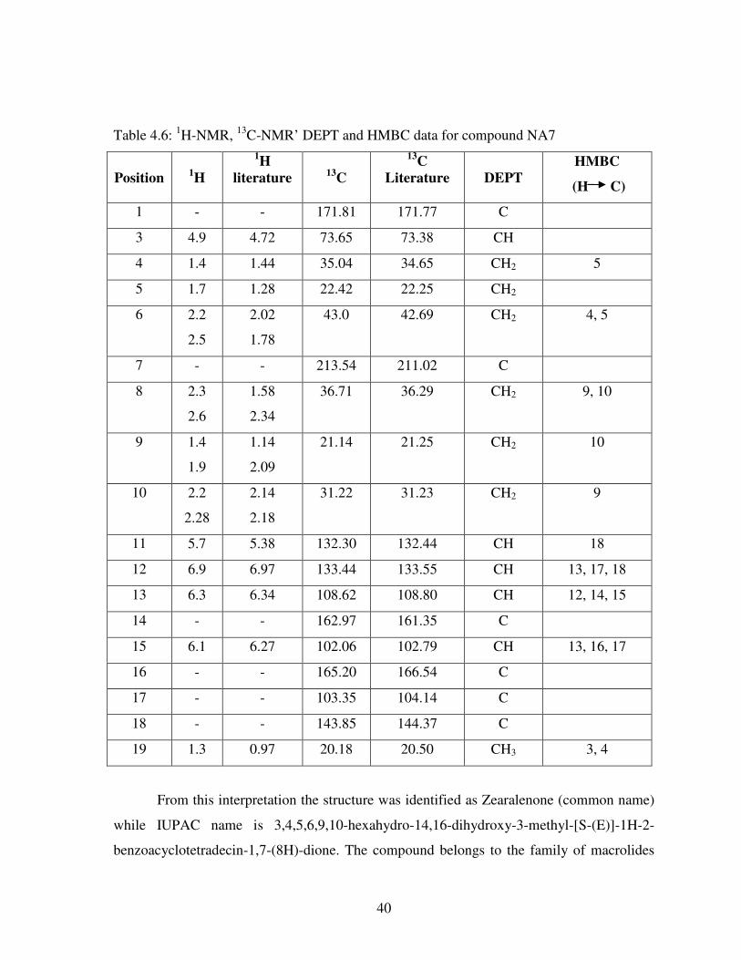
40
Table 4.6: 1H-NMR,
13C-NMR’ DEPT and HMBC data for compound NA7
Position 1H
1H
literature 13
C
13C
Literature DEPT
HMBC
(H C)
1 - - 171.81 171.77 C
3 4.9 4.72 73.65 73.38 CH
4 1.4 1.44 35.04 34.65 CH2 5
5 1.7 1.28 22.42 22.25 CH2
6 2.2
2.5
2.02
1.78
43.0 42.69 CH2 4, 5
7 - - 213.54 211.02 C
8 2.3
2.6
1.58
2.34
36.71 36.29 CH2 9, 10
9 1.4
1.9
1.14
2.09
21.14 21.25 CH2 10
10 2.2
2.28
2.14
2.18
31.22 31.23 CH2 9
11 5.7 5.38 132.30 132.44 CH 18
12 6.9 6.97 133.44 133.55 CH 13, 17, 18
13 6.3 6.34 108.62 108.80 CH 12, 14, 15
14 - - 162.97 161.35 C
15 6.1 6.27 102.06 102.79 CH 13, 16, 17
16 - - 165.20 166.54 C
17 - - 103.35 104.14 C
18 - - 143.85 144.37 C
19 1.3 0.97 20.18 20.50 CH3 3, 4
From this interpretation the structure was identified as Zearalenone (common name)
while IUPAC name is 3,4,5,6,9,10-hexahydro-14,16-dihydroxy-3-methyl-[S-(E)]-1H-2-
benzoacyclotetradecin-1,7-(8H)-dione. The compound belongs to the family of macrolides

41
and had been isolated from a Fusarium species (Pohland et al., 1982). The identity of this
compound was established by comparison of 1H and
13C NMR spectroscopic data with those
reported from literature. The 1H was operated at 500MHz while
13C at 125MHz (Cordier et
al., 1990). The molecular formula from 13
C and 1H data of NA7 was C18H22O5, which gives
eight as the unsaturation number. This information complements to the structure of the
compound shown below and can be accounted for by the aromatic ring, one double bond,
two carbonyls and one lactone ring.
Zearalenone (47) and NOESY correlations
The proposed biosynthetic pathway of Zearalenone shown in figure 4.5 is a relatively
simple structure, which is derived entirely from acetate-malonate units. Head to tail
condensation of nine acetate units synthesizes it via the acetate-malonyl-coenzyme enzyme
system. The aromatic ring is derived through the polyketide synthesis pathway where it
catalysis the biosynthesis of polyketides which are structurally diverse class of natural
products such as antibiotics, toxins and pigments (Blackwell et al., 1985). The cyclization
product formed from a poly-β-keto ester, requiring a variety of reduction processes and
formation of an aromatic ring by aldol condensation near the carboxyl terminus.
O
HO
OH
O
O CH3
13
4
5
6
81011
12
1314
1516
17
18
O
HO
OH CH3
O
O
1 34
5
6
7
1011 8
2
9 7
2
12
1314
15
1617
18
1919
H
H
H
H
H
H
H
H
H
H
H
H
H
9
H
H
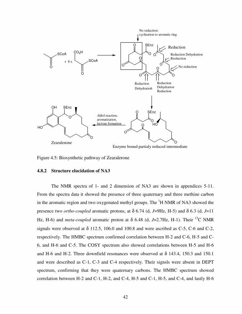
42
SCoA
O
CO2H
SCoA
O
O
O
O
O O
O
O
OO
SEnz
O
O
O
O
O
SEnz
HO
OH
HO
O
SEnz
O
+ 8 x
No reduction:
cyclization to aromatic ring
Reduction
Reduction Dehydration
Rreduction
No reduction
Reduction
Dehydration
Reduction
Reduction
Dehydration
Zearalenone
Aldol reaction,
aromatization,
lactone formation
Enzyme bound partialy reduced intermediate
Figure 4.5: Biosynthetic pathway of Zearalerone
4.8.2 Structure elucidation of NA3
The NMR spectra of 1- and 2 dimension of NA3 are shown in appendices 5-11.
From the spectra data it showed the presence of three quaternary and three methine carbon
in the aromatic region and two oxygenated methyl groups. The 1H NMR of NA3 showed the
presence two ortho-coupled aromatic protons, at δ 6.74 (d, J=9Hz, H-5) and δ 6.3 (d, J=11
Hz, H-6) and meta-coupled aromatic proton at δ 6.48 (d, J=2.7Hz, H-1). Their 13
C NMR
signals were observed at δ 112.5, 106.0 and 100.8 and were ascribed as C-5, C-6 and C-2,
respectively. The HMBC spectrum confirmed correlation between H-2 and C-6, H-5 and C-
6, and H-6 and C-5. The COSY spectrum also showed correlations between H-5 and H-6
and H-6 and H-2. Three downfield resonances were observed at δ 143.4, 150.3 and 150.1
and were described as C-1, C-3 and C-4 respectively. Their signsls were absent in DEPT
spectrum, confirming that they were quaternary carbons. The HMBC spectrum showed
correlation between H-2 and C-1, H-2, and C-4, H-5 and C-1, H-5, and C-4, and lastly H-6
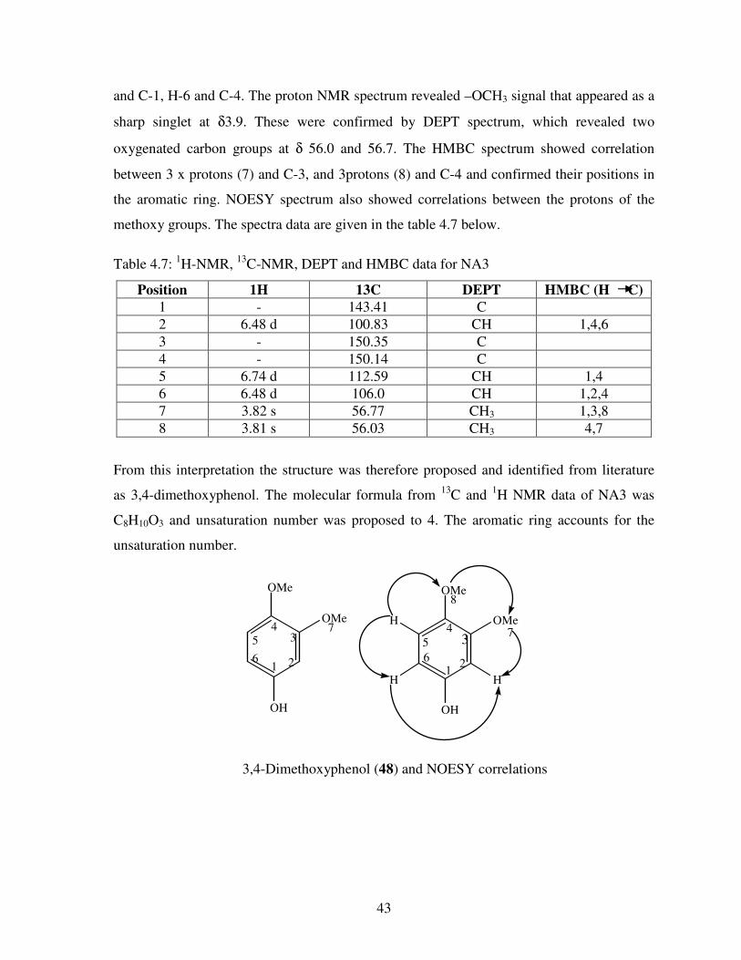
43
and C-1, H-6 and C-4. The proton NMR spectrum revealed –OCH3 signal that appeared as a
sharp singlet at δ3.9. These were confirmed by DEPT spectrum, which revealed two
oxygenated carbon groups at δ 56.0 and 56.7. The HMBC spectrum showed correlation
between 3 x protons (7) and C-3, and 3protons (8) and C-4 and confirmed their positions in
the aromatic ring. NOESY spectrum also showed correlations between the protons of the
methoxy groups. The spectra data are given in the table 4.7 below.
Table 4.7: 1H-NMR,
13C-NMR, DEPT and HMBC data for NA3
Position 1H 13C DEPT HMBC (H C)
1 - 143.41 C
2 6.48 d 100.83 CH 1,4,6
3 - 150.35 C
4 - 150.14 C
5 6.74 d 112.59 CH 1,4
6 6.48 d 106.0 CH 1,2,4
7 3.82 s 56.77 CH3 1,3,8
8 3.81 s 56.03 CH3 4,7
From this interpretation the structure was therefore proposed and identified from literature
as 3,4-dimethoxyphenol. The molecular formula from 13
C and 1H NMR data of NA3 was
C8H10O3 and unsaturation number was proposed to 4. The aromatic ring accounts for the
unsaturation number.
OMe
OMe
OH
OMe
OMe
OH
HH
H4
3
216
57
21
6
57
8
43
3,4-Dimethoxyphenol (48) and NOESY correlations

44
4.9 DISCUSSION
In the screening of the 400 crude extracts of both mycelium and culture filtrate
against the pathogen, thirteen of them gave a zone diameter between 20-40mm in the
bioassay analysis. The activity of the fungal extracts was quite significant as compared with
other crude extracts extracted from native Kenyan plants against F. oxysporium. The
methanol extract of Warbugia ugandensis leaves, Azadirachta indica leaves and seeds, and
Tagetes minuta aerial part gave a zone diameter of 12.0mm, 11.5mm, 10.0mm, 9.0mm and
10.0mm respectively (Rugutt et al., 2006). This shows that the fungi have a potential of
controlling the pathogen. After checking for reproducibility of activity only two crude
extracts (culture extract of strain JO5125 and JO5319) gave a zone diameter greater than
20mm. The strain JO5125 was selected because both culture and mycelium extracts were
active with culture having significant activity.
The strain JO5125 was found to be Fusarium species after molecular markers
technology investigation using 18 SRNA. The difference between strain JO5125 and the
phytopathogen F. oxysporium was that on sub-culturing on PDA media, the mycelium of
strain JO1525 formed pink pigmentation on the plate while the F. oxysporium formed a
purple pigmentation. Also the formae speciales of fungus JO5125 has not been identified.
This is possible as confirmed from literature where Fuchs et al. (1999) and Fravel et al.
(2005) found that non-pathogenic F. oxysporium could effectively suppress Fusarium wilt
of the tomato through a host-mediated mechanism.
Three crude extract were prepared from submerged cultures of fungus JO5125 which
were JO5125 Mex, JO5125 Kex methanol, JO5125 Kex acetone and their MIC
concentrations was found to be 433.3±50ppm, 455.56±52.7ppm and 222.2±44.4ppm
respectively. The acetone eluent culture extract was more active giving the lowest MIC
concentrations as compared to other extracts. Also activity of the intermediates fractions of
the acetone eluent extract were more active and had good separation thus more compounds
were purified from them compared to fractions of the mycelium and methanol eluent
extracts. The MIC from these extracts was quite significant compared with other crude
extracts such as from the essential oils from Curcuma longa that gave complete mycelia

45
inhibition at 2000ppm (Gurdip et al., 2002) and from Flourensia species, which gave total
inhibition at 1500ppm.
The compounds isolated from strain JO5125 were 3,4-dimethoxyphenol and
zearalenone. The two compounds were isolated from acetone eluent extract and labelled as
NA2 for zearalenone and NA3 for 3,4-dimethoxyphenol although zearalenone was also
isolated from mycelium extract labelled as NA6 and as NA7 for methanol eluent extract.
Zearalenone is a white crystalline solid, produced by fermentation of various Fusarium
species such as F. graminearum, F. culmorum, and F. crookwellence. 3,4-dimethoxyphenol
has been produced by various plant species including Croton, Passiffora, lysimachia and
from a range of marine microorganism. The average MIC for 3,4-dimethoxyphenol was
500ppm while for zearalenone was 550±35.4ppm. Comparing the results with the fungicides
used to control the pathogen, Song et al. (2004) found that MIC of the fungicides screened
in vitro on tomato wilt pathogen for seven fungicides, prochlorax, carbendazin, thiram,
toclofos-methyl, hymezazol, azoxystrobin and carboxin were 0.019, 0.235, 26.292, 53.606,
69.961, 144.58, and 154.03ppm respectively. Other antifungal compounds isolated from
plants such as muzigadial (6) was isolated from Warbugia ugandensis gave a MIC of 50ppm
(Rugutt et al., 2006). Rutacridone (10) and its epoxide derivative (11) were found to inhibit
the growth of F. oxysporium at low concentration of 20ppm from ethyl acetate extracts of
the roots of Ruta graveolens (Kumudini et al., 2005). Although the results of the MIC of the
zearalenone and 3,4-dimethoxyphenol are not as significant as the above antifungal
compounds, they also have a potential to control the plant pathogen.
However, zearalenone is a mycotoxin but only becomes injurious to mammals upon
ingestion. From the MIC results obtained zearalenone has been found to be active against
the phytopathogen F. oxysporium. The pathogen infects the tomato plant by penetrating
through root tips or wounds on roots. Therefore if zearalenone has to be applied to control
the pathogen it must be applied onto the soil. Therefore, there is a high probability that
bioaccumulation of the mycotoxin might not appear in the fruits of the crop since it’s not
sprayed directly on the plant but absorbed through the roots. The plant system also has a
mechanism of degrading the toxins since the pesticide residual found in the harvested crop
are of low concentrations. Zearalenone qualifies as environmentally friendly fungicide since
it is from nature, thus biodegradable and can easily be degraded by plants mechanism. The

46
activity can be associated with the lactone ring found in structure of zearalenone. The
lactone rings are major pharmacophoric substructures found in compounds isolated from
fungi. These compounds are fomannosin (20), isocoumarin oospolactone (21),
fuluoferruginin (22), lentinellic acid (23) and 4-chloro-8-hydroxy-6-methoxy-3-
methylisochromanone (25) that have antifungal activity against other plant pathogens. The
other compound 3,4-dimethoxyphenol was also found to be effective in to controlling F.
oxysporium as shown from the MIC results. It is biodegradable since it is from nature and
not toxic. Zearalenone and 3,4-dimethoxyphenol can be potentially used for the
development of new fungicides if zearalenone will have low residual effect on the product
and can be effective agrochemicals.
47
CHAPTER FIVE
5.0 CONCLUSION AND RECOMENDATIONS
5.1 Conclusion
On screening of the crude extracts from both the mycelium and culture broth of the
strains against F. oxysporium, 20 of the extracts screened gave significant activity. This was
as expected that in a random hit rate analysis about 5% of the extracts screened would show
significant and interesting results. The active strains were recultivated again to check for the
reproducibility of activity. One strain serialized JO5125 was selected from the recultivated
strains and crude extract were prepared from the mycelium and culture filtrate using
different solvents. From the Mitsubishi resin liquid-adsorption extraction process for culture
filtrate the acetone eluent crude extract was more potent against F. oxysporium (an in vitro
MIC of 222.2±44.4ppm) compared to that of methanol (MIC 455.56±52.7ppm). This shows
that the extract eluted with less polar solvent (acetone) was more effective in controlling the
pathogen than the extract eluted with more polar (methanol). The mycelium crude extract
showed low efficacy (MIC 433.3±50ppm) against F. oxysporium.
The fractions that had good separation were purified further to give pure compounds.
NMR analysis was done on pure compounds and their spectral data has been provided but
some were in very low yields only the proton NMR studies were possible. They were only
two compounds (3,4-dimethoxyphenol and zearalenone) whose yield were high therefore 1
and 2 dimension NMR spectra analysis was done on them. MIC was determined for 3,4-
dimethoxyphenol and zearalenone where they gave average in vitro MIC of 500 and
550±35.4ppm respectively against the pathogen. The MIC from the pure compounds was
higher as compared to that of the crude extracts. The crude extracts especially the acetone
extract where the two compounds were purified from was more effective due synergistic
effects from the compounds present. These antifungal compounds were found to be mid-
polar from their chemical nature and evidence that the agents responsible for activity are
organic compounds. The results obtained from the crude extracts and pure compounds
supports the use of fungi to produce antifungal compounds. The antifungal compounds
produced can be of low cost, biodegradable and selective in activity and thus fit as agents of

48
food protection. Also the findings that the compounds inhibit F. oxysporium can be
harnessed for the control of tomato fusarial wilts especially in soil interactions to inhibit
xylem blockage. This is possible as in the chemical defence theory in the wild can be
inferred, Fusarium species can inhibit another Fusarium species. Finally, this study further
reaffirms the assertion that there are plenty of untapped resources in the higher fungi, which
could be of great utility to mankind.
5.2 Recommendations
Following recommendations were made in relation to the study:
i. The other fungal strains that showed positive results during screening should be
investigated for the production of compounds that have activity against F. oxysporium.
ii. Broad screening of more basidiomycetes against the F. oxysporium test organism
iii. In vivo analyses of the pure compounds on pot experiments and evaluate the
bioaccumulation of these compounds in the tomato fruits.

49
6.0 REFERENCES
Agrios, G. N. (1998). Plant pathology. Academic press Ltd., 4th edition New York.
Ahmed, I. H., Labuschagne, N. and Korsten, L. (2007). Screening rhizobacteria for
Biological control of Fusarium root and crown rot of sorghum in Ethiopia. Biol. Cont.
40: 97-106
Alabouvette, C. Steinberg, C., Trillas-Gay, M. I., and Cotxarrera, L. (2002). Use of sewage
sludge compost and Trichoderma asperellum isolates to suppress Fusarium wilt of
tomato. Soil Bio. and Biochem. 34: 467-476.
Allen, T. W., Enebak, S. A. and Carey, W. A. (2004). Evaluation of fungicides for control of
species of Fusarium on long leaf pine seed. Crop Prot. 23: 979-982.
Anguiz, R. (1989). Anastomosis group pathogenicity and other characteristics of
Rhizoctonia solani isolated from potatoes in Peru. Plant Dis. 73: 199-201.
Anke, T. and Steglich, W. (1989). β-Methoxyacrylate antibiotics from biological activity to
synthetic analogues: Biologically active molecules – identification, characterization,
and synthesis (SCHLUNEGGAR, U. P., Ed), pp. 9-25. Berlin- Heidelberg: Springer-
Verlag.
Anke, T. and Stelglich, W. (2000). Strobilurins a success story. Angew Chem. Int. Ed. 38:
408-412.
Anke, T., Schram, G., Stelglich, W. and Von Ja Gow, G. (1988). Structure-activity
relationship of natural and synthetic E-β-Methoxyacryalates of the strobilurin and
oudemansin series, in: The roots of modern biochemistry (Kleinkauf, H., Von Dohren,
H., Janicke, L., Eds.), pp. 657-662. Berlin.
Anshu D., Mani U., Babita R., Pant U. C. and Gupta I. J. (1998). Synthesis and
antimicrobial evaluation of some new fluorinated spiro [ [ 1,5 ] -benzothiazepin-2,3'
[ 3'H] -indol] -2'-(1 'H) –ones. J. Flourine Chem. 91: 171-174.
Arnold, A. E., Maynard, Z., Gilbert, G. S., Coley, P. D. And Kursar, T. A. (2000). Are
tropical fungal endophytes hyperdiverse? Ecol. Lett. 3: 267-274.
Bailey, K. L. and Lazarovits, G. (2003). Suppressing soil borne diseases with residue
management and organic amendments. Soil Tillage Res; 72: 169-180.
50
Basset, C., Sherwood, R. T., Kepler, J. A. and Hamilton, P. B. (1967). Production and
biological activity of fomannosin, a toxic sesquiterpene metabolite of Fomes annosum.
Phytopathol. 57: 1046-1052.
Berdy, J. (2003). Are actinomycetes exhausted as a source of secondary metabolites? In
Biotechnologija (ISSN0234-2751) Prot. 9th
Int. Symp. On the Biology of
Actinomycetes. Debanov VG. pp. 13-34.
Bing-Ji, M., Yan, Z., Lian-Zhen, L., He-Min, L., Zhi-Mong, G. and Yuan, R. A. (2008).
New Cyathane-xyloside from the mycelia of Hericium erinaceum. Z. Naturforsch.
63: 1241-1242.
Blackwell, D. A., Greenhalgh, R. and Miller, J. D. (1985). 13
C NMR study of the
biosynthesis of toxins by Fusarium graminearum. J. Biol chem. 260: 4243-4247.
Butler, M. S. (2005). Bioactive molecules from natural products. Nucl. Acid. Res. 34: 678-
683.
Cabello, M. A., Platas, G., Collado, J., Diez, M. T., Martin, I. Vicente, F. Meinz, M., Onishi,
J. C., Douglas, C., Thompson, J., Kurtz, M. B., Schwartz, B. E., Bills, G. F.,
Giacobbe, R. A., Abruzzo, G. K., Flattery, A. M., Kong, L. and Pelaez, F. (2001).
Arundifungin, a novel antifungal compound produced by fungi; biological activity
and taxonomy of the producing organism. Int. Microbial. 4: 493-102.
Cavigelli, M. A., Lengnick, L. L., Buyer, J. S., Fravel, D. R., Handoo, Z. A., McCarty, G.
W., Millner, P. D., Sikora, L. J., Wright, S. E., Vinyard, B. T. and Rabenhorst, M.
(2005). Landscape level variation in soil resources and microbial properties in a no-till
corn field. Appl. Soil Ecol. 29: 99-123.
Chew, P. S. and Hall, R. (1984). Effect of Pythium root rot on yield of white bean Phaseolus
vulgaris. Crop Prot. 3: 423-429.
Chitwood, D. J. (2002). Phytochemical based strategies for nematode control. Ana. Rev.
Phytopathol. 40: 221-249.
Clough, J. M. (1993). The strobilurins,oudemansins, and myxothiazols, fungicidal
derivatives of β-methoxacrylic acid. Nat. Prod. Rep. 10: 565-574.
Cole, M. D., Bridge, P. D., Dellar, J. E., Fellows, L. E., Cornish, M., C. and Anderson, J. C.
(1991). A neo-clerodane balcalensis diternoid from Scutellaceae. Phytochem. 31:
3433-3435.
51
Copping, L. G. and Menn, J. J. (2000). Biopesticides, their action application and efficacy.
Pest Manag. Sci. 56: 651-676.
Cordier, C., Gruselle, M., Jaouen, G., Hughes, D. W. and McGlinchey, M. J. (1990).
Structures of Zearalenone and Zearalenone in solutions: A High-Field NMR and
molecular modeling study. Magn. Reson. Chem. 28: 835-845.
Demain, A. L. (1999). Pharmacologically active secondary metabolites of microorganism.
Appl. Microbial Biotehnol. 52: 455-463.
Dong-Ze, L., Rui-Rui J., Fei, W. and Ji-Kai, L. (2008b). A new spiroaxane sesquiterpene
from cultures of the basidiomycetes Pholiata adiposa. Z. Naturforsch 63b: 111-113.
Dong-Ze, L., Rui-Rui, J., Fei, W. and Ji-Kai, L. (2008a). A novel sesquiterpene from
cultures of the Basidiomycetes Boletus calopus. Z. Naturforsch 63b: 114-116.
Elizabeth, A. B. and Handelsman, J. (1999). Biocontrol of plant diseases. FEBS Microbiol
Lett. 171: 1-9.
Elmer, W. H. and McGovern, R. J. (2004). Efficacies of integrating biologicals with
fungicides for the suppressions of Fusarium wilt of tomato. Crop Prot. 23: 909-914.
Flood, J., (2003). Fusarium wilt of tropical perennial crops: challanges to management.
CABI. Bioscience Egham surrey, United Kingdom.
Fravel, D. R., Deahl,. L., and Stommel, J. R. (2005). Compatibility of the biocontrol fungus.
Fusarium oxysporium. Strain. CS-20 with selected fungucides. Biol. Cont. 34: 165-169
Fuchs, J. G. Moenne-Locioz, Y. and Defago, G. (1999). Ability of non-pathogenic Fusarium
oxysporium Fo47 to protect tomato against Fusarium wilt. Biol. Cont. 14: 105-110.
Grossmann, K. and Retzlaff, G. (1997). Bioregulatory effect of the fungicidal strobilurin
kresoxim-methyl in wheat (Triticum aestivum). Pest. Sci. 50: 11–20.
Gullino, M. L., Leoux, P. and Smith, C. M. (2000). Uses and challenges of novel
compounds for plant disease control. Crop Prot. 19: 1-11.
Gurdip, S., Prakash, S. and Sumitra, M. (2002). Chemical and biocidal investigations on
essential oils of some Indian Curcuma species. Progress in Crystal Growth and
Characterization of Materials 75-81.
Hang, Y. D. and Woodams, E. E. (2003). Control of Fusarium oxysporium by baking soda.
Lendensm.-Wiss. u-Technol. 36: 803-805.
52
Hansen, E. M., Myrold, D. D. and Hamm, P. B. (1996). Effects of soil fumigation and cover
crops on potential pathogens, microbial activity, nitrogen availability, and seedling
quality in tomato nurseries. Phytopathol. 80: 698-740.
Hautzel, R., Anke, H. and Sheldrick, W. S. (1990). Mycenone, a new metabolite from
mycena species TA 87202 (Basidiomycetes) as an inhibitor of isocitrate lyase. J.
Antibiot. 43: 1240-1244.
Hawksworth, D. L. (2001). The magnitude of fungal diversity; the 1.5million species
estimate revised. Mycol. Res. 105: 1422-1432.
Hengkui, L., Yamei, D., Jianxin, W., Changjun, C., Juping, N. and Mingguo, Z. (2008).
JS399-19, a new fungicide against wheat scab. Crop Prot. 27 90-95.
Herbert, J. A. and Marx, D. (1990). Short term control of Panama diseases in South Africa.
Phytophylactica. 22: 339-340.
Herman, G. E. (2000). Myths and dogmas of biocontrol: changes in perception derived from
research on Trichoderma harzianum T-22. Plant Dis. 84: 377-393.
Holloway, P. (2003). Potential of fungal chlorinated metabolites. ARDI Project 00-459.
Phytochem. 36: 35-38.
Hostettman, K. and Martson, A. (2000). Chemistry and pharmacology of natural products. J.
Antibiot. 53: 1153-1166.
Ibadur, R .S., Pravin, K. S., Jaya S. and Jagdamba, S. (2003). Synthesis and Fungicidal
Activity of Novel 4,4’-Bis(2’’-aryl-5’’-methyl/unsubstituted-4’’-oxo-thiazolidin-3’’-
yl) Bibenzyl. J. Agric. Food Chem. 51: 7062-7065.
Jorgensen, L. N., Henriksen, K. E. and Nielsen, G. C. (1999). Adjusting thresholds for
Septoria control in winter wheat using strobilurins. In: Van Ginkel, M., Mc Nab, A.,
Krupinsky, J. (Eds.), Septoria and Stagonospora Diseases of Cereals: A Compilation
of Global Research. CIMMYT, Mexico, DF, pp. 173–175.
Kaeokamnerd, W. (1998). Khao, Yae National Park and its diversity; the importance of
inventory. Proceedings of the Asia-Pasific Mycological conferences on Biodiversity
and Biotechnology, July 6-9. Hua Hin, Thailand.
Klasse, H. J. (2002). Calcium cyanide – An important tool in methyl bromide replacement
strategies. SKW. Trostberg AG, Germany.

53
Knight, V., Sanglier, J., DiTullio, D., Braccili ,S., Bonner, P., Weaters, J., Hughes, D. and
Zhang, L. (2003). Diversifying microbial natural products for drug discovery. Appl
Microbiol 23: 1146-1152.
Kumudini, M. M., Kevin, K. S., David, E. W. and Stephen, O. D. (2005). Algicidal and
Antifungal compounds from roots of Ruta graveolens and synthesis of their analogs.
Phytochem. 66: 2689-2695.
Larkin, R. P. and Fravel, D. R. (2002). Efficacy of various fungal and bacterial biocontrol
organism for Fusarium wilt of tomato. Plant Dis. 83: 1022-1028.
Lasure, L. L. (2000). Screening microbial metabolites for new drugs – Theoretical and
practical issues. J. Antibiot. 53: 1123-1129.
Lauer, U. Anke, T., Sheldrick, W. S., Scherer, A. and Stelglich, W. (1989). Antibiotics from
basidiomycetes. Aleurodiscal: an antifungal sesterpenoids form Aleurodiscus mirabilis
(Berk and Curt.) Hohn. J. Antibiot. 42: 875-882.
McCartney, C., Mercer, P. C., Cooke, L. R. and Fraaije, B. A. (2007). Effects of a
Strobilurins based on disease control, green leaf area, yield and development of
fungicide resistance in Mycosphaerella graminicola in Northern Ireland. Crop Prot.
26: 1272-1280.
Merterrhazy, A. and Bartok, T. (1996). Control of Fusarium by fungicides by fungicides and
its effect on toxincontamination of the grains. Pflanzenschutz-Nachrichten 49: 181-
198.
Mujeebur, R. K. and Shahana, M. K. (2002). Effects of root dip treatment with certain
phosphate solubilizing micro organism on the Fusarium wilt of tomato. Biores.
Techn. 85: 213-215.
Nakajima, M., Itoi, K., Takamatsu, Y., Sato, S. and Takada, M., (1991). A new fungal
metabolite with herbicidal activity. J. Antibiot. 48: 1389-1395.
Nel, B., Stanberg, C., Labuschagne, N. and Viljoen, A., (2006). Evaluation of fungicides and
sterilants for potential application in management of Fusarium wilt of banana. Crop
Prot. 26: 697-705.
Piggut, A. M. and Karuso, P. (2004). Quality, not quantity: The role of natural products and
chemical proteomics in modern drug discovery. Comb. Chem High throughput
series. 7: 607-630.
54
Pohland, A. E., Schuller, P. L. and Steyn, P. S. (1982). Physiochemical Data for some
selected mycotoxins. Pure and Appl. Chem. 54: 2219-2284.
Reuveni, M. (2000). Efficacy of trifloxystrobin (Flint), a new strobilurin fungicide, in
controlling powdery mildews on apple, mango and nectarine, and rust on prune trees.
Crop prot. 19: 335-341.
Ristaino, J. B. and Thomas, W. (2000). Agriculture, methyl bromide and the ozone hole.
Plant Dis. 81: 964-977.
Rodriguez, J. D., Hernandex-castillo, D. Angulo-Sanchez, J. L., Rodriguez-Garaa, R.
Villarreal, Q. J. A. and Lira-Scldiwar, R. H. (2007). Indust. Crops and Prod. 25:
111-116.
Rosa, H. L., Machado, K. M. G., Jacob, C. C., Capaleri, M., Rosa, C. A. and Zani, L. C.
(2003). Screening of Brazillian basidiomycetes for antimicrobial activity. Mem. Inst.
Owswaldo Cruz, Rio de Janeiro. 98: 967-974.
Rugutt, J. K, Ngigi, A. N., Rugutt, K. J. and Ndalut, P. K. (2006). Native Kenyan plants as
possible alternatives to methyl bromide in soil fumigation. Phytomedicine 13: 576–
583
Santos, B. M., Gilreath, J. P., Motis, T. N., Noling, J. W., Jones, J. P. and Norton, J. A.
(2006). Comparing methylbromide alternatives for soilborne diseases nematodes and
weed management in fresh market tomato. Crop Prot. 25: 690-695.
Sauter, H. Ammermann, E, Benolt, R. B., Gold, R. E., Wingert, H. and Schirmer, U. (1996).
Mitochondrial respiration as a target for antifungal: Lessons from research on
strobilurins, in antifungal agents; Discovery and mode of action, pp. 173-191. Oxford:
Bios Scientific Publishers.
Schram, G., Stelglich, W. and Anke, T. (1982). Structure-activity relationship of stobilurins,
oudemansin, and synthetic analogues, Abstact 457, in: Abstracts 13th Int. Congr.
Microbial. Boston.
Sivan, A., Uoko, O. and Chet, I. (1987) Biological control of Fusarium crown rot of tomato
by Trichoderma harzianum under field conditions. Plant dis. 71: 587-592
Song, W., Zhou, L., Yang, C., Cao, X., Zhang, L. and Liu, X. (2004). Tomato Fusarium wilt
and its chemical control strategies in a hydroponic systems. Crop Prot. 23: 243-247.

55
Sparapano, L., Bruno, G., Fierro, O. and Evidente, A. (2004). Studies on structure-activity
relationship of sphaeropsidins A-F, phytotoxins produced by Sphaeropsis sapinea f.
sp. Cuppressi. Phytochem 65: 189-198.
Stark, A., Anke, T., Moeck, U., Steglich, W., Kirtel, A. and Will, G. (1988). Lentinellic
acid, a biologically active protoilludane derivative from lentinellus species
(basidiomycetes). Z. Naturforsch. 43: 177-183.
Strauss, J. and Labuschagne, N. (1993). In vivo and in vitro effect of fungicides on Fusarium
solani associated with citrous roots. Plant Sci. 7: 56-59.
Sudisha, K. N., Amruthesh, S. A., Deepak, N. P., Shetty, B. R., Sarosh, H. and Sheckar, S.
(2005). Comparative efficacy of strobilurin fungicides against downy mildew disease
of pearl millet. Pesticide Biochem. Physiol. 81: 188-197.
Suleman, P., Tohamy, A. M., Saleh, A. A., Madkour, M. A. and Straney ,D. C. (2003).
Variation in sensitivity to tomatine and rishitin among isolates of Fusarium oxysporum
f. sp. lycopersici, and strains not pathogenic to tomato. Physiol Mol Plant Pathol. 48:
131–144.
Tamwa, H. and Mizutani, A. (1999). Mode of action of strobilurins fungicides. J. Pesticide
sci. 24: 189-196.
Thines, E., Anke, H., and Weber, R. W. (2004). Fungal secondary metabolites as inhibitors
of infection-related morphogenesis in phytopathogenic fungi. Mycol. Res. 108: 14-
25.
Thines, E., Eilbert, F., Sterner, O. and Anke, H. (1997). Glisoprenin A, an inhibitor of the
signal transduction pathway leading to appressorium formation in germinating conidia
of Magnaporthe grisea on hydrophobic surfaces. FEMS Microbiol. Lett. 151: 219-224.
Turner, W. B. (1971). Fungal metabolites I. Academic press inc., London. United Kingdom.
Turner, W. B. and Aldridge D. C. (1983) Fungal metabolites II. Academic press, New York.
Vol 2: 631.
U. S. Environmental protection Agency, office of Atmospheric programs Washington DC.
Methyl bromide use background. http://www.epa. Gov/docs/ozone/mbr/background.
Html. (accessed June 2009) path.
Weber, R. W. S., Kappe, R., Paululat, T., Mosker, E. and Anke, H. (2007). Anti-candida
metabolite from endophytic fungi. Phytochem. 68: 886-892.
56
Yan, L., Liu J., Zhang, H. ,Xiangping, Y. and Zhaojie , L. (2006). Stereoselective synthesis
and fungicidal activities of (E)-a-(methoxyimino)-benzeneacetate derivatives
containing 1,3,4-oxadiazole ring. Bioorg. & Med. Chem. Letts. 16: 2278–2282.
Young, M. C., Braga, M. R., Dietrich, S. M., Gottlied, H. E., Trevisan, L. M. and Bolzani,
V. (1992). Fungitoxic non-glycoside iridoids from Alibesta macrophylla. Phytochem.
31: 3433-3435.
Zhanyong, G., Rong, C., Ronye, X., Song, L., Huahua, Y., Wang, P., Cuiping, L. and
Pengcheng, L. (2006). Novel derivatives of chitosan and their antifungal activities in
vitro. Carbohydrate Res. 341: 351-354.
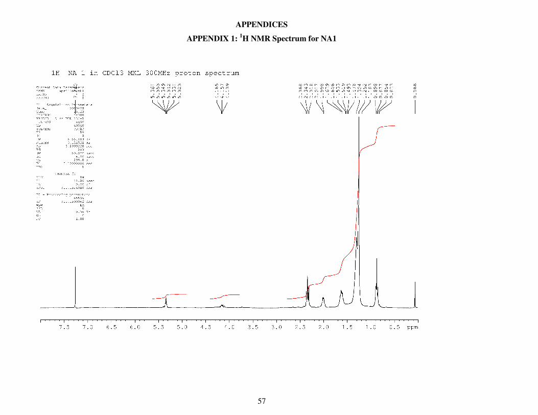
57
APPENDICES
APPENDIX 1: 1H NMR Spectrum for NA1
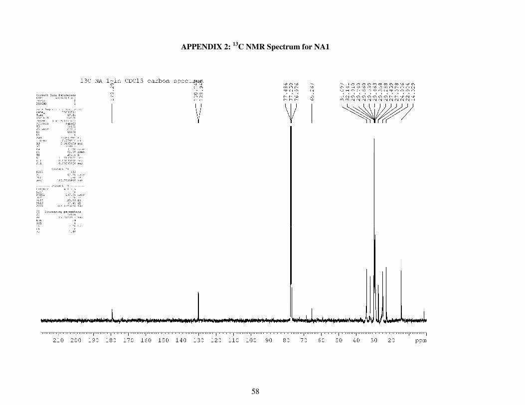
58
APPENDIX 2: 13
C NMR Spectrum for NA1

59
APPENDIX 3: 1H NMR Spectrum for NA2
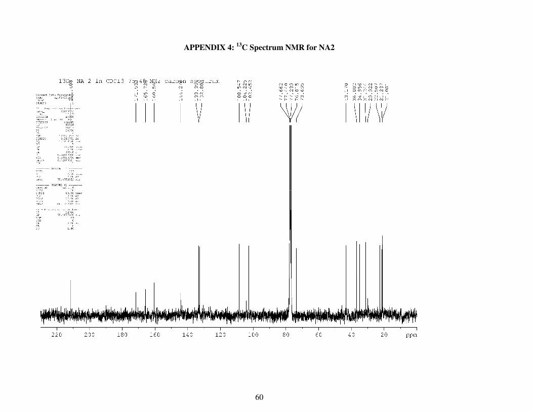
60
APPENDIX 4: 13
C Spectrum NMR for NA2

61
APPENDIX 5: 1H NMR Spectrum for NA3

62
APPENDIX 6: 13
C NMR Spectrum for NA3

63
APPENDIX 7: 1H/
1H COSY NMR Spectrum for NA3

64
APPENDIX 8: DEPT Spectrum for NA3

65
APPENDIX 9: 1H/
13C HMBC Spectrun for NA3

66
APPENDIX10: 1H/
13C HSQC-DEPTSpectrum for NA3
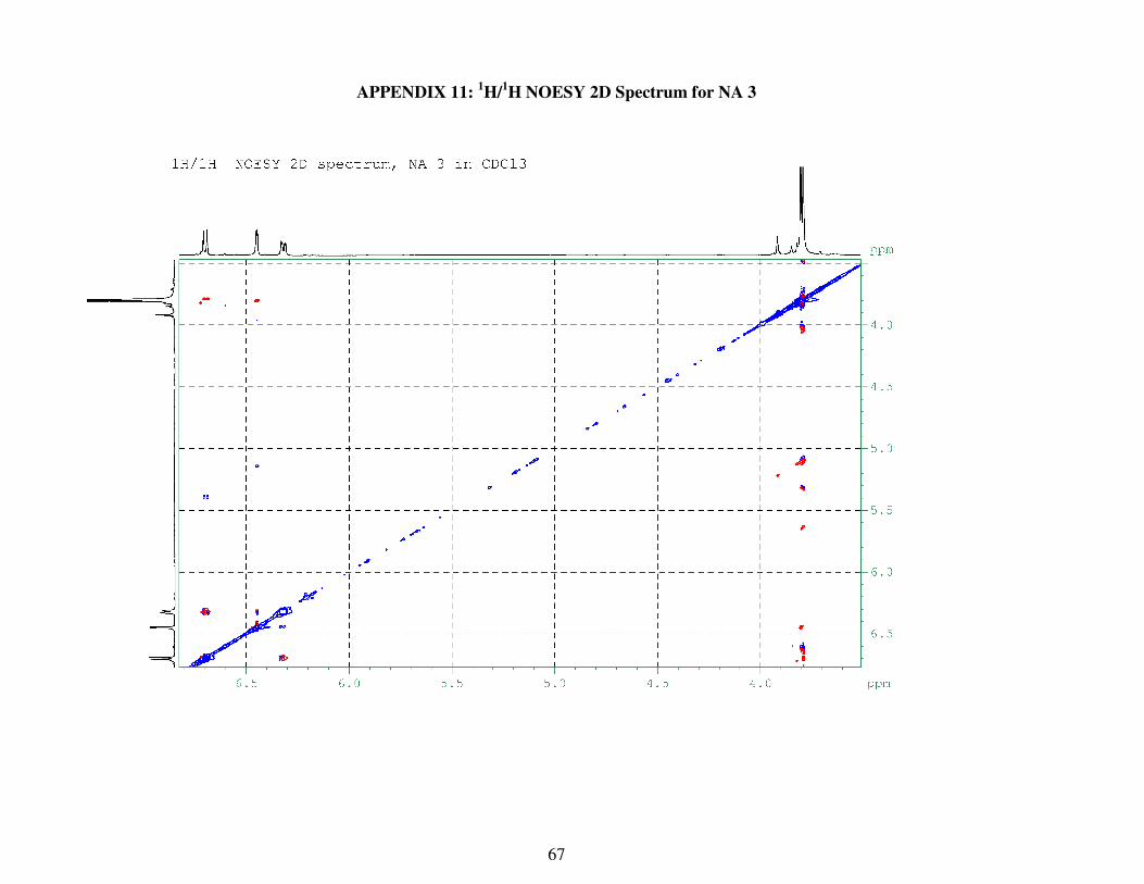
67
APPENDIX 11: 1H/
1H NOESY 2D Spectrum for NA 3
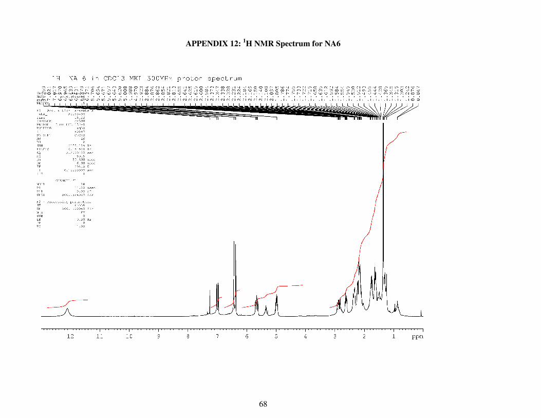
68
APPENDIX 12: 1H NMR Spectrum for NA6

69
APPENDIX 13: 13
C NMR Spectrum for NA6
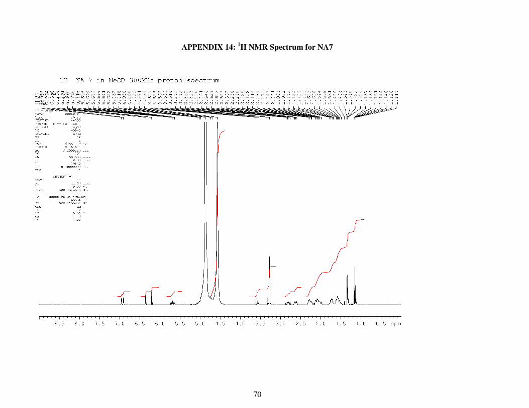
70
APPENDIX 14: 1H NMR Spectrum for NA7
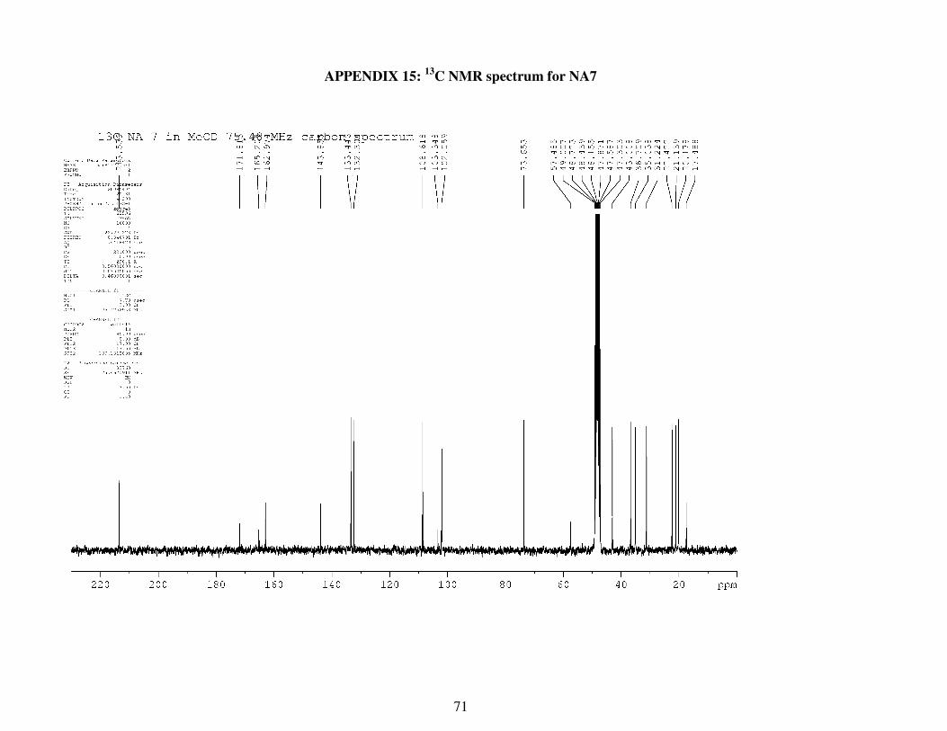
71
APPENDIX 15: 13
C NMR spectrum for NA7
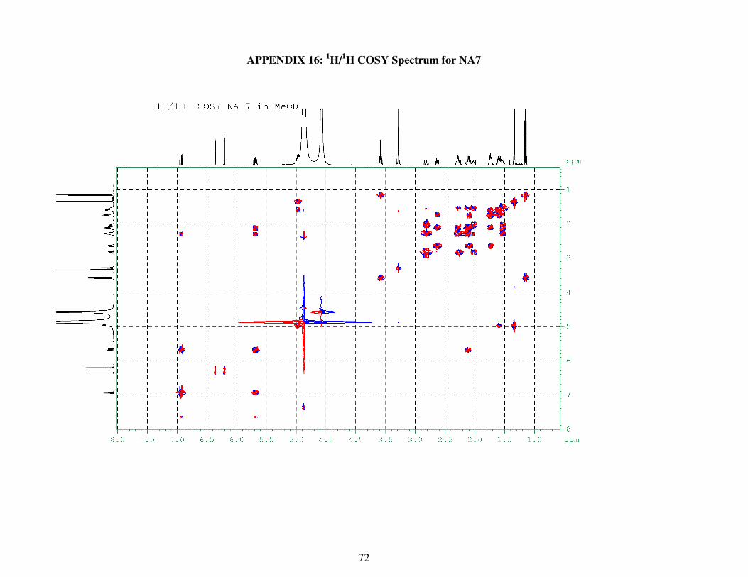
72
APPENDIX 16: 1H/
1H COSY Spectrum for NA7
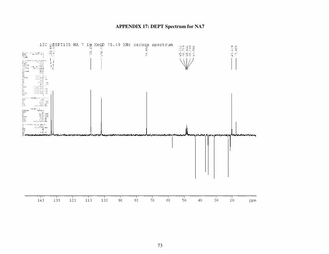
73
APPENDIX 17: DEPT Spectrum for NA7

74
APPENDIX 18: 1H/
13C HMBC Spectrum for NA7

75
APPENDIX 19: 1H/
13C HMBC Spectum for NA7
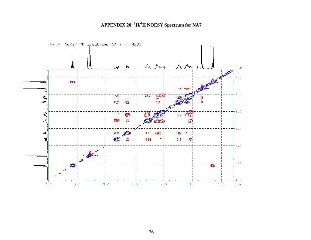
76
APPENDIX 20: 1H/
1H NOESY Spectrum for NA7
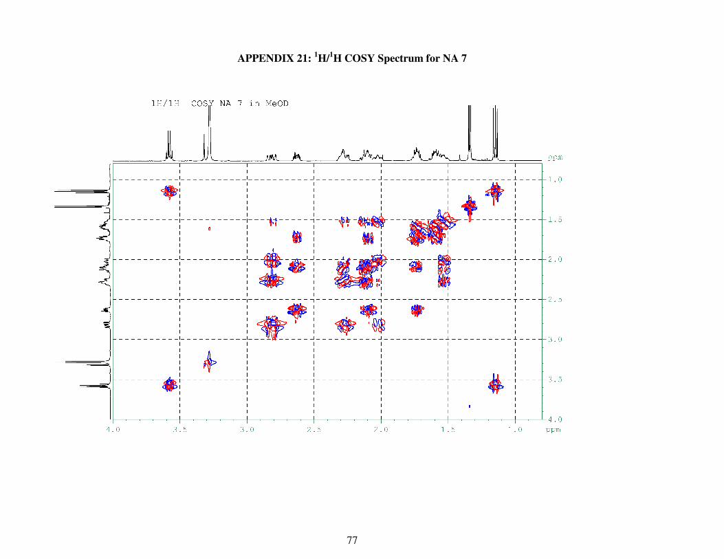
77
APPENDIX 21: 1H/
1H COSY Spectrum for NA 7

78
APPENDIX 22: 1H/
13C HMBC Spectrum for NA7
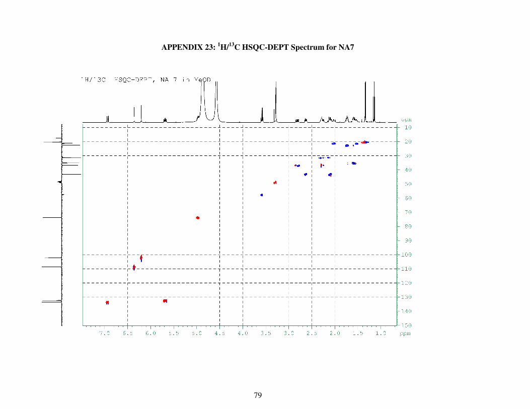
79
APPENDIX 23: 1H/
13C HSQC-DEPT Spectrum for NA7
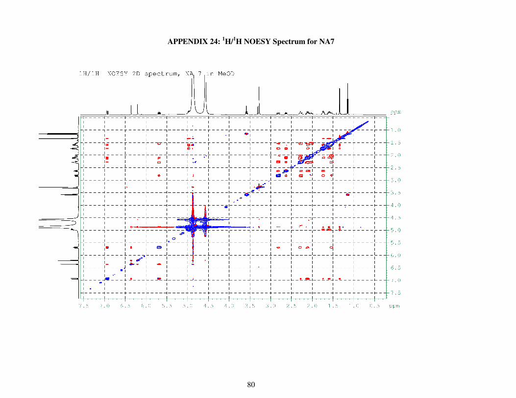
80
APPENDIX 24: 1H/
1H NOESY Spectrum for NA7


