
Role of the medial prefrontal cortex in the extinction of conditioned appetitive behaviour in rats
José Mendoza
A Thesis in the
Department of Psychology
Presented in Partial Fulfillment of the Requirements
For the Degree of Master of Arts (Psychology) at
Concordia University
Montreal, Quebec, Canada
August 2013
©Jose Mendoza
CONCORDIA UNIVERSITY
School of Graduate Studies
This is to certify that the thesis prepared
By: José Mendoza
Entitled: Role of the Medial Prefrontal Cortex in the Extinction of Conditioned Appetitive
Behaviour in Rats
and submitted in partial fulfillment of the requirements for the degree of
Master of Arts (Psychology)
complies with regulations of the University and meets the accepted standards with
respect to originality and quality.
Signed by the final examining committee:
Dr. Wayne Brake Chair
Dr. Uri Shalev Examiner
Dr. James Pfaus Examiner
Dr. Nadia Chaudhri Supervisor
Approved by: _______________________________________
Chair of the Department or Graduate Program Director
_________________________ _________________________
Date Dean of Faculty

iii
Abstract
Role of the medial prefrontal cortex in the extinction of conditioned appetitive behaviour in rats
José Mendoza
The infralimbic medial prefrontal cortex (IL-PFC) has been posited as a common node in
distinct neural circuits that mediate the extinction of appetitive and aversive conditioning.
However, appetitive extinction is typically assessed using instrumental conditioning procedures,
whereas the extinction of aversive conditioning is studied using Pavlovian fear-conditioning. The
role of the IL-PFC in the extinction of appetitive conditioning acquired through Pavlovian
learning remains largely unexplored. The present studies utilized animal models of Pavlovian-
and instrumental-conditioning with sucrose to study the involvement of the IL-PFC in appetitive
extinction. Based on fear-extinction we predicted that inactivating the IL-PFC before extinction
would have minimal effect on within-session extinction, but would impair the storage of
extinction memory. Control studies were conducted in the prelimbic prefrontal cortex (PL-PFC),
which is not involved in extinction. PL-PFC inactivation did not affect the acquisition or recall of
extinction memory. Counter to our predictions, inactivating the IL-PFC facilitated the extinction
of conditioned Pavlovian- and instrumental sucrose-seeking, with no effect on extinction recall
tested 24 hr later. In separate studies, inactivating the IL-PFC during a Pavlovian conditioning
session in which cue presentations were paired with sucrose did not affect cue-elicited behaviour,
but increased responding during inter-trial intervals. The same manipulation performed during
instrumental conditioning did not impact lever pressing for sucrose. These findings contradict a
growing body of literature suggesting that the IL-PFC is important for the acquisition and
consolidation of extinction memory in appetitive conditioning tasks.

iv
Acknowledgements
First and foremost, I would like to thank Dr. Nadia Chaudhri for giving me the
opportunity to grow as a researcher in her lab, and for her valuable guidance and feedback on
this thesis. I would also like to thank Lindsay Sparks and Joanna Sciascia for their friendship and
for being present throughout great and not so great moments. Furthermore, I would like to thank
Dr. Uri Shalev and Dr. Jim Pfaus for accepting to be members of my defence committee and for
their time spent reviewing my thesis.
I would like to express my deepest gratitude to my parents for their encouragement,
support, and for spoiling me with love. Mama, Papa - no existen palabras para expresar como yo
aprecio todo lo que ustedes han hecho por mí, y por el amor que me han dado durante toda mi
vida.
Finally, I would like to thank my beautiful little family that patiently waited for me to
arrive home late from the lab on so many occasions. Merci David pour ton humour et pour les
petits soins et l’amour que tu donne a ton petit frère. Gracias Preciosa por el amor que me das
mismo cuando a veces no me lo merezco. Tu m’as donné le plus beau cadeau du monde et pour
cela je te remercie du fond du cœur. Elias – I dedicate this thesis to you. You have given me a
form of happiness that I did not know existed. I love you hijito lindo.

v
Table of contents
List of figures……………………………………………………………………… vi
General Introduction……………………………………………………….……… 1
General Methods…………………………………………………………...……… 12
Subjects…………………………………………………………….……… 12
Apparatus……………………………………………………………. …… 12
Drugs………………………………………………………………..………13
Surgery………………………………………………………………..…… 13
Intracranial Microinfusions ……………………………………………… 14
Procedure ………………………………………………………….……… 15
Results …………………………………………………………………….……… 19
Discussion………………………………………………………...……………...… 35
References ………………………………………………………………………... 47
Appendix A………………………………………………………………………... 61
Appendix B………………………………………………………………………... 63

vi
List of figures Page
Figure 1. Pavlovian cue-driven sucrose-seeking behaviour across Pavlovian
conditioning, and following M/B or saline infusions in the IL-PFC before a
single extinction session, and across 4 infusion-free recall sessions.
Figure 2. Pavlovian cue-driven sucrose-seeking behaviour across Pavlovian
conditioning, and following M/B or saline infusions in the PL-PFC before a
single extinction session, and across 4 infusion-free recall sessions.
Figure 3. Lever responding for sucrose across instrumental conditioning,
extinction test, and following M/B or saline infusions in the IL-PFC or
PL-PFC before a single extinction session, and across 4 infusion-free
recall sessions.
Figure 4. Mean (± SEM) normalized CS, preCS, and non-CS responses across
Pavlovian conditioning and following M/B or saline infusions in the IL-PFC
before a session in which the CS was paired with sucrose delivery.
Figure 5. Mean (± SEM) lever pressing across instrumental conditioning and
following M/B or saline infusions in the IL-PFC before a session in which active
lever presses delivered sucrose.
Figure 6. Placement of injector tips within the IL-PFC.
31
33
37
39
44
39

1
General Introduction
Pavlovian conditioning is a fundamental form of learning that is important for adaptive
behaviour. The ability to predict, acquire, terminate, or avoid salient environmental events are all
examples of flexibility in behaviour that helps animals to survive. Equally adaptive is the
capacity to withhold behaviour when an expected outcome no longer occurs. This process is
mediated by extinction learning, a form of inhibitory learning that manifests as a gradual
reduction in conditioned behaviour in the absence of an anticipated outcome. In humans, failing
to inhibit responses to environmental stimuli that no longer predict an outcome is a characteristic
observed in drug addicts (Garavan & Hester, 2007; Kiefer & Dinter, 2013) and individuals
suffering from anxiety disorders (Andero & Ressler, 2012; Bouton, Mineka, & Barlow, 2001;
Rothbaum & Davis, 2003). Such behaviour is maladaptive, and may be attributed to
malfunctioning neural circuits that mediate Pavlovian learning. Thus, it is of value to investigate
the neural mechanisms involved in extinction learning, as it may lead to the development of
more efficient therapeutic treatments of addiction and anxiety disorders.
The neural mechanisms that mediate extinction learning have been studied extensively
using animal models (for review see Quirk & Mueller, 2008). Studies investigating the extinction
of Pavlovian conditioned responses typically involve two phases; a conditioning phase and an
extinction phase. During conditioning, training consists of repeatedly pairing a conditioned
stimulus (CS) with an unconditioned stimulus (US). After several CS-US pairings, animals learn
the predictive properties of the CS. This learning is expressed behaviourally in animals when the
CS elicits a conditioned response (CR). Extinction is then conducted by repeatedly presenting
the CS in the absence of the US, which results in a gradual reduction in conditioned responding.
Observing how quickly conditioned responses diminish across CS trials during extinction

2
provides a measure of extinction performance. Additionally, extinction recall can be assessed by
observing the level of conditioned responding recovered during a subsequent extinction session,
as well as by examining how quickly recovered conditioned responses diminish across CS trials.
Studies investigating the neural mechanisms that regulate the acquisition of extinction
manipulate neuronal activity in the brain region of interest immediately before the first extinction
session. By comparison, studies investigating the neural substrates involved in the memory of
extinction manipulate neuronal activity at several time-points immediately, or shortly after the
first extinction session, and assess extinction performance during a recall test session which is
typically conducted either 1 hr or 24 hrs after the initial extinction session.
Instrumental conditioning is the process by which an organism learns to perform an
action to obtain a reinforcer. For example, pressing a lever to obtain a drug-reinforcer is a form
of instrumental conditioning that is typically used in appetitive conditioning. Akin to Pavlovian
conditioned behaviour, instrumental conditioned behaviour can be extinguished by no longer
delivering the reinforcer, causing a reduction in instrumental responses. However, most of the
work on extinction has utilized models of Pavlovian conditioning. Thus, numerous hypotheses
have been proposed to describe the behavioural and neurobiological mechanisms that underlie
extinction within the context of Pavlovian learning. For example, it has been proposed that
conditioned responding decreases during extinction because the CS-US association formed
during Pavlovian conditioning is forgotten, unlearned, or erased (Rescorla & Wagner, 1972).
However, there is evidence against these hypotheses. For instance, extinguished conditioned
responding re-emerges following an extended period of rest (Corty & Coon, 1995; Hammersley,
1992; Tobeña et al., 1993). This phenomenon is known as spontaneous recovery, and is one of
several pieces of evidence suggesting that the CS-US memory that was formed during Pavlovian
3
conditioning is not erased, unlearned, or forgotten during extinction. Instead, it has been
proposed that decreased conditioned responding during extinction is the result of a new
association (CS-no US) that is formed when a CS is repeatedly presented in the absence of the
US (Bouton, 2004). Thus, animals learn to inhibit their behaviour in response to the CS. This
hypothesis suggests that specific neural substrates required for learning the new CS-no US
association are recruited during extinction and are responsible for inhibiting conditioned
behaviour.
Much of what is currently known about the neural mechanisms involved in extinction
originates from fear conditioning studies, in which organisms learn to predict aversive stimuli via
Pavlovian conditioning. Such studies have suggested that the medial prefrontal cortex (mPFC) is
a key component of the neural circuitry that mediates extinction (Morgan, Romanski, & Ledoux,
1993; Milad & Quirk, 2002; Thompson et al., 2010; Sierra-Mercado, Padilla-Coreano, & Quirk,
2011). In addition, subdivisions of the mPFC, including the infralimbic PFC (IL-PFC) and
prelimbic PFC (PL-PFC) have been shown to play differential roles in behaviour. Whereas the
IL-PFC mediates the acquisition and recall of extinction, the PL-PFC has been shown to promote
conditioned responding (Laurent & Westbrook, 2009; Milad, Vidal-Gonzalez, & Quirk, 2004;
Sierra-Mercado et al., 2011; Thompson et al., 2010; Vidal-Gonzalez, Vidal-Gonzalez, Rauch, &
Quirk, 2006).
Converging evidence in fear conditioning studies supports the idea that the IL-PFC
promotes extinction by inhibiting conditioned fear behaviour (Laurent & Westbrook, 2009;
Milad et al., 2004; Sierra-Mercado et al., 2011; Thompson et al., 2010; Vidal Gonzalez et al.,
2006). Similarly, the IL-PFC has been shown to inhibit conditioned appetitive behaviour (Ovari
& Leri, 2008; Lalumiere, Smith, & Kalivas, 2012; Peters, Lalumiere, & Kalivas, 2008). Indeed,

4
it has recently been proposed that the IL-PFC mediates the extinction of behaviour acquired
through either aversive or appetitive conditioning (Peters, Kalivas, & Quirk, 2009). However,
conditioned fear behaviour is typically studied using Pavlovian learning models, whereas
conditioned appetitive behaviour predominantly employs instrumental procedures. Thus, the role
of the IL-PFC in mediating the extinction of conditioned Pavlovian appetitive behaviour has not
yet been examined. The experiments presented in this thesis investigated the neural mechanism
involved in extinction of conditioned Pavlovian and instrumental appetitive behaviour. More
specifically, the role of the IL-PFC and PL-PFC in extinction of a conditioned sucrose-seeking
response in rats was examined. We predicted that IL-PFC, but not PL-PFC inactivation would
delay the acquisition and recall of extinction.
Behavioural evidence that extinction results in new learning
It has previously been suggested that extinction is a process that involves the erasure,
unlearning, or forgetting of a previously formed memory (Rescorla & Wagner, 1972). However,
several behavioural phenomena suggest that extinction involves new learning. These phenomena
include spontaneous recovery, renewal, and reinstatement.
Spontaneous recovery involves a reappearance of a previously extinguished response, and
is induced by passage of time (Corty & Coon, 1995; Hammersley, 1992; Tobeña et al., 1993).
For example, the behaviour of pressing a lever to obtain a reward can be extinguished by
omitting reward delivery. Following a period of rest in which the animal does not have access to
the lever, a recovery in lever-pressing occurs when the lever is presented again. Thus, the sudden
reappearance of the conditioned response indicates that extinction does not eliminate what was
learned during training.
5
Renewal is defined by a recovery in conditioned responding when an organism is
removed from the extinction context. For example, if Pavlovian training is conducted in one
context and extinction is conducted in another, subsequent re-exposure to the Pavlovian training
context results in the reappearance of the extinguished conditioned response. Interestingly,
exposure to a novel context following training and extinction in different contexts also results in
renewal (Bouton, Todd, Vurbic, & Winterbauer, 2011; Neumann & Kitlertsirivatana, 2010). This
context-induced increase in conditioned responding demonstrates that the original CS-US
association is not erased, unlearned, or forgotten during extinction, and that animals learn to
associate the environment in which extinction occurs with the absence of the US. Renewal
studies in particular suggest that extinction results in the formation of a new CS-no US
association, and if extinction is conducted in a context that differs from the training context, then
that association becomes linked to the environment in which extinction occurred (Bouton, 2000,
2002, 2004; Chaudhri, Sahuque, & Janak, 2008).
The reinstatement phenomenon also provides evidence that extinction does not eliminate
conditioned behaviour. Following extinction, conditioned responding can be reactivated by
presenting the organism with different stimuli, including exposure to the US, a US-predictive
cue, or a stressor (Kalivas, Peters, & Knackstedt, 2006; Shalev, Grimm, & Shaham, 2002; Sinha,
Fuse, Aubin, & O’Malley, 2000; Stewart, 2000). For example, an extinguished drug-seeking
response can be restored by presenting the animal with an auditory cue that was previously
paired with drug delivery.
The examples described above indicate that extinction does not ‘erase’ or result in the
‘unlearning’ of the original learning acquired during conditioning. The prevailing interpretation
of these behavioural findings is that repeated presentation of the CS alone during extinction leads

6
to the acquisition of a new inhibitory CS-no US association, causing a gradual reduction in CS-
elicited responses. Thus, extinction is said to involve ‘new learning’ through which organisms
come to inhibit behaviour in response to a CS. A similar explanation is used to describe
extinction of instrumental responses, such that during extinction the organism learns to inhibit
behaviour that no longer results in the expected outcome. Important extensions of the hypothesis
that extinction results in new learning are that (a) there must be underlying neural machinery that
supports this new learning, and that (b) this machinery might be similar to that which is utilized
to form the original associations during conditioning. Considerable effort has gone into
identifying the mechanisms that are important for extinction learning and memory using
Pavlovian fear-conditioning studies, and more recently, appetitive conditioning studies. Research
in both these domains has converged on the medial prefrontal cortex as being an important
region for the acquisition and storage of extinction memory.
Involvement of the IL-PFC in the extinction of conditioned fear behaviour
A number of studies have demonstrated that the infralimbic medial prefrontal cortex (IL-
PFC) is important for inhibiting conditioned fear behaviour during the acquisition of extinction
(Quirk, Russo, Barron, & Lebron, 2000; Sierra-Mercado et al., 2011). For example, Sierra-
Mercado et al. (2011) found that pharmacological inactivation of the IL-PFC delays fear
extinction in rats. To assess the role of the IL-PFC in extinction, rats were initially trained to
associate an auditory tone (CS) with a foot-shock (US) by repeatedly presenting tone-shock
pairings. Prior to the first extinction session in which the tone was repeatedly presented in the
absence of a foot-shock, animals were either infused with a Gamma-aminobutyric acid (GABA)
agonist (muscimol) or saline in the IL-PFC. Inactivating the IL-PFC via muscimol infusions
caused persistent freezing to CS presentations during extinction, resulting in significantly slower

7
extinction compared to controls. Consistent with these findings, activation of IL-PFC neurons via
GABAa antagonist infusions (Thompson et al., 2010) or electrical stimulation (Milad & Quirk,
2002; Milad et al., 2004; Vidal-Gonzalez et al., 2006) has been shown to inhibit conditioned fear
responses during the acquisition of extinction. Together, these findings suggest that neuronal
activity in the IL-PFC is essential for inhibiting conditioned fear behaviour during this
acquisition of extinction learning.
The IL-PFC has also been implicated in the consolidation of extinction memory, and
several studies have found a relationship between IL-PFC function and the recall of extinction.
For example, lesions of the IL-PFC impair extinction recall (Lebron, Milad, & Quirk, 2004;
Quirk et al., 2000). Similarly, pharmacological inactivation of the IL-PFC during the acquisition
of extinction results in elevated conditioned freezing responses during an extinction session on
the following day (Laurent & Westbrook, 2009; Sierra-Mercado et al., 2011), suggesting that the
IL-PFC is essential in the formation of the extinction memory, and that extinction recall is
impaired in the absence of the IL-PFC. In contrast, electrical stimulation of the IL-PFC reduces
conditioned freezing responses during tests for extinction recall, which suggests that activity in
the IL-PFC mediates memory consolidation, and that increasing neuronal activity in this brain
region can strengthen the extinction memory (Milad & Quirk, 2002; Milad et al., 2004). Further,
antagonists of N-methyl-D-aspartate (NMDA) receptors and protein synthesis infused in the IL-
PFC impair extinction recall (Burgos-Robles, Vidal-Gonzalez, Santini, & Quirk, 2007; Santini,
Ge, Ren, Pena, & Quirk, 2004; Sotres-Bayon, Diaz-Mataix, Bush, & Ledoux, 2009). Given that
glutamate binding to NMDA receptors leads to protein synthesis and the formation of long-term
memories (Kandel, 2001), these results provide further support for the hypothesis that extinction
results in the formation of new memories, and that these processes likely occur in the IL-PFC.

8
The PL-PFC has been shown to be important for promoting, rather than inhibiting,
conditioned fear behaviour. Whereas pharmacological inactivation of the IL-PFC causes
persistent conditioned fear behaviour during the acquisition of extinction, inactivation of the PL-
PFC before an extinction session attenuates conditioned freezing responses (Sierra-Mercado et
al., 2011). Moreover, electrical stimulation of PL-PFC neurons during extinction causes
sustained freezing responses, resulting in extinction impairment (Vidal-Gonzalez et al., 2006).
Consistent with this finding, neuronal activity in the PL-PFC after extinction training has been
shown to be significantly correlated with poor extinction performance in rats (Burgos-Robles,
Vidal-Gonzalez, & Quirk, 2009). Unlike the IL-PFC, inactivation of the PL-PFC during
extinction has no effect on extinction recall (Laurent & Westbrook, 2009; Sierra-Mercado et al.,
2011), suggesting a lack of involvement in extinction memory (but see Vidal-Gonzalez et al.,
2006).
Involvement of the IL-PFC in the extinction of conditioned appetitive behaviour
As in fear conditioning, the IL-PFC has been shown to be important for inhibiting
conditioned appetitive behaviour under extinction conditions. For example, lesions to the IL-
PFC enhance spontaneous recovery, renewal, and reinstatement of Pavlovian-conditioned food
seeking behaviour in rats (Rhodes & Killcross, 2004; Rhodes & Killcross, 2007). In line with
these findings, Marchant, Furlong, and McNally (2010) found that IL-PFC neurons are recruited
during extinction, as evidenced by robust c-fos expression in the IL-PFC following extinction of
nose-poking behaviour for alcoholic beer-seeking in rats. In addition, studies employing
pharmacological manipulation of neuronal activity have demonstrated the involvement of the IL-
PFC in the extinction of appetitive instrumental behaviour. Whereas IL-PFC inactivation
reinstates the extinguished behaviour of lever pressing for cocaine (Peters et al., 2008),

9
stimulating IL-PFC neurons suppresses cue-induced reinstatement of cocaine-seeking in rats
(Lalumiere et al., 2012).
Consistent with the findings outlined above, molecular studies have also implicated the
IL-PFC in the formation of extinction memory. Based on evidence that glutamatergic
transmission is required for learning and the formation of memories (Miyamoto, 2006; Rao &
Finkbeiner, 2007; Robbins & Murphy, 2006), investigators have examined the effects of
enhanced glutamate transmission on extinction learning. Several studies have found an
enhancement in the extinction of appetitive behaviour following pharmacological potentiation of
glutamate transmission (Botreau, Paolone, & Stewart, 2006; Cleva, Hicks, Gass, Wischerath, &
Plasters, 2011; Gass & Olive, 2009; Lalumiere et al., 2010; Nic Dhonnchadha et al., 2010).
Moreover, infusing the IL-PFC with the NMDA partial agnoist D-cycloserine (DCS)
immediately after the first extinction session has been shown to enhance extinction recall of
instrumental sucrose-seeking in rats (Peters & De Vries, 2013). Similarly, Lalumiere, Niehoff,
and Kalivas (2010) demonstrated that post-session infusions of the α-amino-3-hydroxy-5-
methyl-4-isoxazolepropionic acid (AMPA) receptor potentiator 4-[2-(phenylsulfonylamino)-
ethylthio]-2,6-difluorophenoxyacetamide (PEPA) over 5 extinction sessions caused a decrease in
active lever responding for cocaine during the last 2 days of extinction training, suggesting that
potentiating glutamatergic transmission in the IL-PFC enhances extinction recall. Overall, these
findings indicate that the IL-PFC is a key brain region responsible for the consolidation of
extinction memory and are consequently in agreement with fear conditioning studies.
10
Specific aims of the present research
Converging evidence from fear and appetitive conditioning studies support the hypothesis
that the IL-PFC has an important role in response inhibition, which is essential for the extinction
of conditioned behaviour. It should be noted that while fear extinction has typically been
examined using Pavlovian fear conditioning, appetitive extinction has largely been studied using
instrumental conditioning procedures. Thus, less is known about the neural mechanisms that
mediate extinction of Pavlovian conditioned reward-seeking behaviour. In addition, the
underlying neural mechanisms that mediate the extinction of appetitive behaviour have mostly
been investigated using drug, but not natural reinforcers.
The experiments in this thesis investigated the role of the IL-PFC in the extinction of
Pavlovian and instrumental conditioned sucrose-seeking behaviour in rats. Given the putative
role of the PL-PFC in promoting conditioned behaviour, the role of the PL-PFC on extinction
was also examined. Rats were given several sessions of Pavlovian conditioning in which a white
noise (CS) was repeatedly paired with the delivery of sucrose (US). Based on the procedures of
Sierra-Mercado et al. (2011), the role of the IL-PFC and PL-PFC in extinction learning was
assessed by pharmacologically inactivating these brain regions before the first extinction session.
On the following day, rats received a subsequent session of extinction to assess the effect of IL-
PFC inactivation on extinction memory .We hypothesized that inactivating the IL-PFC, but not
the PL-PFC, would have an impact on Pavlovian conditioned sucrose-seeking. Given that IL-
PFC inactivation has been shown to impair the acquisition of extinction (Sierra-Mercado et al.,
2011), we predicted that reversible inactivation of the IL-PFC would cause a similar impairment
in extinction learning in the present study. Moreover, based on the observation that activity in the
IL-PFC is essential for the consolidation of the extinction memory (Lalumiere et al., 2010), we

11
expected an impairment in extinction recall in rats that had received M/B in the IL-PFC on the
previous day.
Upon completion of the Pavlovian study described above, animals were given several
instrumental training sessions in which active lever presses delivered sucrose, followed by IL-
PFC or PL-PFC inactivation prior to an initial extinction session. The purpose of this experiment
was to examine the role of these brain areas on the extinction of behaviour acquired through
instrumental conditioning. Based on the finding that IL-PFC inactivation promotes instrumental
cocaine-seeking (Peters et al., 2008), we predicted that inactivation of the IL-PFC would result in
persistent sucrose-seeking, causing an impairment in the acquisition of extinction. Moreover,
extinction memory was tested on the following day by administering a further infusion-free
extinction session. We hypothesized that the IL-PFC would play a significant role in the
consolidation of the extinction memory, and predicted that inactivating IL-PFC on the previous
day would increase instrumental responding for sucrose during extinction recall.
Finally, we evaluated the effect of IL-PFC inactivation on the ability to make port-entries
by inactivating the IL-PFC prior to a Pavlovian conditioning session in which CS trials were
paired with sucrose delivery. Inactivation of the IL-PFC was also conducted prior to a sucrose
self-administration session, in which active lever pressing initiated the delivery of sucrose. We
predicted that there would be no effect of IL-PFC inactivation in well-trained animals, as motor
impairments are typically not observed in the absence of the IL-PFC (McLaughlin & See, 2003;
Fuchs et al., 2005).
12
Method
Subjects
Male Long-Evans rats (Charles River, QC, Canada; N=42; 220-240g on arrival) were
single-housed in plastic shoebox cages (44.5 cm x 25.8 cm x 21.7 cm) containing beta chip
bedding. They were kept in a temperature-controlled room (21° C) on a 12 hr light/dark cycle
with lights on at 7:00 AM. All behavioural procedures were conducted during the light phase.
Rats were handled daily and given a minimum of 7 days to acclimate to the animal colony before
surgery. They had unrestricted access to standard rat chow (Ralston Purina, Canada) and water
throughout the experiments, except as outlined below. All procedures were approved by the
Animal Research Ethics Committee at Concordia University, and are in accordance with
recommendations by the Canadian Council on Animal Care.
Apparatus
Equipment used for behavioural testing was obtained from Med Associates Inc. (St Albans,
VT, USA). Behavioural testing was conducted in operant conditioning chambers (ENV-009A;
32.8 cm x 32.8 cm x 32.8 cm) housed within custom-made, ventilated, sound-attenuating
melamine boxes (53.6 cm x 68.2 cm x 62.8 cm). Each chamber had a clear Plexiglas front door,
back-wall and ceiling, and side-walls made of stainless steel panels. Floors were comprised of
stainless steel bars that extended from front to rear. A waste pan lined with absorbent paper was
located beneath the floor. To prevent rats from manipulating the paper during behavioural
sessions, a stainless steel floor insert comprised of a 0.5-inch grids was placed over the bar floor.
A dual cup liquid receptacle (ENV-200R3AM; 5.3 cm x 3.4 cm x 5.3 cm) was located 2 cm
above the floor in the center of the right wall. One cup in the receptacle was connected via
polyethylene tubing (Tygon; Fisher Scientific, #141691A) to a 20 ml syringe mounted in a pump
13
(PHM-100, 3.33 RPM) located outside the sound-attenuating box. Entries into the receptacle
were measured via infrared detectors (ENV-254-CB) located across the entrance. A retractable
lever (ENV-112BM) was positioned on each side of the receptacle. The center of the left wall
contained a white house-light (75W, 100 mA, ENV-215M) located 30 cm above the bar floor. A
white noise generator (ENV-225SM) and clicker stimulus (ENV-135M) were located to the left
and right of the house-light, respectively. The white noise generator was calibrated to produce a
noise that was 6-8 decibels above background noise (75-78 dB). Stimulus presentations, pump
activation, and extension of levers were controlled by a PC computer using Med PC IV software.
Port entries and lever presses were counted and registered by the same computer using the same
program.
Drugs
Pharmacological inactivation was conducted using the gamma-aminobutyric acid (GABA)
agonists muscimol (Sigma-Aldrich; M1523) and baclofen (Sigma-Aldrich, B5399). A solution
(M/B) was prepared by dissolving 5 mg of muscimol and 93.65 mg of baclofen in 438 ml of
sterile 0.9% saline (0.03 nmol muscimol; 0.3 nmol baclofen). These agonists have been shown to
inhibit neural firing without affecting fibers of passage (Martin & Ghez, 1999; van Duuren et al.,
2007) and the doses used are behaviourally effective in studies on extinction (Peters et al., 2008).
Sucrose (Anachemia Canada Inc., #87688-380) was dissolved in tap water to obtain a final
concentration of 10% (w/v).
Surgery
Surgery was conducted 1-2 weeks after arrival on rats weighing 320-415 g. Animals were
anaesthetized with isoflurane and implanted bilaterally with stainless steel, double-barrelled (1.2
mm apart) guide cannulae (26 gauge, Plastics One, Roanoke, VA; C235G) targeting the IL-PFC
14
(AP = +2.7, ML = ±0.6, DV = -3.1) or PL-PFC (AP = +2.7, ML = ±0.6, DV = -1.6) using
standard stereotaxic procedures. Cannulae were occluded using 33 gauge obturators and secured
to the skull using dental acrylic and four metal screws. After surgery, 2 ml of 0.9% saline and an
analgesic (Anafen; 0.1 ml/kg) were administered by subcutaneous injection. Powdered rat chow
mixed with sugar and tap water was provided post-surgery to promote feeding. Rats received 14
days to recover from surgery before training.
Intracranial microinfusions
Microinfusions were conducted in the room where the operant conditioning chambers were
located. Solutions were infused through a double-barrelled (1.2 mm apart) 33 gauge injector
(Plastics One, Roanoke, VA; C235I), which was connected to 2 Hamilton syringes (10 µl; Fisher
Scientific, 1701 RNR- #14-815-279) via PE-50 tubing (VWR International Co.). Syringes were
placed in a microinfusion pump (Harvard Apparatus, PHD 2000) that infused at a rate of 0.3
µl/min, for a total volume of 0.3 µl. Following the 1 min infusion, the injector was kept inside
the cannula for 2 min to optimize diffusion. Throughout the 3 min microinfusion procedure, rats
were gently restrained to prevent them from detaching the injector. Behavioural testing
commenced 5-20 min after the microinjection.
Sucrose consumption in the home cage
Fourteen days after surgery, a bottle containing 10% sucrose was placed on the home cage.
Rat weights and sucrose consumption were recorded every 24 hr. After 48 hr the bottle was
removed.
15
Experiment 1a. Effect of IL-PFC and PL-PFC inactivation on the extinction of appetitive
Pavlovian conditioning
This study tested the hypothesis that the IL-PFC and PL-PFC have distinct roles in the
extinction of appetitive Pavlovian conditioning.
Twenty-four hours after sucrose exposure in the home cage, rats were handled in the
behaviour testing room for 30 min in order to habituate them to the testing environment. On the
following day, Pavlovian conditioning sessions commenced. Rats received 8 daily 30 min
Pavlovian conditioning sessions (consecutive days; 8:00 AM – 1:00 PM). Each session consisted
of 14 trials in which a 15 sec white-noise CS was paired with the delivery of 0.3 ml of sucrose
into the fluid port for oral consumption. Sucrose delivery began 6 sec after CS onset and co-
terminated with the CS. CS trials were controlled by a variable-time 120 sec schedule. The
house-lights were turned on manually at the start of the session and turned off automatically at
the end of the session. Immediately before Pavlovian training session 4, rats received a sham
microinfusion using the procedure described above, except with an injector that was cut so as not
to protrude beyond the cannula tip, and without any fluid in the lines. In addition, a saline
microinfusion was conducted before training session 6. These infusions were performed to
habituate the rats to the microinfusion procedure.
The acquisition of extinction was examined 24 hr after the last Pavlovian training session.
Rats received a bilateral microinfusion (0.3 µl/hemisphere) of either saline or M/B into the IL-
PFC (Saline, n=7; M/B, n=7) or PL-PFC (Saline, n=7; M/B, n=7) using a between-subjects
design. At 20 min after the infusion they were placed into the operant conditioning chambers for
a 30 min session that was identical to a Pavlovian conditioning session, except that the pumps
were turned off and did not contain sucrose syringes. Extinction memory was tested across 4

16
subsequent daily extinction sessions conducted in the absence of intracranial injections.
Experiment 1b. Effect of IL-PFC and PL-PFC inactivation on the extinction of appetitive
instrumental conditioning
This study tested the hypothesis that the IL-PFC, but not PL-PFC, is important for the
extinction of appetitive instrumental behaviour. Upon completion of the study above, rats from
Experiment 1 underwent the procedures outlined below.
Rats were water deprived for 24 hr and then placed in operant conditioning chambers for
a 12 hr lever-press training session. Session onset was indicated by illumination of the house-
light and extension of the left lever into the chamber. Each lever press resulted in a 0.1 ml
delivery of 10% sucrose into the fluid port on a fixed-ratio 1 (FR1) schedule. After 200 sucrose
deliveries the lever was retracted and the house-light turned off to indicate the end of the session.
Rats were returned to their home-cages where unrestricted access to water was restored.
Sucrose self-administration training began 24 hr after the lever-press training session. In
each daily 45 min session (consecutive days; 8:00 AM – 1:00 PM) responding on the left (active)
lever resulted in the delivery of 0.1 ml of 10% sucrose into the fluid port on an FR1 schedule.
Responding on the right (inactive) lever was recorded but had no programmed consequence.
Because of scheduling constraints, rats with cannulae targeting the IL-PFC received 5 self-
administration training sessions, whereas rats with PL-PFC cannulae placements received 6
training sessions. A saline sham microinfusion was administered before training session 4.
The acquisition of extinction was examined 24 hr after the last sucrose self-
administration session in a session that was identical to a self-administration session, except that
the pumps were turned off and did not contain sucrose syringes. At 20 min before test, rats
received a bilateral microinfusion (0.3 µl/hemisphere) of either saline or M/B into the IL-PFC
17
(Saline, n=7; M/B, n=7) or PL-PFC (Saline, n=6; M/B, n=7) using a between-subjects design. In
order to control for order effects, rats from each group from experiment 1a were equally
distributed into the saline and M/B treatment conditions for this extinction test. Extinction
memory was tested across 4 subsequent daily extinction sessions conducted in the absence of
intracranial injections.
Experiment 2. Effect of IL-PFC inactivation on appetitive Pavlovian and instrumental
conditioning
A separate group of animals were utilized to investigate the impact IL-PFC inactivation
on appetitive conditioning in both Pavlovian and instrumental procedures.
Rats received 8 Pavlovian training sessions as in experiment 1, wherein CS trials were
paired with 10% sucrose. A saline sham microinfusion was conducted prior to training day 7.
Before Pavlovian training session 9, rats received rats received a bilateral microinfusion (0.3
µl/hemisphere) of either saline (n=7) or M/B (n=7) into the IL-PFC using a between subject
design.
After the test described above, rats underwent lever-press training and sucrose self-
administration sessions as described in experiment 2. A sham microinfusion with a cut injector
occurred before session 4 and a saline microinfusion before session 7. The effect of IL-PFC
inactivation on sucrose self-administration was tested on session 9, in which rats received a pre-
session bilateral microinjection of saline (n=7) or M/B (n=7) into the IL-PFC using a between
subject design.
Histological verification of cannulae placements
Following experiments 2 and 3, rats were anesthetized with isoflurane and decapitated.
Brains were removed and immersed in formalin for 24 hr, followed by 25% sucrose for 7 days,
18
and then sectioned on a cryostat (60 microns, coronal). Sections were collected onto glass slides
and stained with cresyl violet. Placement of cannulae and verification of injector tips was
examined using light microscopy for rats utilized in experiment 1 (Fig 1d & 2d) and experiment
2 (Fig 6). Subjects were excluded if 1 or both injector tips were located outside the boundaries of
the IL-PFC or PL-PFC, as delineated in the Paxinos and Watson (1997) rat brain atlas. Based on
this criterion, seven rats with guide cannulae targeting the IL-PFC and 2 rats with PL-PFC
cannulae were excluded from experiment 1 as injectors were located outside the IL-PFC or PL-
PFC, respectively.
Statistical analysis
During Pavlovian conditioning entries into the fluid receptacle (referred to as port-entries)
during each 15 sec CS trial (CS responses) as well as during a 15 sec interval immediately before
each CS (preCS) were recorded. CS responses were normalized to account for differences in
baseline responding by subtracting preCS responses from responses during the corresponding
CS. Port-entries made when the CS was not presented (non-CS responses) were also calculated.
Pavlovian training sessions and extinction recall sessions were analyzed separately using
ANOVA with Session (Pavlovian training sessions 1-8; Recall sessions 1-4) as a within-subject
variable and Group (saline; M/B) as a between-subject variable. Independent samples t-tests
were used to analyze group differences in port-entry responses during the extinction test.
Repeated measures ANOVA was used to analyze port-entry responses per CS trial and latency to
respond to each CS at test with Trial (trials 1-14) as the within-subject variable and Group
(saline; M/B) as the between subject variable.
During instrumental conditioning responses on the left (active) and right (inactive) levers,
and port-entries made into the fluid port were recorded. Sucrose self-administration sessions and
19
recall sessions were analyzed independently using ANOVA with Session (Self-administration
sessions 4-5; Recall sessions 1-4) and Lever (active, inactive) as within-subjects variables and
Group (saline, M/B) as a between-subjects variable. The extinction test session was analyzed
using ANOVA with Lever (active, inactive) as the within-subjects variable, and Group (saline,
M/B) as the between-subjects variable. To further characterize within-session extinction of
instrumental responding, the number of active lever presses made during 1-min time bins across
the test session were analysed using ANOVA with Time (Min 1-45) as a within-subjects variable,
and Group (saline, M/B) as a between-subjects variable. In Experiment 1, one rat was excluded
following instrumental training as it did not learn to self-administer sucrose.
Violations of homogeneity of variance were determined by Mauchly’s test of sphericity and were
corrected using the Greenhouse-Geisser correction. Analyses were conducted using SPSS
software (version 20). The alpha level was set to α = 0.05 for all statistical analyses.
Results
Experiment 1a. Effect of IL-PFC and PL-PFC inactivation on the extinction of appetitive
Pavlovian conditioning
IL-PFC inactivation
Rats learned the association between the CS and sucrose, as shown by an increase in
normalized conditioned port-entries elicited by the CS across session during Pavlovian
conditioning [Fig 1a; Session, F(7,84) = 26.245, p = 0.000]. Both groups acquired Pavlovian
learning at a similar rate [Group, F(1,12) = 0.925, p = 0.854; Group x Session, F(7,84) = 2.210, p
= 0.083]. Compared to saline, inactivating the IL-PFC significantly reduced CS responses during
the first extinction session in which the CS was presented without sucrose [Fig 1a; t(12) = 2.867,
20
p = 0.014]. This effect was specific to CS responses as port-entries made during time intervals
that were not signalled by the CS were not affected by IL-PFC inactivation (Appendix A). There
was no impact of prior IL-PFC inactivation on the subsequent recall of extinction memory.
Responding to the CS decreased across 4 extinction recall sessions [Session, F(3,36) = 41.931, p
= 0.000], with no main effects or interactions with group [Group, F(1,12) = 0.126, p = 0.729;
Group x Session, F(3,36) = 0.733, p = 0.539].
In order to examine the impact of IL-PFC inactivation on within-session extinction
acquisition as well as the within-session expression of extinction memory during recall, a
detailed analysis of extinction sessions 1 (acquisition) and 2 (recall) was conducted. On the first
day of extinction (Fig 1b, left panel), the number of port-entries made during each CS trial
decreased as a function of trial [Trial, F(13,156) = 16.541, p = 0.000]. Compared to saline, IL-
PFC inactivation reduced the number of CS responses overall [Group, F(1,12) = 8.218, p =
0.014]. Interestingly, ANOVA revealed a significant Group x Trial interaction [Group x Trial,
F(13,156) = 2.098, p = 0.017] suggesting that inactivating the IL-PFC caused a more rapid
extinction of CS responses across CS trials. Follow-up t-tests conducted to investigate this
interaction found that there was no difference in the number of port-entries made during the first
2 CS trials in saline or M/B infused rats [Trial 1, t(12) = 1.106, p = 0.290; Trial 2 t(12) = 0.825, p
= 0.425]. However, the number of responses elicited by CS trials 3 and 4 was significantly
reduced following IL-PFC inactivation compared to saline [Trial 3, t(12) = 3.482, p = 0.005;
Trial 4 t(12) = 3.695, p = 0.003]. During extinction recall (Fig 1b, right panel) port-entries during
each CS presentation decreased as a function of trial [Trial, F(13,156) = 14.290, p = 0.000] in
rats from both groups [Group x Trial, F(12,156) = 0.426, p = 0.959; Group x Trial, F(12,156) =
0.426, p = 0.959].
21
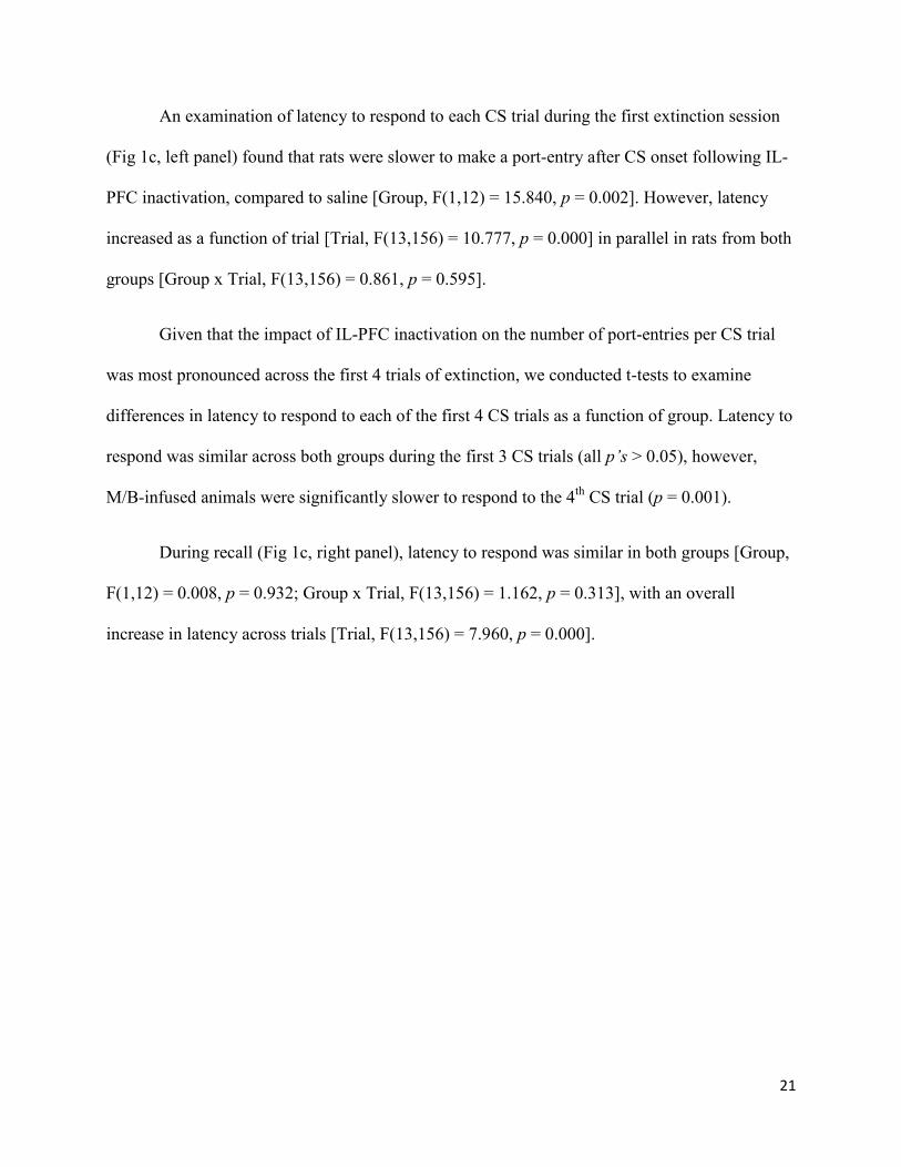
An examination of latency to respond to each CS trial during the first extinction session
(Fig 1c, left panel) found that rats were slower to make a port-entry after CS onset following IL-
PFC inactivation, compared to saline [Group, F(1,12) = 15.840, p = 0.002]. However, latency
increased as a function of trial [Trial, F(13,156) = 10.777, p = 0.000] in parallel in rats from both
groups [Group x Trial, F(13,156) = 0.861, p = 0.595].
Given that the impact of IL-PFC inactivation on the number of port-entries per CS trial
was most pronounced across the first 4 trials of extinction, we conducted t-tests to examine
differences in latency to respond to each of the first 4 CS trials as a function of group. Latency to
respond was similar across both groups during the first 3 CS trials (all p’s > 0.05), however,
M/B-infused animals were significantly slower to respond to the 4th
CS trial (p = 0.001).
During recall (Fig 1c, right panel), latency to respond was similar in both groups [Group,
F(1,12) = 0.008, p = 0.932; Group x Trial, F(13,156) = 1.162, p = 0.313], with an overall
increase in latency across trials [Trial, F(13,156) = 7.960, p = 0.000].

22
*
Pavlovian Conditioning
Po
rt E
ntr
ies
-20
0
20
40
60
80
100
Test Recall
Sessions: 1-8 9 10-13
Infralimbic PFC
M/B n=7
SAL n=7
ANormalized CS
*
*
*
CS Trial
Po
rt E
ntr
ies
0
2
4
6
8
10
12
14
2 4 6 8 10 12 14
Extinction Recall
2 4 6 8 10 12 14
B
^
^
Infralimbic PFC
C
CS Trial
La
ten
cy (
se
c)
0
5
10
15
20
25
2 4 6 8 10 12 14
Extinction Recall
2 4 6 8 10 12 14
Infralimbic PFC
D
Figure 1. Inactivating the IL-PFC before an extinction session in which a Pavlovian, sucrose-
predictive CS was presented without sucrose facilitated the within-session acquisition of
extinction, but had no effect on extinction recall. Rats received Pavlovian conditioning sessions,
followed by a single extinction session that was preceded by an intracranial infusion into the IL-
PFC, and 4 subsequent sessions conducted to assess the recall of extinction memory. In Figures
A, B, and C, filled circles represent rats that received M/B before extinction session 1 (n=7) and
open circles represent rats that received saline (n=7). A Mean (±SEM) normalized CS responses
(CS – PreCS) during Pavlovian conditioning, a single extinction test, and 4 sessions to assess the
recall of extinction. B Mean (±SEM) port-entries made during each CS trial across the
extinction test and the first recall session. C Mean (±SEM) latency to first port-entry response
after the onset of each CS trial during the extinction test and recall session 1. D Placement of
injector tips within the IL-PFC. Distance from bregma is indicated to the right of each coronal
section. Symbols indicate statistical significance from independent samples t-test comparisons:
^P < 0.01, *P < 0.05 significant difference between saline and M/B. Arrows indicate infusions of
saline or M/B in the IL-PFC in this, and subsequent figures.

23
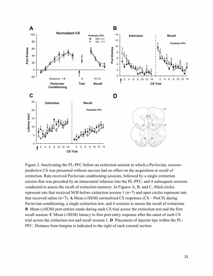
PL-PFC inactivation
Responses to the CS increased across conditioning sessions in rats with guide cannulae
targeting the PL-PFC [Fig 2a; Session, F(7,84) = 30.124, p = 0.000], with no main effects or
interactions involving group [Group, F(1,12) = 0.182, p = 0.677; Group x Session, F(7,84) =
0.703, p = 0.669]. Unlike the IL-PFC, inactivating the PL-PFC had no impact on CS responding
compared to saline at test. [Fig. 3a; t(12) = 1.624, p = 0.130]. Likewise, PL-PFC inactivation had
no impact on responses made outside CS presentations (Appendix A). Furthermore, CS
responses decreased comparably across the 4 extinction recall sessions [Fig 3a; Session, F(3,36)
= 42.569, p = 0.000] in rats from both groups [Group, F(1,12) = 0.909, p = 0.359; Group x
Session, F(3,36) = 0.374, p = 0.640].
That PL-PFC inactivation had no impact on the extinction of CS responding was verified
by a within-session analysis on day 1 of extinction (Fig 2b, left panel), which revealed an overall
decrease in port-entry responses as a function of CS trial [Trial, F(13,156) = 6.793, p = 0.000] in
rats from both groups [Group x Trial, F(13,156) = 0.979, p = 0.475; Group, F(1,12) = 2.572, p =
0.135]. Likewise, during extinction recall (Fig 2b, right panel), port-entries decreased as a
function of trial [Fig 2b; Trial, F(13,156) = 10.587, p = 0.000], and there were no main effects or
interactions involving group [Group, F(1,12) = 0.431, p = 0.524; Group x Trial, F(13,156) =
0.686, p = 0.775].
Latency to make a port-entry response increased across trial during the first extinction
session [Fig 2c, left panel; Trial, F(13,156) = 5.921, p = 0.000], and PL-PFC inactivation had no
impact on this measure [Group, F(1,12) = 1.314, p = 0.274; Group x Trial, F(13,156) = 1.250, p
= 0.249]. Similarly, during extinction recall, latency to make a port-entry increased as a function
of trial [Fig 2c right panel; Trial, F(13,156) = 5.539, p = 0.000], with no main effects or
24
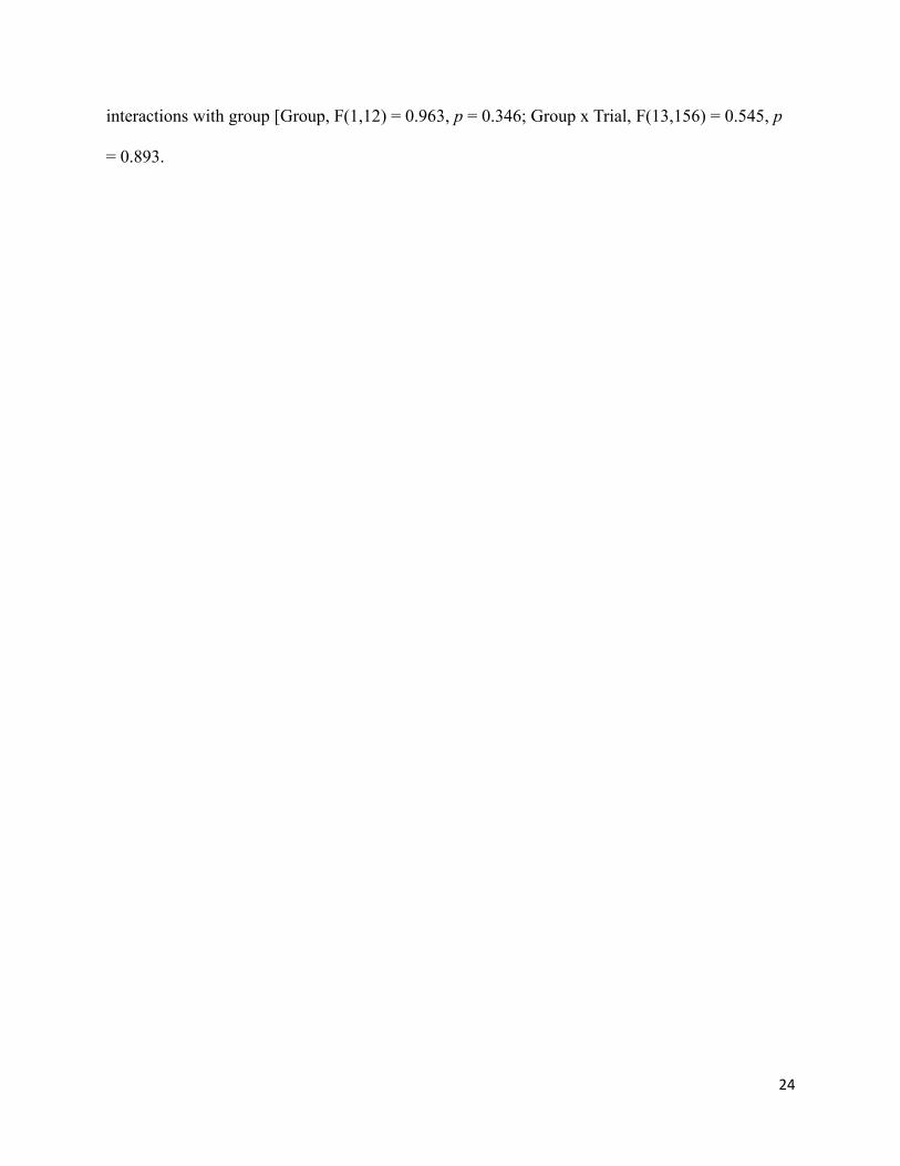
interactions with group [Group, F(1,12) = 0.963, p = 0.346; Group x Trial, F(13,156) = 0.545, p
= 0.893.
25
A
Pavlovian Conditioning
Po
rt E
ntr
ies
-20
0
20
40
60
80
100
Test Recall
Sessions: 1-8 9 10-13
Prelimbic PFC
M/B n=7
SAL n=7
Normalized CS
B
8
CS Trial
Po
rt E
ntr
ies
0
2
4
6
8
10
12
14
2 4 6 8 10 12 14
Extinction Recall
2 4 6 10 12 14
Prelimbic PFC
C
CS Trial
La
ten
cy (
sec)
0
5
10
15
20
25
2 4 6 8 10 12 14
Extinction Recall
2 4 6 8 10 12 14
Prelimbic PFC
D
Figure 2. Inactivating the PL-PFC before an extinction session in which a Pavlovian, sucrose-
predictive CS was presented without sucrose had no effect on the acquisition or recall of
extinction. Rats received Pavlovian conditioning sessions, followed by a single extinction
session that was preceded by an intracranial infusion into the PL-PFC, and 4 subsequent sessions
conducted to assess the recall of extinction memory. In Figures A, B, and C, filled circles
represent rats that received M/B before extinction session 1 (n=7) and open circles represent rats
that received saline (n=7). A Mean (±SEM) normalized CS responses (CS – PreCS) during
Pavlovian conditioning, a single extinction test, and 4 sessions to assess the recall of extinction.
B Mean (±SEM) port-entries made during each CS trial across the extinction test and the first
recall session. C Mean (±SEM) latency to first port-entry response after the onset of each CS
trial across the extinction test and recall session 1. D Placement of injector tips within the PL-
PFC. Distance from bregma is indicated to the right of each coronal section.

26
Experiment 1b. Effect of IL-PFC and PL-PFC inactivation on extinction of appetitive
instrumental conditioning
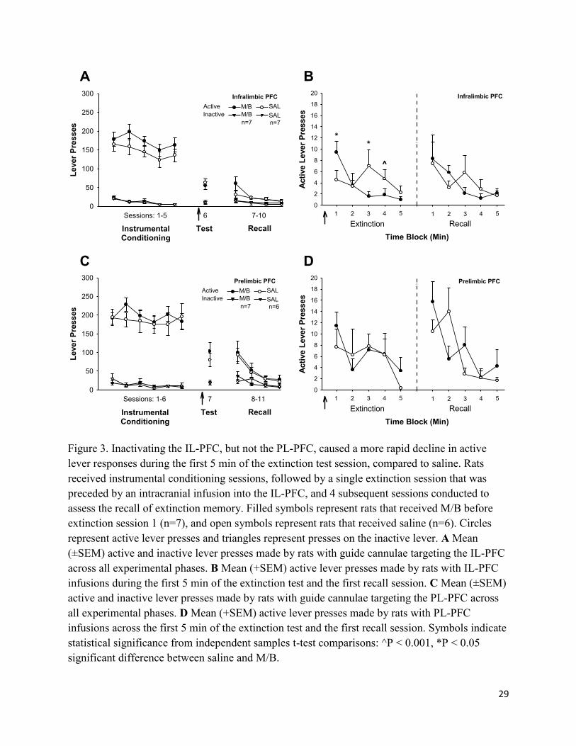
IL-PFC inactivation
Rats learned to discriminate between the active and inactive levers during instrumental
conditioning sessions (Fig 3a). ANOVA was conducted on active and inactive lever responding
across the last 2 sessions of self-administration to verify that responding was stable immediately
before the extinction test. Responding was higher on the active than the inactive lever [Lever,
F(1,12) = 134.491, p = 0.000] in rats from both group [Lever x Group, F(1,12) = 1.271, p =
0.282]. There was no main effect of session [Session, F(1,12) = 1.786, p = 0.206] or group
[F(1,12) = 1.143, p = 0.306], and no statistically significant interactions [Session x Group,
F(1,12) = 0.001, p = 0.971; Session x Lever, F(1,12) = 2.481, p = 0.141; Session x Group x
Lever, F(1,12) = 0.002, p = 0.964].
Compared to saline, inactivating the IL-PFC did not affect lever responses on session 1 of
extinction where active lever responding no longer resulted in sucrose (Fig 3a). Rats responded
more on the active than the inactive lever [Lever, F(1,12) = 60.343, p = 0.000] and there was no
effect of IL-PFC inactivation on responding on either lever [Group, F(1,12) = 0.330, p = 0.330;
Lever x Group, F(1,12) = 0.334, p = 0.574].
Collapsed across 4 recall sessions (Fig 3a) active lever pressing was higher than inactive
lever responding [Lever, F(1,12) = 24.406, p = 0.000]. However, across session there was a
decrease in lever pressing [Session, F(3,36) = 8.829, p = 0.000] in both groups [Group, F(1,12)
= 1.323, p = 0.272; Session x Group, F(3,36) = 1.197, p = 0.325]. There was an across-session
reduction in active lever pressing, but not inactive lever pressing [Session x Lever, F(3,36) =
4.796, p = 0.035] in rats from both groups [Session x Lever x Group, F(3,36) = 2.650, p = 0.063]

27
Active lever responses were averaged into 1-min time-bins across extinction sessions 1 and
2 in order to investigate the effect of IL-PFC inactivation on the acquisition and recall of
extinction, respectively. For clarity, only responding across the first 5 min of each session is
depicted in Figure 3b (see Appendix B for entire session). Active lever responses decreased
across the first 5 min of extinction session 1 [Fig 3b, left panel; Time, F(4,48) = 3.956, p =
0.007]. There was no overall difference in the number of active lever presses as a function of
group [Group, F(1,12) = 1.675, p = 0.220]. However, responding diminished at a different rate
between the groups [Time x Group, F(4,48) = 6.229, p = 0.000]. Follow-up t-tests for
independent samples revealed that at minute 1 of the test session, M/B-infused rats responded
more on the active lever compared to saline-infused rats [t(12) = -2.239, p = 0.045]. At minute 2,
active lever pressing was similar in both groups [t(12) = -0.186, p = 0.856]. However, M/B-
infused rats responded significantly less on the active lever compared with control animals at
minute 3 [t(12) = 2.628, p = 0.022] and minute 4 [t(12) = 3.328, p = 0.006].
During extinction recall (Fig 3b, right panel) there was a near-significant decline in active
lever responses across the first 5 min [Fig 3b; Time, F(4,48) = 3.413, p = 0.066]. However,
unlike extinction session 1, there was no difference between the groups in active lever responses
as a function of time [Group, F(1,12) = 0.015, p = 0.905; Time x Group, F(4,48) = 0.894, p =
0.475].
PL-PFC inactivation
Rats learned to discriminate between the active and inactive levers during instrumental
conditioning sessions (Fig 3c). Overall, rats pressed more on the active than the inactive lever
[Lever, F(1,11) = 101.909, p = 0.000].The number of lever presses did not change across the last

28
2 sessions of instrumental training [Fig 3c; Session, F(1,11) = 0.003, p = 0.954] with no
interactions as a function of group [Group, F(1,11) = 0.016, p = 0.903; Session x Group, F(1,11)
= 2.697, p = 0.129; Session x Lever, F(1,11) = 0.040, p = 0.845; Session x Group x Lever,
F(1,11) = 3.050, p = 0.109].
Compared to saline, PL-PFC inactivation had no impact on lever pressing during extinction
(Fig 3c). Animals continued to discriminate between the active and inactive lever [Lever, F(1,11)
= 29.869, p = 0.000], but there were no main effects or interactions with group [Group, F(1,11) =
0.416, p = 0.532; Group x Lever, F(1,11) = 0.861, p = 0.373].
Across 4 sessions of extinction recall, more responses were made on the active lever than
the inactive lever [Lever, F(3,33) = 15.415, p = 0.000]. However, active lever responses
decreased across session [Lever x Session, F(3,33) = 8.804, p = 0.003], in rats from both groups
[Session x Group, F(3,33) = 0.375, p = 0.652; Session x Lever x Group, F(3,33) = 0.559, p =
0.646]. There was no impact of prior treatment on overall responding [Group, F(1,11) = 0.024, p
= 0.880].
During the first 5 min of extinction session 1 (Fig 3d, left panel) active lever responses
decreased as a function of time [Fig 3d; Time, F(4,44) = 3.205, p = 0.022]. PL-PFC inactivation
had no impact on this measure [Group, F(1,11) = 0.063, p = 0.806; Time x Group, F(4,44) =
0.714, p = 0.587]. Likewise, active lever responses diminished during the first 5 min of
extinction recall [Fig 3d, right panel; Time, F(4,44) = 11.373, p = 0.000]. Prior PL-PFC
inactivation had no effect on active lever pressing during the first 5 min of recall [Group, F(1,11)
= 0.117, p = 0.739], but there was a significant group x time interaction [Group x Time, F(4,44)
= 4.207, p = 0.006]. Follow-up t-tests for independent samples revealed no group differences in
active lever pressing at any time bin during the first 5 min of extinction (all p’s > 0.05).
29
A
InstrumentalConditioning
Le
ver
Pre
sses
0
50
100
150
200
250
300
Test Recall
Sessions: 1-5 6 7-10
Infralimbic PFC
M/B
M/B
SAL
SAL
Active
Inactive
n=7 n=7
B
Time Block (Min)
Acti
ve L
ever
Pre
sses
0
2
4
6
8
10
12
14
16
18
20
Extinction Recall
1 2 3 4 5 1 2 3 4 5
*
^
*
Infralimbic PFC
C
InstrumentalConditioning
Lever
Pre
sses
0
50
100
150
200
250
300
Test Recall
Sessions: 1-6 7 8-11
Prelimbic PFC
M/B
M/B
SAL
SAL
Active
Inactive
n=7 n=6
D
Time Block (Min)
Acti
ve L
ever
Pre
sses
0
2
4
6
8
10
12
14
16
18
20
Extinction Recall
1 2 3 4 5 1 2 3 4 5
Prelimbic PFC
Figure 3. Inactivating the IL-PFC, but not the PL-PFC, caused a more rapid decline in active
lever responses during the first 5 min of the extinction test session, compared to saline. Rats
received instrumental conditioning sessions, followed by a single extinction session that was
preceded by an intracranial infusion into the IL-PFC, and 4 subsequent sessions conducted to
assess the recall of extinction memory. Filled symbols represent rats that received M/B before
extinction session 1 (n=7), and open symbols represent rats that received saline (n=6). Circles
represent active lever presses and triangles represent presses on the inactive lever. A Mean
(±SEM) active and inactive lever presses made by rats with guide cannulae targeting the IL-PFC
across all experimental phases. B Mean (+SEM) active lever presses made by rats with IL-PFC
infusions during the first 5 min of the extinction test and the first recall session. C Mean (±SEM)
active and inactive lever presses made by rats with guide cannulae targeting the PL-PFC across
all experimental phases. D Mean (+SEM) active lever presses made by rats with PL-PFC
infusions across the first 5 min of the extinction test and the first recall session. Symbols indicate
statistical significance from independent samples t-test comparisons: ^P < 0.001, *P < 0.05
significant difference between saline and M/B.

30
Experiment 2. Effect of IL-PFC inactivation on appetitive Pavlovian and instrumental
conditioning
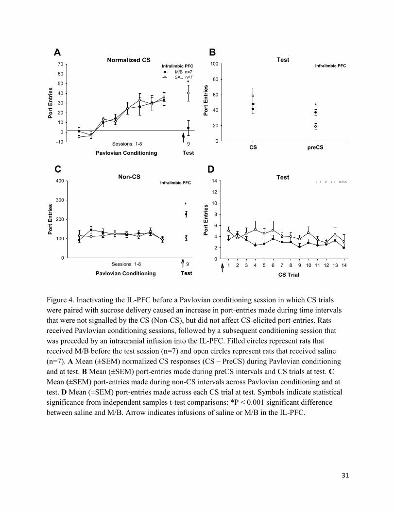
Across training, normalized CS responses increased [Fig 4a; Session, F(7,84) = 16.071, p =
0.000] in rats from both groups [Group, F(1,12) = 0.004, p = 0.951; Session x Group, F(7,84) =
0.239, p = 0.883]. Inactivating the IL-PFC reduced normalized CS responding (Fig 4a),
compared to saline [t(12) = 2.933, p = 0.013]. However, additional analyses (Fig 4b) revealed
that this effect was attributable to IL-PFC inactivation producing a significant increase in preCS
responding [t(12) = -3.101, p = 0.009], with no change in CS responding [t(12) = 1.356, p =
0.200]. Thus, IL-PFC inactivation promoted port-entry responses during time periods that were
not explicitly signalled by the CS. This effect was also evident in an analysis of the number of
responses made during non-CS intervals (Fig 4c). There was no change in non-CS responses
across Pavlovian conditioning as a function of session [Session, F(7,84) = 1.431, p = 0.204] or
group [Group, F(1,12) = 0.093, p = 0.765; Group x Session, F(7,84) = 0.714, p = 0.660].
However, at test IL-PFC inactivation significantly increased non-CS responses, compared to
saline [t(12) = -5.704, p = 0.000].
An analysis of the number of port-entries made during each CS trial (Fig 4d) at test
revealed no impact of IL-PFC inactivation on port-entries per CS trial across the session [Trial,
F(13, 156) = 1.540, p = 0.109; Group, F(1,12) = 1.840, p = 0.200; Group x Trial, F(13,156) =
0.755, p = 0.706].
31
A
Pavlovian Conditioning
Po
rt E
ntr
ies
-10
0
10
20
30
40
50
60
70
Test
Sessions: 1-8 9
Normalized CSInfralimbic PFC
M/B n=7
SAL n=7
Po
rt E
ntr
ies
0
20
40
60
80
100
CS preCS
Infralimbic PFC
B
*
Test
C
Pavlovian Conditioning
Po
rt E
ntr
ies
0
100
200
300
400
Test
Sessions: 1-8 9
Non-CSInfralimbic PFC
D
CS Trial
1 2 3 4 5 6 7 8 9 10 11 12 13 14
Po
rt E
ntr
ies
0
2
4
6
8
10
12
14Infralimbic PFC
M/B n=7
SAL n=7
Test
Figure 4. Inactivating the IL-PFC before a Pavlovian conditioning session in which CS trials
were paired with sucrose delivery caused an increase in port-entries made during time intervals
that were not signalled by the CS (Non-CS), but did not affect CS-elicited port-entries. Rats
received Pavlovian conditioning sessions, followed by a subsequent conditioning session that
was preceded by an intracranial infusion into the IL-PFC. Filled circles represent rats that
received M/B before the test session (n=7) and open circles represent rats that received saline
(n=7). A Mean (±SEM) normalized CS responses (CS – PreCS) during Pavlovian conditioning
and at test. B Mean (±SEM) port-entries made during preCS intervals and CS trials at test. C
Mean (±SEM) port-entries made during non-CS intervals across Pavlovian conditioning and at
test. D Mean (±SEM) port-entries made across each CS trial at test. Symbols indicate statistical
significance from independent samples t-test comparisons: *P < 0.001 significant difference
between saline and M/B. Arrow indicates infusions of saline or M/B in the IL-PFC.
*
*

32
During instrumental training more responses were made on active than the inactive lever
across training [Lever, F(1,12) = 211.155, p = 0.000]. Lever responding remained stable across
self-administration sessions [Session, F(7,84) = 1.239, p = 0.291; Session x Lever, F(7,84) =
1.554, p = 0.161] in rats from both groups [Group, F(1,12) = 0.006, p = 0.938; Session x Group,
F(7,84) = 0.412, p = 0.893; Session x Lever x Group, F(7,84) = 0.601, p = 0.754].
Inactivating the IL-PFC had no effect on instrumental responding for sucrose (Fig 5a). Rats
responded more on the active than the inactive lever [Lever, F(1,12) = 161.477, p = 0.000], with
no impact of IL-PFC inactivation in either measure [Group, F(1,12) = 0.316, p = 0.584; Group x
Lever, F(1,12) = 0.481, p = 0.501]. A detailed examination of the test session (Figure 5b)
indicated that IL-PFC inactivation did not influence within-session responding on either lever
[Time, F(43,516) = 25.108, p = 0.000; Group, F(1,12) = 0.399, p = 0.539; Group x Time, F(43,
516) = 0.756, p = 0.871].

33
A
Instrumental Conditioning
Lever
Pre
sses
0
50
100
150
200
250
300
350
Test
Sessions: 1-8 9
Infralimbic PFC
M/B
M/B
SAL
SAL
Active
Inactive
n=7 n=7
B
Time Block (Min)
5 10 15 20 25 30 35 40 45
Acti
ve L
ever
Pre
sses
0
2
4
6
8
10
12
14
16
18
20Infralimbic PFC
M/B n=7
SAL n=7
Test
Figure 5. Pre-session inactivation of the IL-PFC had no impact on instrumental responding
during a sucrose self-administration session in which pressing on the active lever delivered
sucrose. Rats received instrumental conditioning sessions, followed by a subsequent conditioning
session that was preceded by an intracranial infusion into the IL-PFC. Filled symbols represent
rats that received M/B before extinction session 1 (n=7), and open symbols represent rats that
received saline (n=7). Circles represent active lever presses and triangles represent presses on the
inactive lever. A Mean (±SEM) active and inactive lever presses during instrumental
conditioning and at test. B Mean (±SEM) active lever responses across 1 min time bins at test.
Arrow indicates infusions of saline or M/B in the IL-PFC.

34
Figure 6. Placement of injector tips within the IL-PFC in experiment 2. Distance from bregma is
indicated to the right of each coronal section.

35
Discussion
The present experiments examined the role of the medial prefrontal cortex in the
extinction of Pavlovian and instrumental conditioned sucrose-seeking behaviour. Following
Pavlovian or instrumental conditioning, bilateral, pharmacological inactivation of the IL-PFC or
PL-PFC was conducted prior to extinction in order to assess the involvement of these brain
regions in extinction learning in rats. Inactivation of the IL-PFC caused a more rapid decline in
cue-driven sucrose-seeking behaviour, compared to saline. In contrast, PL-PFC inactivation had
no effect on conditioned responding during extinction, and inactivation of either brain region had
no impact on the recall of extinction when tested on the following day. When examining the role
of the IL-PFC in the extinction of instrumental sucrose-seeking, we found that IL-PFC
inactivation caused a brief increase in active lever pressing at the beginning of the extinction
session, followed by a rapid decline in active lever responding. Unlike the IL-PFC, inactivating
the PL-PFC had no effect on active lever pressing during extinction. When assessing extinction
memory 24 hrs later, neither IL-PFC nor PL-PFC inactivation affected extinction memory. In a
separate experiment, compared to saline, IL-PFC inactivation had no impact on port-entries
elicited by a CS that was paired with sucrose delivery. However, the same inactivation procedure
significantly increased the number of port-entries made during time intervals that were not
signalled by the CS. Inactivating the IL-PFC did not affect active lever pressing during sucrose
self-administration. Thus, IL-PFC inactivation appears to reduce conditioned Pavlovian and
instrumental sucrose-seeking during the acquisition of extinction, and this effect is not attributed
to a motor deficit. Instead, we propose that inactivating the IL-PFC enhances the detection of
change in contingencies, thereby facilitating extinction.

36
Pavlovian & instrumental conditioning
During Pavlovian conditioning, rats learned to associate the CS with sucrose delivery, as
evidenced by a progressive increase in port-entry responses elicited by the CS across Pavlovian
training sessions. During instrumental training, animals pressed more on the active than the
inactive lever, which demonstrates that animals learned the discrimination task. Across training
sessions, there were no group differences in Pavlovian or instrumental conditioned responding as
a function of the treatment administered during future extinction tests.
Infralimbic prefrontal cortex
Numerous studies have demonstrated the involvement of the IL-PFC in the suppression of
conditioned fear (Laurent & Westbrook, 2009; Milad et al., 2004; Tompson et al., 2010; Vidal
Gonzalez et al., 2006) and appetitive behaviour (Ishikawa, Ambroggi, Nicola, & Fields, 2008;
Lalumiere et al., 2012; Peters & De Vries, 2013; Rhodes & Killcross, 2004; Rhodes & Killcross,
2007). The inhibitory role of the IL-PFC in conditioned responding is reflected in studies
investigating the underlying mechanisms that mediate extinction. While enhancing neuronal
activity in the IL-PFC facilitates the acquisition of fear extinction (Thompson et al., 2010; Milad
& Quirk, 2002; Milad et al., 2004; Vidal-Gonzalez et al., 2006), inactivation of the IL-PFC
impairs conditioned fear extinction (Sierra-Mercado et al., 2011). In addition, pharmacological
inactivation of the IL-PFC reinstates extinguished cocaine-seeking behaviour in rats (Peters et
al., 2008), indicating that the IL-PFC is important for suppressing conditioned appetitive
behaviour during extinction. Together, these findings suggest that the IL-PFC is part of the neural
circuitry that mediates the extinction of aversive and appetite behaviour.
It has been proposed that extinction training stimulates the excitatory pathway from IL-

37
PFC to the nucleus accumbens shell (NaShell) and that this pathway is important for inhibiting
conditioned responding for an appetitive cue (Lalumiere et al., 2012; Peters et al., 2009). This
hypothesis is based on the observation that activity in the NaShell sends inhibitory signals to the
ventral pallidum (VP), which is a brain region important for initiating drug-seeking behaviour
(Heimer, Zahm, Churchill, Kalivas, & Wohltmann, 1991; Kalivas, Churchhill, & Romanides,
1999; McFarland & Kalivas, 2001). Thus, activity in the IL-PFC during extinction indirectly
inhibits the VP, thereby causing a reduction in conditioned responding. Alternatively, others have
considered the possibility that the IL-PFC may reduce conditioned responding by preventing PL-
PFC neurons from firing. This hypothesis is based on recent findings whereby optogenetic
stimulation of the IL-PFC inhibited PL-PFC output (Ji & Neugebauer, 2012).
Thus, we predicted that IL-PFC inactivation would delay the acquisition of extinction
during an initial session in which the CS was presented in the absence of sucrose. Instead, we
found that inactivating the IL-PFC diminished the number of CS responses during the first
extinction session. While IL-PFC inactivation had no impact on the number of port-entries made
during the first CS trial, there was a rapid decline in the number of port-entry responses made
during the next 2 CS trials, compared to saline. Similarly, there was no difference in latency to
respond to the first 3 CS trials, however, M/B-infused animals were significantly slower to
respond to the 4th
CS trial. These results suggest that inactivating the IL-PFC facilitated
extinction learning, and contradict fear conditioning findings, in which impaired extinction
learning is observed in the absence of the IL-PFC (Sierra-Mercado et al., 2011). Results from the
present research are all the more surprising given that activation of the IL-PFC has been shown
to suppress conditioned fear responses during the acquisition of extinction (Milad & Quirk,
2002; Milad et al., 2004; Thompson et al., 2010).

38
Given that the role of the IL-PFC in mediating extinction learning has typically been
observed in fear conditioning studies, our results may indicate that the IL-PFC has a different
role in appetitive extinction. To our knowledge, our study is the first to have examined the effect
of IL-PFC inactivation on extinction learning of appetitive behaviour. Other studies have
demonstrated that IL-PFC lesions do not interfere with the acquisition of extinction, but enhance
Pavlovian conditioned food-seeking during tests of reinstatement, renewal, and spontaneous
recovery (Rhodes & Killcross, 2004; Rhodes & Killcross, 2007). However, it is important to note
that in lesion studies Pavlovian conditioning is conducted in the absence of the IL-PFC. Thus,
the absence of the IL-PFC during Pavlovian conditioning may have a significant impact on the
acquisition and expression of extinction.
Our prediction that IL-PFC inactivation would impair the extinction of instrumental
sucrose-seeking was based on the hypothesis that the IL-PFC is a common brain region in
mediating aversive and appetitive extinction (Peters et al., 2009). We found that IL-PFC
inactivation had no impact on the average number of active lever responses during the
acquisition of extinction. Interestingly, an examination of within-session responding revealed
that IL-PFC inactivation caused a burst in active lever responses during the first minute of the
initial extinction session, followed by a more rapid decline in responding, compared to saline.
This finding is consistent with a previous study in which reversible inactivation of the IL-PFC
did not affect the average number of cocaine-seeking responses during the acquisition of
extinction (Peters et al., 2008). However, the authors of this study did not report within-session
behaviour. In designing these experiments, it is important to take into consideration that the
extinction of a conditioned response can be observed in the first few minutes of a session, and
that averaging responses across a test session could potentially mask an effect on extinction

39
learning. Thus, the analysis of within-session responding during extinction of appetitive
behaviour should be not be overlooked.
That IL-PFC inactivation caused a rapid decline in Pavlovian and instrumental sucrose-
seeking during the first extinction suggests that inactivating the IL-PFC may facilitate extinction
learning, possibly by enhancing the ability to detect the change in contingencies during
extinction. As such, the IL-PFC could potentially be responsible for mediating persistence in
responding when an expected outcome fails to occur, as experienced during extinction. This
would explain results from previous studies in which IL-PFC inactivation attenuated
reinstatement of conditioned heroin (Bossert et al., 2010; Rogers & See, 2008) and
methamphetamine seeking in rats (Rocha & Kalivas, 2010). However, others have found that
pharmacological inactivation of the IL-PFC reinstates extinguished instrumental cocaine-seeking
behaviour (Peters et al., 2008), and increasing IL-PFC function via glutamate transmission
enhancement suppresses cue-induced reinstatement of cocaine seeking (Lalumiere et al., 2012).
Given the putative role of glutamate transmission in learning and memory consolidation
(Miyamoto, 2006; Rao & Finkbeiner, 2007), studies that manipulate glutamate transmission may
be more informative than inactivation studies when investigating the underlying neural
mechanisms that mediate extinction learning
Although fear conditioning studies have suggested that the IL-PFC is required for the
consolidation of the extinction memory (Sierra-Mercado et al., 2011), we found that pre-session
inactivation of the IL-PFC does not affect extinction recall. Similarly, IL-PFC inactivation prior
to the extinction of cocaine-seeking has been shown to have no impact on the retention of
extinction (Peters et al., 2008). However, our findings need to be interpreted with caution given
that IL-PFC inactivation was performed before, rather than after extinction. This is important to

40
highlight as memory consolidation of extinction has been shown to occur within the first few
hours after initial extinction learning (Burgos-Robles et al., 2007). Indeed, others have found that
post-extinction inhibition of IL-PFC activity impairs extinction recall, and that increasing IL-
PFC glutamate transmission within this consolidation window enhanced the retention of
extinction learning (Lalumiere et al., 2010).
To investigate whether IL-PFC inactivation impairs the ability to make port-entries, we
inactivated the IL-PFC prior to a Pavlovian conditioning session in which CS trials were paired
with sucrose delivery. We found that inactivation of the IL-PFC had no impact on CS
responding, thereby confirming that animals can execute the Pavlovian task in the absence of the
IL-PFC. We also found that inactivating the IL-PFC did not affect active lever pressing during
operant responding for sucrose in an instrumental conditioning task. These results extend
previous findings that IL-PFC inactivation does not impair locomotor activity (McLaughlin &
See, 2003; Fuchs et al., 2005), and confirm that the decrease in conditioned responding following
IL-PFC inactivation during extinction was not caused by an inability to respond.
Interestingly, IL-PFC inactivation increased the number of port-entries made outside CS
intervals (non-CS). One interpretation of this finding is that inhibiting neuronal activity in the IL-
PFC causes an increase in impulsive behaviour in the presence of a reinforcer. This idea is
further supported by a number of studies showing increased impulsivity in the absence of the IL-
PFC (Chudasama, Passetti, Rhodes, Lopian, Desai, & Robbins, 2003; Murphy, Dalley, &
Robbins, 2005; Tsutui-Kimura et al., 2013). These findings suggest that the role of the IL-PFC in
conditioned responding may depend on the presence or absence of a reinforcer.

41
Prelimbic prefrontal cortex
The role of the PL-PFC in promoting conditioned behaviour has been well established in
studies of fear conditioning. Whereas stimulation of PL-PFC neurons potentiates conditioned
fear behaviour (Vidal-Gonzalez et al., 2006), inactivating these neurons attenuates conditioned
freezing responses during extinction (Laurent & Westbrook, 2009; Sierra-Mercado et al., 2011).
In addition, PL-PFC neurons are activated during early extinction when conditioned fear
behaviour is high, and neuronal activity in the PL-PFC is highly correlated with poor extinction
behaviour in rats (Burgos-Robles et al., 2009). However, unlike the IL-PFC, the PL-PFC does
not appear to be involved in extinction recall (Laurent & Westbrook, 2009; Sierra-Mercado et
al., 2011). Similarly, the PL-PFC has been shown to promote conditioned appetitive behaviour,
as indexed by a reduction in cocaine, alcohol, and methamphetamine drug-seeking during tests
of reinstatement following PL-PFC inactivation (Capriles Rodaros, Sorge, and Stewart, 2003;
Fuchs et al., 2005; McFarland & Kalivas, 2001; McLaughlin & See, 2003; Stefanik et al., 2013;
Willcocks & McNally, 2012). There is also evidence showing that in the absence of the PL-PFC,
animals require more time to reach stable sucrose and food self-administration (Corbit &
Balleine, 2003), which suggests that the PL-PFC is important for initiating reward-seeking
behaviour.
Several hypotheses have been proposed to explain how the PL-PFC promotes conditioned
appetitive behaviour. For example, the PL-PFC may promote appetitive behaviour via
glutamatergic projections to the nucleus accumbens core (Lalumiere & Kalivas, 2008;
McFarland et al., 2003), which in turn projects to the pallidum and activates reward-seeking
behaviour (Peters et al., 2009). Others have proposed that the PL-PFC can initiate reward-
seeking behaviour via its excitatory glutamatergic projections to the basolateral amygdala (BLA;

42
Brinley-Reed, Mascagni, & McDonald, 1995), which is a brain region that has been implicated
in learned stimulus-reward associations (Balleine & Killcross, 2006; Everitt, Cardinal,
Parkinson, & Robbins, 2003; See, 2005). Support for this model comes from findings showing
that enhanced glutamatergic transmission in the BLA facilitates the extinction of a conditioned
preference for a drug-associated context (Schroeder & Packard, 2004), and BLA inactivation
attenuates cue-induced reinstatement of a cocaine-seeking response in rats (Grimm & See, 2000).
Based on the findings described above, we predicted that PL-PFC inactivation would
decrease conditioned responding during the initial extinction session, without affecting
extinction memory when tested on the following day. Contrary to this prediction, PL-PFC
inactivation had no effect on Pavlovian or instrumental conditioned responding during
extinction. In congruence with the literature, there was no impact of PL-PFC inactivation on
extinction recall. As such, our results indicate that the PL-PFC is not involved in the extinction
of conditioned appetitive behaviour. Thus, although the PL-PFC is important for the
reinstatement of reward-seeking behaviour in extinguished animals (Fuchs et al., 2005;
McFarland & Kalivas, 2001; McLaughlin & See, 2003; Willcocks & McNally, 2012), our data
suggests that it does not play a key role in extinction acquisition of conditioned appetitive
behaviour. Indeed, the idea that the PL-PFC is not involved in the extinction of conditioned
reward-seeking is further supported by a study in which PL-PFC lesions did not alter the
extinction of conditioned place preference for a cocaine-associated context (Zavala, Weber, Rice,
Alleweireldt, & Neisewander, 2003).
Methodological considerations
An important consideration in the interpretation of our data is that subjects utilized in our
experiments were Long-Evans rats, whereas most studies that have found the IL-PFC to be
43
important for suppressing conditioned responding used Sprague-Dawley rats (Vidal-Gonzalez et
al., 2006; Milad et al., 2004; Milad & Quirk, 2002; Morgan & Ledoux, 1993; Sierra-Mercado et
al., 2011; Thompson et al., 2010; Vidal-Gonzalez et al., 2006). Evidence exists demonstrating
different outcomes in extinction behaviour between Long-Evans and Sprague-Dawley rats
following IL-PFC lesions. Specifically, it has been found that lesions to the IL-PFC impair
extinction memory in Sprague-Dawley rats, but not in the Long-Evans strain (Chang & Maren,
2010). Though strain type may have influenced the effects of IL-PFC inactivation on extinction
in our experiments, we consider this possibility to be unlikely since others have failed to find a
significant effect on conditioned behaviour during extinction in Sprague-Dawley rats in the
absence of the IL-PFC (Barron, & Lebron, 2000; Peters et al., 2008).
Considering that the IL-PFC has typically been shown to inhibit conditioned cocaine-
seeking (Lalumiere et al., 2012; Peters et al., 2008) but not heroin (Bossert et al., 2010; Rogers &
See, 2008), ecstasy (Ball & Slane, 2012), or methamphetamine (Rocha & Kalivas, 2010) seeking
behaviour, a possible explanation for our findings could be attributed to the type of reinforcer
used. Indeed, it has been proposed that the function of the IL-PFC in conditioned reward-seeking
behaviour may depend on the type of reinforcer that animals are subjected to (Badiani, Belin,
Epstein, Calu, & Shaham, 2011). However, cues associated with natural reinforcers recruit the
same network of neurons as those recruited by drug-associated cues (Schroeder, Binzak, &
Kelley, 2001). In addition, lesions to the IL-PFC potentiate spontaneous recovery, renewal, and
reinstatement of Pavlovian conditioned food-seeking behaviour in rats (Rhodes & Killcross,
2004; Rhodes & Killcross, 2007). Thus, these findings are in line with what has been observed in
experiments using drugs as reinforcers.
Cannulae targeting the IL-PFC in the present experiments passed through the PL-PFC,

44
inadvertently destroying a portion of the PL-PFC. This is important to highlight, as the PL-PFC
has typically been shown to promote conditioned appetitive behaviour ((Fuchs et al., 2005;
McFarland & Kalivas, 2001; McLaughlin & See, 2003; Willcocks & McNally, 2012). Thus, it
can be argued that our finding that IL-PFC inactivation diminished conditioned responding
during extinction was caused by a partial destruction of PL-PFC neurons. Similarly, the M/B
solution infused in the IL-PFC could have made its way into the PL-PFC when removing the
injector, thereby inactivating PL-PFC neurons during the extinction test. However, it is unlikely
that the IL-PFC effect on extinction was caused by the destruction or inactivation of PL-PFC
neurons because infusions of M/B in the PL-PFC had no impact on conditioned responding
during extinction.
Future studies
Our finding that IL-PFC inactivation facilitated the acquisition of extinction is surprising
given that previous fear conditioning studies have observed an opposite effect, whereby
extinction is impaired in the absence of the IL-PFC (Morgan et al., 1993; Sierra-Mercado et al.,
2011). In addition, other studies have demonstrated that stimulating IL-PFC neurons inhibits
conditioned fear behaviour during extinction (Milad et al., 2004; Vidal Gonzalez et al., 2006),
thereby facilitating extinction learning (Thompson et al., 2010). Our data suggests that the role of
the IL-PFC in the acquisition of extinction may depend on the type of behaviour being
extinguished, notably appetitive versus fear behaviour. To further explore this possibility, it
would be of interest to examine the effect of pre-session IL-PFC stimulation on the acquisition of
extinction of a conditioned appetitive response. Impairment in extinction learning by IL-PFC
stimulation would further support our hypothesis that IL-PFC inactivation enhances the
extinction of conditioned appetitive behaviour, and that the IL-PFC promotes persistence in

45
conditioned responding during extinction. Given the novelty of our results, we have yet to fully
understand the neural mechanisms underlying our findings pertaining to the role of the IL-PFC in
the extinction of conditioned sucrose-seeking. The IL-PFC is not the only neural substrate
involved in extinction. Instead, it is part of a larger circuit involving brain regions such as the
BLA, nucleus accumbens, hippocampus, hypothalamus, and ventral pallidum (Gass & Chandler,
2013; Millan, Marchant, & McNally, 2011; Peters et al., 2009; Quirk & Mueller, 2008). Thus,
additional studies are required to further understand how these different brain regions interact
with each other to form a circuit that mediates the extinction of reward-seeking behaviour.
Conclusions
The present experiments investigated the role of the IL-PFC and PL-PFC in the extinction
of Pavlovian and instrumental conditioned sucrose seeking. Contrary to predictions based on fear
extinction studies, we found that IL-PFC inactivation facilitated extinction learning. These
results suggest that the IL-PFC mediates persistence in appetitive conditioned responding during
extinction, and more specifically when expectancy is violated. Moreover, our data demonstrated
that the PL-PFC does not play a central role in the extinction of conditioned appetitive
behaviour, and that neither the IL-PFC nor the PL-PFC appears to be involved in the extinction
memory of conditioned sucrose-seeking. When examining the role of the IL-PFC under
conditions in which CS presentations delivered sucrose, we found that IL-PFC inactivation
caused an increase in impulsivity, which is an effect that has previously been observed in the
absence of the IL-PFC under reinforced conditions. These findings suggest that the IL-PFC and
PL-PFC have more complex roles, other than being important for inhibiting and promoting
conditioned behaviour, respectively. Moreover, our results indicate that the IL-PFC and PL-PFC
may have differential functions in the extinction of aversive and appetite behaviour, and

46
consequently add to the growing literature on neural mechanisms that mediate the extinction of
reward-seeking behaviour.

47
References
Andero, R., & Ressler, K. J. (2012). Fear extinction and BDNF: Translating animal models of
PTSD to the clinic. Genes, Brain, and Behavior, 11(5), 503-512. doi: 10.1111/j.1601-
183X.2012.00801.x; 10.1111/j.1601-183X.2012.00801.x
Badiani, A., Belin, D., Epstein, D., Calu, D., & Shaham, Y. (2011). Opiate versus
psychostimulant addiction: The differences do matter. Nature Reviews.Neuroscience,
12(11), 685-700. doi: 10.1038/nrn3104; 10.1038/nrn3104
Ball, K. T., & Slane, M. (2012). Differential involvement of prelimbic and infralimbic medial
prefrontal cortex in discrete cue-induced reinstatement of 3,4-
methylenedioxymethamphetamine (MDMA; ecstasy) seeking in rats. Psychopharmacology,
224(3), 377-385. doi: 10.1007/s00213-012-2762-5; 10.1007/s00213-012-2762-5
Balleine, B. W., & Killcross, S. (2006). Parallel incentive processing: An integrated view of
amygdala function. Trends in Neurosciences, 29(5), 272-279. doi:
10.1016/j.tins.2006.03.002
Bossert, J. M., Stern, A. L., Theberge, F. R., Cifani, C., Koya, E., Hope, B. T., & Shaham, Y.
(2011). Ventral medial prefrontal cortex neuronal ensembles mediate context-induced
relapse to heroin. Nature Neuroscience, 14(4), 420-422. doi: 10.1038/nn.2758;
10.1038/nn.2758
Botreau, F., Paolone, G., & Stewart, J. (2006). d-cycloserine facilitates extinction of a cocaine-
induced conditioned place preference. Behavioural Brain Research, 172(1), 173-178. doi:
10.1016/j.bbr.2006.05.012
48
Bouton, M. E. (2000). A learning theory perspective on lapse, relapse, and the maintenance of
behavior change. Health Psychology : Official Journal of the Division of Health
Psychology, American Psychological Association, 19(1 Suppl), 57-63.
Bouton, M. E. (2002). Context, ambiguity, and unlearning: Sources of relapse after behavioral
extinction. Biological Psychiatry, 52(10), 976-986.
Bouton, M. E. (2004). Context and behavioral processes in extinction. Learning & Memory
(Cold Spring Harbor, N.Y.), 11(5), 485-494. doi: 10.1101/lm.78804
Bouton, M. E., Mineka, S., & Barlow, D. H. (2001). A modern learning theory perspective on
the etiology of panic disorder. Psychological Review, 108(1), 4-32.
Bouton, M. E., Todd, T. P., Vurbic, D., & Winterbauer, N. E. (2011). Renewal after the
extinction of free operant behavior. Learning & Behavior, 39(1), 57-67. doi:
10.3758/s13420-011-0018-6; 10.3758/s13420-011-0018-6
Brinley-Reed, M., Mascagni, F., & McDonald, A. J. (1995). Synaptology of prefrontal cortical
projections to the basolateral amygdala: An electron microscopic study in the rat.
Neuroscience Letters, 202(1-2), 45-48.
Burgos-Robles, A., Vidal-Gonzalez, I., & Quirk, G. J. (2009). Sustained conditioned responses
in prelimbic prefrontal neurons are correlated with fear expression and extinction failure.
The Journal of Neuroscience : The Official Journal of the Society for Neuroscience, 29(26),
8474-8482. doi: 10.1523/JNEUROSCI.0378-09.2009; 10.1523/JNEUROSCI.0378-09.2009

49
Burgos-Robles, A., Vidal-Gonzalez, I., Santini, E., & Quirk, G. J. (2007). Consolidation of fear
extinction requires NMDA receptor-dependent bursting in the ventromedial prefrontal
cortex. Neuron, 53(6), 871-880. doi: 10.1016/j.neuron.2007.02.021
Capriles, N., Rodaros, D., Sorge, R. E., & Stewart, J. (2003). A role for the prefrontal cortex in
stress- and cocaine-induced reinstatement of cocaine seeking in rats. Psychopharmacology,
168(1-2), 66-74. doi: 10.1007/s00213-002-1283-z
Chang, C. H., & Maren, S. (2010). Strain difference in the effect of infralimbic cortex lesions on
fear extinction in rats. Behavioral Neuroscience, 124(3), 391-397. doi: 10.1037/a0019479;
10.1037/a0019479
Chaudhri, N., Sahuque, L. L., & Janak, P. H. (2008). Context-induced relapse of conditioned
behavioral responding to ethanol cues in rats. Biological Psychiatry, 64(3), 203-210. doi:
10.1016/j.biopsych.2008.03.007; 10.1016/j.biopsych.2008.03.007
Chudasama, Y., Passetti, F., Rhodes, S. E., Lopian, D., Desai, A., & Robbins, T. W. (2003).
Dissociable aspects of performance on the 5-choice serial reaction time task following
lesions of the dorsal anterior cingulate, infralimbic and orbitofrontal cortex in the rat:
Differential effects on selectivity, impulsivity and compulsivity. Behavioural Brain
Research, 146(1-2), 105-119.
Cleva, R. M., Hicks, M. P., Gass, J. T., Wischerath, K. C., Plasters, E. T., Widholm, J. J., &
Olive, M. F. (2011). mGluR5 positive allosteric modulation enhances extinction learning
following cocaine self-administration. Behavioral Neuroscience, 125(1), 10-19. doi:
10.1037/a0022339; 10.1037/a0022339
50
Corbit, L. H., & Balleine, B. W. (2003). The role of prelimbic cortex in instrumental
conditioning. Behavioural Brain Research, 146(1-2), 145-157.
Corty, E. W., & Coon, B. (1995). The extinction of naturally occurring conditioned reactions in
psychoactive substance users: Analog studies. Addictive Behaviors, 20(5), 605-618.
Everitt, B. J., Cardinal, R. N., Parkinson, J. A., & Robbins, T. W. (2003). Appetitive behavior:
Impact of amygdala-dependent mechanisms of emotional learning. Annals of the New York
Academy of Sciences, 985, 233-250.
Fuchs, R. A., Evans, K. A., Ledford, C. C., Parker, M. P., Case, J. M., Mehta, R. H., & See, R. E.
(2005). The role of the dorsomedial prefrontal cortex, basolateral amygdala, and dorsal
hippocampus in contextual reinstatement of cocaine seeking in rats.
Neuropsychopharmacology : Official Publication of the American College of
Neuropsychopharmacology, 30(2), 296-309. doi: 10.1038/sj.npp.1300579
Garavan, H., & Hester, R. (2007). The role of cognitive control in cocaine dependence.
Neuropsychology Review, 17(3), 337-345. doi: 10.1007/s11065-007-9034-x
Gass, J. T., & Chandler, L. J. (2013). The plasticity of extinction: Contribution of the prefrontal
cortex in treating addiction through inhibitory learning. Frontiers in Psychiatry, 4, 46. doi:
10.3389/fpsyt.2013.00046; 10.3389/fpsyt.2013.00046
Gass, J. T., & Olive, M. F. (2009). Positive allosteric modulation of mGluR5 receptors facilitates
extinction of a cocaine contextual memory. Biological Psychiatry, 65(8), 717-720. doi:
10.1016/j.biopsych.2008.11.001; 10.1016/j.biopsych.2008.11.001
51
Grimm, J. W., & See, R. E. (2000). Dissociation of primary and secondary reward-relevant
limbic nuclei in an animal model of relapse. Neuropsychopharmacology : Official
Publication of the American College of Neuropsychopharmacology, 22(5), 473-479. doi:
10.1016/S0893-133X(99)00157-8
Hammersley, R. (1992). Cue exposure and learning theory. Addictive Behaviors, 17(3), 297-300.
Heimer, L., Zahm, D. S., Churchill, L., Kalivas, P. W., & Wohltmann, C. (1991). Specificity in
the projection patterns of accumbal core and shell in the rat. Neuroscience, 41(1), 89-125.
Ishikawa, A., Ambroggi, F., Nicola, S. M., & Fields, H. L. (2008). Contributions of the
amygdala and medial prefrontal cortex to incentive cue responding. Neuroscience, 155(3),
573-584. doi: 10.1016/j.neuroscience.2008.06.037; 10.1016/j.neuroscience.2008.06.037
Kalivas, P. W., Churchill, L., & Romanides, A. (1999). Involvement of the pallidal-
thalamocortical circuit in adaptive behavior. Annals of the New York Academy of Sciences,
877, 64-70.
Kalivas, P. W., Peters, J., & Knackstedt, L. (2006). Animal models and brain circuits in drug
addiction. Molecular Interventions, 6(6), 339-344. doi: 10.1124/mi.6.6.7
Kandel, E. R. (2001). The molecular biology of memory storage: A dialogue between genes and
synapses. Science (New York, N.Y.), 294(5544), 1030-1038. doi: 10.1126/science.1067020
Kiefer, F., & Dinter, C. (2013). New approaches to addiction treatment based on learning and
memory. Current Topics in Behavioral Neurosciences, 13, 671-684. doi:
10.1007/7854_2011_147; 10.1007/7854_2011_147
52
LaLumiere, R. T., & Kalivas, P. W. (2008). Glutamate release in the nucleus accumbens core is
necessary for heroin seeking. The Journal of Neuroscience : The Official Journal of the
Society for Neuroscience, 28(12), 3170-3177. doi: 10.1523/JNEUROSCI.5129-07.2008;
10.1523/JNEUROSCI.5129-07.2008
LaLumiere, R. T., Niehoff, K. E., & Kalivas, P. W. (2010). The infralimbic cortex regulates the
consolidation of extinction after cocaine self-administration. Learning & Memory (Cold
Spring Harbor, N.Y.), 17(4), 168-175. doi: 10.1101/lm.1576810; 10.1101/lm.1576810
LaLumiere, R. T., Smith, K. C., & Kalivas, P. W. (2012). Neural circuit competition in cocaine-
seeking: Roles of the infralimbic cortex and nucleus accumbens shell. The European
Journal of Neuroscience, 35(4), 614-622. doi: 10.1111/j.1460-9568.2012.07991.x;
10.1111/j.1460-9568.2012.07991.x
Laurent, V., & Westbrook, R. F. (2009). Inactivation of the infralimbic but not the prelimbic
cortex impairs consolidation and retrieval of fear extinction. Learning & Memory (Cold
Spring Harbor, N.Y.), 16(9), 520-529. doi: 10.1101/lm.1474609; 10.1101/lm.1474609
Lebron, K., Milad, M. R., & Quirk, G. J. (2004). Delayed recall of fear extinction in rats with
lesions of ventral medial prefrontal cortex. Learning & Memory (Cold Spring Harbor, N.Y.),
11(5), 544-548. doi: 10.1101/lm.78604
Marchant, N. J., Furlong, T. M., & McNally, G. P. (2010). Medial dorsal hypothalamus mediates
the inhibition of reward seeking after extinction. The Journal of Neuroscience : The Official
Journal of the Society for Neuroscience, 30(42), 14102-14115. doi:
10.1523/JNEUROSCI.4079-10.2010; 10.1523/JNEUROSCI.4079-10.2010

53
Martin, J. H., & Ghez, C. (1999). Pharmacological inactivation in the analysis of the central
control of movement. Journal of Neuroscience Methods, 86(2), 145-159.
McFarland, K., & Kalivas, P. W. (2001). The circuitry mediating cocaine-induced reinstatement
of drug-seeking behavior. The Journal of Neuroscience : The Official Journal of the Society
for Neuroscience, 21(21), 8655-8663.
McLaughlin, J., & See, R. E. (2003). Selective inactivation of the dorsomedial prefrontal cortex
and the basolateral amygdala attenuates conditioned-cued reinstatement of extinguished
cocaine-seeking behavior in rats. Psychopharmacology, 168(1-2), 57-65. doi:
10.1007/s00213-002-1196-x
Milad, M. R., & Quirk, G. J. (2002). Neurons in medial prefrontal cortex signal memory for fear
extinction. Nature, 420(6911), 70-74. doi: 10.1038/nature01138
Milad, M. R., Vidal-Gonzalez, I., & Quirk, G. J. (2004). Electrical stimulation of medial
prefrontal cortex reduces conditioned fear in a temporally specific manner. Behavioral
Neuroscience, 118(2), 389-394. doi: 10.1037/0735-7044.118.2.389
Millan, E. Z., Marchant, N. J., & McNally, G. P. (2011). Extinction of drug seeking. Behavioural
Brain Research, 217(2), 454-462. doi: 10.1016/j.bbr.2010.10.037;
10.1016/j.bbr.2010.10.037
Miyamoto, E. (2006). Molecular mechanism of neuronal plasticity: Induction and maintenance
of long-term potentiation in the hippocampus. Journal of Pharmacological Sciences, 100(5),
433-442.
54
Morgan, M. A., Romanski, L. M., & LeDoux, J. E. (1993). Extinction of emotional learning:
Contribution of medial prefrontal cortex. Neuroscience Letters, 163(1), 109-113.
Murphy, E. R., Dalley, J. W., & Robbins, T. W. (2005). Local glutamate receptor antagonism in
the rat prefrontal cortex disrupts response inhibition in a visuospatial attentional task.
Psychopharmacology, 179(1), 99-107. doi: 10.1007/s00213-004-2068-3
Neumann, D. L., & Kitlertsirivatana, E. (2010). Exposure to a novel context after extinction
causes a renewal of extinguished conditioned responses: Implications for the treatment of
fear. Behaviour Research and Therapy, 48(6), 565-570. doi: 10.1016/j.brat.2010.03.002;
10.1016/j.brat.2010.03.002
Nic Dhonnchadha, B. A., Szalay, J. J., Achat-Mendes, C., Platt, D. M., Otto, M. W., Spealman,
R. D., & Kantak, K. M. (2010). D-cycloserine deters reacquisition of cocaine self-
administration by augmenting extinction learning. Neuropsychopharmacology : Official
Publication of the American College of Neuropsychopharmacology, 35(2), 357-367. doi:
10.1038/npp.2009.139; 10.1038/npp.2009.139
Ovari, J., & Leri, F. (2008). Inactivation of the ventromedial prefrontal cortex mimics re-
emergence of heroin seeking caused by heroin reconditioning. Neuroscience Letters, 444(1),
52-55. doi: 10.1016/j.neulet.2008.08.015; 10.1016/j.neulet.2008.08.015
Peters, J., & De Vries, T. J. (2013). D-cycloserine administered directly to infralimbic medial
prefrontal cortex enhances extinction memory in sucrose-seeking animals. Neuroscience,
230, 24-30. doi: 10.1016/j.neuroscience.2012.11.004; 10.1016/j.neuroscience.2012.11.004
55
Peters, J., Kalivas, P. W., & Quirk, G. J. (2009). Extinction circuits for fear and addiction
overlap in prefrontal cortex. Learning & Memory (Cold Spring Harbor, N.Y.), 16(5), 279-
288. doi: 10.1101/lm.1041309; 10.1101/lm.1041309
Peters, J., Kalivas, P. W., & Quirk, G. J. (2009). Extinction circuits for fear and addiction
overlap in prefrontal cortex. Learning & Memory (Cold Spring Harbor, N.Y.), 16(5), 279-
288. doi: 10.1101/lm.1041309; 10.1101/lm.1041309
Peters, J., LaLumiere, R. T., & Kalivas, P. W. (2008). Infralimbic prefrontal cortex is responsible
for inhibiting cocaine seeking in extinguished rats. The Journal of Neuroscience : The
Official Journal of the Society for Neuroscience, 28(23), 6046-6053. doi:
10.1523/JNEUROSCI.1045-08.2008; 10.1523/JNEUROSCI.1045-08.2008
Quirk, G. J., & Mueller, D. (2008). Neural mechanisms of extinction learning and retrieval.
Neuropsychopharmacology : Official Publication of the American College of
Neuropsychopharmacology, 33(1), 56-72. doi: 10.1038/sj.npp.1301555
Quirk, G. J., Russo, G. K., Barron, J. L., & Lebron, K. (2000). The role of ventromedial
prefrontal cortex in the recovery of extinguished fear. The Journal of Neuroscience : The
Official Journal of the Society for Neuroscience, 20(16), 6225-6231.
Quirk, G. J., Russo, G. K., Barron, J. L., & Lebron, K. (2000). The role of ventromedial
prefrontal cortex in the recovery of extinguished fear. The Journal of Neuroscience : The
Official Journal of the Society for Neuroscience, 20(16), 6225-6231.

56
Rao, V. R., & Finkbeiner, S. (2007). NMDA and AMPA receptors: Old channels, new tricks.
Trends in Neurosciences, 30(6), 284-291. doi: 10.1016/j.tins.2007.03.012
Rescorla, R.A. & Wagner, A.R. (1972) A theory of Pavlovian conditioning: Variations in the
effectiveness of reinforcement and nonreinforcement. In A.H. Black & W.F. Prokasy (Eds.),
Classical conditioning II: current research, (64–99). New York: Appleton-Century-Crofts.
Rhodes, S. E., & Killcross, A. S. (2007). Lesions of rat infralimbic cortex enhance renewal of
extinguished appetitive pavlovian responding. The European Journal of Neuroscience,
25(8), 2498-2503. doi: 10.1111/j.1460-9568.2007.05486.x
Rhodes, S. E., & Killcross, S. (2004). Lesions of rat infralimbic cortex enhance recovery and
reinstatement of an appetitive pavlovian response. Learning & Memory (Cold Spring
Harbor, N.Y.), 11(5), 611-616. doi: 10.1101/lm.79704
Robbins, T. W., & Murphy, E. R. (2006). Behavioural pharmacology: 40+ years of progress,
with a focus on glutamate receptors and cognition. Trends in Pharmacological Sciences,
27(3), 141-148. doi: 10.1016/j.tips.2006.01.009
Rocha, A., & Kalivas, P. W. (2010). Role of the prefrontal cortex and nucleus accumbens in
reinstating methamphetamine seeking. The European Journal of Neuroscience, 31(5), 903-
909. doi: 10.1111/j.1460-9568.2010.07134.x; 10.1111/j.1460-9568.2010.07134.x
Rogers, J. L., Ghee, S., & See, R. E. (2008). The neural circuitry underlying reinstatement of
heroin-seeking behavior in an animal model of relapse. Neuroscience, 151(2), 579-588. doi:
10.1016/j.neuroscience.2007.10.012

57
Rothbaum, B. O., & Davis, M. (2003). Applying learning principles to the treatment of post-
trauma reactions. Annals of the New York Academy of Sciences, 1008, 112-121.
Santini, E., Ge, H., Ren, K., Pena de Ortiz, S., & Quirk, G. J. (2004). Consolidation of fear
extinction requires protein synthesis in the medial prefrontal cortex. The Journal of
Neuroscience : The Official Journal of the Society for Neuroscience, 24(25), 5704-5710.
doi: 10.1523/JNEUROSCI.0786-04.2004
Schroeder, B. E., Binzak, J. M., & Kelley, A. E. (2001). A common profile of prefrontal cortical
activation following exposure to nicotine- or chocolate-associated contextual cues.
Neuroscience, 105(3), 535-545.
Schroeder, J. P., & Packard, M. G. (2004). Facilitation of memory for extinction of drug-induced
conditioned reward: Role of amygdala and acetylcholine. Learning & Memory (Cold Spring
Harbor, N.Y.), 11(5), 641-647. doi: 10.1101/lm.78504
See, R. E. (2005). Neural substrates of cocaine-cue associations that trigger relapse. European
Journal of Pharmacology, 526(1-3), 140-146. doi: 10.1016/j.ejphar.2005.09.034
Shalev, U., Grimm, J. W., & Shaham, Y. (2002). Neurobiology of relapse to heroin and cocaine
seeking: A review. Pharmacological Reviews, 54(1), 1-42.
Sierra-Mercado, D., Padilla-Coreano, N., & Quirk, G. J. (2011). Dissociable roles of prelimbic
and infralimbic cortices, ventral hippocampus, and basolateral amygdala in the expression
and extinction of conditioned fear. Neuropsychopharmacology : Official Publication of the

58
American College of Neuropsychopharmacology, 36(2), 529-538. doi:
10.1038/npp.2010.184; 10.1038/npp.2010.184
Sinha, R., Fuse, T., Aubin, L. R., & O'Malley, S. S. (2000). Psychological stress, drug-related
cues and cocaine craving. Psychopharmacology, 152(2), 140-148.
Sotres-Bayon, F., Diaz-Mataix, L., Bush, D. E., & LeDoux, J. E. (2009). Dissociable roles for
the ventromedial prefrontal cortex and amygdala in fear extinction: NR2B contribution.
Cerebral Cortex (New York, N.Y.: 1991), 19(2), 474-482. doi: 10.1093/cercor/bhn099;
10.1093/cercor/bhn099
Stefanik, M. T., Moussawi, K., Kupchik, Y. M., Smith, K. C., Miller, R. L., Huff, M. L., . . .
LaLumiere, R. T. (2013). Optogenetic inhibition of cocaine seeking in rats. Addiction
Biology, 18(1), 50-53. doi: 10.1111/j.1369-1600.2012.00479.x; 10.1111/j.1369-
1600.2012.00479.x
Stewart, J. (2000). Pathways to relapse: The neurobiology of drug- and stress-induced relapse to
drug-taking. Journal of Psychiatry & Neuroscience : JPN, 25(2), 125-136.
Thompson, B. M., Baratta, M. V., Biedenkapp, J. C., Rudy, J. W., Watkins, L. R., & Maier, S. F.
(2010). Activation of the infralimbic cortex in a fear context enhances extinction learning.
Learning & Memory (Cold Spring Harbor, N.Y.), 17(11), 591-599. doi:
10.1101/lm.1920810; 10.1101/lm.1920810

59
Tobena, A., Fernandez-Teruel, A., Escorihuela, R. M., Nunez, J. F., Zapata, A., Ferre, P., &
Sanchez, R. (1993). Limits of habituation and extinction: Implications for relapse prevention
programs in addictions. Drug and Alcohol Dependence, 32(3), 209-217.
Tsutsui-Kimura, I., Ohmura, Y., Izumi, T., Kumamoto, H., Yamaguchi, T., Yoshida, T., &
Yoshioka, M. (2013). Milnacipran enhances the control of impulsive action by activating
D(1)-like receptors in the infralimbic cortex. Psychopharmacology, 225(2), 495-504. doi:
10.1007/s00213-012-2835-5; 10.1007/s00213-012-2835-5
van Duuren, E., van der Plasse, G., van der Blom, R., Joosten, R. N., Mulder, A. B., Pennartz, C.
M., & Feenstra, M. G. (2007). Pharmacological manipulation of neuronal ensemble activity
by reverse microdialysis in freely moving rats: A comparative study of the effects of
tetrodotoxin, lidocaine, and muscimol. The Journal of Pharmacology and Experimental
Therapeutics, 323(1), 61-69. doi: 10.1124/jpet.107.124784
Vidal-Gonzalez, I., Vidal-Gonzalez, B., Rauch, S. L., & Quirk, G. J. (2006). Microstimulation
reveals opposing influences of prelimbic and infralimbic cortex on the expression of
conditioned fear. Learning & Memory (Cold Spring Harbor, N.Y.), 13(6), 728-733. doi:
10.1101/lm.306106
Willcocks, A. L., & McNally, G. P. (2013). The role of medial prefrontal cortex in extinction and
reinstatement of alcohol-seeking in rats. The European Journal of Neuroscience, 37(2), 259-
268. doi: 10.1111/ejn.12031; 10.1111/ejn.12031
60
Zavala, A. R., Weber, S. M., Rice, H. J., Alleweireldt, A. T., & Neisewander, J. L. (2003). Role
of the prelimbic subregion of the medial prefrontal cortex in acquisition, extinction, and
reinstatement of cocaine-conditioned place preference. Brain Research, 990(1-2), 157-164.

61
Appendix A
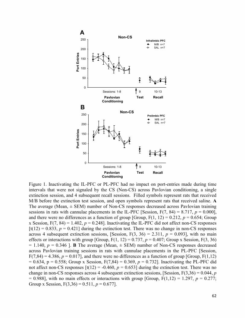
62
Pavlovian Conditioning
Po
rt E
ntr
ies
0
50
100
150
200
250
Test Recall
Sessions: 1-8 9 10-13
Infralimbic PFC
M/B n=7
SAL n=7
ANon-CS
B
Pavlovian Conditioning
Po
rt E
ntr
ies
0
50
100
150
200
250
Test Recall
Sessions: 1-8 9 10-13
Prelimbic PFC
M/B n=7
SAL n=7
Non-CS
Figure 1. Inactivating the IL-PFC or PL-PFC had no impact on port-entries made during time
intervals that were not signaled by the CS (Non-CS) across Pavlovian conditioning, a single
extinction session, and 4 subsequent recall sessions. Filled symbols represent rats that received
M/B before the extinction test session, and open symbols represent rats that received saline. A
The average (Mean, ± SEM) number of Non-CS responses decreased across Pavlovian training
sessions in rats with cannulae placements in the IL-PFC [Session, F(7, 84) = 8.717, p = 0.000],
and there were no differences as a function of group [Group, F(1, 12) = 0.212, p = 0.654; Group
x Session, F(7, 84) = 1.402, p = 0.248]. Inactivating the IL-PFC did not affect non-CS responses
[t(12) = 0.833, p = 0.421] during the extinction test. There was no change in non-CS responses
across 4 subsequent extinction sessions, [Session, F(3, 36) = 2.311, p = 0.093], with no main
effects or interactions with group [Group, F(1, 12) = 0.737, p = 0.407; Group x Session, F(3, 36)
= 1.140, p = 0.346 ]. B The average (Mean, ± SEM) number of Non-CS responses decreased
across Pavlovian training sessions in rats with cannulae placements in the PL-PFC [Session,
F(7,84) = 4.386, p = 0.017], and there were no differences as a function of group [Group, F(1,12)
= 0.634, p = 0.558; Group x Session, F(7,84) = 0.369, p = 0.732]. Inactivating the PL-PFC did
not affect non-CS responses [t(12) = -0.460, p = 0.653] during the extinction test. There was no
change in non-CS responses across 4 subsequent extinction sessions, [Session, F(3,36) = 0.044, p
= 0.988], with no main effects or interactions with group [Group, F(1,12) = 1.297, p = 0.277;
Group x Session, F(3,36) = 0.511, p = 0.677].

63
Appendix B

64
**
*
A
Time Block (Min)
Acti
ve L
ever
Pre
sses
0
2
4
6
8
10
12
14
16
18
20
5 10 15 20 25 30 35 40 451 5 10 15 20 25 30 35 40 451
Extinction Recall
Infralimbic PFC
M/B n=7
SAL n=7
B
Time Block (Min)
Acti
ve L
ever
Pre
sses
0
2
4
6
8
10
12
14
16
18
20
5 10 15 20 25 30 35 40 451 5 10 15 20 25 30 35 40 451
Extinction Recall
Prelimbic PFC
M/B n=7
SAL n=6
Figure 2. Inactivating the IL-PFC before an extinction session caused a rapid decline in active
lever pressing during the extinction test session, but not during recall. Responses on the active
lever during extinction diminished as a function of time following infusions in the IL-PFC
[Time, F(43, 516) = 3.038, p = 0.000] or PL-PFC [Time F(43,473) = 2.100, p = 0.000]. There
was no main effect of group on active lever presses following infusions in the IL-PFC [Group,
F(1, 12) = 2.634, p = 0.131] or in the PL-PFC [Group F(1,11) = 0.655, p = 0.435]. However,
M/B infusions caused a more rapid decrease in active lever pressing in rats with cannulae
targeting the IL-PFC [Group x Time, F(43, 516) = 1.674, p = 0.006], but not in rats with PL-PFC
canulae placements [Time x Group F(43,473) = 0.882, p = 0.686]. During recall, active lever
pressing diminished as a function of time following infusions in the IL-PFC [Time F(43,516) =
3.243, p = 0.000] or in the PL-PFC [Time F(43,473) = 3.066, p = 0.000]. There was no main
effect of group on active lever presses following infusions in the IL-PFC [Group F(1,12) =
2.115, p = 0.171] or in the PL-PFC [Group F(1,11) = 0.035, p = 0.855]. A significant group x
time interaction was found in rats with PL-PFC infusions [Time x Group F(43,473) = 1.505, p =
0.024] but not in rats with infusions in the IL-PFC [Time x Group F(43,473) = 0.882, p = 0.686].
Mean (± SEM) active lever-pressing following M/B (filled symbols) or saline (open symbols)
infusions in the IL-PFC (A) or PL-PFC (B) across the extinction test and recall session 1.


