ROLE OF ENZYMATICALLY MODIFIED LOW-DENSITY LIPOPROTEIN (E-LDL) ON MACROPHAGE GENE
EXPRESSION
DISSERTATION ZUR ERLANGUNG DES DOKTORGRADES DER
NATURWISSENSCHAFTEN (DR.RER.NAT.) DER NATURWISSENSCHAFTLICHEN
FAKULTÄT III – BIOLOGIE UND VORKLINISHCE MEDIZINE DER UNIVERSITÄT
REGENSBURG
vorgelegt von
Quoc Chinh, Duong Regensburg, Juli 2007
All the experimental works have been carried out at the Institute for Clinical
Chemistry and Laboratory Medicine, University of Regensburg under the direction of
Prof. Dr. Gerd Schmitz.
Submission date: 17.07.2007
Board of examiners: Prof. Dr. Herbert Tschochner (chairman) Prof. Dr. Egghard Holler (1st. supervisor) Prof. PD. Dr. Charalampos Aslanidis (2nd. supervisor) Prof. Dr. Stephan Schneuwly (3rd. examiner)
Prof. Dr. Richard Warth (add. examiner)


Table of contents
Page
Acknowledgment i Summary (English/Deutsch) ii 1 INTRODUCTION 1 1.1. ROLE OF MONOCYTES AND MACROPHAGES IN ATHEROSCLEROSIS 1 1.2. ENZYMATIC MODIFICATION OF LOW-DENSITY LIPOPROTEIN 3 1.3. BIOCHEMISTRY, CELL ORGANELLE DISTRIBUTION AND REGULATION OF
PHOSPHOLIPID PATHWAYS 3 1.4. THE ROLE OF BIOLOGICALLY ACTIVE DERIVATIVES OF THE GLYCERO-
PHOSPHOLIPID AND AMINOPHOSPHOLIPID PATHWAYS 6
1.4.1. Biological role of lysophosphatidylcholine 6
1.4.2. Biological role of lysophosphatidic acid 8
1.4.3. Biological role of ceramide 10
1.5.4. Biological role of sphingosine 13
1.4.5. Biological role of sphingosine-1-phosphate 16
1.4.6. Biological role of sphingosylphosphorylcholine 18
1.5. THE LYSOPHOSPHOLIPID RECEPTORS 20
1.5.1. G protein-coupled receptors for lysophospholipids (GPCRs) 20
1.5.2. Scavenger receptors 26
1.6. AFFYMETRIX DNA MICROARRAYS 26 1.7. AIMS OF THE STUDY 29 2 MATERIALS AND METHODS 30 2.1. MATERIALS 30 2.2. DONORS FOR MONOCYTES ISOLATION 33
2.2.1. Density gradient centrifugation of heparinized blood samples 33
2.2.2. Elutriation of monocytes 34
2.3. ISOLATION AND MODIFICATION OF LIPOPROTEINS 35 2.3.1. Isolation of lipoproteins 35
2.3.2. Enzymatic modification of low-density lipoprotein (E-LDL) 35
2.4. MONOCYTE CELL CULTURE 36 2.4.1. Cell culture 36
2.4.1.1. Foam cells induction and removal of cholesterol by HDL3 loading 36
2.4.1.2. Lysophospholipids stimulation of monocyte-derived macrophages 36
2.4.2. Cells harvesting 37
2.5. TOTAL RNA ISOLATION FROM MONOCYTES AND MACROPHAGES 37 2.6. RNA QUALITY ASSESSMENT AND QUANTITATION 39 2.7. AFFYMETRIX DNA MICROARRAY GENE EXPRESSION ANALYSIS 40 2.8. GENE EXPRESSION ANALYSIS BY QUANTITATIVE RT-PCR 41
2.8.1. Reverse transcription reaction (First strand cDNA synthesis) 41
2.8.2. Relative quantitation of gene expression with TaqMan® PCR 42
2.9. TOTAL CELL LYSATES PREPARATION AND WESTERN BLOT ANALYSIS OF PROTEINS 44

2.9.1 Preparation of total cell lysates 44
2.9.2 Western blot analysis of protein 45
3 RESULTS 48 3.1. ANALYSIS OF MARKER GENES IN MONOCYTES, MACROPHAGES,
CHOLESTEROL LOADED/DELOADED MACROPHAGES 48 3.1.1. Expression of scavenger receptors, Cla-1 and CD36, in monocytes, monocyte-
derived macrophages and E-LDL treated macrophages 48
3.1.2. Adipophilin is a sensitive marker for lipid loading in human blood monocytes 50
3.1.3. Expression analysis of ATP-binding cassette transporter 2 gene (ABCA2) in
monocytes, macrophages and foam cells 51
3.2. ANALYSIS OF LYSOPHOSPHOLIPID RECEPTORS EXPRESSION IN MONOCYTES, MACROPHAGES, AND CHOLESTEROL-LOADED/ -DELOADED MACROPHAGES 52
3.2.1. mRNA expression of lysophospholipid receptors in human monocytes 52
3.2.2. mRNA expression of the lysophospholipid receptors during in-vitro differentiation of
monocytes to macrophage 54
3.2.3. mRNA expression of the lysophospholipid receptors during foam cell formation and
lipid deloading 54
3.2.4. Protein expression of the lysophospholipid receptors 55
3.3. DNA MICROARRAY EXPERIMENTS AND BIOSTATICTICS ANALYSIS OF GENES REGULATED BY E-LDL AND LYSOPHOSPHOLIPIDS 56
3.3.1. Principles of large-scale gene expression analysis by DNA microarray 56
3.3.2. Expression analysis of control genes with DNA microarray 58
3.3.3 Biostatistic ranking of regulated genes in microarray analysis 59
3.4. DNA MICROARRAY ANALYSIS OF MACROPHAGE GENE EXPRESSION REGULATION IN LITERATURE-BASED PATHWAYS 60
3.4.1 Assemble of genes into literature-based pathways and categories 60
3.4.2. Candidate genes and pathways regulation in stimulated macrophages 63
3.4.2.1. Analysis of cholesterol metabolism pathway in stimulated macrophages 63
3.4.2.2 Analysis of ABC transporter genes expression in lipid-stimulated macro-phages 64
4 DISCUSSION 66 Citations 75 Appendix A: Abbreviations 96 Appendix B: List of figures 97 Appendix C: List of tables 98 Publications and presentations 99 Erklärung 100

Acknowledgment
Acknowledgment First of all, I would like to express my gratitude to Prof. Dr. Gerd Schmitz at Institute for
Clinical Chemistry, University of Regensburg, for providing me the position and all
excellent working conditions to carry out my study. I would like to deeply thank Prof. Dr.
Egghard Holler at the Biology Faculty, University of Regensburg, for being my first
supervisor and for his supports during my study.
From the bottom of my heart, I would like to express my deepest thanks to Prof. PD.
Dr. Charalampos Aslanidis for not only being supervisor of my work but also for
supporting me on all aspects of my living in Regensburg. Because of him I could have
the chance to study in Germany, and also without his patience and helps through these
years, I could never have come this far.
I would like to express my special thanks to PD. Dr. Christa Büchler, as our group
leader, for sharing her excellent knowledge and skills. I would especially thank PD. Dr.
Thomas Langmann for all interesting discussion that we had, for his excellent ideas
and supports, also his help in correction of my thesis writing. Taking this occasion, I
also would like to thank Dr. Wolfgang E Kaminski, Dr. Mirko Ritter, Dr. Salim Maa-
Bared, Dr. Michael Kapinsky, Cornelia Hasenknopf, Sylvia Kirchner-Luft, Markus
Solleder and all other friends and colleagues at the Institute for Clinical Chemistry and
from other institutes for their friendships and collaboration.
I am also grateful to Prof. Dr. Stephan Schneuwly, Prof. Dr. Herbert Tschochner and
Prof. Dr. Richard Warth at the Faculty of Biology, University of Regensburg, for their
supports.
Above all, I indebted my dearest parents, my parents-in-law, my brother and sister, who
never give up encouraging me and care so much about me. This work is also dedicated
to my lovely wife, Hang, and my lovely son, Tuan, who are always being patient and
providing me lots of strength to overcome those difficult years.
i

Summary
Summary
Monocyte-derived macrophage gene regulation plays an important role not only in
pathogenesis of atherosclerosis but also in many other inflammatory diseases, such as,
cirrhosis, rheumatoid arthritis, glomerulosclerosis, pulmonary fibrosis and chronic
pancreatitis. In addition, bioactive lipids as derivatives of lysophospholipids have been
remarkably evidenced to contribute to many pathophysiological stages of these
diseases. Therefore, the aim of this thesis work is to analyze global gene expression of
monocyte-derived macrophages under the modulation of selected of bioactive lipids,
including ceramide, S1P, SPC, LPA and LPA, and modified low-density lipoprotein as
the source of the these bioactive lipids. Results archived from CD36 and Cla-1 analysis
in phagocytic differentiation and foam cell formation was further confirmed the higher
atherogenic properties of enzymatically modifification LDL, compared to other type of
modifications (acetylation, oxidation). Furthermore, these results also supported the
hypothesis of an autoregulatory loop for enhanced cholesterol uptake and provide a
link between this modified LDL and the HDL metabolism. In addition, analyses of
adipophilin and ABCA2 were also revealed that adipophilin could be considered as a
new sensitive marker for lipid loading of human monocytes/macrophages as well as
ABCA2 could play a role in intracellular LDL-derived free cholesterol trafficking and lipid
homeostasis. Expression of the 13 up-to-date G-protein-coupled receptors for bioactive
lipid derivatives; S1P, LPA, LPC, SPC in human monocytes/macrophages was also
investigated. The serum-free phagocytic differentiation model was able to eliminate the
discrepancy arrived from other studies. Since these bioactive lipid derivatives are
products of platelets and involve in many cellular processes, these results suggested
the importance of further study on cross-talk between platelets and monocytes/
macrophages. DNA chip analysis provided an overview of cellular effects of the above-
mentioned bioactive lipids and cholesterol loading on macrophages global genes
expression. The effects of cholesterol loading (E-LDL) on cholesterol metabolism
pathways and homeostasis were investigated in-details. Interestingly, exogenous free
cholesterol loading of human macrophages could lead to a complete blockage of
cellular cholesterol metabolism pathway triggered by SREBP-2, while triacylglycerides
and fatty acids metabolism pathways triggered by SREBP-1c were not influenced. The
study also reported the expression of ABC transporter A2, A3, A8, B4, C9, D2, G1 and
G4 in human macrophages and suggested the roles of ABCA2, ABCB4 and ABCG1 in
macrophages cellular lipid rheostat.
ii
Zusammenfassung
Zusammenfassung Genregulation in Makrophagen spielt eine grosse Rolle in der Pathogenese der
Atherosklerose und weiteren inflamatorischen Erkrankungen wie Zirrhose,
Rheumatoider Arthritis, Glomerulosklerose, Lungenfibrose und chronischer
Pankreatitis. Lysophospholipid-Derivate und andere bioaktive Lipide scheinen
verschiedene Schritte dieser Erkrankungen zu modulieren. Ein Ziel dieser Arbeit war
deshalb die globale Analyse der Genexpression von Makrophagen unter den Einfluß
bioaktiver Lipide einschließlich Ceramiden, S1P, SPC, LPA und LPA, und modifizierten
LDL-Lipoproteinen.
Die durchgeführten DNA Chip Analysen zeigten einen dominanten Einfluss von
bioaktive Lipiden auf die globale Genexpression der Cholesterinaufnahme.
Expressionsanalysen von CD36 und Cla-1 bei der phagozytären Differenzierung und
Schaumzell-Bildung bestätigen die hohe atherogene Eigenschaft von enzymatisch
modifiziertem LDL gegenüber acetyliertem und oxydiertem LDL. Ausserdem
unterstützen diese Ergebnisse die Hypothese einer autoregulatorischen Schleife der
Cholesterin-Aufnahme mit dem HDL-Metabolismus.
Weitere Analyse zeigten, dass Adipopholin ein Marker der Aufnahme von Lipiden in
humane Monozyten/Makrophagen ist und, dass ABCA2 eine Rolle im intrazellulären
Cholesterintransport spielt.
Abschliessend wurde in dieser Arbeit die Expression von 13 G-Protein-gekoppelten
Rezeptoren für bioaktive Lipid Derivate (S1P, LPA, LPC, SPC) in humanen
Monozyten/Makrophagen untersucht.
iii
Introduction
1. INTRODUCTION 1.1. ROLE OF MONOCYTES AND MACROPHAGES IN ATHEROSCLEROSIS Cardiovascular disease is currently the leading cause of illness and death in
industrialized countries and predicted to be the pre-eminent health problem worldwide
[1]. Atherosclerosis, a progressive disease characterized by the accumulation of lipids
and fibrous elements in the large arteries, constitutes the single most important
contributor to cardiovascular disease. This multiple-phase, decade-spanning of
progressive pathological alteration of large and medium-sized elastic and muscular
arteries may be developed since first decade of life [2].
Pathological studies in the last decades have revealed a defined series of changes in
blood vessels during atherogenesis and demonstrated that blood-derived inflammatory
cells, particularly monocytes/macrophages, play a key role in atherosclerosis. The
development and progression of atherosclerosis is displayed in Figure 1.1 and can be
shortly summarized as follows:
- Lipoproteins from circulating blood infiltrate the intima and accumulate in the
arterial wall leading to diffuse intimal thickening.
- In response to lipid accumulation, blood monocytes migrate into thickened
intimal area and differentiate to macrophages. Subsequently, macrophages
undergo foam cell transformation by taking up lipids, leading to the
development of fatty streak lesions. These events are accompanied by
proliferation and migration of smooth muscle cells from the media into the
intima.
- Local proliferation of specific macrophage populations.
- The centers of atherosclerotic plaques are surrounded by a dense population of
foam cells leading to necrotic events.
- As consequence, plaque disruption occurs in the shoulder region, where
macrophages have accumulated, leading to ulceration or arterial occlusion.
- Thrombosis occurs in the ulcer of the advanced complicated lesions, often
accompanied by calcification in and around the atheromatous core.
1

Introduction
A B
C D
Russell Ross, 1999 [2] Figure 1.1: The development of atherosclerosis in the artery wall. (A) Endothelial dysfunction leading to its high permeability for lipoproteins and other plasma
constituents. (B) Fatty streak formation is caused by migration of monocytes, macrophages and
foam cell transformation. (C) Formation of advanced complicated atherosclerotic lesion. Fatty
streaks progress to intermediate and advanced lesions and tend to form fibrous caps that walls
off the lesion from the lumen. (D) Disruption of the fibrous cap or ulceration of the fibrous
plaque. These events can rapidly lead to thrombosis and usually occurs at the site of thinning of
the fibrous cap that covers the advanced lesion.
Monocytes and macrophages are not only playing important roles in atherosclerosis but
are also involved in other chronic inflammatory diseases, such as liver cirrhosis,
rheumatoid arthritis, glomerulosclerosis, pulmonary fibrosis and chronic pancreatitis [2].
Therefore, gene expression analysis of these cells, under certain pathological
circumstances and/or disease models, is currently under intensive research.
2

Introduction
1.2. ENZYMATIC MODIFICATION OF LOW-DENSITY LIPOPROTEIN Macrophages transform into atherogenic foam cells by taking up modified low-density
lipoproteins (LDL) but not native LDL. To date, LDL has been reported to be altered
either by oxidation (Ox-LDL), chemical modification (e.g. Ac-LDL) [3;4] or enzymatic
modification (E-LDL) [5;6]. In vitro enzymatically modification of LDL was reported by
Bhakdi et al., using the combination of trypsin, cholesteryl esterase and neuraminidase.
Enzymatic modification converts native LDL into an atherogenic moiety that is
remarkably similar to lipid deposits in atherosclerotic lesions [7-10]. Likewise, in vitro E-
LDL has complement-activating capacity [7-10] and a higher potential to induce
macrophage foam cell transformation compared to Ac-LDL and Ox-LDL [11].
Therefore, in vitro induction of macrophages using E-LDL might be considered as
promising laboratory model for atherogenesis studies.
1.3. BIOCHEMISTRY, CELL ORGANELLE DISTRIBUTION AND REGULATION
OF PHOSPHOLIPID PATHWAYS Phospholipids are the major cell and organelle membrane lipids. Phospholipids and
their derivatives form the building blocks of biological membranes and play important
roles in proliferation, migration and programmed cell death. Phospholipids are
constructed from four molecules; fatty acids, a platform where fatty acids are attached,
a phosphate and an alcohol attached to the phosphate.
Glycerophospholipids (or phosphoglycerides) are phospholipids that contain glycerol.
In glycerophospholipids, the hydroxyl groups at C-1 and C-2 of glycerol are esterified to
the carboxyl groups of two fatty acid chains. The C-3 hydroxyl group of glycerol back-
bone is esterified to phosphoric acid. Phosphatidate (diacylglycerol 3-phosphate), the
simplest glycerophospholipid, is present in a small amount in membranes but acts as
the key intermediate in biosynthesis of other glycerophospholipids. In that process, the
phosphate group of phosphatidate becomes esterified to the hydroxyl group of several
alcohols. The common alcohol moieties of glycerophospholipids are serine, ethanol-
amine, choline, glycerol and inositol.
Glycerophospholipids are synthesized in aminoglycero-phospholipid synthesis, which
consists of phosphatidylserine (PS) pathway and Kennedy pathway (figure 1.2). PS,
phosphatidylethanolamine (PE), phosphatidylinositol (PI), phosphatidylcholine (PC) and
phosphatidic acid are the most predominant phospholipids in endoplasmic reticulum.
3

Introduction
Pool size of phosphatidic acid makes up only 1-2% of the endoplasmic reticulum
membrane lipid, but the flux through this pool is extremely high due to its function as
precursor for other phospholipids and triacylglycerol [12].
PEtn: Phosphoethanolamine
PCho: Phosphocholine
Figure 1.2: Biosynthesis pathways and interconversion of PS, PE and PC. PS is synthesized in bacteria, yeast, mammals from serine in endoplasmic reticulum (ER) and
disseminated throughout the cell for the assembly of new organelles [13]. PS can be converted
to phosphatidylethanolamine (PE) by decarboxylation and PC is subsequently formed by
methylation of PE. (Adapted from Voelker [14]).
Although endoplasmic reticulum synthesizes a dominant amount, Golgi is also
contributing to the production of these phospholipids. Significant rates of PC and PI
synthesis are found in the Golgi [15-17]. In addition, the mitochondria also synthesize
their own pool of 3-phosphatidylglycerol that is believed to act as precursor for
mitochondrial phosphatidylglycerol (PG) and cardiolipin [18] and these two
phospholipids are retained within the mitochondria.
Following the synthesis, these phospholipids must be transported to membranes that
lack the synthetic machinery to generate their own lipids. This transportation, for many
4
Introduction
years, is believed to be carried out by phospholipid transfer-exchange proteins [19].
Recent findings, however, indicate that the transport of these phospholipids between
organelles follows some specialized routes that are independent from those followed by
membrane proteins [12]. The potential route that could account for above-mentioned
observation is the movement via specialized zones between subcellular membranes. A
mitochondria-associated membrane (MAM) of endoplasmic reticulum (ER) was
evidenced as the way that PS is imported to mitochondria [20].
Interorganelle distribution of these phospholipids starts with PS synthesis in the
endoplasmic reticulum and the mitochondria-associated membrane. Nascent PS is
transported to mitochondria and Golgi-vacuoles via the pathways, designated PSTA
and PSTB respectively. The transportation of PS to mitochondria is an ATP-dependent
process in mammalian systems. This has been demonstrated for both intact and
permeabilized cells [20-22]. Within mitochondria and Golgi, PS is metabolized to PE by
PS decarboxylase (Psd1p or Psd2p). The PE can be retained within mitochondria and
Golgi or exported to other organelles via pathways PEEA and/or PEEB [12].
Methylation of PE by methyltransferases (Pem1p and Pem2p) in endoplasmic reticulum
leads to the formation of PC. In addition, the Golgi can synthesize PC and the ER can
synthesize PE, PC using ethanolamine and/or choline precursors following the
Kennedy pathway (figure 1.3).
To date, the mechanism of transporting phospholipids from their sites of synthesis to
the plasma membrane has not been completely understood but probably involves both
vesicular and cytosolic protein-mediated transfer mechanisms [23].
5
Introduction
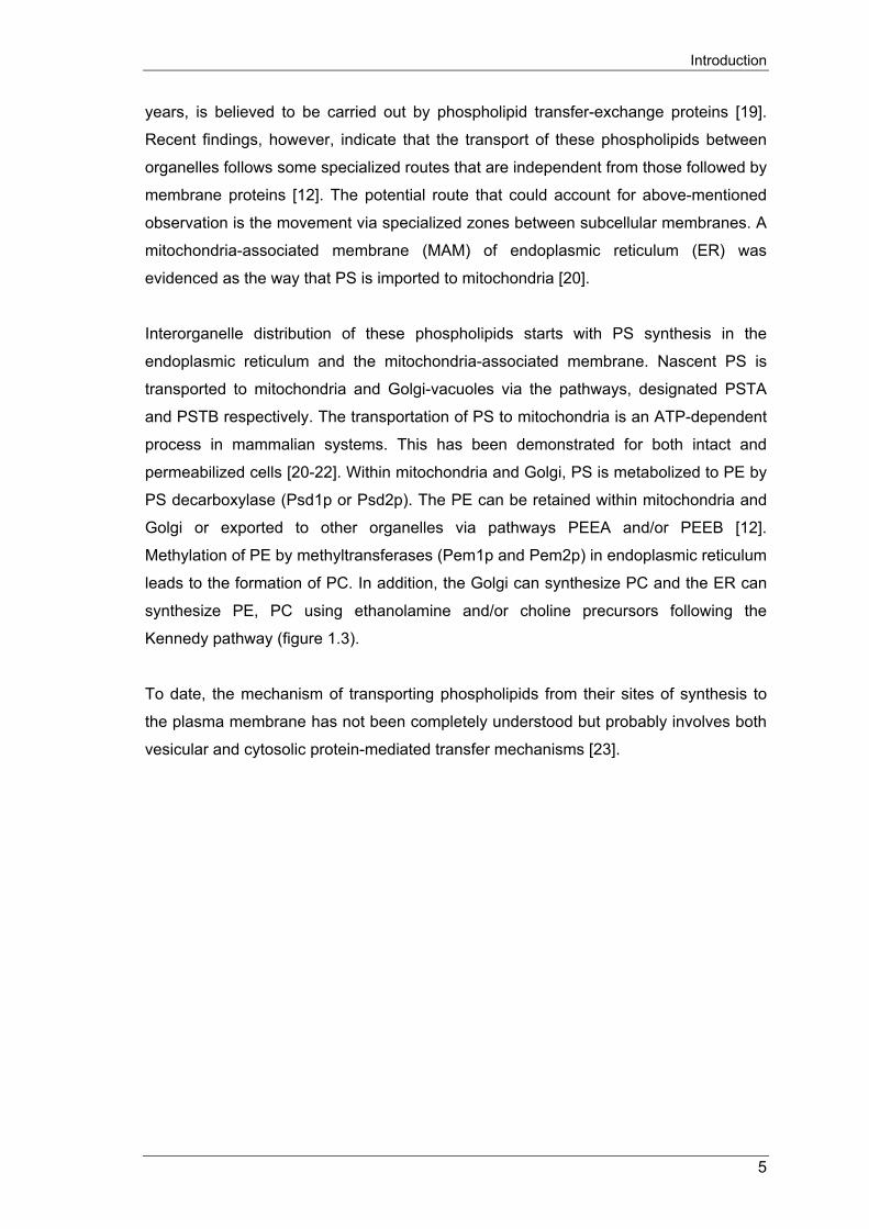
Figure 1.3: Biosynthesis and trafficking of aminoglycerophospholipids. PS is synthesized in the endoplasmic reticulum (ER) and transported to the mitochondrial-
associated membrane (MAM), which then delivers it to the outer membrane of the mitochondria
(M) by an ATP-dependent process. The lipid is then transported to the mitochondrial inner
membrane where it is converted to PE by PS decarboxylase I. PS is also transported from the
ER to the Golgi (G) by an unknown mechanism, where it is converted to PE by PS
decarboxylase II. The mechanism by which PE and PS is transported to the plasma membrane
(PM) is not clear, but could involve vesicular or protein-mediated mechanisms [24].
1.4. THE ROLE OF BIOLOGICALLY ACTIVE DERIVATIVES OF THE GLYCERO-
PHOSPHOLIPID AND AMINOPHOSPHOLIPID PATHWAYS 1.4.1. Biological role of lysophosphatidylcholine Lysophosphatidylcholine (LPC), a naturally occurring lysophospholipid, serves as a
highly active biological molecule involved in many cellular processes. LPC has been
reported to play an important role in atherosclerotic lesion development [25;26] and in
chronic inflammatory diseases [25;27;28]. LPC is a normal constituent of body fluids,
including blood and ascites [29] but is greatly elevated in hyperlipidemic low-density
lipoprotein (LDL) and in atherosclerotic lesions from humans and experimental animals
compared to unaffected tissues [30-32].
6

Introduction
Molecular species of LPC, distinguished by the length and saturation level of their acid
chains, are the metabolites of cell membrane-derived PC. This conversion is the result
of hydrolysis of PC by hormone-activated cytosolic phospholipase A2 (PLA2) [33].
Among the different forms of PLA2, secretory forms of PLA2 (sPLA2) are released from
macrophages and other cell types at the site on inflammation and tissue injury [34].
LPC is also derived from PC-containing lipoproteins as the result of oxidative
processes via the action of endogenous PLA2 [35].
LPC exists in various concentrations (5-180µM) [29] and physiological forms, including
the free form, micellar, and bound to LDL or hydrophobic serum proteins (albumin) [36].
Under physiological conditions, plasma LPC is present mainly in albumin- and
lipoprotein-bound forms. These forms may remain active in some nonreceptor-
mediated functions, such as delivery of fatty acids and choline to tissues, but may be in
the inactive forms for receptor-mediated activities [29;36]. Besides, different
concentrations of LPC present in various cellular and tissue systems (i.e., different LPC
compartmentations) may differently regulate cellular functions [37]. It has also been
shown that serum proteins neutralize the toxicity of LPC [38]. Zhu et al. showed that in
the presence of molar concentration of BSA, the ability to elicit an increase in
intracellular calcium ([Ca2++]i) of LPC via its receptor, GPR4, was greatly diminished
[39]. Biological effects and signaling properties of LPC have been most extensively
investigated in vivo in cell types related to atherosclerosis, including endothelial cells
(EC), smooth muscle cells (SMC), monocyte/macrophage and lymphocytes [25;40;41].
LPC has been shown to activate phospholipase C (PLC) [42] and protein kinase C
(PKC) [40], induces [Ca2++]i [42;43] and activates or inhibits mitogen-activated protein
(MAP) kinase [44;45]. Although there are more lipid molecules reported as components
of oxidized LDL (oxLDL) (i.e., 7β-hydroperoxylcholesterol and 4-hydroxynonenal)
[46;47], recent findings indicated that LPC is the primary lipid responsible for oxidative
LDL-mediated alterations in endothelial functions [48].
Recent evidence indicated that LPC plays a dual function as an atherogenic or an anti-
atherogenic agent [41]. Due to its various physiological forms, it may be important to
determine whether this dual function is related to receptor-mediated vs. nonreceptor-
mediated effects, receptor compartmentalization and/or is mediated via different types
of receptors. Mouse models have shown that LPC stimulates phospholipase D (PLD)
activity that in turn elevates generation of other second messengers such as
phosphatidic acid, lysophosphatidic acid and diacylglycerol, all of which are considered
responsible for many cellular processes including atherogenesis and inflammation [49].
7

Introduction
Other findings also reported that LPC might be responsible for Ox-LDL-induced
apoptosis [50], showing a possible new role in endothelial dysfunctions in pathogenesis
of atherosclerosis. LPC induces endothelial cell suicide by sensitizing endothelial cell to
Fas-mediated apoptosis [51]. These findings add to the understanding of
atherosclerosis but the mechanisms of LPC-induced apoptosis have not been fully
elucidated.
Recently, three G protein-coupled receptors (GPCRs), OGR1/GPR68, GPR4 and G2A,
are identified as receptors for LPC [39;52;53]. However, GPR4 and OGR1 have higher
affinity to sphingosylphosphorylcholine (SPC) than LPC. Ligand-induced increase in
[Ca2++]i, particularly released from intracellular stores, is characteristic of many GPCRs
including OGR1, G2A and GPR4. Furthermore, these receptors function as
immunosuppressor genes (G2A) [54], tumor suppressor regulated genes (OGR1)
involved in apoptosis [55]. These findings are still preliminary and the receptor-
mediated signal transduction of LPC needs further investigation.
1.4.2. Biological role of lysophosphatidic acid Lysophosphatidic acid (LPA) is a bioactive lipid controlling a large number of cellular
responses through the activation of specific GPCRs. LPA is present in several body
fluids (serum, plasma, aqueous humor) and can also be secreted by several cell types,
including platelets, fibroblasts, adipocytes and cancer cells [56]. LPA is the simplest
phospholipid that contains long chain fatty acids (C16-C24). Production of LPA involves
a number of enzyme activities, extracellularly (sPLA2, PLD) and intracellularly (cytosolic
PLA2, glycerol-3-phosphate acyltransferase, monoacylglycerol kinase). Although
platelets are described as the main source of LPA, several studies have revealed the
existence of LPA in other cells (cancer cells, fibroblasts, adipocytes) and lipoproteins
under physiopathological conditions [56].
In the vascular system, activation of platelets upon the injury of endothelial cells leads
to release of LPA. In activated platelets, PLC and diacyl glycerol kinase are contributed
to the conversion of phosphoinositides to phosphatidic acid (PA). PA is then degraded
to LPA by PLA2. Recent findings indicated that the production of LPA by platelets is
sensitive to extracellular calcium [57-59]. Moreover, albumin binds LPA with high
affinity and detection of LPA in medium is possible only in the presence of albumin.
Other studies also revealed that biological activity of serum albumin on the stimulation
of chloride efflux in Xenopus oocytes and actin stress fiber formation and cell
8

Introduction
proliferation is due to its high content of LPA [60-62]. Another source of LPA may result
from the conversion of LPC that accumulated in lipoproteins which contain unsaturated
fatty acids. This conversion is carried out by PLD present in plasma. Whereas native
LDL contains only a small amount of LPA, mild oxidation of LDL leads to the large
contents of LPA. This explains why LPA is not only present in circulating blood but also
accumulates in the intima of human atherosclerotic lesions in comparison to normal
arterial tissues [63].
LPA may be one of the factors involved in the permeability of endothelium, one of the
first steps in atherosclerosis Recent studies indicated that LPA and LPA present in
mildly Ox-LDL are responsible for actin stress fiber formation, contraction of endothelial
cells and intercellular gap formation, processes leading to endothelial permeability [63-
70]. The decrease of the endothelial barrier function requires LPA concentrations
above 1µM and exposure time for several hours [64]. This has been observed in
human umbilical venous endothelial cells, human aortic endothelial cells, porcine brain
capillary endothelial cells and the human endothelial cell lines [64;65;68-70].
Endothelial dysfunction may drive endothelial cells to undergo apoptosis and leads to
plaque erosion and intravascular thrombosis without plaque rupture [71;72]. In other
studies, LPA, however, has been found to stabilize the integrity of endothelial
monolayer [73;74]. This contradictory observation may be explained by the different
effects of LPA on different species, origin of endothelial cells and/or differences in cell
culture conditions leading to expression of LPA receptors.
LPA is also reported to act directly on vascular smooth muscle cells (VSMCs) or
indirectly by stimulating the release of vasoactive substances from endothelium.
Platelet-derived LPA up-regulates the production of endothelin-1, a VSMC-contracting
substance, in endothelial cells derived from rat aorta and piglet cerebral microvessels
in vitro [75;76] and synergizes with endothelin-1 reinforcing vasoconstriction in animal
models in vivo [77]. At high concentrations (10-100µM), LPA stimulates the proliferation
of VSMCs and fibroblasts. The stimulatory or inhibitory effects of LPA on VSMCs
migration depend on their phenotype and also the type of matrix on which the cells
grow. Low concentrations of unsaturated LPA species (EC50: 20-30nM) induce VSMCs
differentiation, migration and proliferation, whereas saturated LPA species are inactive.
Interestingly, the differentiated and de-differentiated VSMCs are expressing only LPA
receptor 1 and 3 (LPA1 and LPA3) but not LPA receptor 2 (LPA2) and the de-
differentiation induced by LPA is mediated by activation of ERK- and p38 MAP kinases.
9
Introduction
These unsaturated LPA species which induced phenotypic modulation of VSMCs are
implicated as potent atherogenic factors [78].
In addition, LPA also shows its effects on adhesion molecules and monocyte adhesion.
LPA (10µM) increased the expression of E-selectin and vascular cell adhesion
molecule 1 (VCAM-1) on endothelial cell surface and stimulated monocyte binding.
Expression of these molecules have been found to be stimulated by LPA via its
receptors (LPA1, 2 and 3 or old nomenclature; EDG-2, EDG-4 and EDG-7, respectively)
and inhibited by NPTyrPA and NPSerPA as LPA receptor antagonists [79-81]. These
effects of LPA may be due to activation of the NF-kB transcription factor [82]. The
increase of intracellular concentration of calcium, stimulation of chemotaxis and
haptotactic migration of human monocytes were reported to be induced by LPA (at
millimolar concentration) [83] via its receptors (Table 3).
Similar to sphingosine-1-phosphate, LPA is also having effects on endothelial cell
migration and proliferation, which are important for vascular repair and re-
endothelialization of the carotid, coronary or peripheral arteries after surgical
endarterectomy, balloon angioplasty or stent implantation [84-86].
1.4.3. Biological role of ceramide Ceramide belongs to the group of sphingosine-based lipid second messenger
molecules that are involved in diverse cellular responses to exogenous stimuli. These
cellular responses, including proliferation, differentiation and apoptosis, are believed as
results of the coupling of ceramide to different signaling cascades in both stimulus and
cell-type specific manners [87;88].
Ceramide can be synthesized de novo by condensation of serine and palmitoyl-CoA to
form ketosphinganine, which is subsequently reduced to sphinganine
(dihydrosphingosine) [89]. In the presence of ceramide synthase (sphinganine N-acyl
transferase), sphinganine is catalyzed to form dihydroceramide and subsequently is
catalyzed by dihydroceramide desaturase to form ceramide. Although it is known that
dihydroceramide is generated in endoplasmic reticulum and in the mitochondria, the
subcellular localization of the dihydroceramide desaturase has not been determined.
This enzyme appears to be crucial because ceramide, but not dihydroceramide, is
biologically active. Also, ceramide synthase is reported to be a stimulus-responsive
enzyme. Several studies indicated that prolonged activation of ceramide can be
10

Introduction
induced by daunorubicin treatment of some cell types, such as P388, U937 and HL-60
cells [90;91]. Ceramide can also be generated by breakdown of glycosphingolipid
complexes through acid hydrolases although the regulation of this enzyme by
exogenous stimuli is still unknown.
Activation of the sphingomyelin cycle subsequently increases the intracellular ceramide
levels and leading to various cellular effects of ceramide, depending on the cellular
complement of its effectors, activities of synthesizing and catabolizing enzymes as well
as the subcellular localization in which ceramide is generated. There are several
ceramide effector enzymes that have been identified. One of those is ceramide
activated protein phosphatase (CAPP), which has been identified as a member of 2A
class of protein phosphatases (PP2A). Fishbein et al. reported that PP2A, as the
ceramide effector, mediates ceramide-induced growth arrest in yeast [92] and this has
been confirmed by gene knockout studies [93]. Another ceramide effector enzyme is
protein kinase known as ceramide acitvated protein (CAP) kinase [94]. It is known that
CAP kinase participates in TNF inflammatory responses by activating Raf and
subsequently the MAP kinase cascade [95]. Also, Raf itself may act as a direct target of
ceramide in interleukin-1 signaling [96]. Therefore, it is suggested that CAP and Raf
may be responsible for mediating ceramide activation of the MAP kinase pathway in
response to inflammatory cytokines. Protein kinase Cζ is also reported to directly bind
ceramide and modulate the cellular response to TNF [97]. Interestingly, kinase
suppressor of Ras (KSR), Raf and protein kinase Cζ all have a cysteine-rich domain,
which has been recently hypothesized to play a role in the binding of ceramide to these
kinases [98]. While CAPPs, CAP kinase and Raf have been reported to play a role in
growth arrest and inflammatory cytokine signaling, there is no decisive evidence
provided to demonstrate the role of these ceramide effectors in apoptosis.
Signaling through the sphingomyelin pathway as a second messenger, via distinct
receptors (CD28, CD95, TNF-α, IL-1β, progesterol, γ-interferon), ceramide shows
diverse cellular functions, including fibroblasts proliferation, promyelocytes
differentiation, inhibition of the respiratory burst in human neutrophils, survival of T9
glioma cells and regulation of apoptosis [87;88;99]. Ceramide analogue such as cell-
permeable C6-ceramide was first shown to have anti-proliferation effects in HL-60 cells,
while cell-permeable C2-ceramide induced HL-60 cells differentiation towards the
monocytic lineage and mimics the action of agonists, such as 1α,25-dihyroxyvitamin
D3, IFNγ and TNF-α [94;100]. In addition, mimicking of the effects of TNF-α is also
11

Introduction
resulting from the induction of apoptosis by exogenously addition of ceramide to cells
[101] (figure 1.4).
Figure 1.4: Signaling via ceramide Activation of the acid sphingomyelinase results in hydrolysis of sphingomyelin, the release of
ceramide, and the transformation of rafts to small ceramide-enriched microdomains that
spontaneously fuse to a large ceramide-enriched membrane platform. SM, sphingomyelin;
ASM, acid sphingomyelinase; AC, acid ceramidase; I, intracellular; E, extracellular. (Gulbins
[102])
To date, the most highlighted importance of ceramide is its role in apoptosis, a
physiological form of cell death in order to control cell populations. Apoptosis has been
reported to be induced by receptor-mediated, stress stimuli and growth factor-
deprivation-mediated processes. CD95, which may serve as paradigm for receptor-
mediated apoptosis, has been shown to activate acid sphingomyelinase and trigger the
release of ceramide. CD95 also leads to the formation of ceramide-enriched membrane
platforms that mediate clustering of the receptor [103-105], which has been shown in
several cell types [106]. Clustering of CD95 in ceramide-enriched membranes, as has
been reported in several studies, is required for the production of apoptosis. In those
studies, acid sphingomyelinase-deficient cells fail to release ceramide on CD95
stimulation and therefore resist apoptosis, while re-addition of natural C16-ceramide to
12

Introduction
these cells lead to apoptosis [104;105]. Likewise, acid sphingomyelinase deficient
animals were protected from TNF-α-induced apoptosis of hepatocytes, hepatic failure
and death [107].
In addition, the acid sphingomyelinase and ceramide-enriched membrane platforms are
not only restricted to receptor-mediated apoptosis but also cause non-receptor-
mediated apoptosis, or stress stimuli-trigger cell death. In particular, the acid
sphingomyelinase has been shown to be central for the induction of cell death by
gamma-irradiation and UV-A, UV-C light [108-111]. Other chemotherapeutic reagents,
such as cisplatin and doxorubicin, were shown to stimulate acid sphingomyelinase that
trigger the release of ceramide, formation of ceramide-enriched membrane platforms
which served to cluster CD95 and, therefore, excluded death of tumor cells [112;113].
Ceramide is also found to play roles in growth factor-deprivation-mediated apoptosis. It
was recently shown that several drugs, such as desiparmine and imiparmine [114],
prevented apoptosis induced by inhibition of intergrins, while inhibition of αvβ3/ αvβ5
intergrins results in activation of acid sphingomyelinase and release of ceramide
leading to protection of cell death.
Acid sphingomyelinase has been also reported by Tilly et al. to play role in
developmental death in mice [113]. In mice as well as in human females, approximately
80% of all oocytes undergo cell death until birth and genetic deficiency of acid
sphingomyelinase prevented and delayed developmental apoptosis of oocytes thus
leading to hyperplasia at birth. This shows a fundamental function of acid
sphingomyelinase in oocytes apoptosis.
1.4.4. Biological role of sphingosine Sphingosine, as the intermediate metabolite of ceramide and sphingosine-1-phosphate
(S1P), plays a key role in balancing the intracellular levels of ceramide and
sphingosine-1-phosphate leading to either survival or death of cells. In the
sphingomyelin pathway, sphingosine can be generated during metabolization of
ceramide by ceramidase in the early stages of apoptosis. Sphingosine has been found
to induce apoptosis when added exogenously to many cell types. Sphingosine can be
further phosphorylated by sphingosine kinase to form S1P, which acts as a signaling
molecule that antagonizes ceramide-mediate apoptosis processes (figure 1.5).
13

Introduction
Figure 1.5: Sphingolipids metabolism
Sphingosine is generated from ceramide by ceramidase as well as phosphorylated to
sphingosine-1-phosphate by sphingosine kinase.
The role of sphingosine in apoptosis was initially supported by the study of Igarashi et
al. [115]. In this study, human neutrophils treated with TNF-α showed an increase in
the level of both, ceramide and sphingosine during the first 60 min. prior to any
apoptotic morphological changes. The increase of ceramide is more rapid than that of
sphingosine suggesting that formed ceramide has been degenerated to sphingosine
after cells were treated with TNF-α. The increased intracellular level of sphingosine
reaches up to 5-10µM and a similar amount of exogenous added sphingosine both
showed to induce apoptosis in neutrophils, mimicking TNF-α [115]. Even though
ceramide and sphingosine are interconvertible metabolites, sphingosine induces
apoptosis without being converted to ceramide inasmuch as the ceramide synthase
inhibitor fumonisin B1 dose not affect sphingosine-induced apoptosis in several cell
types, including HL-60, U937, Jurkat, TF1 erythroleukemic and Hep3B hepatoma cells
[116-120].
Sphingosine is demonstrated to mediate apoptosis in leukemic cells or solid cancer cell
lines via various mechanisms. Among those, a large body of evidence has been
accumulated regarding the role of MAPKs. The p38 MAPK subgroups, known to
present one of two independent parallel MAPK pathways, were found strongly activated
by sphingosine in neutrophils. This finding suggested that sphingosine-mediated
apoptosis could be p38 MAPK-dependent [121]. Other studies also demonstrated a
14

Introduction
strong and virtually complete inhibition of p44-ERK1/p42-ERK2 activity in leukemic and
solid cancer cells by sphingosine [117;122;123], implicating the direct contribution of
sphingosine to ERK1/ERK2’s regulation properties of cell proliferation. Furthermore,
sphingosine is also reported to stimulate the cleavage of poly ADP-ribose polymerase
(PARP), a well-known target for executioner caspases that plays a central role in
executing mammalian cell apoptosis [120;124] (figure 1.6).
Figure 1.6: Mechanisms of apoptosis regulation by sphingosine [125] Cyt c, cytochrome c; tBax, truncated Bax; tBid, truncated Bid; ERK, extracellular-regulated
protein kinase; JNK, c-Jun N-terminal kinase; Rsk, ribosomal S6 kinase; cPKC, conventional
protein kinase C isoforms; nPKC, novel protein kinase C isoforms; pRb, retinoblastoma gene
product. Pro-apoptotic elements are represented in red background, and anti-apoptotic ones in
green.
In addition to its dependence to MAPK cascades and the caspases, sphingosine is also
believed to mediate apoptosis through the mitochondrial pathways. These include the
decrease of Bcl-2, Bcl-xL enforced expression that abolished apoptosis [126-128],
triggering of executioner caspases activation and cytochrome c exit by sphingosine
[118;129]. Also, the involvement of other members of the pro-apoptotic Bcl-2 family,
such as Bax, Bid and Bad, to sphingosine apoptosis signaling could implicate an
alternative mechanism, while during apoptosis, sphingosine is shown to induce
truncation of Bid and Bax [49;118;119]. Another direction of sphingosine-induced
apoptosis may be its association to the Akt/Rsk/Bad pathway. Recently, several studies
showed that sphingosine-induced apoptosis associates with inhibition of basal-/serum-
15
Introduction
stimulated Akt kinase activity in hepatoma cells [124] and with inhibition of Bad
phosphorylation, through inhibition of a PKC/Rsk pathway [130-133].
At last, while ceramide and sphingosine are considered as pro-apoptotic molecules, its
metabolite, S1P, promotes cell survival in response to apoptotic stimuli such as TNF-α,
Fas ligation, serum deprivation, cell-permeable ceramides [134-140]. In this
‘ceramide/S1P rheostat’, sphingosine kinase and S1P phosphatase play the important
roles that determine whether cells die or survive.
1.4.5. Biological role of sphingosine-1-phosphate Sphingosine-1-phosphate (S1P) appears as a normal lipid constituent of human
plasma and serum [141]. It has been shown to elicit a great variety of responses,
including stimulation of cell proliferation, survival, motility and cytoskeleton in a large
number of cell types. S1P is also acting as an intracellular second messenger and
extracellular mediator.
The formation of S1P is a dynamic balance between sphingosine kinase and S1P-
lyase. Sphingosine kinase, which exists in cytosol and cell membranes, catalyzes
sphingosine to S1P [142;143]. Whereas, S1P lyase cleaves S1P into fatty aldehyde
and phosphoethanolamine and possibly is the key enzyme that keeps intracellular S1P
levels low [144]. In addition to S1P lyase, S1P phosphatase may also play a role in the
attenuation of S1P by converting S1P into sphingosine [145]. S1P may also be
synthesized by hydrolysis of sphingosylphosphorylcholine (SPC) catalyzed by
lysophospholipase D or by deacylation of ceramide-1-phosphate [146]. These,
however, have never been reported in biological systems. Extracellularly, S1P has
been reported to be synthesized and released by blood platelets [141]. The
accumulation of S1P in platelets is due to the highly active level of sphingosine kinase
and its activity is not depending on cell activation by physiological antagonists [147-
149]. Extracellularly release of S1P from blood platelets is following the activation of
protein kinase C directly or indirectly by stimulation with thrombin, 12-O-tetradecanoyl-
phorbol 13-acetate or 1-oleoyl-2-acetyl-glycerol. Concentration of S1P in plasma and
serum are very high; 190pmol/ml and 480pmol/ml, respectively [141]. As same as LPA,
plasma S1P is also bound to albumin and this keeps S1P metabolically stable and
might prevent it to elicit activities. S1P was also reported to be present in ascites from
patients with ovarian cancer cells. In addition, high activity of sphingosine kinase was
also found in other peripheral blood cells, including erythocytes, neutrophils and
16

Introduction
mononuclear cells [150]. Considering the great variety of S1P-responsive cell types
and wide distribution of S1P receptor expression, it may suggest that S1P is released
from various cell types to influence target cells in paracrine or autocrine fashions [151].
Furthermore, many studies focusing on diverse biological effects of S1P released from
blood platelets have demonstrated that S1P is involved in a variety of physiological and
pathophysiological processes, including thrombosis, hemostasis, angiogenesis and
atherosclerosis [83;151;152].
S1P has been reported to induce intracellular calcium mobilization, shape change
reaction as well as primary aggregation in platelets. The effect of S1P on platelets
requires a high concentration and much weaker than that of its metabolite, LPA [149].
The physiological role of S1P as an autocrine stimulator of platelets remains to be
further investigated. In contrast to its week effects on platelets, S1P has been shown to
have dramatic effects on endothelial cells. Its receptor transcripts, S1P1 (EDG-1) and
S1P3 (EDG-3), at high and low level respectively, were reported to be expressed in
human umbilical vein endothelial cells (HUVECs) [153;154]. S1P, at as low as
nanomolar concentration, reportedly induces a variety of endothelial responses. S1P,
as the intracellular second messenger, was reported to induce Ca2+ mobilization,
MAPK activation and Rho family of small G protein activation [153-158], as confirmed
by transfection studies [159-161]. Functionally, S1P stimulates endothelial cell survival
or proliferation through its Gi-coupled receptor, S1P1. In addition, S1P also induces
adherent junction assembly, migration, capillary tube formation and promotion of
angiogenesis [153-155;157]. As extracellular mediator, S1P is found to induce
expression of adhesion molecules such as E-selectin and VCAM-1. S1P, but not its
metabolites, ceramide or sphingosine, is able to mimic TNF-α to induce adhesion
molecules that activate NF-κB [135;162]. In addition, S1P receptors are also reported
to respond to changes in fluid shear stress that modulates vascular structure, function
and plays an important role in the pathogenesis of vascular diseases such as
atherosclerosis, hypertension and restenosis. S1P1 mRNA level in endothelial cells was
confirmed to markedly increase in response to fluid flow [143;163].
Transfection studies were reported that S1P, via its S1P2 receptor, induces Ca2+
mobilization, MAPK activation and adenylyl cyclase activation in human aortic smooth
muscle cells [164-166]. While endothelial cell migration is induced by S1P, vascular
smooth muscle cell migration (induced by PDGF) is demonstrated to be inhibited by
this bioactive lipid [166]. This cell type-dependent discrepancy in S1P effects on cell
17
Introduction
migration is intriguing and maybe explained by different expression patterns of S1P
receptor subtypes. Kon J. et al. described that S1P1 and S1P3 receptors, expressed in
endothelial cells, are able to induce migration of this cell type in response to S1P,
whereas S1P2 failed to promote migration activity. Among S1P receptor subtypes, S1P2
exerted the most potent effect on adenylyl cyclase stimulation leading to the inhibition
of smooth muscle cell migration [165]. The discrepancy in S1P effects on cell migration,
however, requires further investigation.
1.4.6. Biological role of sphingosylphosphorylcholine In contrast to S1P and LPA, only little information is available regarding the formation,
degradation and biological action of sphingosylphosphorylcholine (SPC). However, it
has been shown to be involved in many cellular processes and is produced under
physiological and pathological conditions. Recently, SPC was detected in normal rabbit
plasma and serum [167]. Its higher concentration in serum, compared to that in plasma,
suggests that SPC is also formed during blood clotting. Similar to other bioactive lipids,
SPC is also found to be associated with high-density lipoprotein (HDL) [168;169].
Despite its presence in plasma, SPC seems to undergo a clearance mechanism during
circulation [170]. The pathways of SPC production under physiological conditions have
still to be analyzed. SPC may also be produced by sphingomyelin deacylase in certain
pathological conditions. It has been found accumulated in Niemann-Pick disease,
which is characterized by a deficiency of sphingomyelinase [171;172]. Other data
suggest that SPC also accumulated in atopic dermatitis [173], in ascites caused by
ovarian cancer and in certain meningeomas [174].
As with S1P, SPC has been reported to promote both intracellular and extracellular
actions. Many of SPC actions are shared by its structurally related S1P [175-178]
suggesting that these two lipids have common cites of action. S1P-GPCRs are
believed to be activated by SPC, although a much higher concentration is needed,
compared to that of S1P. The best-studied intracellular action of SPC is release of Ca2+
from intracellular stores such as microsomes [179], permeabilized smooth muscle cells
[180], and permeabilized EA.hy926 human endothelial cells [181]. The release of Ca2+
that is observed in permeabilized cells and microsomes is reported to require as high
as micromolar concentrations of SPC (EC50 of 3-6µM). In some studies, it is reported to
be up to 50µM of SPC [179;180]. At these concentrations, SPC promotes Ca2+
mobilization by activating S1P-GPCRs. These two actions of SPC have been studied
separately as reported for HEK 293 cells; Extracellular SPC induced a stereospecific
18

Introduction
Ca2+ mobilization in intact cells most likely via activation of S1P-GPCRs, whereas in
permeabilized cells it caused Ca2+ release at same potency and efficacy [182]. Other
cells such as CFPAC-1 (a pancreatic duct adenocarcinoma cell line) and CFNP9o (an
immortalized airway epithelial cell line) had no responses to SPC at concentrations of
10µM and 20µM, respectively [183;184]. This may suggest that the threshold of action
of SPC in these cells is much higher.
Nevertheless, there are still open questions raised about intracellular SPC-induced
Ca2+ release as the exact target of SPC’s action is not known and, more importantly,
the physiological significance of in-vitro observed actions is unclear. Compared to its
structurally related S1P, SPC’s intracellular formation is not reported or takes as long
as several hours, which is too slow to induce rapid process of Ca2+ mobilization [183].
Furthermore, SPC’s contractile effect in permeabilized smooth muscle cells is not
inhibited by pretreatment with thapsigagin or an antibody against myosin light chain,
both of which block inositol 1,4,5-trisphosphate (IP3)-induced contraction, but sensitive
to protein kinase C and to an antibody against MAPK [185]. Besides, the effects of
GPCRs are preserved during smooth muscle cells permeabilization and therefore it can
not be stated whether SPC acted as intracellular or extracellular mediator [186].
SPC have been shown to share some actions with S1P [175-178], however it also
exerts biological actions that are not shared by S1P such as inhibition of Ca2+ influx in
GH4C1 cells [187], induction of superoxide anions formation in HL-60 granulocytes
[188] and stimulation of inositol phosphate production in airway epithelial cells [189].
This suggested that, besides the signal transduction via S1P-GPCRs, SPC may act on
its own receptors. Initially, OGR1, GPR4, GPR12 and G2A are reported as receptors
for SPC [39;53;190]. However, Ludwig et al. recently have demonstrated that GPR4
and OGR1 are not responsive to SPC [191]. This raised a question about the identity of
these receptors and needed to be further investigated. Therefore, one can assume that
SPC acts only on OGR1 (high-affinity) and G2A (lower affinity) compared to that of LPC
[52]. In addition to its direct action to release Ca2+ from intracellular stores,
extracellularly applied SPC also induces rapid increase in intracellular Ca2+
concentration ([Ca2+]i) in a number of cell types, including vascular endothelial cells and
VSMCs [192;193]. The SPC-induced [Ca2+]i is pertussis toxin sensitive and can be
observed with G2A receptor [52], interestingly, in the absence of inositol phosphate
production [188]. SPC is also demonstrated to act on cell growth as it is reported in
quiescent 3T3 fibroblasts [194] and other cells such as endothelial cells [192],
19

Introduction
keratinocytes [195] and VSMCs [193], as well as play roles in cell migration [196;197],
adhesion, stress fiber formation and cytoskeleton rearrangements [198].
Beside the extra and intracellular mediator functions, SPC also shows its effects in
tissues and organs. The induction of [Ca2+]i in vascular endothelial cells [199] and
VSMCs [193;200] and induction of endothelial cells migration and differentiations [196]
by SPC clearly showed that it has an influence on vascular tone. Cells of immune
system are recently reported to be activated and stimulated by SPC. HL-60 leukemia
cells and human neutrophils are activated by SPC [188;201]. In HEY cells, SPC is also
reported to increase mRNA levels of IL-8 at the concentration 1-10µM.
1.5. THE LYSOPHOSPHOLIPID RECEPTORS 1.5.1. G protein-coupled receptors for lysophospholipids (GPCRs)
Lysophospholipid receptors form a super family of GPCRs, a large family of plasma
membrane receptors, activated by a wide variety of ligands, including proton, H+ ion,
small molecules, odorants, peptides, proteins and lipids. GPCRs are used ubiquitously
and widely for signal transduction across the plasma membranes. Lysophospholipid
receptors as well as other GPCRs, share a common structure that is encoded by seven
membrane spanning helix containing proteins. The basic structure motif of these
receptors form an extracellular and/or transmembrane ligand binding pocket and an
intracellular domain that interacts with signal transducer molecules. The most
prominent family being the heterotrimeric G proteins (Gs, Gi/o, Gq/11, G12/13 and G16).
Ligand binding to these receptors results in their conformation changes and therefore
allows productive interaction with the G proteins and other effector molecules. It is
believed that the basic structural motif of seven transmembrane helices is well suited
for cell-cell communication events that utilize diffusible mediators. Lysophospholipid
receptors as well as most of lipid receptors belong to the Rhodopsin super family of
GPCRs. Herein, only a group of bioactive lysophospholipid receptors is being
discussed, including receptors for S1P, LPA, LPC, SPC.
As discussed in previous chapters, lysophospholipids are bioactive lipids emerged as
important mediators and involved in various physiological actions as well as cellular
effects. Many of these functions associate to the appropriate GPCRs. To date, there
are fifteen mammalian GPCRs identified as high affinity cellular surface receptors for
these lysophospholipids: LPA (LPA1-5), S1P (S1P1-5, GPR3 and GPR12), LPC (G2A,
20

Introduction
GPR4) and SPC (OGR1, GPR4, GPR12 and G2A). Receptors for S1P and LPA are
also known as endothelial differentiation gene (EDG) receptors.
Figure 1.7: Evolutionary dendrogram of lysophospholipid receptors Evolutionary tree made by comparison of lysophospholipid receptor amino acid sequences,
archived from NCBI database, using HUSAR bioinformatics lab software.
21

Introduction
LPA receptors are reported in many studies to meditate LPA downstream signaling,
including MAPK activation, adenylyl cyclase (AC) inhibition/activation, phospholipase C
(PLC) activation/ Ca2+ mobilization, arachidonic acid release, Akt/PKB activation and
the activation of small GTPase, Rho, Rac, and Ras (figure 1.8). LPA receptors are
found to be expressed in various tissues such as; brain, heart, placenta, colon, small
intestine, prostate, testis, ovary, pancreas, spleen, kidney, skeletal muscle, and thymus
[202]. So far, five receptors (LPA1- LPA5) have been identified for LPA and three of
those (LPA1-LPA3, previously named as EDG-2, 4, 7) share a high amino acid
sequence similarity.
LPA1 was the first to be identified as high affinity receptor for LPA [203]. Functional
analysis using mammalian heterologous receptor expression system revealed the
multi-functionality of this receptor [204;205]. LPA1 coupled with three types of G
proteins; Gi/o, Gq and G12/13 [206;207] and activation of these proteins leads to LPA-
mediated induction of many cellular responses including proliferation, serum-response
element (SRE) activation, MAPK activation, AC inhibition, PLC activation, Ca2+
mobilization, Akt activation and Rho activation [204;205].
LPA2 was identified by homology search of orphan GPCR from GenBank. LPA2 was
also found to couple with Gi/o, Gq and G12/13 in order to mediate LPA-induced cellular
signaling. Targeted deletion of LPA2 in mice does not result in any obvious phenotypic
abnormality but significant loss of normal LPA signaling (e.g., PLC activation, Ca2+
mobilization and tress fiber formation) is observed in primary cultures of mouse
embryonic fibroblasts (MEFs) [208]. Creation of LPA1(-/-)/LPA2
(-/-) double-null mice
showed the absence or severe reduction of many LPA-induced responses which
include proliferation, AC inhibition, PLC activation, Ca2+ mobilization, JNK and Akt
activation and stress fiber formation while LPA2(-/-) itself does not show any similar
effects [208].
LPA3 was isolated as an orphan GPCR gene by degenerated polymerase chain
reaction (PCR)-based cloning and homology search [209;210]. LPA3 distinct from LPA1
and LPA2 due to its capability of coupling with only Gi/o and Gq but not G12/13 [207]. LPA3
is much less responsive to LPA species, which contain saturate acyl chain, in
comparison to LPA1 and LPA2 [209;210]. LPA3 also shares common affinity to LPA to
induce PLC activation, Ca2+ mobilization, AC inhibition/ activation and MAPK activation
[207;209;210] although its effects on AC likely depends on cell types and expression
levels.
22
Introduction
LPA4 (or GPR23) was identified from orphan GPCR gene libraries within another
evolutionary branch of the LP receptor superfamily [211]. Unlike the other three LPA
receptors, LPA4 is found to be encoded by only a single exon and shared greater
similarity to platelet-activating factor GPCR. Like other LPA receptors, LPA4 mediates
LPA-induced Ca2+ mobilization and cAMP accumulation and possibly couples to Gs
protein to activate AC, although its capability to couple to other G proteins is not clear.
A new LPA receptor named as LPA5 (or GPR92) was identified by Lee et al. [212]. In
this study, LPA5 was shown to share 35% of similarity to LPA4 but lower identities
compared to LPA1-3. LPA5 is capable to couple to Gq and to increase cAMP levels,
while LPA1-3 and LPA4 signaling by coupling to Gi and Gs, respectively. Although LPA5-
mediated signaling is relevant to normal function is likely in concert with other
previously identified LPA receptors, however, its expression at single cell types is
unclear.
As for LPA receptors, S1P receptors have been extensively investigated because of
their importance in mediating S1P signaling. There are eight receptors identified for
S1P including S1P1-S1P5 (previously named as EDG-1, 5, 3, 6, 8) and GPR3, 6, 12.
S1P1 was the first identified S1P receptor, isolated initially as an orphan GPCR in
human endothelial cells. The S1P1 gene contains two exons but the entire coding
region is located in exon 2 [204]. S1P1 is primarily coupled to Gi/o protein and mediates
S1P-induced MAPK activation, AC inhibition, PLC activation, Ca2+ mobilization, cell
aggregation, Rac and Rho activation and cell migration (figure 1.8) [204;213-215].
There is evidence suggesting the involvement of PDGF-induced cellular responses to
S1P1 signaling. PDGF-induced Src activation, focal adhesion kinase activation and cell
migration are defective in S1P1 null mice, whereas neither PDGF receptor
autophosphorylation nor DNA synthesis are altered in this setting [216;217]. Also,
PDGF activates sphingosine kinase, inducing its membrane translocation which may
lead to an increase of S1P levels in local areas where the S1P1 is presented [216-219].
In addition, immunoprecipitation experiments suggested a possible protein interaction
between DPGF and S1P1 [218;219]. Other studies also revealed the role of S1P1 in
embryonic development [220;221].
The second S1P receptor, S1P2 was isolated as an orphan GPCR gene from rat
cardiovascular and nervous system and is encoded by a single exon. Similar to S1P3,
S1P4 and S1P5, S1P2 appears as a high-affinity receptor for S1P and low-affinity
23

Introduction
receptor for SPC. S1P2 couples with Gi/o, Gq and possibly Gs proteins and meditates
S1P-induced cell proliferation, survival, cell rounding, activation of SRE, MAPK, AC,
PLC and Rho as well as Ca2+ mobilization [213-215]. In contrast to S1P1, S1P2 inhibits
Rac activity and therefore prevents cell migration [222]. The S1P-induced Rho
activation is significantly impaired, whereas PLC activation and Ca2+ mobilization
remained intact in S1P2 knock-out MEFs, suggesting that S1P2 is critical for Rho
activation but not for PLC activation and Ca2+ mobilization [223].
S1P3 is also an orphan GPCR gene isolated by screening of a human genomic library
[224]. Similar to S1P2, S1P3 is also encoded by a single exon and couples to Gi/o, Gq,
G12/13 and maybe also to Gs to mediate S1P-induced cell proliferation, cell survival, cell
rounding, MAPK activation, SRE activation, AC activation/inhibition, PLC activation,
Ca2+ mobilization, Rho activation, Rac activation, and cell migration [213-215].
Targeted disruption of S1P3 in mice showed no obvious phenotypic abnormality but
significant loss of S1P signaling especially PLC activation and Ca2+ mobilization are
observed in S1P3(-/-) MEFs [223;225].
S1P4 was isolated from in vitro differentiated human and murine dendritic cells [226] as
an orphan GPCR gene. It couples with Gi/o, G12/13, and possibly Gs, and mediates S1P-
induced MAPK activation, PLC activation, Ca2+ mobilization, AC activation, Rho
activation, cytoskeletal rearrangement (including stress fiber formation and cell
rounding), and cell motility [225;227-229]. To date, the in vivo roles and functions of
S1P4 are still unclear.
S1P5 was isolated as an orphan GPCR gene from rat pheochromocytoma 12 cells
[230] and is encoded by a single exon. Like the other S1P receptors S1P5 couples with
Gi/o and G12/13 to mediate S1P-induced AC inhibition and Ca2+ mobilization but unlike
others, S1P5 mediates the inhibition of MAPK activation/cell proliferation [225;231-233].
As for S1P4, physiological roles of S1P5 are still under investigation.
The newly identified receptors GPR3 and GPR12 have been reported as receptors for
S1P [234] while other experiments showed that GPR12 has a higher affinity to SPC
and therefore it is considered as a receptor for SPC rather than S1P [235]. GPR3 has
been demonstrated to couple not only with Gs but also Gi/o proteins and therefore
exhibits constitutive signaling towards the Gs and Gi/o pathways [236].
24

Introduction
Figure 1.8: Biological roles of receptors for LPA and S1P LPA and S1P receptors coupled to G-proteins to activate multiple pathways
In addition to its low-affinity receptors (S1P1-S1P5), there are three receptors including
OGR1, GPR4 and GPR12 that have been investigated as high-affinity receptors for
SPC. All of these receptors were isolated as the orphan GPCR genes and reported as
receptors for SPC [39;53;190]. Nevertheless, recent studies from Ludwig and
Uhlenbrock pointed out that OGR1 and GPR12 showing no response to SPC, and
therefore, raised questions about the identity of OGR1 and GPR12 [191;237]. These
receptors all couple with Gi/o protein and mediate SPC-induced Ca2+ mobilization, while
OGR1 additionally couples with Gq to activate MAPK and GPR12 couples with Gs to
activate AC [237]. GPR4 is reported to mediate cell proliferation, SRE activation and
cell mobility [39]. These SPC receptors are demonstrated to be widely expressed in
human tissues; ovary, liver, lung, kidney, heart, placenta, brain, spleen, testis, small
intestine, and peripheral blood lymphocytes [39;238-240]. However, the identity of
25

Introduction
these receptors must be clarified beyond provisional classification as receptors for
SPC.
The last receptor, G2A, has been confirmed as low-affinity for SPC but high-affinity for
LPC. G2A was also an orphan GPCR gene delivered from mouse bone marrow cells
[55]. G2A appears to couple with G12/13 to mediate LPC-induced activation of small
GTPase (Rho, Rac and Ras), stress fiber formation and SRE activation [241;242]. It
also mediates Ca2+ mobilization and MAPK activation in a PTX-sensitive manner,
suggesting a coupling with Gi/o. G2A, additionally, it can mediate cell migration and
apoptosis [52;243]. Recently, G2A has been reported to mediate LPC-induced
activation of PLC and AC in the PTX-sensitive fashion, implicating a possibility to
couple with Gq and Gs [243]. G2A expression is restricted to lymphoid tissue, such as
thymus, spleen and bone marrow in mice [241]. G2A has been reported to play roles in
immunological function as well as lymphocyte homeostasis [54].
1.5.2. Scavenger receptors Scavenger receptors are involved in many processes including innate immune
response, removal of apoptotic cells, transportation of long-chain fatty acids, mediating
collagen and thrombospondin action as well as the development of atherosclerosis.
CD36 (an 88kDa class B scavenger receptor or platelet glycoprotein IV) was identified
as a facilitator of fatty acid uptake and inhibitor of fatty acid transport. CD36 is
expressed in endothelial cells, adipocytes, smooth and skeletal muscle cells,
cardiomyocytes, platelets, monocytes, and macrophages. CD36 recognizes and binds
many ligands, such as oxLDLs, long-chain fatty acids, collagen, thrombospondin 1,
apoptotic cells, anionic phospholipids, and Plasmodium falciparum-infected
erythrocytes. The class B scavenger receptor family also includes the HDLs receptor
type B class I (SR-BI or Cal-1) that is expressed in a wide range of tissues such as
adrenal gland, liver, testis as well as monocytes/ macrophages and is involved in the
selective uptake of cholesterol ester, recognized modified lipoproteins and plays a role
in HDL metabolism [244;245]. CD36 and Cla-1 share a hairpin membrane topology with
two transmembrane domains and with both termini in the cytoplasm [246;247].
26

Introduction
1.6. AFFYMETRIX DNA MICROARRAYS To manage the large quantity of gene expression analysis of monocytes-differentiated
macrophages and stimulated macrophages in this thesis work, a set of Affymetrix
GeneChipTM microarrays, Human Genome U133A, was employed. Using this
GeneChipTM microarray set, the expression of approximately 14,800 human gene
transcripts were screened in parallel, under different in vitro stimulations.
GeneChipTM microarrays contain of millions of oligonucleotides (referred to probes),
chemically synthesized and at specific locations on a small coated quartz surface.
GeneChipTM microarrays are produced using photolithographic manufacturing process
derived from the integration of semiconductor fabrication techniques, solid phase
chemistry, combinatorial chemistry, molecular biology and sophisticated robotics.
The photolithographic manufacturing process begins with 5-inch square quartz wafer.
At first, the wafer is covered by a light-sensitive chemical compound that prevents the
binding of the first nucleotide of DNA probe being created. Lithographic masks are then
used to either block or transmit light onto specific locations to allow the coupling. When
the surface is flooded with the solution containing either adenine, thymine, cytosine, or
guanine, and coupling occurs only in those regions on the glass that have been
unblock through illumination. The bound nucleotide also bears a light-sensitive
protecting group that is able to be blocked or unblocked in the next synthesizing cycle.
The process is cycled until the probes reach their full length, usually 25 nucleotides
(figure 1.9). Commercially available arrays are typically manufactured at a density of
over 1.3 million unique features (the locations where specific probe is synthesized) per
array.
The 25-mer probes on Affymetrix microarray are designed with highest specificity and
are able to distinguish one sequence from billions of similar sequences. For each probe
on the array that perfectly matches to its complement sequence in the sample,
Affymetrix also built a paired mismatch probe which contains a mismatch nucleotide
directly in the middle of the probe sequence. While the perfect match probe gives
measurable fluorescence mean, the mismatch probe allows detecting and eliminating
any false or contaminating fluorescence within that measurement. Therefore, the
mismatch probe is served as internal control of the perfect match probe for the
quantification and subtraction of any spurious signals, i.e. cross hybridization, from a
gene expression analysis.
27
Introduction
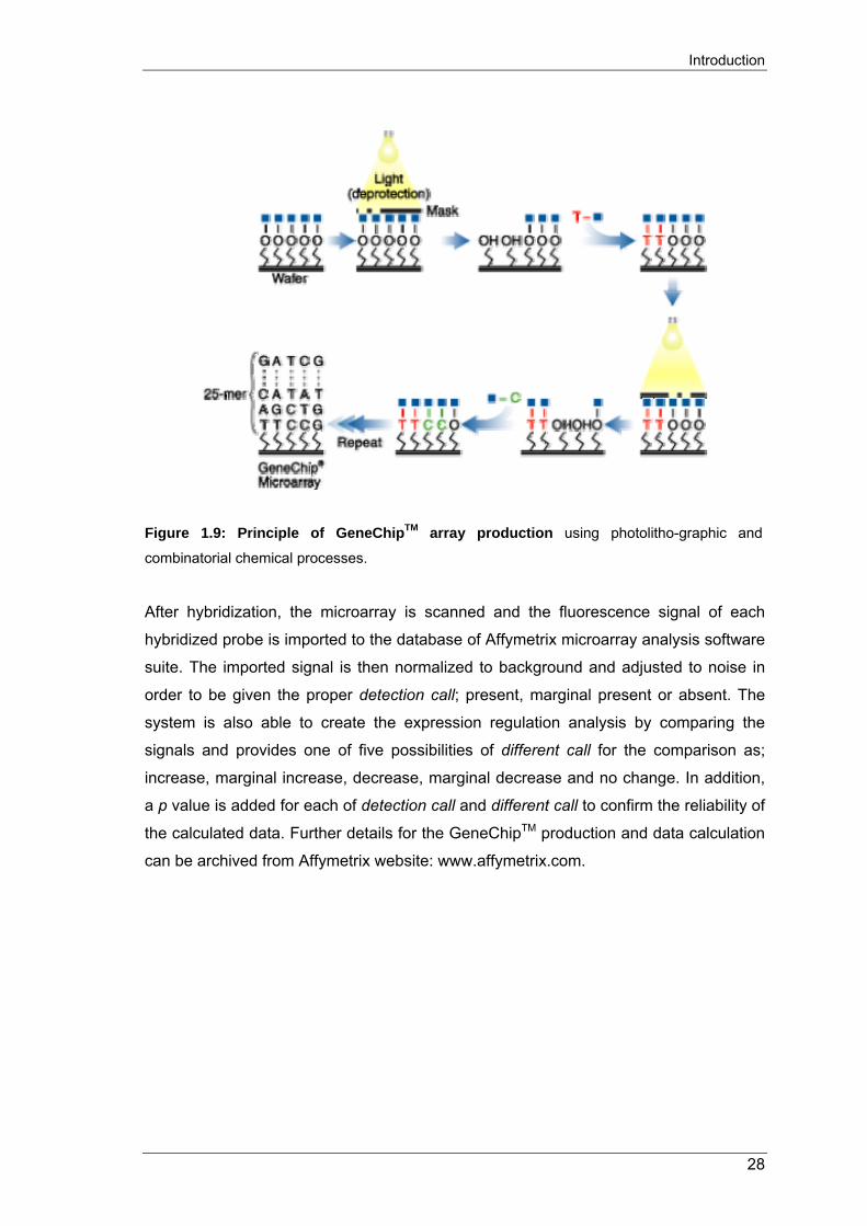
Figure 1.9: Principle of GeneChipTM array production using photolitho-graphic and
combinatorial chemical processes.
After hybridization, the microarray is scanned and the fluorescence signal of each
hybridized probe is imported to the database of Affymetrix microarray analysis software
suite. The imported signal is then normalized to background and adjusted to noise in
order to be given the proper detection call; present, marginal present or absent. The
system is also able to create the expression regulation analysis by comparing the
signals and provides one of five possibilities of different call for the comparison as;
increase, marginal increase, decrease, marginal decrease and no change. In addition,
a p value is added for each of detection call and different call to confirm the reliability of
the calculated data. Further details for the GeneChipTM production and data calculation
can be archived from Affymetrix website: www.affymetrix.com.
28

Introduction
1.7. AIMS OF THE STUDY
o To analyze the expression of marker genes in macrophages using our own in
vitro phagocytic differentiation and macrophage foam cell formation models.
o To analyze the expression of lysophospholipid receptors on human monocytes,
macrophages and cholesterol loaded/de-loaded macrophages.
o To analyze global gene expression of macrophages during in vitro foam cell
formation and bioactive lipids stimulation using Affymetrix DNA chip.
o To analyze more specific monocyte/macrophage gene expression using known
literature pathways.
29

Materials and Methods
2. MATERIALS AND METHODS 2.1. MATERIALS Table 2.1: List of used reagents and kits
Reagent and kit Provider
Agarose Biozym, Hameln, Germany
Ampicillin Roche, Mannheim, Germany
Bacto-Agar Difco-Laboratories, Detroit, USA
Bacto-Trypton Difco-Laboratories, Detroit, USA
Bacto-Yeast extracts Difco-Laboratories, Detroit, USA
Blue-dyed Phagobeads (0,8 ?m) Sigma, Deisenhofen, Germany
BSA (Lipid-free) Sigma, Deisenhofen, Germany
DABCO (Triethylendiamin) Sigma, Deisenhofen, Germany
EDTA (Di-sodium) Pharmacia Biotech, Freiburg, Germany
Fluoromount-G Southern Biotech, Birmingham, USA
Fluoesbrite microparticles Polysciences, Eppelheim, Germany
H2O Nuclease-free Promega, Madison, AL, USA
Kanamycin Roche, Ingelheim, Germany
L-glutamine Gibco BRL, Berlin, Germany
Lubrol WX Serva, Heidelberg, Germany
MEM (Non-essential Amino acid) Gibco BRL, Berlin, Germany
Minimal SD Agar Base Clontech, Palo Alto, USA
Nickel-Chelating Resin GenoTech Biosciences, Lohmar, Germany
Penicillin/Streptomycin Gibco BRL, Berlin, Germany
Polyvinyl alcohol Sigma, Deisenhofen, Germany
Protease inhibitors Calbiochem, Bad Soden, Germany
Protein A-Dynabeads Dynal, Hamburg, Germany
Second strand buffer Invitrogen, Glasgow, UK
Triton X-100 Boehringer, Mannheim, Germany
Urea Pharmacia Biotech, Freiburg, Germany
1 kb Ladder, DNA Gibco BRL, Berlin, Germany
100 bp Ladder, DNA Pharmacia, Freiburg, Germany
AMV-Reverse Transcriptase Promega, Madison USA
BCA Protein Assay Kit Pierce, Rockford, IL, USA
Cell Line Nucleofector Kit Amaxa GmbH, Cologne, Germany
30
Materials and Methods
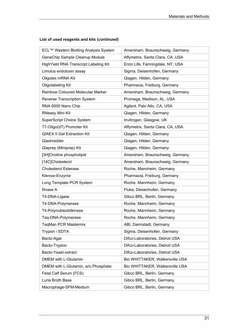
List of used reagents and kits (continued)
ECL™ Western Blotting Analysis System Amersham, Braunschweig, Germany
GeneChip Sample Cleanup Module Affymetrix, Santa Clara, CA, USA
HighYield RNA Transcript Labeling Kit Enzo Life, Farmingdale, NY, USA
Limulus endotoxin assay Sigma, Deisenhofen, Germany
Oligotex mRNA Kit Qiagen, Hilden, Germany
Oligolabeling Kit Pharmacia, Freiburg, Germany
Rainbow Coloured Molecular Marker Amersham, Braunschweig, Germany
Reverse Transcription System Promega, Madison, AL, USA
RNA 6000 Nano Chip Agilent, Palo Alto, CA, USA
RNeasy Mini Kit Qiagen, Hilden, Germany
SuperScript Choice System Invitrogen, Glasgow, UK
T7-Oligo(dT) Promoter Kit Affymetrix, Santa Clara, CA, USA
QIAEX II Gel Extraction Kit Qiagen, Hilden, Germany
Qiashredder Qiagen, Hilden, Germany
Qiaprep (Miniprep) Kit Qiagen, Hilden, Germany
[3H]Choline phospholipid Amersham, Braunschweig, Germany
[14C]Cholesterol Amersham, Braunschweig, Germany
Cholesterol Esterase Roche, Mannheim, Germany
Klenow-Enzyme Pharmacia, Freiburg, Germany
Long Template PCR System Roche, Mannheim, Germany
Rnase A Fluka, Deisenhofen, Germany
T4-DNA-Ligase Gibco BRL, Berlin, Germany
T4-DNA-Polymerase Roche, Mannheim, Germany
T4-Polynukleotidkinase Roche, Mannheim, Germany
Taq-DNA-Polymerase Roche, Mannheim, Germany
TaqMan PCR Mastermix ABI, Darmstadt, Germany
Trypsin / EDTA Sigma, Deisenhofen, Germany
Bacto-Agar Difco-Laboratories, Detroit USA
Bacto-Trypton Difco-Laboratories, Detroit USA
Bacto-Yeast extract Difco-Laboratories, Detroit USA
DMEM with L-Glutamin Bio WHITTAKER, Walkersville USA
DMEM with L-Glutamin, w/o Phosphate Bio WHITTAKER, Walkersville USA
Fetal Calf Serum (FCS) Gibco BRL, Berlin, Germany
Luria Broth Base Gibco BRL, Berlin, Germany
Macrophage-SFM-Medium Gibco BRL, Berlin, Germany
31
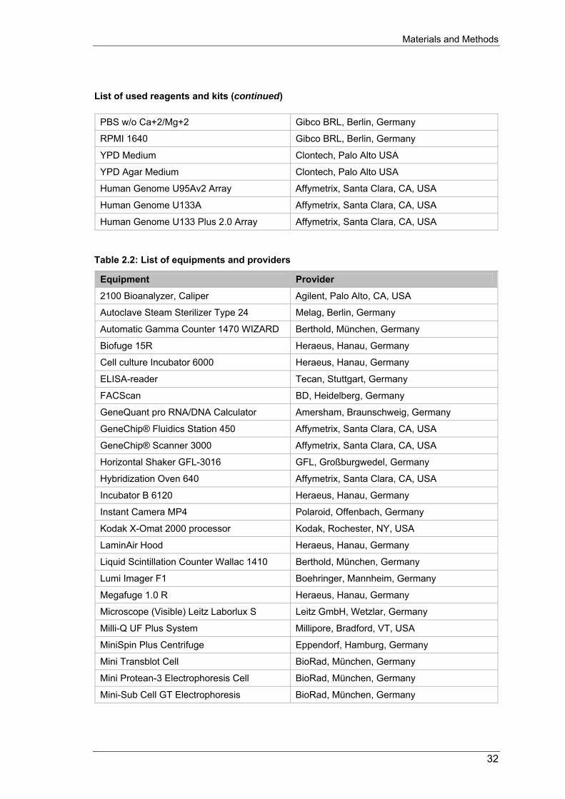
Materials and Methods
List of used reagents and kits (continued)
PBS w/o Ca+2/Mg+2 Gibco BRL, Berlin, Germany
RPMI 1640 Gibco BRL, Berlin, Germany
YPD Medium Clontech, Palo Alto USA
YPD Agar Medium Clontech, Palo Alto USA
Human Genome U95Av2 Array Affymetrix, Santa Clara, CA, USA
Human Genome U133A Affymetrix, Santa Clara, CA, USA
Human Genome U133 Plus 2.0 Array Affymetrix, Santa Clara, CA, USA Table 2.2: List of equipments and providers
Equipment Provider
2100 Bioanalyzer, Caliper Agilent, Palo Alto, CA, USA
Autoclave Steam Sterilizer Type 24 Melag, Berlin, Germany
Automatic Gamma Counter 1470 WIZARD Berthold, München, Germany
Biofuge 15R Heraeus, Hanau, Germany
Cell culture Incubator 6000 Heraeus, Hanau, Germany
ELISA-reader Tecan, Stuttgart, Germany
FACScan BD, Heidelberg, Germany
GeneQuant pro RNA/DNA Calculator Amersham, Braunschweig, Germany
GeneChip® Fluidics Station 450 Affymetrix, Santa Clara, CA, USA
GeneChip® Scanner 3000 Affymetrix, Santa Clara, CA, USA
Horizontal Shaker GFL-3016 GFL, Großburgwedel, Germany
Hybridization Oven 640 Affymetrix, Santa Clara, CA, USA
Incubator B 6120 Heraeus, Hanau, Germany
Instant Camera MP4 Polaroid, Offenbach, Germany
Kodak X-Omat 2000 processor Kodak, Rochester, NY, USA
LaminAir Hood Heraeus, Hanau, Germany
Liquid Scintillation Counter Wallac 1410 Berthold, München, Germany
Lumi Imager F1 Boehringer, Mannheim, Germany
Megafuge 1.0 R Heraeus, Hanau, Germany
Microscope (Visible) Leitz Laborlux S Leitz GmbH, Wetzlar, Germany
Milli-Q UF Plus System Millipore, Bradford, VT, USA
MiniSpin Plus Centrifuge Eppendorf, Hamburg, Germany
Mini Transblot Cell BioRad, München, Germany
Mini Protean-3 Electrophoresis Cell BioRad, München, Germany
Mini-Sub Cell GT Electrophoresis BioRad, München, Germany
32

Materials and Methods
List of equipments and providers (continued)
pH-Meter pH537 WTW, Weilheim, Germany
Precision Balance Sartorius MD BA 200 Sartorius, Göttingen, Germany
Power Supply PAC 300 BioRad, München, Germany
Princeton MicroMax CCD-1317-K/1 Roper Scientific, Trenton, NJ, USA
Shaking Incubator GFL-3032 GFL, Großburgwedel, Germany
Shaking Water Bath Julabo SW-20C Julabo, Seelbach, Germany
Spectrophotometer UV/VIS Lambda 2 Perkin Elmer, Überlingen, Germany
Stirrer with Heating Surface IKAMAG Labor Center, Nürnberg, Germany
SpeedVaq Alpha RVC Christ, Osterode, Germany
Sysmex Micro-Cell Counter F-300 Digitana AG, Hamburg, Germany Thermocycler Gene Amp PCR System 9600 Perkin Elmer, Uberlingen, Germany
Thermomixer Comfort Eppendorf, Hamburg, Germany
Ultrasonic Disintegrator Soniprep 150 MSE, Watford Herts, United Kingdom
Ultracentrifuge (fixed angle) J2-21 M/E Beckman, München, Germany
Ultracentrifuge L-70 Beckman, München, Germany
Ultracentrifuge Optima TLX Beckman, München, Germany
Vortex-Mixer REAX 2000 Heidolph, Kelheim, Germany
Zeiss Axiovert S-100 Spectral Microscope Carl Zeiss, Goettingen, Germany
ECL Hyperfilm Amersham, Braunschweig, Germany
Fluorotrans Membrane (PVDF) Pall Filtron GmbH, Dreieich, Germany
Instant Picture Film Typ 667 Polaroid, Offenbach, Germany
X-Ray Films Biomax Kodak, Rochester, NY, USA 2.2. DONORS FOR MONOCYTES ISOLATION Leukocyte-enriched apheresates or heparinized blood samples were obtained from
healthy, normolipidemic, from 25 to 45 years of age volunteers, bearing the
apolipoprotein E3/E3 genotype after informed consent.
2.2.1. Density gradient centrifugation of heparinized blood samples Heparinized blood samples were subjected to the following procedures within 4hrs after
donation. Mononuclear cells, comprising monocytes and lymphocytes, were isolated
from 3 to 15ml of whole blood diluted with an equal volume of PBS (w/o Ca2+, Mg2+)
through 35 min. of brakeless centrifugation at 600g upon a layer of Histopaque-1077.
33

Materials and Methods
The upper plasma layer was removed and a white band of mononuclear cells was
quantitatively isolated from the gradients mid. After 2 washing steps with PBS (w/o
Ca2+, Mg2+) each followed by centrifugation at 425g the cells were resuspended in a
small volume (1-2ml) of PBS (w/o Ca2+, Mg2+) and the concentration of the suspension
was determined with a sysmex counter.
2.2.2. Elutriation of monocytes Suspensions enriched in human peripheral blood leukocytes were isolated by
leukapheresis in a Spectra cell-separator (Gambro BCT), supplemented with the
anticoagulant ADCA and diluted with an equal volume of PBS (w/o Ca2+, Mg2+). The
diluted apheresate was then subjected to counterflow centrifugation (J2-MC centrifuge
with JE-6B Rotor, Beckmann) [248] using buffers and centrifuge adjustment as follows:
Running buffer: PBS (w/o Ca2+, Mg2+)
1.0 % (v/v) penicillin / streptomycin-solution
0.5 % (w/v) BSA
0.1 % (w/v) Glucose
Centrifugation protocol:
Rotation: 2040 rpm. Rotor temperature: 15°C
Flow rate (ml/min) Volume of fraction (ml)* Loading 7 -
Pre-run 9 150
Fractions 1 - 4 12 50
Fractions 5 - 8 15 50
Fractions 9 - 12 18 50
Fractions 13 - 16 20 50
Fractions 17 - 19 22 50
Fractions 20 - 22 24 50
*In case of a marked erythrocyte contamination of the apheresate it may be necessary
to enlarge the volume of initial fractions till the content of erythrocytes substantially
decreases.
34

Materials and Methods
The monocyte content of each fraction was determined using flow cytometry based on
the different scatter properties of lymphocytes, granulocytes and monocytes Fractions
that were devoid of granulocytes with a monocyte content of at least 90% of leukocytes
were pooled, washed with PBS (w/o Ca2+, Mg2+) and resuspended in culture medium.
The concentration of the suspension was determined with a sysmex counter.
2.3. ISOLATION AND MODIFICATION OF LIPOPROTEINS 2.3.1. Isolation of lipoproteins Lipoproteins were isolated from human plasma or serum by sequential preparative
ultracentrifugation in KBr gradients according to [249] followed by extensive dialysis
and filter sterilization. All lipoprotein concentrations mentioned are protein
concentrations determined by Lowry´s method [250]. Lipoprotein fractions were stored
at 4°C and used within two weeks from end of dialysis.
2.3.2. Enzymatic modification of low-density lipoprotein (E-LDL) To gain the same atherogenic properties of LDL found in atherosclerotic plaque,
isolated LDL was enzymatically modified before loading to cells.
o As described in [9]. Briefly, LDL was diluted to 2 mg/ml protein in PBS (w/o
Ca2+, Mg2+). Enzyme treatment was conducted with trypsin (6.6µg/ml) and
cholesterol esterase (40µg/ml) for 2 h at 37°C. Subsequently, the pH of the
solution was adjusted to 5.5 by addition of morpholinoethane sulfonic acid
(MES) buffer, pH 5.0. Finally, neuraminidase (79mU/ml) and Mg-ascorbate
solution (30 mg/ml) were added for 14 h at 37°C.
o As described in [251]. Briefly, LDL was diluted to 2 mg/ml protein in PBS (w/o
Ca2+, Mg2+). Enzyme treatment was conducted with trypsin (6.6µg/ml) and
cholesterol esterase (40µg/ml) for 24-36 h at 37°C.
These two preparation procedures led to similar results concerning lipid loading
potency and competed with each other for binding and uptake with identical potencies
(data not shown). The absence of oxidation products in E-LDL was verified by the
determination of thiobarbituric acid-reactive substances (TBARS) to quantify lipid
peroxidation products [5]. Modified lipoproteins were stored at 4°C and used within a
35

Materials and Methods
week. During LDL preparation and subsequent modification, general precautions were
taken to avoid lipopolysaccharide (LPS) contamination.
2.4. MONOCYTE CELL CULTURE 2.4.1. Cell culture After isolation, monocytes were seeded at 106 cell/ml in macrophage serum-free
medium supplemented with monocyte-colony stimulating factor (M-CSF, 50ng/ml) on
plastic Petri dishes (10cm diameter). Monocytes were incubated for up to 4 days at
37°C/ 5% CO2 (Heraeus 6000 Incubator, Heraeus Instruments) to induce phagocytic
differentiation, as described by Stohr et al. [252]. Atherogenic lipoprotein (E-LDL)
and/or other stimuli were added according to the two following independent
experiments:
2.4.1.1. Foam cells induction and removal of cholesterol by HDL3 loading Macrophages at day 4 were induced to foam cells by adding of 40µg/ml atherogenic
lipoprotein (E-LDL) and incubated for 24h. Subsequently, the culture medium was
refreshed and supplemented with 100µg HDL3 for 24h in 5% CO2 at 37°C.
Foam celMacrophage Deloaded Macrophage
Monocytes
M-CSF E-LDL HDL
4 days 24 hours 24 hours
Figure 2.1: In vitro differentiation of monocyte to macrophage, foam cell induction and cholesterol deloading of foam cells.
36
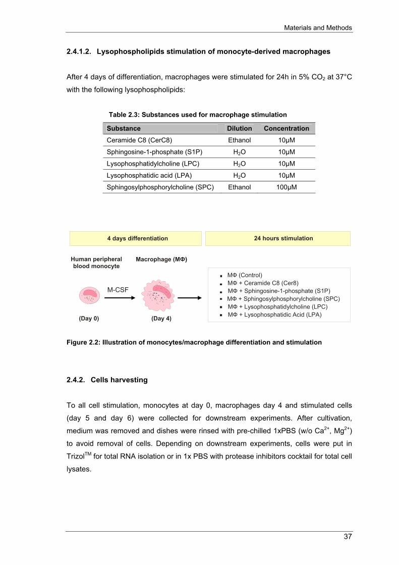
Materials and Methods
2.4.1.2. Lysophospholipids stimulation of monocyte-derived macrophages
After 4 days of differentiation, macrophages were stimulated for 24h in 5% CO2 at 37°C
with the following lysophospholipids:
Table 2.3: Substances used for macrophage stimulation
Substance Dilution Concentration
Ceramide C8 (CerC8) Ethanol 10µM
Sphingosine-1-phosphate (S1P) H2O 10µM
Lysophosphatidylcholine (LPC) H2O 10µM
Lysophosphatidic acid (LPA) H2O 10µM
Sphingosylphosphorylcholine (SPC) Ethanol 100µM
Human peripheral blood monocyte
(Day 0)
Macrophage (MФ)
(Day 4)
M-CSF
24 hours stimulation 4 days differentiation
MФ (Control)MФ + Ceramide C8 (Cer8) MФ + Sphingosine-1-phosphate (S1P)
MФ + Lysophosphatidylcholine (LPC) MФ + Lysophosphatidic Acid (LPA)
MФ + Sphingosylphosphorylcholine (SPC)
Figure 2.2: Illustration of monocytes/macrophage differentiation and stimulation
2.4.2. Cells harvesting To all cell stimulation, monocytes at day 0, macrophages day 4 and stimulated cells
(day 5 and day 6) were collected for downstream experiments. After cultivation,
medium was removed and dishes were rinsed with pre-chilled 1xPBS (w/o Ca2+, Mg2+)
to avoid removal of cells. Depending on downstream experiments, cells were put in
TrizolTM for total RNA isolation or in 1x PBS with protease inhibitors cocktail for total cell
lysates.
37
Materials and Methods
2.5. TOTAL RNA ISOLATION FROM MONOCYTES AND MACROPHAGES Reagents: 1xPBS
TrizolTM
Ethanol Chloroform Isopropanol RNase-free water
Protocol:
o After culture, discard medium and rinse dishes with pre-chilled 1xPBS o (The cultured medium could be spin, discard cell pellet and store at –80°C for
ELISA analysis) o Add 1ml Trizol to dish (approx. 1x10⁷ cells) and harvest cells by cell-scraper.
Cell-suspension then could be stored at –80°C for at least 30 min. o Fast thaw cell-in-Trizol then resuspend cells by syringe (3-5 times) and leave at
RT for 2 min. o Apply about 700µl of cell suspension to the QIAshreder column (QIAGEN
79654) and spin at max. speed for 2min. Collect solution to a new, fresh tube and leave at RT for 5 min. (the QIAshreder column could be used 2-3 times).
o Divide the cells suspension to the centrifuge tube (maximum 2/3 of the tube’s total vol.)
o Add chloroform (1/5 vol. of cell suspension), mix by inverting the tube for few times. Incubate tube at RT for 2 min.
o Spin at max. speed (15000 rpm) for 15 min. Collect aqueous phase to the new, fresh tube and put on ice.
o Add 1/5 vol. of Chloroform and mix by inverting few times. Incubate tube at RT for 2 min.
o Spin at max. speed (15000 rpm) for 15 min. Collect aqueous phase to the new, fresh tube and put on ice.
o Precipitate RNA by isopropanol (0.7 vol. of cell suspension), invert gently and leave on ice for at least 10 min.
o Spin at max. speed (15000 rpm) for 30 min. Discard solution and carefully wash pellet with 70% pre-chilled ethanol (do not vortex). And spin again at max. speed for 15 min. Repeat this step if necessary.
o Carefully discard ethanol and dry pellet under hood. Resuspend pellet with RNAse-free water (approx. 50μl per 1x10⁷ cells).
o Control RNA quality and quantity by spectrophotometer and/or running on 1.2% formamide gel.
38
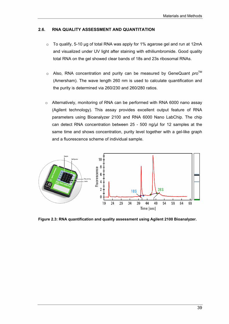
Materials and Methods
2.6. RNA QUALITY ASSESSMENT AND QUANTITATION
o To qualify, 5-10 µg of total RNA was apply for 1% agarose gel and run at 12mA
and visualized under UV light after staining with ethiliumbromide. Good quality
total RNA on the gel showed clear bands of 18s and 23s ribosomal RNAs.
o Also, RNA concentration and purity can be measured by GeneQuant proTM
(Amersham). The wave length 260 nm is used to calculate quantification and
the purity is determined via 260/230 and 260/280 ratios.
o Alternatively, monitoring of RNA can be performed with RNA 6000 nano assay
(Agilent technology). This assay provides excellent output feature of RNA
parameters using Bioanalyzer 2100 and RNA 6000 Nano LabChip. The chip
can detect RNA concentration between 25 - 500 ng/µl for 12 samples at the
same time and shows concentration, purity level together with a gel-like graph
and a fluorescence scheme of individual sample.
Figure 2.3: RNA quantification and quality assessment using Agilent 2100 Bioanalyzer.
39

Materials and Methods
2.7. AFFYMETRIX DNA MICROARRAY GENE EXPRESSION ANALYSIS The procedure of cRNA samples for GeneChip array hybridization is as following:
Figure 2.4: principle of GeneChipTM cRNA probe labeling
o First-strand cDNA was synthesized from 10 μg of total RNA of each simulation,
using using 50 μM of T7-oligo-dT primer and 400U of SuperScript II RT
(Invitrogen Life Technologies SuperScript Choice system).
o Second-strand cDNA was synthesized 10U of T4 DNA polymerase using using
T7-oligo(dT) primer (Life Technologies).
o cDNA is cleaned by the Gene Chip Sample Cleanup Module (Affymetrix).
40
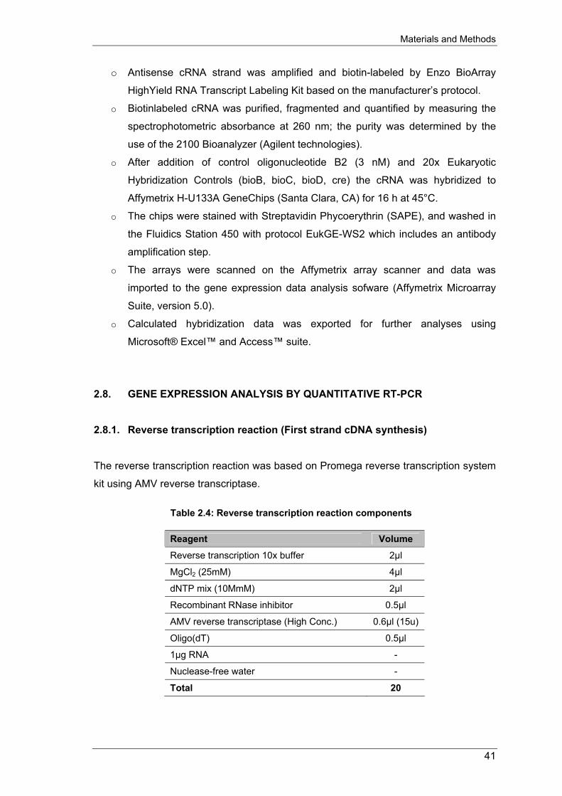
Materials and Methods
o Antisense cRNA strand was amplified and biotin-labeled by Enzo BioArray
HighYield RNA Transcript Labeling Kit based on the manufacturer’s protocol.
o Biotinlabeled cRNA was purified, fragmented and quantified by measuring the
spectrophotometric absorbance at 260 nm; the purity was determined by the
use of the 2100 Bioanalyzer (Agilent technologies).
o After addition of control oligonucleotide B2 (3 nM) and 20x Eukaryotic
Hybridization Controls (bioB, bioC, bioD, cre) the cRNA was hybridized to
Affymetrix H-U133A GeneChips (Santa Clara, CA) for 16 h at 45°C.
o The chips were stained with Streptavidin Phycoerythrin (SAPE), and washed in
the Fluidics Station 450 with protocol EukGE-WS2 which includes an antibody
amplification step.
o The arrays were scanned on the Affymetrix array scanner and data was
imported to the gene expression data analysis sofware (Affymetrix Microarray
Suite, version 5.0).
o Calculated hybridization data was exported for further analyses using
Microsoft® Excel™ and Access™ suite.
2.8. GENE EXPRESSION ANALYSIS BY QUANTITATIVE RT-PCR 2.8.1. Reverse transcription reaction (First strand cDNA synthesis) The reverse transcription reaction was based on Promega reverse transcription system
kit using AMV reverse transcriptase.
Table 2.4: Reverse transcription reaction components
Reagent Volume
Reverse transcription 10x buffer 2µl
MgCl2 (25mM) 4µl
dNTP mix (10MmM) 2µl
Recombinant RNase inhibitor 0.5µl
AMV reverse transcriptase (High Conc.) 0.6µl (15u)
Oligo(dT) 0.5µl
1µg RNA -
Nuclease-free water -
Total 20
41

Materials and Methods
Protocol:
o Place 1µg of total RNA in a microcentrifuge tube and add nuclease-free water
up to the volume of 10.4µl and incubate at 70°C for 10 min. Spin briefly and
place the tube on ice.
o Prepare the mixture of reagents (table 2.4) and add to the RNA microcentrifuge
tube above. Mix well and incubate the reaction at 42°C for 15 min.
o Heat the reaction tube at 99°C for 5 min. and then incubate at 0-5°C for 5 min.
2.8.2. Relative quantitation of gene expression with TaqMan® PCR
o The relative quantification of gene expression analysis was carried out on ABI
PRISM® 7900HT Sequence Detection System (Applied Biosystems). The
amplification and detection of the target gene sequence based on the gene-
specific primers, TaqMan florescent probe using TaqMan Universal PCR Master
Mix (UMM). UMM 2x stock solution contains heat-activated AmpliTaq Gold DNA
Polymerase, AmpErase UNG for carryover prevention, dNTPs (containing
dUTP), Passive Reference Dye (ROX) and optimized buffer components.
o Messenger RNA sequences of target genes were archived from GenBank and
gene-specific primers and probes were designed using Primer ExpressTM
software version 2.0 (Applied Biosystems) which optimized for TaqMan
quantitative PCR assays. The primers and probes were then ordered from
MWG-Biotech. TaqMan probe for target gene is labeled with two different
fluorescence dyes; 6-carboxylflourescein (FAM) at 5’ end and 6-
carboxyltetramethylrhodamine at 3’ end (TAMRA). For GAPDH endogenous
control, the 3’ end of its probe was labeled with VICTM. Using combination of the
two dyes (FAM/TAMRA or VIC/TAMRA) enables the system to detect and count
every single amplicon formed after every cycle.
Reagent:
o TaqMan GAPDH control (human)
o TaqMan universal PCR master mix
Protocol:
o Prepare the serial dilution of cDNA as following: 50, 25, 12.5, 6.125 and 3.0625
ng for calibration curve generation. These can be a pool of samples cDNA or
another cDNA from monocytes/macrophages. H2O used as negative control.
42

Materials and Methods
o For target gene quantification, 25 or 50ng of sample cDNA was used as
template.
o Each run was performed in triplicates for both calibration curve generation and
target gene quantification (table 2.5 and 2.6).
Table 2.5: Reaction mixture for target gene
Reagent Volume
cDNA (2-50ng) 5µl
Master mix 10µl
Forward primer (18µM) 1µl
Reverse primer (18µM) 1µl
Labeled TaqMan probe (5µM) 1µl
Nuclease-free water 2µl
Total 20µl
Table 2.6: Reaction mixture for GPADH control
Reagent Volume
cDNA (2-50ng) 5µl
Master mix 10µl
GAPDH control 1µl
Nuclease-free water 4µl
Total 20µl
o The running protocol as follow:
Table2.7: PCR program
PCR step Temp. (°C) Time No. of cycle
1. Carryover decontamination via UNG 50 2 min. 1
2. AmpliTaq Gold pre-activation 90 10 min. 1
3. Melting step 90 15 sec.
4. Anneal/extend step (combined) 60 1 min. 40
o Data analysis was performed with Sequence Detector Software (SDS) 2.0
(Applied Biosystems). Calibration curve generation was generated for every
sample which showed ΔRn on the y axis (where Rn is the fluorescence
emission intensity of the reporter dye normalized to a passive reference, and
ΔRn is the Rn of no reacted samples minus that of reacted sample) against
cycle number on the x axis. From each amplification plot, a threshold cycle (CT)
43

Materials and Methods
value was calculated, which is defined as cycle at which a statistically significant
increase in ΔRn is first detected and is display in the graph as the intercept
point of the amplification curve with the threshold.
o The obtained CT values were used further analysis by Microsoft® Excel™. The
CT value of each target gene was used to calculate the initial input amount of
cDNA template. Thereafter the results were normalized to endogenous control,
GAPDH. At last, the fold changes were calculated by comparison of the
expression of target gene upon different stimulations.
2.9. TOTAL CELL LYSATES PREPARATION AND WESTERN BLOT ANALYSIS
OF PROTEINS 2.9.1. Preparation of total cell lysates Total cell lysates were prepared from monocytes and macrophages as following:
Reagents
Protease inhibitors: Leupeptin 25µM
Aprotinin 25µM
AEBSF 25µM
Protein lysate buffer (100ml): 1M Tris 1ml
0.5M EDTA 0.4ml
5M NaCl 3ml
10% Brij 96 8.75ml
10% NP40 1.25ml
Protease inhibitors 150µl
ddH2O up to 100ml
2x loading buffer (10ml): Brommophenol blue pinch
0.5M Tris pH 6.8 2.5 ml
Glycerol 2.0 ml
44
Materials and Methods
10% SDS 2.0 ml
ddH2O 2.5 ml
Beta-mecaptoethanol 1.0 ml
Protocol:
o After cell culture, cell culture medium was removed and dishes were rinse with
pre-chilled 1x PBS and collect cells to 1.5 ml centrifuge tube by cell scraper.
Centrifuge the tube at 10000 rpm, 4°C for 7 min., discard PBS and add 500µl
(approx. 107 cells) of lysate buffer.
o Sonicate the cell pellet for 45 seconds and keep the pellet on ice for 2 min.
Repeat sonication for 2 more times.
o Centrifuge at 14000 rpm, 4°C and transfer protein lysates to a new fresh tube.
o Keep protein lysates at -80°C for long storage.
2.9.2. Western blot analysis of protein Material and equipment:
o PVDF membrane
o Whatman 3MM paper
o Mini trans-blot cell (Bio-Rad)
o Tris-HCl Ready gell system (Bio-Rad)
o Full-Range Rainbow™ protein molecular marker (Amersham: RPN800)
o ECL plus western blotting detection reagents (Amersham: RPN21323)
Solution
o 5x AA running buffer: (1000 ml)
15g Tris
72g Glycin
5g SDS (or 250 ml 10% SDS)
ddH2O up to 1000 ml
o Transfer buffer
100 ml MeOH
3.03g Tris
14.4g Glycin
ddH2O up to 1000 ml
45

Materials and Methods
o 1x PBS: 1 mM sodium phosphate, 15 mM NaCl pH 7.4
o PBS/Tween: PBS + 0.05% Tween-20 (500µl/1l)
Procedure:
Protein separation:
o Boil protein samples and marker for 5 min. and put immediately on ice. Spin
down briefly before use.
o Apply the amount of 10 – 100 ng of protein samples and 10 ng of marker to
Tris-HCl ready gel and run for approx. 1 hour, at 25mA/gel.
o After running, rinse the gel briefly with water and put in transfer buffer
Western blotting and probing of antibodies:
o Blotting of protein was carried out according to manufacture’s guidance (Bio-
Rad)
o After blotting, remove membrane and rinse blot several times with PBS/Tween.
o Block the blot in 5% milk/PBS/Tween for 2 hours, at room temperature on
shaker.
o Primary antibody probing: dilute antibody (according to product guidance) in
5% milk/PBS incubate with blot for 2 hours at room temperature on shaker.
o Wash the blot 3 times for 10 min. in PBS/Tween
o Secondary antibody probing: dilute antibody (according to product guidance) in
5% milk/PBS, incubate for 2 hours at room temperature on shaker. o Wash the blot 3 times for 10 min. in PBS/Tween
Detection:
o Incubate blot in diluted detection reagent complex for 5 min. at room
temperature. Drain off excess detection reagent with a dry tissue, wrap up the
blot and gently smooth out air bubbles.
o Expose the autoradiography film for different time period to detect the protein
bands.
Stripping and re-probing of blots:
o Add approx. 25 ml stripping buffer to the blot, incubate in 50°C water bath for
30 min. with occasional agitation.
o Wash blot 2 times for 10 min. in PBS/Tween
46

Materials and Methods
o Incubate (optional) the blot with detection reagent and expose to film to ensure
that the blot has been adequately stripped
o Block blot in 5% milk/PBS/Tween for 2 hours at room temperature on shaker.
47

Results
3. RESULTS 3.1. ANALYSIS OF MARKER GENES IN MONOCYTES, MACROPHAGES,
CHOLESTEROL LOADED/DELOADED MACROPHAGES 3.1.1. Expression of scavenger receptors, Cla-1 and CD36, in monocytes,
monocyte-derived macrophages and E-LDL treated macrophages Scavenger receptor, CD36, is known to be expressed in monocytes and its expression
is elevated during in vitro and in vivo macrophage differentiation and Ox-LDL-induced
foam cell formation [253;254]. In addition, scavenger receptor type B, class I (SR-BI, or
Cla-1) that sharing a high homology to CD36 is also hypothesized to increase its
expression during macrophage differentiation. Therefore, the expression of these
receptors was subjected to analyze with in vitro models of M-CSF-induced macrophage
differentiation and E-LDL-induced macrophage foam cell formation.
For the analysis of Cla-1, freshly isolated human monocytes were differentiated to
macrophages in serum-free medium, supplemented with M-CSF. After 4 days of
differentiation, total RNA and total cell lysate from differentiated macrophages were
prepared. Northern blot and western blot analyses of above-mentioned samples
showed that Cla-1 was weakly expressed in monocytes but induced its expression in
monocyte-derived macrophages (figure 3.1a,b). GAPDH probe and β-actin protein
were used as control of northern and western analyses, respectively.
48

Results
Figure 3.1: Induce mRNA expression of Cla-1 in differentiated macrophages. (A) mRNA expression of
Cla-1 in monocytes and monocyte-derived macrophage.
(B) Protein expression of Cla-1 in monocytes, monocyte-
derived macrophages and other cells
Also, the expression alteration of CD36 scavenger receptor by E-LDL was investigated.
In this experiment, monocyte-derived macrophages were incubated with E-LDL
according to three different time-points (2, 4, 24 hours of incubation). The quantitative
RT-PCR results showed that the expression of CD36 was highly elevated in
macrophages incubated with E-LDL compared to macrophages incubated with enzyme
cocktail (EC, combination of enzymes that used to modified native LDL) as control. In
contrast, the expression of CD36 in macrophages was partially elevated by E-LDL in
comparison to that of native LDL-stimulated in macrophages. These results also
revealed that the E-LDL-induced expression of CD36 is time-dependent. Control run of
RNA from enzyme cocktails incubation showed no changes in macrophages
expression of CD36.
Figure 3.2: Elevation of macrophage CD36 expression by E-LDL.
49

Results
3.1.2. Adipophilin is a sensitive marker for lipid loading in human blood monocytes
Expression of adipophilin, or adipose differentiation related protein, is shown to be
elevated during Ox-LDL-induced foam cell formation [255]. Therefore, it could be
considered as a marker gene for in vitro induction of foam cell formation.
At first, the expression of adipophilin on human monocytes was analyzed. Monocytes
were freshly isolated from whole blood of 11 healthy individuals. Total cell proteins of
these cells were subjected to test the expression of adipophilin using western blot
analysis. As the results, adipophilin was found to be expressed in monocytes from four
donors (3, 6, 9, 11). However, the results showed an unequal amount of total proteins
from isolated monocytes among individuals and explained the differences in expression
of adipophilin in monocytes among different donors. To control, the blot was stripped
and hybridized with β-actin (figure 3.3a)
Figure 3.3a: Expression of adipophilin in human monocytes
Also, several publications as well as results of our group experiment indicated that E-
LDL is more similar to atherogenic LDL isolated from atherogenic lesions than other
types of modified lipoprotein (ox-LDL, ac-LDL) [9;256] and rapidly induced foam cell
formation in monocytes and macrophages. Therefore, the regulation of adipophilin in
macrophages treated with E-LDL has been tested. In this experiment, freshly isolated
monocytes were differentiated to macrophages using M-CSF, incubated with E-LDL
(40µg/ml) and cell sample harvesting according to time periods (2, 4 and 24 hours of
incubation). The quantitative RT-PCR analysis showed that adipophilin expression was
induced by E-LDL when compared to macrophages differentiated by M-CSF. The
expression was up-regulated and time dependent, while the incubation with enzyme
cocktails (EC) did not change its expression. In this experiment, GAPDH was used as
control gene (figure 3.3b).
50
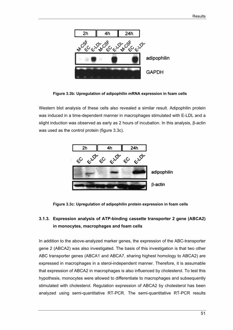
Results
Figure 3.3b: Upregulation of adipophilin mRNA expression in foam cells
Western blot analysis of these cells also revealed a similar result. Adipophilin protein
was induced in a time-dependent manner in macrophages stimulated with E-LDL and a
slight induction was observed as early as 2 hours of incubation. In this analysis, β-actin
was used as the control protein (figure 3.3c).
Figure 3.3c: Upregulation of adipophilin protein expression in foam cells
3.1.3. Expression analysis of ATP-binding cassette transporter 2 gene (ABCA2)
in monocytes, macrophages and foam cells In addition to the above-analyzed marker genes, the expression of the ABC-transporter
gene 2 (ABCA2) was also investigated. The basis of this investigation is that two other
ABC transporter genes (ABCA1 and ABCA7, sharing highest homology to ABCA2) are
expressed in macrophages in a sterol-independent manner. Therefore, it is assumable
that expression of ABCA2 in macrophages is also influenced by cholesterol. To test this
hypothesis, monocytes were allowed to differentiate to macrophages and subsequently
stimulated with cholesterol. Regulation expression of ABCA2 by cholesterol has been
analyzed using semi-quantitative RT-PCR. The semi-quantitative RT-PCR results
51

Results
showed that ABCA2 is slightly induced in macrophages compared to monocytes but
show a very high expression in macrophages treated with cholesterol (E-LDL). The
house-keeping gene (GAPDH) was used as a control gene in this experiment. The
result confirmed the hypothesis that ABCA2 expression in human macrophages is
regulated by cholesterol loading of macrophages.
Figure 3.4: Upregulation of ABCA2 expression in monocyte-derived macrophages and cholesterol-loaded macrophages
Notes:
- The Cla-1 and CD36 results were collaborated to the publication of Buechler
and Ritter, BBRC, 1999 [257] and Kapinsky, ATVB, 2001 [11], respectively.
- The adipophilin results were contributed to the publication of Buechler, BBA,
2001 [258].
- The ABCA2 results were contributed to the publication of Kaminski, BBRC,
2001 [259].
3.2. ANALYSIS OF LYSOPHOSPHOLIPID RECEPTORS EXPRESSION IN
MONOCYTES, MACROPHAGES, AND CHOLESTEROL-LOADED/ -DELOADED MACROPHAGES
3.2.1. mRNA expression of lysophospholipid receptors in human monocytes To investigate the mRNA expression levels of lysophospholipid receptors in human
monocytes, an equal amount of total RNA from 4 different donors were employed for
TaqMan® quantitative RT-PCR. The standard curve for each of receptors was
generated from a pool of RNAs from all donors. The final results were calculated to
standard curve and normalized to the GAPDH gene expression as the endogenous
52

Results
control (Fig. 3.5). Experimental results showed that the mRNAs of all tested
lysophospholipid receptors were detected in human blood monocytes. Of those, G2A
showed the highest mRNA expression level and the strength of the expression followed
by S1P3, S1P4, GPR3, LPA2, S1P1, LPA4, LPA1, GPR6, GPR12, S1P2, S1P5 and LPA3.
mRNA transcript from LPA3 was also detected but expressed about fivefold lower than
that of G2A. Also, the standard deviation given in figure 3.5 resembles the variance of
the lysophospholipid receptor mRNAs abundance from the four different investigated
donors. These data show that monocytes of different donors express similar levels of
the lysophospholipid receptors mRNAs with less than 2 folds difference.
Table 3.1: Primers and probes for TaqMan® RT-PCR analysis of lysophospholipid receptors
Receptor * Forward primer 5’-3’ Hybridization probe 5’-3’ Reverse primer 5’-3’
S1P1 TGCGGGAAGGGAGTATGTTT TGGCCCTGTCAGCCTCCG CGATGGCGAGGAGACTGAAC
S1P2 GCCTCTCTACGCCAAGCATTA CTGTGCGTGGTGACCATCTTCTCCAT TTGAGCGGACCACGCAGTA
S1P3 TGATTGTGGTGAGCGTGTTCA CGCCTGCTGGTCCCCACTCTTC GGCCACATCAATGAGGAAGAG
S1P4 GGAGCCCCTGTCCAGCAT TCCAGCGTGCGGAGCATCTGAA TCCACACGCAAGACTGCAA
S1P5 GCTTGCTCCACTGTCTTGCC TCGCCTTCGTGGGCATCC TAGAGTGCACAGATAGCGGCC
LPA1 CAATACTCGGAGACTGACTCTTAGC ATGGCTCCTGCGTCAGGGCCT CCGTCAGGCTGGTGTCAAT
LPA2 GGCTGTGAGTCCTGCAATGTC AGGCCAACTCACTGGTCAATGCTGC TCTCAGCATCTCGGCAAGAGT
LPA3 CTTGACTGCTTCCCTCACCAA TTGCTGGTTATCGCCGTGGAGAGG CGCATCCTCATGATTGACATG
G2A GCCATCCCTCTCTCCATCATC CCTTCACCAACCACCGGATTTTCAGG TGCTTCACCTTGGCCTTCTG
GPR3 GCTCCCTGCCACCTACAACTC CTACGCCTTCCGCAACCAGGAT GACAGCCCACAGCACTTTCTG
GPR6 ACTCCATGATCAATCCCATCATCTA CGCAACCAGGAGATCCAGGCG GGCACTTTGGACTGGAAACAG
GPR12 TCTCGGTGTCCTTCCTCTTCAT TGCGCTCATGCATCAGCTCTAC GGCGTGCCTCATCACAATCT
* LPA4 probe was ordered as an Assay-On-Demand probe from Applied Biosystems,
Darmstadt, Germany.
Figure 3.5: mRNA expression of lysophospholipid receptors in human monocytes
53

Results
3.2.2. mRNA expression of the lysophospholipid receptors during in-vitro
differentiation of monocytes to macrophage In order to analyze the mRNA expression of these lysophospholipid receptors during
the phagocytic differentiation of macrophages, monocytes from above-mentioned
donors were allowed to differentiate to macrophages for four days in a serum-free
medium using M-CSF. Total RNA from those cells was isolated and the mRNA
expression was determined by TaqMan® RT-PCR using standard curves for each
receptor. After normalization to the housekeeping gene GAPDH, fold changes of
mRNA expression of these receptors in macrophages compared to the corresponding
monocytes were calculated by division of the corresponding values. As it is shown in
figure 3.6, the lysophospholipid receptors expressed differently during macrophage
differentiation and the value 1 represented for a no-change line. The mRNA of GPR3
and G2A were 3 and 2.5 folds down-regulated in macrophages, respectively, when
compared to non-differentiated monocytes. Other receptors, including S1P1, S1P2,
S1P3 S1P4, S1P5, GPR6, GPR12, LPA1, LPA2, LPA3 and LPA4 showed a less than 2.5-
fold changes of the mRNA expression levels (figure 3.6).
3.2.3. mRNA expression of the lysophospholipid receptors during foam cell
formation and lipid deloading To further investigate the effects of modified lipoprotein on lysophospholipid receptors,
differentiated macrophages were incubated with E-LDL (40 µg/ml, 24h) and
subsequently incubated with purified HDL3 (100 µg/ml, 24h) to allow lipid efflux.
Experimental results showed that LPA3 and LPA4 mRNAs were found induced in E-LDL
treated cells, mRNA levels were, respectively, 2.7-fold and 4.2-fold increased in E-LDL
laden cells compared to 4 days differentiated macrophages. However, LPA3 and LPA4
were not found reduced in HDL3 treated foam cells (figure 3.6). The mRNAs of the
remaining receptors showed no regulation upon E-LDL loading and HDL3 de-loading.
This indicates that the mRNAs of the lysophospholipid receptors are not significantly
regulated by sterols or fatty acids.
54

Results
Figure 3.6: mRNA expression of lysophospholipid receptors in human differentiated macrophages, foam cells and HDL3-treated macrophages.
3.2.4. Protein expression of the lysophospholipid receptors To investigate the expression of the lysophospholipid receptors on the protein level in
this in vitro model, immunoblots with currently available antibodies were performed.
Protein isolated from the prostate (BD Biosciences, CA, USA) was used as positive
control and β-actin abundance as a loading control. Cell lysates were prepared from
monocytes, 4-day differentiated macrophages, foam cells and foam cells treated with
HDL3. In addition, 4-day differentiated macrophages were treated with HDL3 without
prior E-LDL treatment to investigate the effects of HDL3 on differentiated macrophages.
The experiments were performed with cells lysates from at least 2 different donors.
Immunoblots results showed that S1P1, S1P2, S1P4, LPA1 and LPA2 expressed in
monocytes and the expression was similar in monocytes, macrophages, foam cells and
HDL3 treated foam cells. S1P5 and LPA3 proteins could not be detected in any of the
stimulated cells. S1P3 is not expressed in monocytes but is strongly induced during
differentiation, whereas incubation with E-LDL or HDL3 showed a reduced abundance
of S1P3 when compared to macrophages. LPA1 is already expressed in monocytes but
down-regulated in macrophages, foam cells and HDL3 treated macrophages. LPA2
protein expression was equal in all stimulations.
55

Results
Figure 3.7: Immunoblots of lysophospholipid receptors in monocytes, macrophages, foam cells and HDL3-treated macrophages.
3.3. DNA MICROARRAY EXPERIMENTS AND BIOSTATICTICS ANALYSIS OF
GENES REGULATED BY E-LDL AND LYSOPHOSPHOLIPIDS 3.3.1. Principles of large-scale gene expression analysis by DNA microarray To identify the regulation of genes and pathways in foam cell induced by E-LDL and in
macrophages stimulated by lysophospholipids, the Affymetrix GeneChipTM U133A was
employed. The principle microarray analysis is shown below.
56

Results
Figure 3.8: Experimental model of microarray analysis of macrophages
Total RNAs from healthy donors were used for lysophospholipid stimulation and DNA
microarray analysis. The cRNAs were labeled with streptavidin phycoethrin fluorescent
and hybridized to the chip. After scanning, the result of hybridizations were archived as
high-resolution images and analyzed with the Affymetrix Microarray Suite 5.0TM. Gene
information (including name, UniGene, chromosome localization, and cellular function)
was then merged to every probeset on the chip to generate the complete gene
database for further analysis.
In this experimental model, the expression level of the gene of interest during
stimulation was represented by its mRNA accumulation level conserved during cRNA
synthesis and fluorescent labeling. Subsequently, the accumulation level of the probes
those hybridized to the DNA microarray was detected in terms of mean fluorescent of
the probeset. Finally, the change of expression patterns of genes was calculated
according to the comparison of mean fluorescent among different stimulations. The
regulation of gene expression was calculated as following:
57
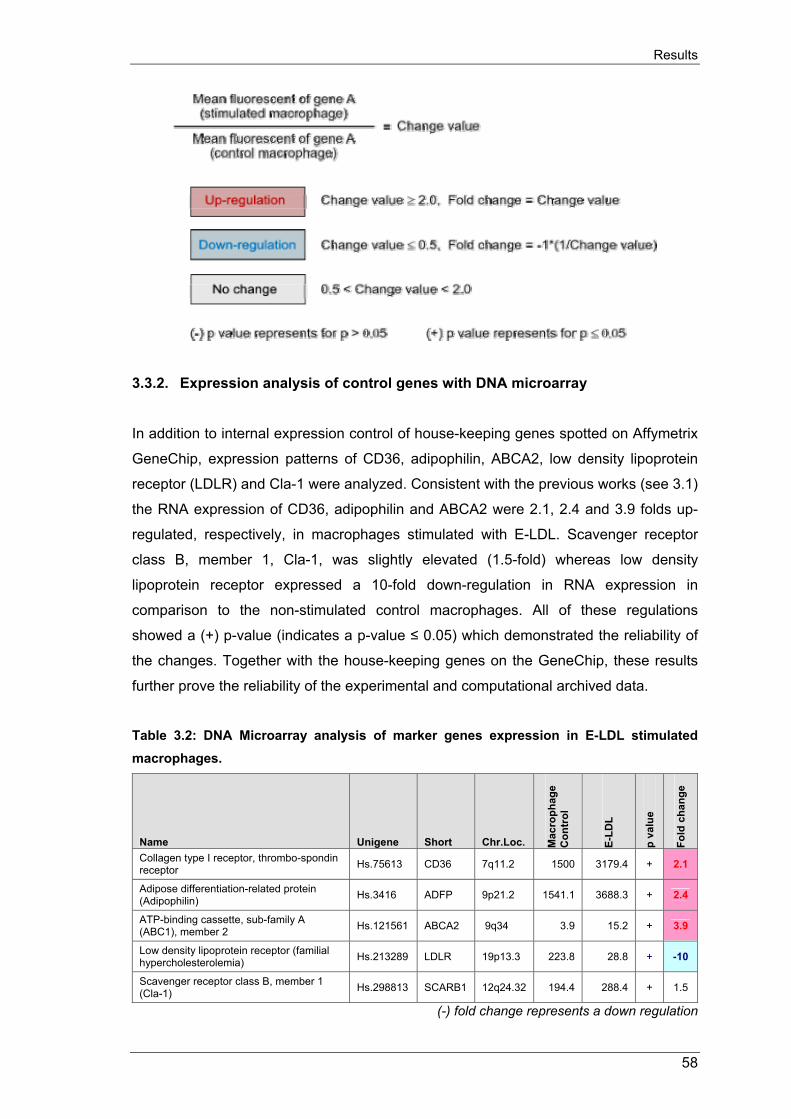
Results
3.3.2. Expression analysis of control genes with DNA microarray In addition to internal expression control of house-keeping genes spotted on Affymetrix
GeneChip, expression patterns of CD36, adipophilin, ABCA2, low density lipoprotein
receptor (LDLR) and Cla-1 were analyzed. Consistent with the previous works (see 3.1)
the RNA expression of CD36, adipophilin and ABCA2 were 2.1, 2.4 and 3.9 folds up-
regulated, respectively, in macrophages stimulated with E-LDL. Scavenger receptor
class B, member 1, Cla-1, was slightly elevated (1.5-fold) whereas low density
lipoprotein receptor expressed a 10-fold down-regulation in RNA expression in
comparison to the non-stimulated control macrophages. All of these regulations
showed a (+) p-value (indicates a p-value ≤ 0.05) which demonstrated the reliability of
the changes. Together with the house-keeping genes on the GeneChip, these results
further prove the reliability of the experimental and computational archived data.
Table 3.2: DNA Microarray analysis of marker genes expression in E-LDL stimulated macrophages.
Name Unigene Short Chr.Loc. Mac
roph
age
Con
trol
E-LD
L
p va
lue
Fold
cha
nge
Collagen type I receptor, thrombo-spondin receptor Hs.75613 CD36 7q11.2 1500 3179.4 + 2.1
Adipose differentiation-related protein (Adipophilin) Hs.3416 ADFP 9p21.2 1541.1 3688.3 + 2.4
ATP-binding cassette, sub-family A (ABC1), member 2 Hs.121561 ABCA2 9q34 3.9 15.2 + 3.9
Low density lipoprotein receptor (familial hypercholesterolemia) Hs.213289 LDLR 19p13.3 223.8 28.8 + -10
Scavenger receptor class B, member 1 (Cla-1) Hs.298813 SCARB1 12q24.32 194.4 288.4 + 1.5
(-) fold change represents a down regulation
58

Results
3.3.3. Biostatistic ranking of regulated genes in microarray analysis The Affymetrix GeneChipTM U133A contains 22,283 gene transcripts (duplicates
excluded). As the results archived from DNA microarray analyses, there were n=2962
(13%) genes found to be regulated by E-LDL stimulation. Similarly, n=1701 (7.6%)
genes found to be regulated by ceramide, n=1762 (7.9%) genes for S1P, n=2019
(9.1%) genes for SPC, n=1876 (8.4%) genes for LPC and n=1721 (7.7%) genes for
LPA. Among these, E-LDL and SPC were having higher influence on macrophage
genes than that of ceramide, S1P, LPC and LPA. Furthermore, the calculation also
revealed that these stimuli suppressed more genes than elevated them. The
percentage of suppressed genes ranges from 54% (LPA) to 73% (E-LDL) of total
regulated genes. In total, there were n=5534 gene transcripts in macrophages found to
be significantly regulated upon at least one of the stimulations (table 3.3).
Table 3.3: Genes regulated by individual stimulation in macrophages
Stimuli Up Down Total Percentage
E-LDL 797 2165 2962 13% Ceramide 617 1084 1701 7.6% S1P 569 1193 1762 7.9% SPC 878 1141 2019 9.1% LPC 534 1252 1786 8.4% LPA 788 933 1721 7.7%
In addition, statistic analysis of regulated genes revealed that there is remarkable
number of genes being influenced by two or more stimuli. Therefore, it is necessary to
look at the role of structure-related and/or function-related stimuli in genes regulation.
For this purpose, four groups of genes were assembled to ceramide/S1P, S1P/SPC,
LPC/LPA and S1P/LPA biostatistics ranking lists. Subsequently, five sub-groups were
generated for each of comparison, including common up, common down, specific up,
specific down and inversely regulated genes (table 3.5).
59

Results
Table 3.4: Global genes co-regulated by different stimulations in macrophages
Common up
Common down
Specific up
Specific down
Inversely regulated Sum
Cer/S1P 253 442 62 Cer 327 617 S1P
291 714
2706
S1P/LPA 275 365 75 S1P 271 776 LPA
461 545
2768
LPC/LPA 265 382 84 LPC 250 808 LPA
461 529
2779
S1P/SPC 287 385 75 S1P 263 752 SPC
535 737
3034
Table 3.5: Pathway genes co-regulated by different stimulations in macrophages
Common up
Common down
Specific up
Specific down
Inversely regulated Total
Cer/S1P 137 203 34 Cer 142 324 S1P
129 325
1294
S1P/LPA 128 175 33 S1P 146 346 LPA
216 313
1357
LPC/LPA 110 195 39 LPC 144 415 LPA
232 289
1424
3.4. DNA MICROARRAY ANALYSIS OF MACROPHAGE GENE EXPRESSION
REGULATION IN LITERATURE-BASED PATHWAYS 3.4.1. Assemble of genes into literature-based pathways and categories In order to have more insights of the gene regulation in macrophages, the GeneChip-
presented genes were group into different pathway-related and/or structure-related
categories. These pathway-defined and category-arrangement were performed
according to literature surveys, database searches and unpublished data from our
laboratory groups. The current assemble table having 65 pathways/categories (n=5673
genes), exclude unidentified-pathway genes and unknown transcripts. There are
duplicates of gene entries in the pathways list due to the multiple contributions of
numerous genes to different pathways.
60
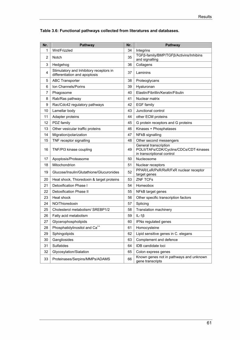
Results
Table 3.6: Functional pathways collected from literatures and databases.
Nr. Pathway Nr. Pathway 1 Wnt/Frizzled 34 Integrins
2 Notch 35 TGFβ-family/BMP/TGFβ/Activins/Inhibins and signalling
3 Hedgehog 36 Collagens
4 Stimulatory and Inhibitory receptors in differentiation and apoptosis 37 Laminins
5 ABC Transporter 38 Proteoglycans 6 Ion Channels/Porins 39 Hyaluronan 7 Phagosome 40 Elastin/Fibrillin/Keratin/Fibulin 8 Rab/Ras pathway 41 Nuclear matrix 9 Rac/Cdc42 regulatory pathways 42 EGF family
10 Lamellar body 43 Junctional control 11 Adapter proteins 44 other ECM proteins 12 PDZ family 45 G protein receptors and G proteins 13 Other vesicular traffic proteins 46 Kinases + Phosphatases 14 Migration/polarization 47 NFkB signalling 15 TNF receptor signalling 48 Other second messengers
16 TNF/PI3 kinase coupling 49 General transcription POLII/TAFs/CDK/Cyclins/CDCs/CDT-kinases in transcriptional control
17 Apoptosis/Proteasome 50 Nucleosome 18 Mitochondrion 51 Nuclear receptors
19 Glucose/Insulin/Glutathione/Glucuronides 52 PPAR/LxR/PxR/RxR/FxR nuclear receptor target genes
20 Heat shock, Thioredoxin & target proteins 53 ZNF TCFs 21 Detoxification Phase I 54 Homeobox 22 Detoxification Phase II 55 NFkB target genes 23 Heat shock 56 Other specific transcription factors 24 NO/Thioredoxin 57 Splicing 25 Cholesterol metabolism/ SREBP1/2 58 Translation machinery 26 Fatty acid metabolism 59 IL-1β 27 Glycerophospholipids 60 IFNα regulated genes 28 Phosphatidylinositol and Ca++ 61 Homocysteine 29 Sphingolipids 62 Lipid sensitive genes in C. elegans 30 Gangliosides 63 Complement and defence 31 Sulfatides 64 IDB candidate loci 32 Glycosylation/Sialation 65 Colon express genes
33 Proteinases/Serpins/MMPs/ADAMS 66 Known genes not in pathways and unknown gene transcripts
61
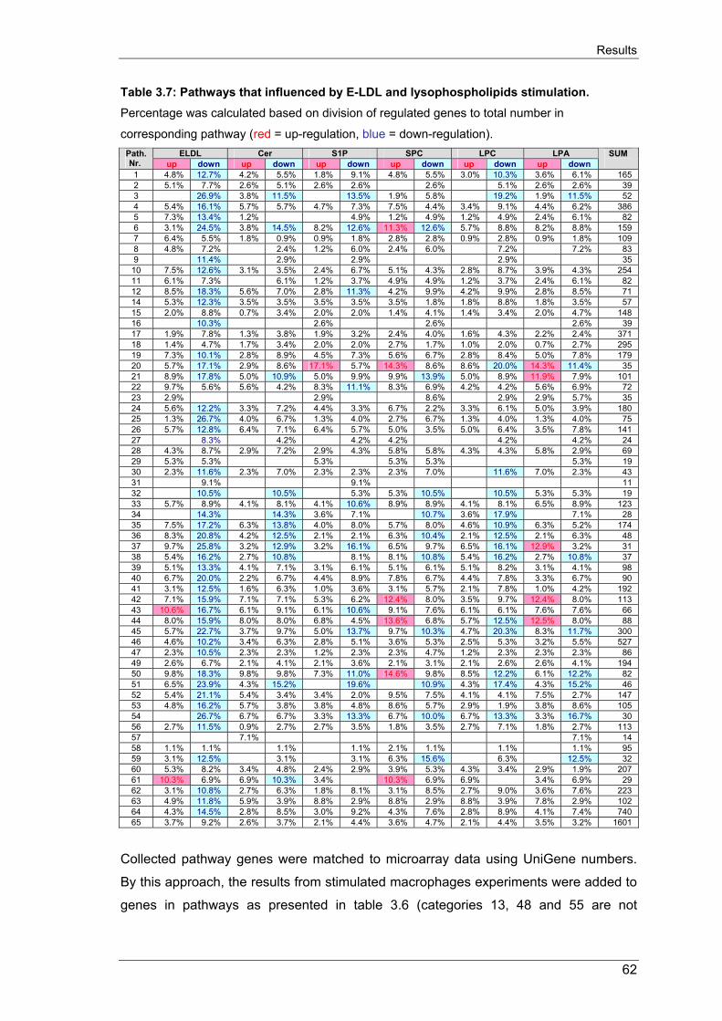
Results
Table 3.7: Pathways that influenced by E-LDL and lysophospholipids stimulation. Percentage was calculated based on division of regulated genes to total number in
corresponding pathway (red = up-regulation, blue = down-regulation). ELDL Cer S1P SPC LPC LPA Path.
Nr. up down up down up down up down up down up down SUM
1 4.8% 12.7% 4.2% 5.5% 1.8% 9.1% 4.8% 5.5% 3.0% 10.3% 3.6% 6.1% 165 2 5.1% 7.7% 2.6% 5.1% 2.6% 2.6% 2.6% 5.1% 2.6% 2.6% 39 3 26.9% 3.8% 11.5% 13.5% 1.9% 5.8% 19.2% 1.9% 11.5% 52 4 5.4% 16.1% 5.7% 5.7% 4.7% 7.3% 7.5% 4.4% 3.4% 9.1% 4.4% 6.2% 386 5 7.3% 13.4% 1.2% 4.9% 1.2% 4.9% 1.2% 4.9% 2.4% 6.1% 82 6 3.1% 24.5% 3.8% 14.5% 8.2% 12.6% 11.3% 12.6% 5.7% 8.8% 8.2% 8.8% 159 7 6.4% 5.5% 1.8% 0.9% 0.9% 1.8% 2.8% 2.8% 0.9% 2.8% 0.9% 1.8% 109 8 4.8% 7.2% 2.4% 1.2% 6.0% 2.4% 6.0% 7.2% 7.2% 83 9 11.4% 2.9% 2.9% 2.9% 35 10 7.5% 12.6% 3.1% 3.5% 2.4% 6.7% 5.1% 4.3% 2.8% 8.7% 3.9% 4.3% 254 11 6.1% 7.3% 6.1% 1.2% 3.7% 4.9% 4.9% 1.2% 3.7% 2.4% 6.1% 82 12 8.5% 18.3% 5.6% 7.0% 2.8% 11.3% 4.2% 9.9% 4.2% 9.9% 2.8% 8.5% 71 14 5.3% 12.3% 3.5% 3.5% 3.5% 3.5% 3.5% 1.8% 1.8% 8.8% 1.8% 3.5% 57 15 2.0% 8.8% 0.7% 3.4% 2.0% 2.0% 1.4% 4.1% 1.4% 3.4% 2.0% 4.7% 148 16 10.3% 2.6% 2.6% 2.6% 39 17 1.9% 7.8% 1.3% 3.8% 1.9% 3.2% 2.4% 4.0% 1.6% 4.3% 2.2% 2.4% 371 18 1.4% 4.7% 1.7% 3.4% 2.0% 2.0% 2.7% 1.7% 1.0% 2.0% 0.7% 2.7% 295 19 7.3% 10.1% 2.8% 8.9% 4.5% 7.3% 5.6% 6.7% 2.8% 8.4% 5.0% 7.8% 179 20 5.7% 17.1% 2.9% 8.6% 17.1% 5.7% 14.3% 8.6% 8.6% 20.0% 14.3% 11.4% 35 21 8.9% 17.8% 5.0% 10.9% 5.0% 9.9% 9.9% 13.9% 5.0% 8.9% 11.9% 7.9% 101 22 9.7% 5.6% 5.6% 4.2% 8.3% 11.1% 8.3% 6.9% 4.2% 4.2% 5.6% 6.9% 72 23 2.9% 2.9% 8.6% 2.9% 2.9% 5.7% 35 24 5.6% 12.2% 3.3% 7.2% 4.4% 3.3% 6.7% 2.2% 3.3% 6.1% 5.0% 3.9% 180 25 1.3% 26.7% 4.0% 6.7% 1.3% 4.0% 2.7% 6.7% 1.3% 4.0% 1.3% 4.0% 75 26 5.7% 12.8% 6.4% 7.1% 6.4% 5.7% 5.0% 3.5% 5.0% 6.4% 3.5% 7.8% 141 27 8.3% 4.2% 4.2% 4.2% 4.2% 4.2% 24 28 4.3% 8.7% 2.9% 7.2% 2.9% 4.3% 5.8% 5.8% 4.3% 4.3% 5.8% 2.9% 69 29 5.3% 5.3% 5.3% 5.3% 5.3% 5.3% 19 30 2.3% 11.6% 2.3% 7.0% 2.3% 2.3% 2.3% 7.0% 11.6% 7.0% 2.3% 43 31 9.1% 9.1% 11 32 10.5% 10.5% 5.3% 5.3% 10.5% 10.5% 5.3% 5.3% 19 33 5.7% 8.9% 4.1% 8.1% 4.1% 10.6% 8.9% 8.9% 4.1% 8.1% 6.5% 8.9% 123 34 14.3% 14.3% 3.6% 7.1% 10.7% 3.6% 17.9% 7.1% 28 35 7.5% 17.2% 6.3% 13.8% 4.0% 8.0% 5.7% 8.0% 4.6% 10.9% 6.3% 5.2% 174 36 8.3% 20.8% 4.2% 12.5% 2.1% 2.1% 6.3% 10.4% 2.1% 12.5% 2.1% 6.3% 48 37 9.7% 25.8% 3.2% 12.9% 3.2% 16.1% 6.5% 9.7% 6.5% 16.1% 12.9% 3.2% 31 38 5.4% 16.2% 2.7% 10.8% 8.1% 8.1% 10.8% 5.4% 16.2% 2.7% 10.8% 37 39 5.1% 13.3% 4.1% 7.1% 3.1% 6.1% 5.1% 6.1% 5.1% 8.2% 3.1% 4.1% 98 40 6.7% 20.0% 2.2% 6.7% 4.4% 8.9% 7.8% 6.7% 4.4% 7.8% 3.3% 6.7% 90 41 3.1% 12.5% 1.6% 6.3% 1.0% 3.6% 3.1% 5.7% 2.1% 7.8% 1.0% 4.2% 192 42 7.1% 15.9% 7.1% 7.1% 5.3% 6.2% 12.4% 8.0% 3.5% 9.7% 12.4% 8.0% 113 43 10.6% 16.7% 6.1% 9.1% 6.1% 10.6% 9.1% 7.6% 6.1% 6.1% 7.6% 7.6% 66 44 8.0% 15.9% 8.0% 8.0% 6.8% 4.5% 13.6% 6.8% 5.7% 12.5% 12.5% 8.0% 88 45 5.7% 22.7% 3.7% 9.7% 5.0% 13.7% 9.7% 10.3% 4.7% 20.3% 8.3% 11.7% 300 46 4.6% 10.2% 3.4% 6.3% 2.8% 5.1% 3.6% 5.3% 2.5% 5.3% 3.2% 5.5% 527 47 2.3% 10.5% 2.3% 2.3% 1.2% 2.3% 2.3% 4.7% 1.2% 2.3% 2.3% 2.3% 86 49 2.6% 6.7% 2.1% 4.1% 2.1% 3.6% 2.1% 3.1% 2.1% 2.6% 2.6% 4.1% 194 50 9.8% 18.3% 9.8% 9.8% 7.3% 11.0% 14.6% 9.8% 8.5% 12.2% 6.1% 12.2% 82 51 6.5% 23.9% 4.3% 15.2% 19.6% 10.9% 4.3% 17.4% 4.3% 15.2% 46 52 5.4% 21.1% 5.4% 3.4% 3.4% 2.0% 9.5% 7.5% 4.1% 4.1% 7.5% 2.7% 147 53 4.8% 16.2% 5.7% 3.8% 3.8% 4.8% 8.6% 5.7% 2.9% 1.9% 3.8% 8.6% 105 54 26.7% 6.7% 6.7% 3.3% 13.3% 6.7% 10.0% 6.7% 13.3% 3.3% 16.7% 30 56 2.7% 11.5% 0.9% 2.7% 2.7% 3.5% 1.8% 3.5% 2.7% 7.1% 1.8% 2.7% 113 57 7.1% 7.1% 14 58 1.1% 1.1% 1.1% 1.1% 2.1% 1.1% 1.1% 1.1% 95 59 3.1% 12.5% 3.1% 3.1% 6.3% 15.6% 6.3% 12.5% 32 60 5.3% 8.2% 3.4% 4.8% 2.4% 2.9% 3.9% 5.3% 4.3% 3.4% 2.9% 1.9% 207 61 10.3% 6.9% 6.9% 10.3% 3.4% 10.3% 6.9% 6.9% 3.4% 6.9% 29 62 3.1% 10.8% 2.7% 6.3% 1.8% 8.1% 3.1% 8.5% 2.7% 9.0% 3.6% 7.6% 223 63 4.9% 11.8% 5.9% 3.9% 8.8% 2.9% 8.8% 2.9% 8.8% 3.9% 7.8% 2.9% 102 64 4.3% 14.5% 2.8% 8.5% 3.0% 9.2% 4.3% 7.6% 2.8% 8.9% 4.1% 7.4% 740 65 3.7% 9.2% 2.6% 3.7% 2.1% 4.4% 3.6% 4.7% 2.1% 4.4% 3.5% 3.2% 1601
Collected pathway genes were matched to microarray data using UniGene numbers.
By this approach, the results from stimulated macrophages experiments were added to
genes in pathways as presented in table 3.6 (categories 13, 48 and 55 are not
62

Results
presented in this table due to no current update is available). The result table 3.6
summarized the contribution of pathways regulation of the stimuli in macrophages.
Differ from other stimulation, E-LDL remarkably showing its influences on 45 pathways
and most of them were down-regulated.
3.4.2. Candidate genes and pathways regulation in stimulated macrophages 3.4.2.1. Analysis of cholesterol metabolism pathway in stimulated macrophages Lipid homeostasis in vertebrate cells is regulated by a family of membrane-bound
transcription factor named as sterol regulatory element-binding proteins (SREBPs). The
mammalian genome encodes three SREBP isoforms, designed SREBP-1a, -1c and
SREBP-2. These three isoforms directly activate the expression of more than 30 genes
dedicated to the synthesis and uptake of cholesterol, fatty acids, triglycerides,
phospholipids as well as the NADPH cofactors required to synthesis of these
molecules. Using DNA microarray, the synthesis pathways of these molecules in lipid-
stimulated macrophages have been analyzed. Total of n=75 related genes were
screened and n=30 genes have been found to be regulated in these stimulations (table
3.8).
There are two pathways that activated by SREBP-1c and SREBP-2. The microarray
analysis revealed that enzymatically-modified LDL was having highest influences in
comparison to other stimuli and E-LDL showing its most effects on the cholesterol
metabolism pathway that activated by SREBP-2. E-LDL suppressed the expression of
SREBP-2 activator and almost all related enzymes with only an exception of HMG CoA
synthase. HMG CoA synthase isoform 1 was down-regulated by E-LDL, ceramide and
SPC while isoform 2 was up-regulated by E-LDL and ceramide. Contradictory with
SREBP-2 activated pathway, expression of SREBP-1c and most of SREBP-1c
activated enzymes were not influenced by any of tested stimuli. Ceramide, S1P, SPC
and LPC were regulated several genes but not seemed to alter the synthesis pathway.
In addition to the suppression of SREBP-2, SERBP cleavage-activating protein (SCAP)
as escort protein for SERBP and sensor of sterols was not regulated in any
stimulations.
63

Results
Table 3.8: Cholesterol metabolism pathway genes regulated by lipid stimulation of macrophages. (red = up-regulation, blue = down-regulation, (-) for down-regulation, value =
fold change).
Name Unigene Short name ChromLoc
ELD
L vs
. Ctr
l
Cer
8 vs
. Ctr
l
S1P
vs. C
trl
SPC
vs. C
trl
LPC
vs.
Ctr
l
LPA
vs.
Ctr
l
SREBP cleavage-activating protein Hs.78442 SCAP 3p21.31 1.2 -1.3 1.1 1.2 1.0 -1.3 sterol regulatory element binding transcription factor 2 Hs.108689 SREBF2 22q13 -2.0 -1.1 -1.1 1.0 -1.1 -1.3
acetyl-Coenzyme A acetyltransferase 2 (acetoacetyl Coenzyme A thiolase) Hs.278544 ACAT2 6q25.3-
q26 -2.5 -1.1 -1.1 1.0 1.0 1.0
3-hydroxy-3-methylglutaryl-Coenzyme A synthase 1 (soluble) Hs.77910 HMGCS1 5p14-p13 -3.3 -2.0 -1.7 -2.0 -1.4 -1.7
3-hydroxy-3-methylglutaryl-Coenzyme A synthase 2 (mitochondrial) Hs.59889 HMGCS2 1p13-p12 2.0 3.5 1.1 1.5 1.9 -2.0
3-hydroxy-3-methylglutaryl-Coenzyme A reductase Hs.11899 HMGCR 5q13.3-q14 -2.0 -1.1 1.1 -1.1 1.1 -1.1
mevalonate kinase (mevalonic aciduria) Hs.130607 MVK 12q24 -3.3 -1.7 -1.1 -1.4 3.9 -1.7
mevalonate (diphospho) decarboxylase Hs.3828 MVD 16q24.3 1.2 2.3 2.4 2.1 1.7 1.8
isopentenyl-diphosphate delta isomerase Hs.76038 IDI1 10p15.3 -5.0 -1.1 1.0 -1.3 1.0 1.0
farnesyl-diphosphate farnesyltransferase 1 Hs.48876 FDFT1 8p23.1-p22 -5.0 1.0 1.0 -1.1 1.0 1.0
squalene epoxidase Hs.71465 SQLE 8q24.1 -5.0 -1.4 -1.1 -1.3 1.0 1.0 Human lanosterol 14-alpha demethylase (CYP51P2) processed pseudogene Hs.226213 -3.3 -1.4 -1.1 -1.3 -1.3 -1.3
7-dehydrocholesterol reductase Hs.11806 DHCR7 11q13.2-q13.5 -3.3 -1.3 1.1 1.2 -1.1 -1.1
low density lipoprotein receptor (familial hypercholesterolemia) Hs.213289 LDLR 19p13.3 -10.0 -1.3 1.0 -1.4 1.0 -1.1
sterol O-acyltransferase (acyl-Coenzyme A: cholesterol acyltransferase) 1 Hs.14553 SOAT1 1q25 1.1 -1.4 1.2 -2.5 1.0 1.0
malic enzyme 1, NADP(+)-dependent, cytosolic Hs.14732 ME1 6q12 -2.5 1.2 1.1 1.4 1.0 1.1
hexokinase 2 Hs.198427 HK2 2p13 -2.0 -1.3 1.1 -1.1 -1.1 -1.1
insulin induced gene 1 Hs.56205 INSIG1 7q36 -5.0 -1.3 1.1 -1.1 1.0 -1.1 peroxisomal trans 2-enoyl CoA reductase; putative short chain alcohol dehydrogenase Hs.281680 PECR 2q35 1.4 1.2 -2.0 1.3 1.2 -1.7
proprotein convertase subtilisin/kexin type 1 Hs.78977 PCSK1 5q15-q21 -1.1 -1.1 -1.3 2.6 1.3 2.7
proprotein convertase subtilisin/kexin type 1 inhibitor Hs.256311 PCSK1N Xp11.23 -3.3 -3.3 -3.3 1.5 -1.7 -2.5
proprotein convertase subtilisin/kexin type 2 Hs.93164 PCSK2 20p11.2 -2.5 -1.4 -1.4 -1.1 -2.0 -1.7
proprotein convertase subtilisin/kexin type 7 Hs.32978 PCSK7 11q23-q24 1.7 -2.5 1.1 -1.4 -1.7 1.2
elongation of very-long-chain-fatty-acids like 2 Hs.246107 ELOVL2 6p22.3 -1.3 -3.3 -5.0 -3.3 -5.0 -2.0 Sterol regulatory element binding transcription factor 1 Hs.592123 SREBPF1 17p11.2 -1.7 -1.3 1.0 1.1 -1.4 1.1
acetyl-Coenzyme A carboxylase alpha Hs.7232 ACACA 17q21 -1.7 -3.3 1.1 -2.5 1.1 1.2
acetyl-Coenzyme A carboxylase beta Hs.183857 ACACB 12q24.1 -1.1 2.5 -1.1 1.3 1.0 -1.3
fatty acid desaturase 1 Hs.132898 FADS1 11q12.2-q13.1 -2.5 -1.1 1.0 1.0 -1.1 1.0
fatty acid desaturase 2 Hs.184641 FADS2 11q12-q13.1 -10.0 -1.1 1.0 1.2 -1.3 1.3
fatty-acid-Coenzyme A ligase, long-chain 4 Hs.81452 FACL4 Xq22.3-q23 -2.0 -1.3 -1.1 -2.0 -1.3 1.2
fatty-acid-Coenzyme A ligase, long-chain 6 Hs.14945 FACL6 5q31 -2.5 -2.0 -1.4 1.1 -2.0 -2.0
3.4.2.2. Analysis of ABC transporter genes expression in lipid-stimulated macro-
phages In addition to the previously described ABCA2 gene, a list of ABC transporter genes
family regulated in stimulation of macrophages has been created. Table 3.9 showing
ABC genes that regulated by E-LDL, ceramide, S1P, SPC, LPC and LPA. 35 genes
from the total of 51 ABC gene members of 7 subfamilies have been analyzed with
microarrays and n=15 (43%) genes from 5 subfamilies found to be regulated during
64

Results
macrophages stimulations. Interestingly, several ABC genes have been found highly up
or down regulated in lipid stimulated macrophage. ABCA2, known to transport
estramustine and steroids, was 3.9-fold up-regulated. ABCA8, known for drug
transportation, was 7-fold up-regulated. Whereas, ABCD2, as a very-long-chain fatty
acid transporter, was 10 and 3.3 folds down-regulated by E-LDL and SPC, respectively.
Other genes; ABCA3 (surfactant phospholipids transporter), ABCA12 and ABCB9 were
3.3 folds down-regulated by E-LDL and LPA.
Table 3.9: Regulation of ABC transporter genes upon stimulation (red = up-regulation, blue = down-regulation, (-) for down-regulation, value = fold change).
Name Unigene Short name ChromLoc
ELD
L vs
. Ctr
l
Cer
8 vs
. Ctr
l
S1P
vs. C
trl
SPC
vs.
Ctr
l
LPC
vs.
Ctr
l
LPA
vs.
Ctr
l
ATP-binding cassette, sub-family A (ABC1), member 2 Hs.121561 ABCA2 9q34 3.9 -1.1 1.9 1.1 1.1 1.8
ATP-binding cassette, sub-family A (ABC1), member 3 Hs.26630 ABCA3 16p13.3 -3.3 -2.0 1.1 -1.3 -1.1 -1.1
ATP-binding cassette, sub-family A (ABC1), member 6 Hs.15780 ABCA6 17q24.3 -1.7 -1.4 -1.1 -2.0 -2.0 -1.1
ATP-binding cassette, sub-family A (ABC1), member 8 Hs.38095 ABCA8 17q24 7.0 1.3 -2.0 -1.4 -1.3 2.2
ATP-binding cassette, sub-family A (ABC1), member 12 Hs.134585 ABCA12 2q35 -1.4 -1.0 -1.4 1.2 -1.7 -3.3
ATP-binding cassette, sub-family B (MDR/TAP), member 1 Hs.21330 ABCB1 7q21.1 -1.7 -1.7 -1.1 -2.5 1.2 1.1
ATP-binding cassette, sub-family B (MDR/TAP), member 4 Hs.73812 ABCB4 7q21.1 3.1 -1.0 -1.7 1.1 -1.7 -2.0
ATP-binding cassette, sub-family B (MDR/TAP), member 8 Hs.118634 ABCB8 7q36 1.3 -1.4 1.6 1.2 2.2 -2.0
ATP-binding cassette, sub-family B (MDR/TAP), member 9 Hs.6129 ABCB9 12q24 -1.1 -1.0 1.6 1.3 -1.3 -3.3
ATP-binding cassette, sub-family C (CFTR/MRP), member 9 Hs.248960 ABCC9 12p12.1 -2.0 -1.1 -1.1 1.1 -1.3 1.1
ATP-binding cassette, sub-family D (ALD), member 2 Hs.117852 ABCD2 12q11-q12 -10.0 1.4 -1.0 -3.3 1.3 1.2
ATP-binding cassette, sub-family G (WHITE), member 1 Hs.10237 ABCG1 21q22.3 2.2 1.2 1.6 1.5 -1.3 1.4
ATP-binding cassette, sub-family G (WHITE), member 2 Hs.194720 ABCG2 4q22 1.6 -1.7 -2.0 -1.4 -1.0 -1.1
ATP-binding cassette, sub-family G (WHITE), member 4 Hs.126378 ABCG4 11q23.3 -2.0 -1.1 -1.0 -1.4 -1.3 -1.1 ATP-binding cassette, sub-family G (WHITE), member 5 (sterolin 1) Hs.132992 ABCG5 2p21 1.2 2.2 1.7 2.1 1.3 1.4
65

Discussion
4. DISCUSSION Monocyte-derived macrophage gene regulation plays an important role not only in
pathogenesis of atherosclerosis but also in many other inflammatory diseases, such as,
cirrhosis, rheumatoid arthritis, glomerulosclerosis, pulmonary fibrosis and chronic
pancreatitis. In addition, bioactive lipids as derivatives of lysophospholipids have been
remarkably evidenced to contribute to many pathophysiological stages of these
diseases. For example, lysophospholipids are significantly involved in pathogenesis of
atherosclerosis by modulating a broad spectrum of processes including platelet
aggregation, cell growth and differentiation as well as recruitment of vascular cells.
Therefore, the aim of this thesis work is to analyze global gene expression of
monocyte-derived macrophages under the modulation of selected of bioactive lipids,
including ceramide, S1P, SPC, LPA and LPA, and modified low-density lipoprotein as
the source of the these bioactive lipids. All experiments were performed with human
peripheral blood monocytes from ApoE 3 homozygous phenotype from healthy donors.
Phagocytic differentiation was carried out in serum-free culture media to minimize
potential artefacts.
Expression analysis of marker genes in in vitro phagocytic differentiation and foam cell
formation models.
Scavenger receptors (CD36, Cla-1), adipophilin and ABCA2 were analyzed as marker
genes to test the in vitro model of phagocytic differentiation and foam cell formation
induced by enzymatic modified lipoprotein.
Phagocytic differentiation is accompanied by enhanced cholesterol uptake activity and
reported with the accumulation of number of cell surface receptors including scavenger
receptors, as receptors for lipoproteins. CD36 and its homologue, Cla-1 (SR-BI), are
both detected in monocytes and macrophages. CD36 recognizes and binds many
ligands, including modified LDL. Expression of CD36 is reported to be elevated during
in vivo macrophage differentiation and Ox-LDL-induced foam cell formation [253;254].
From my experiments, CD36 was abundantly expressed in monocytes and strongly up-
regulated during phagocytic differentiation. Similar to current literatures addressed on
atherogenesis, E-LDL incubation of monocyte-derived macrophages enhanced the
expression of CD36 on both mRNA and protein levels, while other modified LDL (Ac-
LDL, Ox-LDL) had the reverse effects [11]. Our collaborated study published by
Kapinsky et al. also revealed that E-LDL but not Ac-LDL or Ox-LDL induced foam cell
66

Discussion
formation in freshly isolated peripheral blood monocytes. Similarly, Cla-1 was also
detected to be highly up-regulated during macrophage differentiation and in E-LDL
loaded macrophages, while it was hardly detected in monocytes [11]. Recent findings
also revealed that Cla-1 involved in the selective uptake of cholesterol ester,
recognized modified lipoproteins and played a role in HDL metabolism [244;245].
Therefore, these results support the hypothesis of an autoregulatory loop for enhanced
cholesterol uptake and provide a link between this modified LDL and the HDL
metabolism.
The excess lipid deposition in macrophages has been reported to modulate expression
of several genes, including adipophilin. Adipophilin or adipose differentiation related
protein (ADRP) is currently reported to be abundance in macrophage-rich areas of
atherosclerotic lesions. Its expression in atherosclerotic plagues was calculated as 3.5-
fold higher compared to healthy areas of the same arteries. In their study, Larigauderie
et al. showed that the stimulation of adipophilin expression by Ac-LDL promotes
triglycerides and cholesterol storage and reduces cholesterol efflux [260]. In my study,
the expression of adipophilin was analyzed with E-LDL-induced macrophage foam cell
formation. Levels of adipophilin mRNA and protein were rapidly elevated within 2 hours
and approached its peak at 24 hours of E-LDL incubations. Other study also revealed
that the expression of adipophilin is increased in Ox-LDL-induced foam cell formation of
human macrophage. However, the induction of adipophilin expression by Ox-LDL was
not detected within 6 hours of incubation [255]. Therefore, these data suggested a high
atherogenic potent of E-LDL and adipophilin could be considered as a sensitive marker
for lipid loading in human blood monocytes.
Together with ABCA1, ABCA4 and ABCA7, ABCA2 is found to be expressed in human
monocytes/macrophages and enhanced expression in macrophages treated with
cholesterol. In line with these findings, my experiments also revealed that ABCA2 is
already expressed in human peripheral blood monocytes and elevated its expression
during phagocytic differentiation. In addition, the high level of accumulation of ABCA2
was observed in foam cells induced by E-LDL compared to macrophages. The
repeated experiment using DNA chip (chapter 3.4.2.2) also confirmed an upregulation
of ABCA2 during foam cell formation induced by enzymatically modified LDL. These
data also assume that ABAC2 plays a role in intracellular trafficking of LDL-derived free
cholesterol and cholesterol homeostasis.
67

Discussion
All together, the above-mentioned data strongly indicate that the in vitro model of
phagocytic differentiation and E-DLD-induced foam cell formation reflects the in vivo
processes occurred during the development of atherosclerosis.
Expression analysis of lysophospholipid receptors.
To date, there are 13 G-protein-coupled receptors have been identified to recognize
and bind to S1P, SPC, LPA and LPC. However, only a few studies have been
published investigating the expression of these receptors in human blood monocytes
and macrophages, as major cells involved in the pathogenesis of atherosclerosis. In
one study, mRNA expression of LPA1, LPA2 and S1P1, S1P2, S1P4 were reported in
human monocytes and macrophages whereas S1P3 and S1P5 mRNAs could not be
detected using RT-PCR [261]. Another study reported the expression of S1P1, LPA1,
LPA2 and LPA3 mRNAs in human alveolar macrophages but no mRNA for S1P2, S1P3,
S1P4 and S1P5 were detected in these cells [262]. These results showed a
contradictory in the expression of S1P2, S1P4 and LPA3 and it become unclear weather
these receptors are expressed in macrophages or not. However, these macrophages
have been cultured in the present of serum, which already contains high concentration
of S1P and LPA. This may, at least in part, explain the contradictory of the above-
mentioned results. The in vitro model of phagocytic differentiation in this thesis work
was developed with no additional serum to investigate the expression of these
receptors in both mRNA and protein levels. The advantage of mRNA expression
analysis with TaqMan RT-PCR is to allow relative quantify the gene expression using
calibration curves thus eliminate the varying efficiencies of single PCR reaction. The
mRNAs of all 13 lysophospholipid receptors could be amplified in freshly isolated
human monocytes but no significant regulation detected in further stimulation of
monocytes and/or macrophages. With the exception of LPA4, 5, GPR3, 4, 12 and G2A
(commercial antibody unavailable), the protein expression analysis revealed the
present of S1P1, 2, 4 and LPA1, 2 in monocytes and macrophages. In addition, Rikitake et
al. reported an expression of G2A protein in human and rabbit macrophages as well as
in murine atherosclerotic lesion [263]. Expression analysis of these receptors using
microarray was not giving any significant data as the detected mean florescence were
generally low.
68

Discussion
Expression analysis of global macrophages genes with DNA microarray.
Large scale analysis has revealed an overview of macrophages global gene expression
under the stimulations of the above-mentioned lysophospholipids. Herein, however, the
discussion is only restricted to the effects of modified lipoprotein (E-LDL) on genes
related to cholesterol metabolism pathways and ABC transporters during macrophage
foam cell formation.
The lipid homeostasis in vertebrate cells is regulated by a family of membrane-bound
transcription factors designated as sterol regulatory element-binding transcription
factors, or proteins (SREBPs). Although the mammalian genome encodes three
SREBP isoforms but only SREBP-1c and SREBP-2 are having predominant roles in
regulation of cholesterol metabolism. Despite the fact that at high expression level, all
three SREBP isoforms could activate all genes listed in fig. 3.9, the SREBP-1c is
preferentially activate the fatty acid and lipoproteins biosynthesis and SREBP-2
preferentially activate cholesterol biosynthesis pathways, at normal physiological
expression level.
The regulation activity of SREBPs depends on an escort protein SCAP (SREBP
cleavage-activating protein) and two enzymes designate Site-1 and Site-2 protease. At
normal stage, SREBPs are synthesized as inactive precursor, bound to endoplasmic
reticulum. When cells become depleted in cholesterol, SCAP escorts SREBPs to Golgi
apparatus, where it is cleaved by the two proteases and translocated to nucleus to
activate the promoter and/or enhancer on multiple target genes of the metabolism
pathways. The lipid homeostasis activity of SREBPs is manifested when cellular
cholesterol content rises up. In which, the SCAP/SERBPs complex is no longer
incorporated into ER transport vesicles and therefore SREBPs remained inactivated. In
knockout and transgenic mice studies the role of SREBPs in cholesterol regulation is
referred to SCAP as the sensor [264]. Mice with disrupted SCAP gene exhibited a fall
of 70-80% of cholesterol and fatty acid synthesis in liver. In cultured cells, the
processing of SERBP is reported to be inhibited by sterols by suppression of SCAP
activity. Transgenic mice with a single amino acid substitution in the sterol-sensing
domain (D443N) overproduced cholesterol. In addition, transgenic mice carried this
mutation in liver also exhibited a similar phonotype. The levels of nucleus SREBP-1
and 2 were elevated leading to an increase of expression of all SREBP target genes
even when these animals are fed with cholesterol-rich diet. Therefore, it is assumable
that SCAP activity in vivo is normally under partial inhibition by endogenous
69

Discussion
cholesterol. Thus the synthesis of cholesterol and fatty acids is partially repressed in
the liver.
In my study, the role SREBP in lipid metabolism of human macrophages was
investigated. Although both of SREBP isoforms 1c and 2 were detected in human
macrophages, their expression levels in E-LDL-induced foam cells were not similar.
Interestingly, mRNA expression SERBP-2 isoform is suppressed by exogenous
cholesterol leading to a complete downregulation of almost all target genes of the
cholesterol synthesis pathway (fig. 3.9). With the only exception of SMGCS2, which
was 2-fold upregulated, other SREBP target genes were 2 to 5-fold downregulated in
E-LDL loading macrophages. Thus, loading of E-LDL is completely closing down this
pathway and it further confirmed by additional downregulation of malic enzyme 1 (ME1)
that support energy for the cholesterol metabolism pathway.
Figure 3.9: Regulation of cholesterol metabolism pathway genes in simulated macrophages. (red = up-regulation, blue = down-regulation, NC = no change, text in the boxes
represent the stimulation). Figure adapted from [265].
70

Discussion
It is known that SREBPs enhance lipid synthesis but it also stimulate LDL receptor
expression [266], so their net effect on plasma lipoprotein levels depend on a balance
between opposing effects. Mice that have an overexpressed or underexpressed of
SREBPs showing a reduction of plasma levels of lipoproteins. Similarly, transgenic
mice that overexpressed SREBPs also reduce its level of cholesterol and triglycerides
in comparison to control, although they massively overproduced fatty acids and/or
cholesterol. These observations could be explained that these lipids have been rapidly
degraded in a process that mediated by LDL receptor. Mice that have a LDL-receptor
deficiency exhibit a ten-fold of cholesterol and triglyceride induction levels. In
conformity with the above conclusions, mRNA expression analyses in this thesis work
revealed a 10-fold suppression of LDL-receptor during foam cell formation of
macrophages. Thus, exogenous cholesterol turns down cholesterol metabolism
pathways and also partially prevents cholesterol influx by LDL-receptor suppression in
macrophages.
However, these mRNA expression analyses also revealed no change of fatty acid and
triglycerides metabolism pathways in human macrophages upon extracellular lipid
loading, except the suppression of conversion of monounsaturated fatty acids to fatty
acyl CoA, triggered by FADS1, FADS2 and FACoA ligase 4 and 6.
Cellular cholesterols homeostasis is also regulated by several mechanisms involved
high-density lipoproteins, apolipoproteins (apoA-I, apoE) and lipid-regulated ABC
transporters. ABC transporters translocate a broad spectrum of molecules across cell
membranes, including amino acids, carbohydrates, peptides, vitamins, ions,
xenobiotics and lipids [267] and therefore, exert various cellular functions. Many of
inherited diseases have been found to be caused or associated with mutations that
occurred within ABC transporter genes family. Among these are familial HDL deficiency
(or Tangier disease) cause by a mutation of ABCA1 [268;269] and accumulation of
dietary cholesterol in sitosterolemia that account for ABCG5/ABCG8 [270;271] Herein,
the regulation of these ABC transporters in human macrophage foam cells induced by
exogenous E-LDL loading were investigated. From 35 ABC genes represented on the
DNA chip, there were 8 genes regulated during foam cell formation, including ABCA2,
ABCA3, ABCA8, ABCB4, ABCC9, ABCD2, ABCG1, and ABCG4.
Among those, ABCG1 is sharing highest functional similarity to the ABCA1 gene, the
first ABC transporter found to be expressed in human macrophage [272]. While ABCA1
exhibited no response to lipid loading of macrophages, ABCG1 showed a 2.2 fold
71

Discussion
upregulation in these cells. Initially, ABCG1 was reported to be expressed in human
monocytes and its expression elevated during phagocytic differentiation as well as
upon sterol-loading of macrophages using acetylated LDL. The sterol response of
ABCG1 was further confirmed by an experiment in which cholesterol deloading of
macrophages with HDL3 lead to a suppression of ABCG1 mRNA and protein [273].
Furthermore, the induction of ABCG1 expression is not only observed with Ac-LDL but
also other types of modified LDL such as ox-LDL and E-LDL, while native LDL and free
cholesterol exhibited no effects on ABCG1 expression [274;275]. The role of ABCG1 in
cholesterol efflux was initially reported by our Institute, when ABCG1-specific antisense
oligonucleotides caused a significantly impaired cholesterol and phospholipid efflux to
HLD3 [273]. Recently, other investigations reported that ABCG1 can function either
alone or in combination with other ABCG-family members, specifically ABCG4, to
regulate cellular lipid efflux. DNA chip experiments from this thesis work exhibited an
inversed regulation of ABCG1 and ABCG4 upon cholesterol loading. However, this
may be explained by the very low expression of ABGC4 in macrophages in comparison
to that of ABCG1.
Table 3.9: Regulation of ABC transporter genes upon stimulation (red = up-regulation, blue = down-regulation, (-) for down-regulation, value = fold change).
Name Unigene Short name ChromLoc
ELD
L vs
. Ctr
l
Cer
8 vs
. Ctr
l
S1P
vs. C
trl
SPC
vs.
Ctr
l
LPC
vs.
Ctr
l
LPA
vs.
Ctr
l ATP-binding cassette, sub-family A (ABC1), member 2 Hs.121561 ABCA2 9q34 3.9 -1.1 1.9 1.1 1.1 1.8
ATP-binding cassette, sub-family A (ABC1), member 3 Hs.26630 ABCA3 16p13.3 -3.3 -2.0 1.1 -1.3 -1.1 -1.1
ATP-binding cassette, sub-family A (ABC1), member 8 Hs.38095 ABCA8 17q24 7.0 1.3 -2.0 -1.4 -1.3 2.2
ATP-binding cassette, sub-family B (MDR/TAP), member 4 Hs.73812 ABCB4 7q21.1 3.1 -1.0 -1.7 1.1 -1.7 -2.0
ATP-binding cassette, sub-family C (CFTR/MRP), member 9 Hs.248960 ABCC9 12p12.1 -2.0 -1.1 -1.1 1.1 -1.3 1.1
ATP-binding cassette, sub-family D (ALD), member 2 Hs.117852 ABCD2 12q11-q12 -10.0 1.4 -1.0 -3.3 1.3 1.2
ATP-binding cassette, sub-family G (WHITE), member 1 Hs.10237 ABCG1 21q22.3 2.2 1.2 1.6 1.5 -1.3 1.4
ATP-binding cassette, sub-family G (WHITE), member 4 Hs.126378 ABCG4 11q23.3 -2.0 -1.1 -1.0 -1.4 -1.3 -1.1
Table 3.10: ABC genes transported molecules and functions.
Gene Domain structure Tissue expression, cellular location Known or putative transported molecule
ABCA2 (TMD-ABC)2 Brain Estramustine, steroids
ABCA3 (TMD-ABC)2 Lung Surfactant phospholipids
ABCA8 (TMD-ABC)2 Ovary xenobiotic
ABCB4 (TMD-ABC)2 Liver, apical membrane Phosphatidylcholine
ABCC9 TMD0-(TMD-ABC)2 Heart, muscle Sulfonylureas
ABCD2 TM-ABC Peroxisomes Very-long-chain fatty acids
ABCG1 ABC-TMD Ubiquitous Phospholipids, cholesterol
ABCG4 ABC-TMD Liver
72

Discussion
Our DNA chip analysis also exhibited the regulation of other ABC transporter genes
that are predominantly expressed in different tissues. Among those, ABCA3 and
ABCB4 are reported to play role in cellular lipid rheostat. ABCA3 is predominantly
expressed in lung and localizes to the limiting membrane of lamellar bodies in alveolar
type II cells. Mutations in ABCA3 gene cause a neonatal respiratory failure of humans.
ABCA3 is reported to transport pulmonary surfactant and is involved in the
accumulation of phospholipids and cholesterol in lipid vesicles. Nevertheless, my data
showed a 3.3-fold downregulation of ABCA3 in macrophages upon lipid loading, which
could be potentially explained by its low expression in macrophages. Inversely, ABCB4
was 3.1-fold elevated upon lipid loading of those macrophages. It is however reported
to be expressed predominantly in liver rather than in macrophages (table 3.10).
Similarly, the results from ABCA8, ABCC9 and ABCD2 might be technical artefacts
since no expression has been reported in monocytes/macrophages using other
techniques.
Conclusions
Results archived from CD36 and Cla-1 analysis in phagocytic differentiation and foam
cell formation was further confirmed the higher atherogenic properties of enzymatically
modifification LDL, compared to other type of modifications (acetylation, oxidation).
Furthermore, these results also support the hypothesis of an autoregulatory loop for
enhanced cholesterol uptake and provide a link between this modified LDL and the
HDL metabolism. In addition, analyses of adipophilin and ABCA2 were also revealed
that adipophilin could be considered as a new sensitive marker for lipid loading of
human monocytes/macrophages as well as the role of ABCA2 in intracellular LDL-
derived free cholesterol trafficking and lipid homeostasis.
The expression of up-to-date G-protein-coupled receptor superfamily for the bioactive
lipid derivatives; S1P, LPA, LPC, SPC in human monocytes/macrophages was also
investigated. The serum-free phagosytic differentiation model was able to eliminate the
discrepancy arrived from previous studies. Since these bioactive lipid derivatives are
products of platelets and involve in many cellular processes, results archived from this
thesis work would sugguest the importance of further study on cross-talk between
platelets and monocytes/ macrophages.
DNA chip analysis provided an overview of cellular effects of the above-mentioned
bioactive lipids and cholesterol loading on macrophages global genes expression. Due
73

Discussion
to the limit of the thesis work, only the effects of cholesterol loading (E-LDL) on
cholesterol metabolism pathways and homeostasis were investigated in-details.
Interestingly, exogenous free cholesterol loading of human macrophages could lead to
a complete blockage of cellular cholesterol metabolism pathway triggered by SREBP-2,
while triacylglycerides and fatty acids metabolism pathways triggered by SREBP-1c
were not influenced. The study also reported the expression of ABC transporter A2, A3,
A8, B4, C9, D2, G1 and G4 in human macrophages and suggested the roles of
ABCA2, ABCB4 and ABCG1 in macrophages cellular lipid rheostat.
74

Citations
Citations [1] Murray CJ, Lopez AD. Global mortality, disability, and the contribution of risk
factors: Global Burden of Disease Study. Lancet 1997; 349: 1436-1442.
[2] Ross R. Atherosclerosis--an inflammatory disease. N. Engl. J. Med. 1999; 340: 115-126.
[3] Brown MS, Basu SK, Falck JR, Ho YK, Goldstein JL. The scavenger cell pathway for lipoprotein degradation: specificity of the binding site that mediates the uptake of negatively-charged LDL by macrophages. J. Supramol. Struct. 1980; 13: 67-81.
[4] Goldstein JL, Ho YK, Basu SK, Brown MS. Binding site on macrophages that mediates uptake and degradation of acetylated low density lipoprotein, producing massive cholesterol deposition. Proc. Natl. Acad. Sci. U. S. A 1979; 76: 333-337.
[5] Steinbrecher UP, Parthasarathy S, Leake DS, Witztum JL, Steinberg D. Modification of low density lipoprotein by endothelial cells involves lipid peroxidation and degradation of low density lipoprotein phospholipids. Proc. Natl. Acad. Sci. U. S. A 1984; 81: 3883-3887.
[6] Steinberg D, Parthasarathy S, Carew TE, Khoo JC, Witztum JL. Beyond cholesterol. Modifications of low-density lipoprotein that increase its atherogenicity. N. Engl. J. Med. 1989; 320: 915-924.
[7] Simionescu N, Vasile E, Lupu F, Popescu G, Simionescu M. Prelesional events in atherogenesis. Accumulation of extracellular cholesterol-rich liposomes in the arterial intima and cardiac valves of the hyperlipidemic rabbit. Am. J. Pathol. 1986; 123: 109-125.
[8] Seifert PS, Hugo F, Tranum-Jensen J, Zahringer U, Muhly M, Bhakdi S. Isolation and characterization of a complement-activating lipid extracted from human atherosclerotic lesions. J. Exp. Med. 1990; 172: 547-557.
[9] Bhakdi S, Dorweiler B, Kirchmann R, Torzewski J, Weise E, Tranum-Jensen J, Walev I, Wieland E. On the pathogenesis of atherosclerosis: enzymatic transformation of human low density lipoprotein to an atherogenic moiety. J. Exp. Med. 1995; 182: 1959-1971.
[10] Torzewski M, Klouche M, Hock J, Messner M, Dorweiler B, Torzewski J, Gabbert HE, Bhakdi S. Immunohistochemical demonstration of enzymatically modified human LDL and its colocalization with the terminal complement complex in the early atherosclerotic lesion. Arterioscler. Thromb. Vasc. Biol. 1998; 18: 369-378.
[11] Kapinsky M, Torzewski M, Buchler C, Duong CQ, Rothe G, Schmitz G. Enzymatically degraded LDL preferentially binds to CD14(high) CD16(+) monocytes and induces foam cell formation mediated only in part by the class B scavenger-receptor CD36. Arterioscler. Thromb. Vasc. Biol. 2001; 21: 1004-1010.
75
Citations
[12] Voelker DR. New perspectives on the regulation of intermembrane glycerophospholipid traffic. J. Lipid Res. 2003; 44: 441-449.
[13] Bell RM, Ballas LM, Coleman RA. Lipid topogenesis. J. Lipid Res. 1981; 22: 391-403.
[14] Voelker DR. Interorganelle transport of aminoglycerophospholipids. Biochim. Biophys. Acta 2000; 1486: 97-107.
[15] Vance JE, Vance DE. Does rat liver Golgi have the capacity to synthesize phospholipids for lipoprotein secretion? J. Biol. Chem. 1988; 263: 5898-5909.
[16] Henneberry AL, Wright MM, McMaster CR. The major sites of cellular phospholipid synthesis and molecular determinants of Fatty Acid and lipid head group specificity. Mol. Biol. Cell 2002; 13: 3148-3161.
[17] Leber A, Hrastnik C, Daum G. Phospholipid-synthesizing enzymes in Golgi membranes of the yeast, Saccharomyces cerevisiae. FEBS Lett. 1995; 377: 271-274.
[18] Chakraborty TR, Vancura A, Balija VS, Haldar D. Phosphatidic acid synthesis in mitochondria. Topography of formation and transmembrane migration. J. Biol. Chem. 1999; 274: 29786-29790.
[19] Wirtz KW. Phospholipid transfer proteins. Annu. Rev. Biochem. 1991; 60: 73-99.
[20] Voelker DR. Characterization of phosphatidylserine synthesis and translocation in permeabilized animal cells. J. Biol. Chem. 1990; 265: 14340-14346.
[21] Voelker DR. Disruption of phosphatidylserine translocation to the mitochondria in baby hamster kidney cells. J. Biol. Chem. 1985; 260: 14671-14676.
[22] Voelker DR. Phosphatidylserine translocation to the mitochondrion is an ATP-dependent process in permeabilized animal cells. Proc. Natl. Acad. Sci. U. S. A 1989; 86: 9921-9925.
[23] Sprong H, van der SP, van Meer G. How proteins move lipids and lipids move proteins. Nat. Rev. Mol. Cell Biol. 2001; 2: 504-513.
[24] Balasubramanian K, Schroit AJ. Aminophospholipid asymmetry: A matter of life and death. Annu. Rev. Physiol 2003; 65: 701-734.
[25] Chisolm GM, III, Chai Y. Regulation of cell growth by oxidized LDL. Free Radic. Biol. Med. 2000; 28: 1697-1707.
[26] Lusis AJ. Atherosclerosis. Nature 2000; 407: 233-241.
[27] Yokota T, Hansson GK. Immunological mechanisms in atherosclerosis. J. Intern. Med. 1995; 238: 479-489.
[28] Murugesan G, Fox PL. Role of lysophosphatidylcholine in the inhibition of endothelial cell motility by oxidized low density lipoprotein. J. Clin. Invest 1996; 97: 2736-2744.
76

Citations
[29] Croset M, Brossard N, Polette A, Lagarde M. Characterization of plasma unsaturated lysophosphatidylcholines in human and rat. Biochem. J. 2000; 345 Pt 1: 61-67.
[30] Portman OW, Alexander M. Lysophosphatidylcholine concentrations and metabolism in aortic intima plus inner media: effect of nutritionally induced atherosclerosis. J. Lipid Res. 1969; 10: 158-165.
[31] Portman OW, Illingworth DR. Metabolism of lysolecithin in vivo and in vitro with particular emphasis on the arterial wall. Biochim. Biophys. Acta 1974; 348: 136-144.
[32] Portman OW, Illingworth DR. Lysolecithin binding to human and squirrel monkey plasma and tissue components. Biochim. Biophys. Acta 1973; 326: 34-42.
[33] McKean ML, Smith JB, Silver MJ. Formation of lysophosphatidylcholine by human platelets in response to thrombin. Support for the phospholipase A2 pathway for the liberation of arachidonic acid. J. Biol. Chem. 1981; 256: 1522-1524.
[34] Bingham CO, III, Austen KF. Phospholipase A2 enzymes in eicosanoid generation. Proc. Assoc. Am. Physicians 1999; 111: 516-524.
[35] Chisolm GM, III, Penn MS. Oxidized lipoproteins and atherosclerosis. In: Fuster V, Ross R, Topol E, editors. Atherosclerosis and coronary disease. 129-149. 1996. New York, Raven Press. 1996.
Ref Type: Catalog
[36] Kabarowski JH, Xu Y, Witte ON. Lysophosphatidylcholine as a ligand for immunoregulation. Biochem. Pharmacol. 2002; 64: 161-167.
[37] Xu Y. Sphingosylphosphorylcholine and lysophosphatidylcholine: G protein-coupled receptors and receptor-mediated signal transduction. Biochim. Biophys. Acta 2002; 1582: 81-88.
[38] Mochizuki M, Zigler JS, Jr., Russell P, Gery I. Serum proteins neutralize the toxic effect of lysophosphatidyl choline. Curr. Eye Res. 1982; 2: 621-624.
[39] Zhu K, Baudhuin LM, Hong G, Williams FS, Cristina KL, Kabarowski JH, Witte ON, Xu Y. Sphingosylphosphorylcholine and lysophosphatidylcholine are ligands for the G protein-coupled receptor GPR4. J. Biol. Chem. 2001; 276: 41325-41335.
[40] Prokazova NV, Zvezdina ND, Korotaeva AA. Effect of lysophosphatidylcholine on transmembrane signal transduction. Biochemistry (Mosc. ) 1998; 63: 31-37.
[41] Hara S, Shike T, Takasu N, Mizui T. Lysophosphatidylcholine promotes cholesterol efflux from mouse macrophage foam cells. Arterioscler. Thromb. Vasc. Biol. 1997; 17: 1258-1266.
[42] Okajima F, Sato K, Tomura H, Kuwabara A, Nochi H, Tamoto K, Kondo Y, Tokumitsu Y, Ui M. Stimulatory and inhibitory actions of lysophosphatidylcholine, depending on its fatty acid residue, on the phospholipase C/Ca2+ system in HL-60 leukaemia cells. Biochem. J. 1998; 336 ( Pt 2): 491-500.
77

Citations
[43] Ogita T, Tanaka Y, Nakaoka T, Matsuoka R, Kira Y, Nakamura M, Shimizu T, Fujita T. Lysophosphatidylcholine transduces Ca2+ signaling via the platelet-activating factor receptor in macrophages. Am. J. Physiol 1997; 272: H17-H24.
[44] Bassa BV, Roh DD, Vaziri ND, Kirschenbaum MA, Kamanna VS. Lysophosphatidylcholine activates mesangial cell PKC and MAP kinase by PLCgamma-1 and tyrosine kinase-Ras pathways. Am. J. Physiol 1999; 277: F328-F337.
[45] Rikitake Y, Kawashima S, Yamashita T, Ueyama T, Ishido S, Hotta H, Hirata K, Yokoyama M. Lysophosphatidylcholine inhibits endothelial cell migration and proliferation via inhibition of the extracellular signal-regulated kinase pathway. Arterioscler. Thromb. Vasc. Biol. 2000; 20: 1006-1012.
[46] Dimmeler S, Hermann C, Zeiher AM. Apoptosis of endothelial cells. Contribution to the pathophysiology of atherosclerosis? Eur. Cytokine Netw. 1998; 9: 697-698.
[47] Chisolm GM, Ma G, Irwin KC, Martin LL, Gunderson KG, Linberg LF, Morel DW, DiCorleto PE. 7 beta-hydroperoxycholest-5-en-3 beta-ol, a component of human atherosclerotic lesions, is the primary cytotoxin of oxidized human low density lipoprotein. Proc. Natl. Acad. Sci. U. S. A 1994; 91: 11452-11456.
[48] Kita T, Kume N, Ishii K, Horiuchi H, Arai H, Yokode M. Oxidized LDL and expression of monocyte adhesion molecules. Diabetes Res. Clin. Pract. 1999; 45: 123-126.
[49] Gomez-Munoz A, O'Brien L, Hundal R, Steinbrecher UP. Lysophosphatidylcholine stimulates phospholipase D activity in mouse peritoneal macrophages. J. Lipid Res. 1999; 40: 988-993.
[50] Takahashi M, Okazaki H, Ogata Y, Takeuchi K, Ikeda U, Shimada K. Lysophosphatidylcholine induces apoptosis in human endothelial cells through a p38-mitogen-activated protein kinase-dependent mechanism. Atherosclerosis 2002; 161: 387-394.
[51] Sata M, Walsh K. Endothelial cell apoptosis induced by oxidized LDL is associated with the down-regulation of the cellular caspase inhibitor FLIP. J. Biol. Chem. 1998; 273: 33103-33106.
[52] Kabarowski JH, Zhu K, Le LQ, Witte ON, Xu Y. Lysophosphatidylcholine as a ligand for the immunoregulatory receptor G2A. Science 2001; 293: 702-705.
[53] Xu Y, Zhu K, Hong G, Wu W, Baudhuin LM, Xiao Y, Damron DS. Sphingosylphosphorylcholine is a ligand for ovarian cancer G-protein-coupled receptor 1. Nat. Cell Biol. 2000; 2: 261-267.
[54] Le LQ, Kabarowski JH, Weng Z, Satterthwaite AB, Harvill ET, Jensen ER, Miller JF, Witte ON. Mice lacking the orphan G protein-coupled receptor G2A develop a late-onset autoimmune syndrome. Immunity. 2001; 14: 561-571.
[55] Weng Z, Fluckiger AC, Nisitani S, Wahl MI, Le LQ, Hunter CA, Fernal AA, Le Beau MM, Witte ON. A DNA damage and stress inducible G protein-coupled receptor blocks cells in G2/M. Proc. Natl. Acad. Sci. U. S. A 1998; 95: 12334-12339.
78

Citations
[56] Pages C, Simon MF, Valet P, Saulnier-Blache JS. Lysophosphatidic acid synthesis and release. Prostaglandins Other Lipid Mediat. 2001; 64: 1-10.
[57] Billah MM, Lapetina EG, Cuatrecasas P. Phospholipase A2 activity specific for phosphatidic acid. A possible mechanism for the production of arachidonic acid in platelets. J. Biol. Chem. 1981; 256: 5399-5403.
[58] Eichholtz T, Jalink K, Fahrenfort I, Moolenaar WH. The bioactive phospholipid lysophosphatidic acid is released from activated platelets. Biochem. J. 1993; 291 ( Pt 3): 677-680.
[59] Gerrard JM, Robinson P. Identification of the molecular species of lysophosphatidic acid produced when platelets are stimulated by thrombin. Biochim. Biophys. Acta 1989; 1001: 282-285.
[60] Tigyi G, Miledi R. Lysophosphatidates bound to serum albumin activate membrane currents in Xenopus oocytes and neurite retraction in PC12 pheochromocytoma cells. J. Biol. Chem. 1992; 267: 21360-21367.
[61] Tigyi G, Henschen A, Miledi R. A factor that activates oscillatory chloride currents in Xenopus oocytes copurifies with a subfraction of serum albumin. J. Biol. Chem. 1991; 266: 20602-20609.
[62] Ridley AJ, Hall A. The small GTP-binding protein rho regulates the assembly of focal adhesions and actin stress fibers in response to growth factors. Cell 1992; 70: 389-399.
[63] Siess W, Zangl KJ, Essler M, Bauer M, Brandl R, Corrinth C, Bittman R, Tigyi G, Aepfelbacher M. Lysophosphatidic acid mediates the rapid activation of platelets and endothelial cells by mildly oxidized low density lipoprotein and accumulates in human atherosclerotic lesions. Proc. Natl. Acad. Sci. U. S. A 1999; 96: 6931-6936.
[64] Nieuw Amerongen GP, Vermeer MA, van Hinsbergh VW. Role of RhoA and Rho kinase in lysophosphatidic acid-induced endothelial barrier dysfunction. Arterioscler. Thromb. Vasc. Biol. 2000; 20: E127-E133.
[65] Essler M, Retzer M, Bauer M, Heemskerk JW, Aepfelbacher M, Siess W. Mildly oxidized low density lipoprotein induces contraction of human endothelial cells through activation of Rho/Rho kinase and inhibition of myosin light chain phosphatase. J. Biol. Chem. 1999; 274: 30361-30364.
[66] Essler M, Retzer M, Bauer M, Zangl KJ, Tigyi G, Siess W. Stimulation of platelets and endothelial cells by mildly oxidized LDL proceeds through activation of lysophosphatidic acid receptors and the Rho/Rho-kinase pathway. Inhibition by lovastatin. Ann. N. Y. Acad. Sci. 2000; 905: 282-286.
[67] Cross MJ, Roberts S, Ridley AJ, Hodgkin MN, Stewart A, Claesson-Welsh L, Wakelam MJ. Stimulation of actin stress fibre formation mediated by activation of phospholipase D. Curr. Biol. 1996; 6: 588-597.
[68] Vouret-Craviari V, Boquet P, Pouyssegur J, Obberghen-Schilling E. Regulation of the actin cytoskeleton by thrombin in human endothelial cells: role of Rho proteins in endothelial barrier function. Mol. Biol. Cell 1998; 9: 2639-2653.
79

Citations
[69] Schulze C, Smales C, Rubin LL, Staddon JM. Lysophosphatidic acid increases tight junction permeability in cultured brain endothelial cells. J. Neurochem. 1997; 68: 991-1000.
[70] Hirase T, Kawashima S, Wong EY, Ueyama T, Rikitake Y, Tsukita S, Yokoyama M, Staddon JM. Regulation of tight junction permeability and occludin phosphorylation by Rhoa-p160ROCK-dependent and -independent mechanisms. J. Biol. Chem. 2001; 276: 10423-10431.
[71] Rossig L, Dimmeler S, Zeiher AM. Apoptosis in the vascular wall and atherosclerosis. Basic Res. Cardiol. 2001; 96: 11-22.
[72] Mallat Z, Tedgui A. Current perspective on the role of apoptosis in atherothrombotic disease. Circ. Res. 2001; 88: 998-1003.
[73] Alexander JS, Patton WF, Christman BW, Cuiper LL, Haselton FR. Platelet-derived lysophosphatidic acid decreases endothelial permeability in vitro. Am. J. Physiol 1998; 274: H115-H122.
[74] English D, Kovala AT, Welch Z, Harvey KA, Siddiqui RA, Brindley DN, Garcia JG. Induction of endothelial cell chemotaxis by sphingosine 1-phosphate and stabilization of endothelial monolayer barrier function by lysophosphatidic acid, potential mediators of hematopoietic angiogenesis. J. Hematother. Stem Cell Res. 1999; 8: 627-634.
[75] Chua CC, Hamdy RC, Chua BH. Upregulation of endothelin-1 production by lysophosphatidic acid in rat aortic endothelial cells. Biochim. Biophys. Acta 1998; 1405: 29-34.
[76] Yakubu MA, Leffler CW. Regulation of ET-1 biosynthesis in cerebral microvascular endothelial cells by vasoactive agents and PKC. Am. J. Physiol 1999; 276: C300-C305.
[77] Yakubu MA, Liliom K, Tigyi GJ, Leffler CW. Role of lysophosphatidic acid in endothelin-1- and hematoma-induced alteration of cerebral microcirculation. Am. J. Physiol 1997; 273: R703-R709.
[78] Hayashi K, Takahashi M, Nishida W, Yoshida K, Ohkawa Y, Kitabatake A, Aoki J, Arai H, Sobue K. Phenotypic modulation of vascular smooth muscle cells induced by unsaturated lysophosphatidic acids. Circ. Res. 2001; 89: 251-258.
[79] Rizza C, Leitinger N, Yue J, Fischer DJ, Wang DA, Shih PT, Lee H, Tigyi G, Berliner JA. Lysophosphatidic acid as a regulator of endothelial/leukocyte interaction. Lab Invest 1999; 79: 1227-1235.
[80] Bittman R, Swords B, Liliom K, Tigyi G. Inhibitors of lipid phosphatidate receptors: N-palmitoyl-serine and N-palmitoyl-tyrosine phosphoric acids. J. Lipid Res. 1996; 37: 391-398.
[81] Liliom K, Bittman R, Swords B, Tigyi G. N-palmitoyl-serine and N-palmitoyl-tyrosine phosphoric acids are selective competitive antagonists of the lysophosphatidic acid receptors. Mol. Pharmacol. 1996; 50: 616-623.
[82] Palmetshofer A, Robson SC, Nehls V. Lysophosphatidic acid activates nuclear factor kappa B and induces proinflammatory gene expression in endothelial cells. Thromb. Haemost. 1999; 82: 1532-1537.
80

Citations
[83] Siess W. Athero- and thrombogenic actions of lysophosphatidic acid and sphingosine-1-phosphate. Biochim. Biophys. Acta 2002; 1582: 204-215.
[84] Lee H, Goetzl EJ, An S. Lysophosphatidic acid and sphingosine 1-phosphate stimulate endothelial cell wound healing. Am. J. Physiol Cell Physiol 2000; 278: C612-C618.
[85] Paik JH, Chae S, Lee MJ, Thangada S, Hla T. Sphingosine 1-phosphate-induced endothelial cell migration requires the expression of EDG-1 and EDG-3 receptors and Rho-dependent activation of alpha vbeta3- and beta1-containing integrins. J. Biol. Chem. 2001; 276: 11830-11837.
[86] Liu F, Verin AD, Wang P, Day R, Wersto RP, Chrest FJ, English DK, Garcia JG. Differential regulation of sphingosine-1-phosphate- and VEGF-induced endothelial cell chemotaxis. Involvement of G(ialpha2)-linked Rho kinase activity. Am. J. Respir. Cell Mol. Biol. 2001; 24: 711-719.
[87] Spiegel S, Foster D, Kolesnick R. Signal transduction through lipid second messengers. Curr. Opin. Cell Biol. 1996; 8: 159-167.
[88] Pushkareva M, Obeid LM, Hannun YA. Ceramide: an endogenous regulator of apoptosis and growth suppression. Immunol. Today 1995; 16: 294-297.
[89] Merrill AH, Jr., Jones DD. An update of the enzymology and regulation of sphingomyelin metabolism. Biochim. Biophys. Acta 1990; 1044: 1-12.
[90] Bose R, Verheij M, Haimovitz-Friedman A, Scotto K, Fuks Z, Kolesnick R. Ceramide synthase mediates daunorubicin-induced apoptosis: an alternative mechanism for generating death signals. Cell 1995; 82: 405-414.
[91] Boland MP, Foster SJ, O'Neill LA. Daunorubicin activates NFkappaB and induces kappaB-dependent gene expression in HL-60 promyelocytic and Jurkat T lymphoma cells. J. Biol. Chem. 1997; 272: 12952-12960.
[92] Fishbein JD, Dobrowsky RT, Bielawska A, Garrett S, Hannun YA. Ceramide-mediated growth inhibition and CAPP are conserved in Saccharomyces cerevisiae. J. Biol. Chem. 1993; 268: 9255-9261.
[93] Nickels JT, Broach JR. A ceramide-activated protein phosphatase mediates ceramide-induced G1 arrest of Saccharomyces cerevisiae. Genes Dev. 1996; 10: 382-394.
[94] Mathias S, Dressler KA, Kolesnick RN. Characterization of a ceramide-activated protein kinase: stimulation by tumor necrosis factor alpha. Proc. Natl. Acad. Sci. U. S. A 1991; 88: 10009-10013.
[95] Yao B, Zhang Y, Delikat S, Mathias S, Basu S, Kolesnick R. Phosphorylation of Raf by ceramide-activated protein kinase. Nature 1995; 378: 307-310.
[96] Huwiler A, Brunner J, Hummel R, Vervoordeldonk M, Stabel S, van den BH, Pfeilschifter J. Ceramide-binding and activation defines protein kinase c-Raf as a ceramide-activated protein kinase. Proc. Natl. Acad. Sci. U. S. A 1996; 93: 6959-6963.
81
Citations
[97] Lozano J, Berra E, Municio MM, Diaz-Meco MT, Dominguez I, Sanz L, Moscat J. Protein kinase C zeta isoform is critical for kappa B-dependent promoter activation by sphingomyelinase. J. Biol. Chem. 1994; 269: 19200-19202.
[98] van Blitterswijk WJ. Hypothesis: ceramide conditionally activates atypical protein kinases C, Raf-1 and KSR through binding to their cysteine-rich domains. Biochem. J. 1998; 331 ( Pt 2): 679-680.
[99] Ballou LR, Laulederkind SJ, Rosloniec EF, Raghow R. Ceramide signalling and the immune response. Biochim. Biophys. Acta 1996; 1301: 273-287.
[100] Okazaki T, Bielawska A, Bell RM, Hannun YA. Role of ceramide as a lipid mediator of 1 alpha,25-dihydroxyvitamin D3-induced HL-60 cell differentiation. J. Biol. Chem. 1990; 265: 15823-15831.
[101] Jarvis WD, Kolesnick RN, Fornari FA, Traylor RS, Gewirtz DA, Grant S. Induction of apoptotic DNA damage and cell death by activation of the sphingomyelin pathway. Proc. Natl. Acad. Sci. U. S. A 1994; 91: 73-77.
[102] Gulbins E, Li PL. Physiological and pathophysiological aspects of ceramide. Am. J. Physiol Regul. Integr. Comp Physiol 2006; 290: R11-R26.
[103] Cifone MG, De Maria R, Roncaioli P, Rippo MR, Azuma M, Lanier LL, Santoni A, Testi R. Apoptotic signaling through CD95 (Fas/Apo-1) activates an acidic sphingomyelinase. J. Exp. Med. 1994; 180: 1547-1552.
[104] Cremesti A, Paris F, Grassme H, Holler N, Tschopp J, Fuks Z, Gulbins E, Kolesnick R. Ceramide enables fas to cap and kill. J. Biol. Chem. 2001; 276: 23954-23961.
[105] Grassme H, Jekle A, Riehle A, Schwarz H, Berger J, Sandhoff K, Kolesnick R, Gulbins E. CD95 signaling via ceramide-rich membrane rafts. J. Biol. Chem. 2001; 276: 20589-20596.
[106] Fanzo JC, Lynch MP, Phee H, Hyer M, Cremesti A, Grassme H, Norris JS, Coggeshall KM, Rueda BR, Pernis AB, Kolesnick R, Gulbins E. CD95 rapidly clusters in cells of diverse origins. Cancer Biol. Ther. 2003; 2: 392-395.
[107] Garcia-Ruiz C, Colell A, Mari M, Morales A, Calvo M, Enrich C, Fernandez-Checa JC. Defective TNF-alpha-mediated hepatocellular apoptosis and liver damage in acidic sphingomyelinase knockout mice. J. Clin. Invest 2003; 111: 197-208.
[108] Paris F, Fuks Z, Kang A, Capodieci P, Juan G, Ehleiter D, Haimovitz-Friedman A, Cordon-Cardo C, Kolesnick R. Endothelial apoptosis as the primary lesion initiating intestinal radiation damage in mice. Science 2001; 293: 293-297.
[109] Santana P, Pena LA, Haimovitz-Friedman A, Martin S, Green D, McLoughlin M, Cordon-Cardo C, Schuchman EH, Fuks Z, Kolesnick R. Acid sphingomyelinase-deficient human lymphoblasts and mice are defective in radiation-induced apoptosis. Cell 1996; 86: 189-199.
[110] Charruyer A, Grazide S, Bezombes C, Muller S, Laurent G, Jaffrezou JP. UV-C light induces raft-associated acid sphingomyelinase and JNK activation and translocation independently on a nuclear signal. J. Biol. Chem. 2005; 280: 19196-19204.
82

Citations
[111] Zhang Y, Mattjus P, Schmid PC, Dong Z, Zhong S, Ma WY, Brown RE, Bode AM, Schmid HH, Dong Z. Involvement of the acid sphingomyelinase pathway in uva-induced apoptosis. J. Biol. Chem. 2001; 276: 11775-11782.
[112] Lacour S, Hammann A, Grazide S, Lagadic-Gossmann D, Athias A, Sergent O, Laurent G, Gambert P, Solary E, Dimanche-Boitrel MT. Cisplatin-induced CD95 redistribution into membrane lipid rafts of HT29 human colon cancer cells. Cancer Res. 2004; 64: 3593-3598.
[113] Morita Y, Perez GI, Paris F, Miranda SR, Ehleiter D, Haimovitz-Friedman A, Fuks Z, Xie Z, Reed JC, Schuchman EH, Kolesnick RN, Tilly JL. Oocyte apoptosis is suppressed by disruption of the acid sphingomyelinase gene or by sphingosine-1-phosphate therapy. Nat. Med. 2000; 6: 1109-1114.
[114] Erdreich-Epstein A, Tran LB, Cox OT, Huang EY, Laug WE, Shimada H, Millard M. Endothelial apoptosis induced by inhibition of integrins alphavbeta3 and alphavbeta5 involves ceramide metabolic pathways. Blood 2005; 105: 4353-4361.
[115] Ohta H, Yatomi Y, Sweeney EA, Hakomori S, Igarashi Y. A possible role of sphingosine in induction of apoptosis by tumor necrosis factor-alpha in human neutrophils. FEBS Lett. 1994; 355: 267-270.
[116] Jarvis WD, Fornari FA, Traylor RS, Martin HA, Kramer LB, Erukulla RK, Bittman R, Grant S. Induction of apoptosis and potentiation of ceramide-mediated cytotoxicity by sphingoid bases in human myeloid leukemia cells. J. Biol. Chem. 1996; 271: 8275-8284.
[117] Jarvis WD, Fornari FA, Jr., Auer KL, Freemerman AJ, Szabo E, Birrer MJ, Johnson CR, Barbour SE, Dent P, Grant S. Coordinate regulation of stress- and mitogen-activated protein kinases in the apoptotic actions of ceramide and sphingosine. Mol. Pharmacol. 1997; 52: 935-947.
[118] Cuvillier O, Edsall L, Spiegel S. Involvement of sphingosine in mitochondria-dependent Fas-induced apoptosis of type II Jurkat T cells. J. Biol. Chem. 2000; 275: 15691-15700.
[119] Isogai C, Murate T, Tamiya-Koizumi K, Yoshida S, Ito T, Nagai H, Kinoshita T, Kagami Y, Hotta T, Hamaguchi M, Saito H. Analysis of bax protein in sphingosine-induced apoptosis in the human leukemic cell line TF1 and its bcl-2 transfectants. Exp. Hematol. 1998; 26: 1118-1125.
[120] Hung WC, Chang HC, Chuang LY. Activation of caspase-3-like proteases in apoptosis induced by sphingosine and other long-chain bases in Hep3B hepatoma cells. Biochem. J. 1999; 338 ( Pt 1): 161-166.
[121] Frasch SC, Nick JA, Fadok VA, Bratton DL, Worthen GS, Henson PM. p38 mitogen-activated protein kinase-dependent and -independent intracellular signal transduction pathways leading to apoptosis in human neutrophils. J. Biol. Chem. 1998; 273: 8389-8397.
[122] Jarvis WD, Fornari FA, Jr., Tombes RM, Erukulla RK, Bittman R, Schwartz GK, Dent P, Grant S. Evidence for involvement of mitogen-activated protein kinase, rather than stress-activated protein kinase, in potentiation of 1-beta-D-
83

Citations
arabinofuranosylcytosine-induced apoptosis by interruption of protein kinase C signaling. Mol. Pharmacol. 1998; 54: 844-856.
[123] Sakakura C, Sweeney EA, Shirahama T, Hagiwara A, Yamaguchi T, Takahashi T, Hakomori S, Igarashi Y. Selectivity of sphingosine-induced apoptosis. Lack of activity of DL-erythyro-dihydrosphingosine. Biochem. Biophys. Res. Commun. 1998; 246: 827-830.
[124] Chang HC, Tsai LH, Chuang LY, Hung WC. Role of AKT kinase in sphingosine-induced apoptosis in human hepatoma cells. J. Cell Physiol 2001; 188: 188-193.
[125] Cuvillier O. Sphingosine in apoptosis signaling. Biochim. Biophys. Acta 2002; 1585: 153-162.
[126] Sakakura C, Sweeney EA, Shirahama T, Hakomori S, Igarashi Y. Suppression of bcl-2 gene expression by sphingosine in the apoptosis of human leukemic HL-60 cells during phorbol ester-induced terminal differentiation. FEBS Lett. 1996; 379: 177-180.
[127] Shirahama T, Sakakura C, Sweeney EA, Ozawa M, Takemoto M, Nishiyama K, Ohi Y, Igarashi Y. Sphingosine induces apoptosis in androgen-independent human prostatic carcinoma DU-145 cells by suppression of bcl-X(L) gene expression. FEBS Lett. 1997; 407: 97-100.
[128] Matsubara S, Ozawa M. Expression of alpha-catenin in alpha-catenin-deficient cells increases resistance to sphingosine-induced apoptosis. J. Cell Biol. 2001; 154: 573-584.
[129] Cuvillier O, Nava VE, Murthy SK, Edsall LC, Levade T, Milstien S, Spiegel S. Sphingosine generation, cytochrome c release, and activation of caspase-7 in doxorubicin-induced apoptosis of MCF7 breast adenocarcinoma cells. Cell Death. Differ. 2001; 8: 162-171.
[130] Bertolotto C, Maulon L, Filippa N, Baier G, Auberger P. Protein kinase C theta and epsilon promote T-cell survival by a rsk-dependent phosphorylation and inactivation of BAD. J. Biol. Chem. 2000; 275: 37246-37250.
[131] Tan Y, Ruan H, Demeter MR, Comb MJ. p90(RSK) blocks bad-mediated cell death via a protein kinase C-dependent pathway. J. Biol. Chem. 1999; 274: 34859-34867.
[132] Hannun YA, Loomis CR, Merrill AH, Jr., Bell RM. Sphingosine inhibition of protein kinase C activity and of phorbol dibutyrate binding in vitro and in human platelets. J. Biol. Chem. 1986; 261: 12604-12609.
[133] Hannun YA, Bell RM. Functions of sphingolipids and sphingolipid breakdown products in cellular regulation. Science 1989; 243: 500-507.
[134] Cuvillier O, Pirianov G, Kleuser B, Vanek PG, Coso OA, Gutkind S, Spiegel S. Suppression of ceramide-mediated programmed cell death by sphingosine-1-phosphate. Nature 1996; 381: 800-803.
[135] Xia P, Wang L, Gamble JR, Vadas MA. Activation of sphingosine kinase by tumor necrosis factor-alpha inhibits apoptosis in human endothelial cells. J. Biol. Chem. 1999; 274: 34499-34505.
84

Citations
[136] Cuvillier O, Levade T. Sphingosine 1-phosphate antagonizes apoptosis of human leukemia cells by inhibiting release of cytochrome c and Smac/DIABLO from mitochondria. Blood 2001; 98: 2828-2836.
[137] Cuvillier O, Rosenthal DS, Smulson ME, Spiegel S. Sphingosine 1-phosphate inhibits activation of caspases that cleave poly(ADP-ribose) polymerase and lamins during Fas- and ceramide-mediated apoptosis in Jurkat T lymphocytes. J. Biol. Chem. 1998; 273: 2910-2916.
[138] Edsall LC, Pirianov GG, Spiegel S. Involvement of sphingosine 1-phosphate in nerve growth factor-mediated neuronal survival and differentiation. J. Neurosci. 1997; 17: 6952-6960.
[139] Perez GI, Knudson CM, Leykin L, Korsmeyer SJ, Tilly JL. Apoptosis-associated signaling pathways are required for chemotherapy-mediated female germ cell destruction. Nat. Med. 1997; 3: 1228-1232.
[140] Kleuser B, Cuvillier O, Spiegel S. 1Alpha,25-dihydroxyvitamin D3 inhibits programmed cell death in HL-60 cells by activation of sphingosine kinase. Cancer Res. 1998; 58: 1817-1824.
[141] Yatomi Y, Igarashi Y, Yang L, Hisano N, Qi R, Asazuma N, Satoh K, Ozaki Y, Kume S. Sphingosine 1-phosphate, a bioactive sphingolipid abundantly stored in platelets, is a normal constituent of human plasma and serum. J. Biochem. (Tokyo) 1997; 121: 969-973.
[142] Spiegel S. Sphingosine 1-phosphate: a prototype of a new class of second messengers. J. Leukoc. Biol. 1999; 65: 341-344.
[143] Kohama T, Olivera A, Edsall L, Nagiec MM, Dickson R, Spiegel S. Molecular cloning and functional characterization of murine sphingosine kinase. J. Biol. Chem. 1998; 273: 23722-23728.
[144] van Veldhoven PP, Mannaerts GP. Sphingosine-phosphate lyase. Adv. Lipid Res. 1993; 26: 69-98.
[145] Mandala SM, Thornton R, Tu Z, Kurtz MB, Nickels J, Broach J, Menzeleev R, Spiegel S. Sphingoid base 1-phosphate phosphatase: a key regulator of sphingolipid metabolism and stress response. Proc. Natl. Acad. Sci. U. S. A 1998; 95: 150-155.
[146] van Veldhoven PP, Foglesong RJ, Bell RM. A facile enzymatic synthesis of sphingosine-1-phosphate and dihydrosphingosine-1-phosphate. J. Lipid Res. 1989; 30: 611-616.
[147] Yatomi Y, Yamamura S, Ruan F, Igarashi Y. Sphingosine 1-phosphate induces platelet activation through an extracellular action and shares a platelet surface receptor with lysophosphatidic acid. J. Biol. Chem. 1997; 272: 5291-5297.
[148] Stoffel W, Hellenbroich B, Heimann G. Properties and specificities of sphingosine kinase from blood platelets. Hoppe Seylers. Z. Physiol Chem. 1973; 354: 1311-1316.
[149] Yatomi Y, Ruan F, Hakomori S, Igarashi Y. Sphingosine-1-phosphate: a platelet-activating sphingolipid released from agonist-stimulated human platelets. Blood 1995; 86: 193-202.
85

Citations
[150] Yang L, Yatomi Y, Miura Y, Satoh K, Ozaki Y. Metabolism and functional effects of sphingolipids in blood cells. Br. J. Haematol. 1999; 107: 282-293.
[151] Yatomi Y, Ozaki Y, Ohmori T, Igarashi Y. Sphingosine 1-phosphate: synthesis and release. Prostaglandins Other Lipid Mediat. 2001; 64: 107-122.
[152] English D, Brindley DN, Spiegel S, Garcia JG. Lipid mediators of angiogenesis and the signalling pathways they initiate. Biochim. Biophys. Acta 2002; 1582: 228-239.
[153] Lee MJ, Thangada S, Claffey KP, Ancellin N, Liu CH, Kluk M, Volpi M, Sha'afi RI, Hla T. Vascular endothelial cell adherens junction assembly and morphogenesis induced by sphingosine-1-phosphate. Cell 1999; 99: 301-312.
[154] Wang F, Van Brocklyn JR, Hobson JP, Movafagh S, Zukowska-Grojec Z, Milstien S, Spiegel S. Sphingosine 1-phosphate stimulates cell migration through a G(i)-coupled cell surface receptor. Potential involvement in angiogenesis. J. Biol. Chem. 1999; 274: 35343-35350.
[155] Lee OH, Kim YM, Lee YM, Moon EJ, Lee DJ, Kim JH, Kim KW, Kwon YG. Sphingosine 1-phosphate induces angiogenesis: its angiogenic action and signaling mechanism in human umbilical vein endothelial cells. Biochem. Biophys. Res. Commun. 1999; 264: 743-750.
[156] Lee OH, Lee DJ, Kim YM, Kim YS, Kwon HJ, Kim KW, Kwon YG. Sphingosine 1-phosphate stimulates tyrosine phosphorylation of focal adhesion kinase and chemotactic motility of endothelial cells via the G(i) protein-linked phospholipase C pathway. Biochem. Biophys. Res. Commun. 2000; 268: 47-53.
[157] Hisano N, Yatomi Y, Satoh K, Akimoto S, Mitsumata M, Fujino MA, Ozaki Y. Induction and suppression of endothelial cell apoptosis by sphingolipids: a possible in vitro model for cell-cell interactions between platelets and endothelial cells. Blood 1999; 93: 4293-4299.
[158] Miura Y, Yatomi Y, Rile G, Ohmori T, Satoh K, Ozaki Y. Rho-mediated phosphorylation of focal adhesion kinase and myosin light chain in human endothelial cells stimulated with sphingosine 1-phosphate, a bioactive lysophospholipid released from activated platelets. J. Biochem. (Tokyo) 2000; 127: 909-914.
[159] Okamoto H, Takuwa N, Gonda K, Okazaki H, Chang K, Yatomi Y, Shigematsu H, Takuwa Y. EDG1 is a functional sphingosine-1-phosphate receptor that is linked via a Gi/o to multiple signaling pathways, including phospholipase C activation, Ca2+ mobilization, Ras-mitogen-activated protein kinase activation, and adenylate cyclase inhibition. J. Biol. Chem. 1998; 273: 27104-27110.
[160] Okamoto H, Takuwa N, Yatomi Y, Gonda K, Shigematsu H, Takuwa Y. EDG3 is a functional receptor specific for sphingosine 1-phosphate and sphingosylphosphorylcholine with signaling characteristics distinct from EDG1 and AGR16. Biochem. Biophys. Res. Commun. 1999; 260: 203-208.
[161] Windh RT, Lee MJ, Hla T, An S, Barr AJ, Manning DR. Differential coupling of the sphingosine 1-phosphate receptors Edg-1, Edg-3, and H218/Edg-5 to the G(i), G(q), and G(12) families of heterotrimeric G proteins. J. Biol. Chem. 1999; 274: 27351-27358.
86
Citations
[162] Xia P, Gamble JR, Rye KA, Wang L, Hii CS, Cockerill P, Khew-Goodall Y, Bert AG, Barter PJ, Vadas MA. Tumor necrosis factor-alpha induces adhesion molecule expression through the sphingosine kinase pathway. Proc. Natl. Acad. Sci. U. S. A 1998; 95: 14196-14201.
[163] Takada Y, Kato C, Kondo S, Korenaga R, Ando J. Cloning of cDNAs encoding G protein-coupled receptor expressed in human endothelial cells exposed to fluid shear stress. Biochem. Biophys. Res. Commun. 1997; 240: 737-741.
[164] Gonda K, Okamoto H, Takuwa N, Yatomi Y, Okazaki H, Sakurai T, Kimura S, Sillard R, Harii K, Takuwa Y. The novel sphingosine 1-phosphate receptor AGR16 is coupled via pertussis toxin-sensitive and -insensitive G-proteins to multiple signalling pathways. Biochem. J. 1999; 337 ( Pt 1): 67-75.
[165] Kon J, Sato K, Watanabe T, Tomura H, Kuwabara A, Kimura T, Tamama K, Ishizuka T, Murata N, Kanda T, Kobayashi I, Ohta H, Ui M, Okajima F. Comparison of intrinsic activities of the putative sphingosine 1-phosphate receptor subtypes to regulate several signaling pathways in their cDNA-transfected Chinese hamster ovary cells. J. Biol. Chem. 1999; 274: 23940-23947.
[166] Bornfeldt KE, Graves LM, Raines EW, Igarashi Y, Wayman G, Yamamura S, Yatomi Y, Sidhu JS, Krebs EG, Hakomori S, . Sphingosine-1-phosphate inhibits PDGF-induced chemotaxis of human arterial smooth muscle cells: spatial and temporal modulation of PDGF chemotactic signal transduction. J. Cell Biol. 1995; 130: 193-206.
[167] Liliom K, Sun G, Bunemann M, Virag T, Nusser N, Baker DL, Wang DA, Fabian MJ, Brandts B, Bender K, Eickel A, Malik KU, Miller DD, Desiderio DM, Tigyi G, Pott L. Sphingosylphosphocholine is a naturally occurring lipid mediator in blood plasma: a possible role in regulating cardiac function via sphingolipid receptors. Biochem. J. 2001; 355: 189-197.
[168] Nofer JR, Fobker M, Hobbel G, Voss R, Wolinska I, Tepel M, Zidek W, Junker R, Seedorf U, von Eckardstein A, Assmann G, Walter M. Activation of phosphatidylinositol-specific phospholipase C by HDL-associated lysosphingolipid. Involvement in mitogenesis but not in cholesterol efflux. Biochemistry 2000; 39: 15199-15207.
[169] Nofer JR, Levkau B, Wolinska I, Junker R, Fobker M, von Eckardstein A, Seedorf U, Assmann G. Suppression of endothelial cell apoptosis by high density lipoproteins (HDL) and HDL-associated lysosphingolipids. J. Biol. Chem. 2001; 276: 34480-34485.
[170] Meyer zu HD, Himmel HM, Jakobs KH. Sphingosylphosphorylcholine-biological functions and mechanisms of action. Biochim. Biophys. Acta 2002; 1582: 178-189.
[171] Strasberg PM, Callahan JW. Lysosphingolipids and mitochondrial function. II. Deleterious effects of sphingosylphosphorylcholine. Biochem. Cell Biol. 1988; 66: 1322-1332.
[172] Rodriguez-Lafrasse C, Vanier MT. Sphingosylphosphorylcholine in Niemann-Pick disease brain: accumulation in type A but not in type B. Neurochem. Res. 1999; 24: 199-205.
87

Citations
[173] Imokawa G, Takagi Y, Higuchi K, Kondo H, Yada Y. Sphingosylphosphorylcholine is a potent inducer of intercellular adhesion molecule-1 expression in human keratinocytes. J. Invest Dermatol. 1999; 112: 91-96.
[174] Seijo L, Merchant TE, van der Ven LT, Minsky BD, Glonek T. Meningioma phospholipid profiles measured by 31P nuclear magnetic resonance spectroscopy. Lipids 1994; 29: 359-364.
[175] Spiegel S, Milstien S. Sphingolipid metabolites: members of a new class of lipid second messengers. J. Membr. Biol. 1995; 146: 225-237.
[176] Spiegel S, Milstien S. Functions of a new family of sphingosine-1-phosphate receptors. Biochim. Biophys. Acta 2000; 1484: 107-116.
[177] Pyne S, Pyne NJ. Sphingosine 1-phosphate signalling in mammalian cells. Biochem. J. 2000; 349: 385-402.
[178] Meyer zu HD, van Koppen CJ, Jakobs KH. Molecular diversity of sphingolipid signalling. FEBS Lett. 1997; 410: 34-38.
[179] Kindman LA, Kim S, McDonald TV, Gardner P. Characterization of a novel intracellular sphingolipid-gated Ca(2+)-permeable channel from rat basophilic leukemia cells. J. Biol. Chem. 1994; 269: 13088-13091.
[180] Ghosh TK, Bian J, Gill DL. Intracellular calcium release mediated by sphingosine derivatives generated in cells. Science 1990; 248: 1653-1656.
[181] Kim S, Lakhani V, Costa DJ, Sharara AI, Fitz JG, Huang LW, Peters KG, Kindman LA. Sphingolipid-gated Ca2+ release from intracellular stores of endothelial cells is mediated by a novel Ca(2+)-permeable channel. J. Biol. Chem. 1995; 270: 5266-5269.
[182] Meyer zu HD, Niederdraing N, Neumann E, Frode R, Lass H, van Koppen CJ, Jakobs KH. Discrimination between plasma membrane and intracellular target sites of sphingosylphosphorylcholine. Eur. J. Pharmacol. 1998; 354: 113-122.
[183] Orlati S, Porcelli AM, Hrelia S, Lorenzini A, Rugolo M. Intracellular calcium mobilization and phospholipid degradation in sphingosylphosphorylcholine-stimulated human airway epithelial cells. Biochem. J. 1998; 334 ( Pt 3): 641-649.
[184] Orlati S, Hrelia S, Rugolo M. Pertussis toxin- and PMA-insensitive calcium mobilization by sphingosine in CFPAC-1 cells: evidence for a phosphatidic acid-dependent mechanism. Biochim. Biophys. Acta 1997; 1358: 93-102.
[185] Bitar KN, Yamada H. Modulation of smooth muscle contraction by sphingosylphosphorylcholine. Am. J. Physiol 1995; 269: G370-G377.
[186] Todoroki-Ikeda N, Mizukami Y, Mogami K, Kusuda T, Yamamoto K, Miyake T, Sato M, Suzuki S, Yamagata H, Hokazono Y, Kobayashi S. Sphingosylphosphorylcholine induces Ca(2+)-sensitization of vascular smooth muscle contraction: possible involvement of rho-kinase. FEBS Lett. 2000; 482: 85-90.
88

Citations
[187] Tornquist K, Pasternack M, Kaila K. Sphingosine derivatives inhibit depolarization-evoked calcium entry in rat GH4C1 cells. Endocrinology 1995; 136: 4894-4902.
[188] van Koppen CJ, Meyer zu HD, Zhang C, Laser KT, Jakobs KH. A distinct G(i) protein-coupled receptor for sphingosylphosphorylcholine in human leukemia HL-60 cells and human neutrophils. Mol. Pharmacol. 1996; 49: 956-961.
[189] Orlati S, Porcelli AM, Hrelia S, Rugolo M. Sphingosylphosphorylcholine and sphingosine-1-phosphate mobilize cytosolic calcium through different mechanisms in human airway epithelial cells. Cell Calcium 1998; 23: 387-394.
[190] Ignatov A, Lintzel J, Hermans-Borgmeyer I, Kreienkamp HJ, Joost P, Thomsen S, Methner A, Schaller HC. Role of the G-protein-coupled receptor GPR12 as high-affinity receptor for sphingosylphosphorylcholine and its expression and function in brain development. J. Neurosci. 2003; 23: 907-914.
[191] Ludwig MG, Vanek M, Guerini D, Gasser JA, Jones CE, Junker U, Hofstetter H, Wolf RM, Seuwen K. Proton-sensing G-protein-coupled receptors. Nature 2003; 425: 93-98.
[192] Sun L, Xu L, Henry FA, Spiegel S, Nielsen TB. A new wound healing agent--sphingosylphosphorylcholine. J. Invest Dermatol. 1996; 106: 232-237.
[193] Chin TY, Chueh SH. Sphingosylphosphorylcholine stimulates mitogen-activated protein kinase via a Ca2+-dependent pathway. Am. J. Physiol 1998; 275: C1255-C1263.
[194] Desai NN, Spiegel S. Sphingosylphosphorylcholine is a remarkably potent mitogen for a variety of cell lines. Biochem. Biophys. Res. Commun. 1991; 181: 361-366.
[195] Wakita H, Matsushita K, Nishimura K, Tokura Y, Furukawa F, Takigawa M. Sphingosylphosphorylcholine stimulates proliferation and upregulates cell surface-associated plasminogen activator activity in cultured human keratinocytes. J. Invest Dermatol. 1998; 110: 253-258.
[196] Boguslawski G, Lyons D, Harvey KA, Kovala AT, English D. Sphingosylphosphorylcholine induces endothelial cell migration and morphogenesis. Biochem. Biophys. Res. Commun. 2000; 272: 603-609.
[197] Rumenapp U, Lummen G, Virchow S, Hanske J, Meyer zu HD, Jakobs KH. Sphingolipid receptor signaling and function in human bladder carcinoma cells: inhibition of LPA- but enhancement of thrombin-stimulated cell motility. Naunyn Schmiedebergs Arch. Pharmacol. 2000; 361: 1-11.
[198] Seufferlein T, Rozengurt E. Sphingosylphosphorylcholine rapidly induces tyrosine phosphorylation of p125FAK and paxillin, rearrangement of the actin cytoskeleton and focal contact assembly. Requirement of p21rho in the signaling pathway. J. Biol. Chem. 1995; 270: 24343-24351.
[199] Meyer zu HD, van Koppen CJ, Windorfer B, Himmel HM, Jakobs KH. Calcium signalling by G protein-coupled sphingolipid receptors in bovine aortic endothelial cells. Naunyn Schmiedebergs Arch. Pharmacol. 1996; 354: 397-403.
89
Citations
[200] Meyer zu HD, Lass H, Kuchar I, Lipinski M, Alemany R, Rumenapp U, Jakobs KH. Stimulation of intracellular sphingosine-1-phosphate production by G-protein-coupled sphingosine-1-phosphate receptors. Eur. J. Pharmacol. 2001; 414: 145-154.
[201] Okajima F, Kondo Y. Pertussis toxin inhibits phospholipase C activation and Ca2+ mobilization by sphingosylphosphorylcholine and galactosylsphingosine in HL60 leukemia cells. Implications of GTP-binding protein-coupled receptors for lysosphingolipids. J. Biol. Chem. 1995; 270: 26332-26340.
[202] An S, Bleu T, Hallmark OG, Goetzl EJ. Characterization of a novel subtype of human G protein-coupled receptor for lysophosphatidic acid. J. Biol. Chem. 1998; 273: 7906-7910.
[203] Hecht JH, Weiner JA, Post SR, Chun J. Ventricular zone gene-1 (vzg-1) encodes a lysophosphatidic acid receptor expressed in neurogenic regions of the developing cerebral cortex. J. Cell Biol. 1996; 135: 1071-1083.
[204] Fukushima N, Ishii I, Contos JJ, Weiner JA, Chun J. Lysophospholipid receptors. Annu. Rev. Pharmacol. Toxicol. 2001; 41: 507-534.
[205] Contos JJ, Ishii I, Chun J. Lysophosphatidic acid receptors. Mol. Pharmacol. 2000; 58: 1188-1196.
[206] Fukushima N, Kimura Y, Chun J. A single receptor encoded by vzg-1/lpA1/edg-2 couples to G proteins and mediates multiple cellular responses to lysophosphatidic acid. Proc. Natl. Acad. Sci. U. S. A 1998; 95: 6151-6156.
[207] Ishii I, Contos JJ, Fukushima N, Chun J. Functional comparisons of the lysophosphatidic acid receptors, LP(A1)/VZG-1/EDG-2, LP(A2)/EDG-4, and LP(A3)/EDG-7 in neuronal cell lines using a retrovirus expression system. Mol. Pharmacol. 2000; 58: 895-902.
[208] Contos JJ, Ishii I, Fukushima N, Kingsbury MA, Ye X, Kawamura S, Brown JH, Chun J. Characterization of lpa(2) (Edg4) and lpa(1)/lpa(2) (Edg2/Edg4) lysophosphatidic acid receptor knockout mice: signaling deficits without obvious phenotypic abnormality attributable to lpa(2). Mol. Cell Biol. 2002; 22: 6921-6929.
[209] Bandoh K, Aoki J, Hosono H, Kobayashi S, Kobayashi T, Murakami-Murofushi K, Tsujimoto M, Arai H, Inoue K. Molecular cloning and characterization of a novel human G-protein-coupled receptor, EDG7, for lysophosphatidic acid. J. Biol. Chem. 1999; 274: 27776-27785.
[210] Im DS, Heise CE, Harding MA, George SR, O'Dowd BF, Theodorescu D, Lynch KR. Molecular cloning and characterization of a lysophosphatidic acid receptor, Edg-7, expressed in prostate. Mol. Pharmacol. 2000; 57: 753-759.
[211] Noguchi K, Ishii S, Shimizu T. Identification of p2y9/GPR23 as a novel G protein-coupled receptor for lysophosphatidic acid, structurally distant from the Edg family. J. Biol. Chem. 2003; 278: 25600-25606.
[212] Lee CW, Rivera R, Gardell S, Dubin AE, Chun J. GPR92 as a new G12/13 and Gq coupled lysophosphatidic acid receptor that increases cAMP: LPA5. J. Biol. Chem. 2006.
90
Citations
[213] Kluk MJ, Hla T. Signaling of sphingosine-1-phosphate via the S1P/EDG-family of G-protein-coupled receptors. Biochim. Biophys. Acta 2002; 1582: 72-80.
[214] Siehler S, Manning DR. Pathways of transduction engaged by sphingosine 1-phosphate through G protein-coupled receptors. Biochim. Biophys. Acta 2002; 1582: 94-99.
[215] Takuwa Y. Subtype-specific differential regulation of Rho family G proteins and cell migration by the Edg family sphingosine-1-phosphate receptors. Biochim. Biophys. Acta 2002; 1582: 112-120.
[216] Rosenfeldt HM, Hobson JP, Maceyka M, Olivera A, Nava VE, Milstien S, Spiegel S. EDG-1 links the PDGF receptor to Src and focal adhesion kinase activation leading to lamellipodia formation and cell migration. FASEB J. 2001; 15: 2649-2659.
[217] Hobson JP, Rosenfeldt HM, Barak LS, Olivera A, Poulton S, Caron MG, Milstien S, Spiegel S. Role of the sphingosine-1-phosphate receptor EDG-1 in PDGF-induced cell motility. Science 2001; 291: 1800-1803.
[218] Waters C, Sambi B, Kong KC, Thompson D, Pitson SM, Pyne S, Pyne NJ. Sphingosine 1-phosphate and platelet-derived growth factor (PDGF) act via PDGF beta receptor-sphingosine 1-phosphate receptor complexes in airway smooth muscle cells. J. Biol. Chem. 2003; 278: 6282-6290.
[219] Alderton F, Rakhit S, Kong KC, Palmer T, Sambi B, Pyne S, Pyne NJ. Tethering of the platelet-derived growth factor beta receptor to G-protein-coupled receptors. A novel platform for integrative signaling by these receptor classes in mammalian cells. J. Biol. Chem. 2001; 276: 28578-28585.
[220] Liu Y, Wada R, Yamashita T, Mi Y, Deng CX, Hobson JP, Rosenfeldt HM, Nava VE, Chae SS, Lee MJ, Liu CH, Hla T, Spiegel S, Proia RL. Edg-1, the G protein-coupled receptor for sphingosine-1-phosphate, is essential for vascular maturation. J. Clin. Invest 2000; 106: 951-961.
[221] Allende ML, Yamashita T, Proia RL. G-protein-coupled receptor S1P1 acts within endothelial cells to regulate vascular maturation. Blood 2003; 102: 3665-3667.
[222] Okamoto H, Takuwa N, Yokomizo T, Sugimoto N, Sakurada S, Shigematsu H, Takuwa Y. Inhibitory regulation of Rac activation, membrane ruffling, and cell migration by the G protein-coupled sphingosine-1-phosphate receptor EDG5 but not EDG1 or EDG3. Mol. Cell Biol. 2000; 20: 9247-9261.
[223] Ishii I, Ye X, Friedman B, Kawamura S, Contos JJ, Kingsbury MA, Yang AH, Zhang G, Brown JH, Chun J. Marked perinatal lethality and cellular signaling deficits in mice null for the two sphingosine 1-phosphate (S1P) receptors, S1P(2)/LP(B2)/EDG-5 and S1P(3)/LP(B3)/EDG-3. J. Biol. Chem. 2002; 277: 25152-25159.
[224] Yamaguchi F, Tokuda M, Hatase O, Brenner S. Molecular cloning of the novel human G protein-coupled receptor (GPCR) gene mapped on chromosome 9. Biochem. Biophys. Res. Commun. 1996; 227: 608-614.
91

Citations
[225] Ishii I, Friedman B, Ye X, Kawamura S, McGiffert C, Contos JJ, Kingsbury MA, Zhang G, Brown JH, Chun J. Selective loss of sphingosine 1-phosphate signaling with no obvious phenotypic abnormality in mice lacking its G protein-coupled receptor, LP(B3)/EDG-3. J. Biol. Chem. 2001; 276: 33697-33704.
[226] Graler MH, Bernhardt G, Lipp M. EDG6, a novel G-protein-coupled receptor related to receptors for bioactive lysophospholipids, is specifically expressed in lymphoid tissue. Genomics 1998; 53: 164-169.
[227] Yamazaki Y, Kon J, Sato K, Tomura H, Sato M, Yoneya T, Okazaki H, Okajima F, Ohta H. Edg-6 as a putative sphingosine 1-phosphate receptor coupling to Ca(2+) signaling pathway. Biochem. Biophys. Res. Commun. 2000; 268: 583-589.
[228] Van Brocklyn JR, Graler MH, Bernhardt G, Hobson JP, Lipp M, Spiegel S. Sphingosine-1-phosphate is a ligand for the G protein-coupled receptor EDG-6. Blood 2000; 95: 2624-2629.
[229] Graler MH, Grosse R, Kusch A, Kremmer E, Gudermann T, Lipp M. The sphingosine 1-phosphate receptor S1P4 regulates cell shape and motility via coupling to Gi and G12/13. J. Cell Biochem. 2003; 89: 507-519.
[230] Glickman M, Malek RL, Kwitek-Black AE, Jacob HJ, Lee NH. Molecular cloning, tissue-specific expression, and chromosomal localization of a novel nerve growth factor-regulated G-protein- coupled receptor, nrg-1. Mol. Cell Neurosci. 1999; 14: 141-152.
[231] Im DS, Heise CE, Ancellin N, O'Dowd BF, Shei GJ, Heavens RP, Rigby MR, Hla T, Mandala S, McAllister G, George SR, Lynch KR. Characterization of a novel sphingosine 1-phosphate receptor, Edg-8. J. Biol. Chem. 2000; 275: 14281-14286.
[232] Malek RL, Toman RE, Edsall LC, Wong S, Chiu J, Letterle CA, Van Brocklyn JR, Milstien S, Spiegel S, Lee NH. Nrg-1 belongs to the endothelial differentiation gene family of G protein-coupled sphingosine-1-phosphate receptors. J. Biol. Chem. 2001; 276: 5692-5699.
[233] Niedernberg A, Scherer CR, Busch AE, Kostenis E. Comparative analysis of human and rat S1P(5) (edg8): differential expression profiles and sensitivities to antagonists. Biochem. Pharmacol. 2002; 64: 1243-1250.
[234] Lee H, Liao JJ, Graeler M, Huang MC, Goetzl EJ. Lysophospholipid regulation of mononuclear phagocytes. Biochim. Biophys. Acta 2002; 1582: 175-177.
[235] Takeya H, Gabazza EC, Aoki S, Ueno H, Suzuki K. Synergistic effect of sphingosine 1-phosphate on thrombin-induced tissue factor expression in endothelial cells. Blood 2003; 102: 1693-1700.
[236] Graler MH, Goetzl EJ. Lysophospholipids and their G protein-coupled receptors in inflammation and immunity. Biochim. Biophys. Acta 2002; 1582: 168-174.
[237] Uhlenbrock K, Gassenhuber H, Kostenis E. Sphingosine 1-phosphate is a ligand of the human gpr3, gpr6 and gpr12 family of constitutively active G protein-coupled receptors. Cell Signal. 2002; 14: 941-953.
92

Citations
[238] Xu Y, Casey G. Identification of human OGR1, a novel G protein-coupled receptor that maps to chromosome 14. Genomics 1996; 35: 397-402.
[239] Mahadevan MS, Baird S, Bailly JE, Shutler GG, Sabourin LA, Tsilfidis C, Neville CE, Narang M, Korneluk RG. Isolation of a novel G protein-coupled receptor (GPR4) localized to chromosome 19q13.3. Genomics 1995; 30: 84-88.
[240] Saeki Y, Ueno S, Mizuno R, Nishimura T, Fujimura H, Nagai Y, Yanagihara T. Molecular cloning of a novel putative G protein-coupled receptor (GPCR21) which is expressed predominantly in mouse central nervous system. FEBS Lett. 1993; 336: 317-322.
[241] Zohn IE, Klinger M, Karp X, Kirk H, Symons M, Chrzanowska-Wodnicka M, Der CJ, Kay RJ. G2A is an oncogenic G protein-coupled receptor. Oncogene 2000; 19: 3866-3877.
[242] Kabarowski JH, Feramisco JD, Le LQ, Gu JL, Luoh SW, Simon MI, Witte ON. Direct genetic demonstration of G alpha 13 coupling to the orphan G protein-coupled receptor G2A leading to RhoA-dependent actin rearrangement. Proc. Natl. Acad. Sci. U. S. A 2000; 97: 12109-12114.
[243] Lin P, Ye RD. The lysophospholipid receptor G2A activates a specific combination of G proteins and promotes apoptosis. J. Biol. Chem. 2003; 278: 14379-14386.
[244] Krieger M. Scavenger receptor class B type I is a multiligand HDL receptor that influences diverse physiologic systems. J. Clin. Invest 2001; 108: 793-797.
[245] Silver DL, Tall AR. The cellular biology of scavenger receptor class B type I. Curr. Opin. Lipidol. 2001; 12: 497-504.
[246] Matsuzawa Y. Adipocytokines and metabolic syndrome. Semin. Vasc. Med. 2005; 5: 34-39.
[247] Hajri T, Abumrad NA. Fatty acid transport across membranes: relevance to nutrition and metabolic pathology. Annu. Rev. Nutr. 2002; 22: 383-415.
[248] Muller G, Kerkhoff C, Hankowitz J, Pataki M, Kovacs E, Lackner KJ, Schmitz G. Effects of purinergic agents on human mononuclear phagocytes are differentiation dependent. Implications for atherogenesis. Arterioscler. Thromb. 1993; 13: 1317-1326.
[249] Lindgren FT, Adamson GL, Jenson LC, Wood PD. Lipid and lipoprotein measurements in a normal adult American population. Lipids 1975; 10: 750-756.
[250] Lowry OH, Rosebrough NJ, Farr AL, Randall RJ. Protein measurement with the folin phenol reagent. J. Biol. Chem. 1951; 193: 265-275.
[251] Bhakdi S, Torzewski M, Klouche M, Hemmes M. Complement and atherogenesis: binding of CRP to degraded, nonoxidized LDL enhances complement activation. Arterioscler. Thromb. Vasc. Biol. 1999; 19: 2348-2354.
[252] Stohr J, Schindler G, Rothe G, Schmitz G. Enhanced upregulation of the Fc gamma receptor IIIa (CD16a) during in vitro differentiation of ApoE4/4 monocytes. Arterioscler. Thromb. Vasc. Biol. 1998; 18: 1424-1432.
93

Citations
[253] Huh HY, Pearce SF, Yesner LM, Schindler JL, Silverstein RL. Regulated expression of CD36 during monocyte-to-macrophage differentiation: potential role of CD36 in foam cell formation. Blood 1996; 87: 2020-2028.
[254] Nicholson AC, Han J, Febbraio M, Silversterin RL, Hajjar DP. Role of CD36, the macrophage class B scavenger receptor, in atherosclerosis. Ann. N. Y. Acad. Sci. 2001; 947: 224-228.
[255] Wang X, Reape TJ, Li X, Rayner K, Webb CL, Burnand KG, Lysko PG. Induced expression of adipophilin mRNA in human macrophages stimulated with oxidized low-density lipoprotein and in atherosclerotic lesions. FEBS Lett. 1999; 462: 145-150.
[256] Bhakdi S. Complement and atherogenesis: the unknown connection. Ann. Med. 1998; 30: 503-507.
[257] Buechler C, Ritter M, Quoc CD, Agildere A, Schmitz G. Lipopolysaccharide inhibits the expression of the scavenger receptor Cla-1 in human monocytes and macrophages. Biochem. Biophys. Res. Commun. 1999; 262: 251-254.
[258] Buechler C, Ritter M, Duong CQ, Orso E, Kapinsky M, Schmitz G. Adipophilin is a sensitive marker for lipid loading in human blood monocytes. Biochim. Biophys. Acta 2001; 1532: 97-104.
[259] Kaminski WE, Piehler A, Pullmann K, Porsch-Ozcurumez M, Duong C, Bared GM, Buchler C, Schmitz G. Complete coding sequence, promoter region, and genomic structure of the human ABCA2 gene and evidence for sterol-dependent regulation in macrophages. Biochem. Biophys. Res. Commun. 2001; 281: 249-258.
[260] Larigauderie G, Furman C, Jaye M, Lasselin C, Copin C, Fruchart JC, Castro G, Rouis M. Adipophilin enhances lipid accumulation and prevents lipid efflux from THP-1 macrophages: potential role in atherogenesis. Arterioscler. Thromb. Vasc. Biol. 2004; 24: 504-510.
[261] Fueller M, Wang dA, Tigyi G, Siess W. Activation of human monocytic cells by lysophosphatidic acid and sphingosine-1-phosphate. Cell Signal. 2003; 15: 367-375.
[262] Hornuss C, Hammermann R, Fuhrmann M, Juergens UR, Racke K. Human and rat alveolar macrophages express multiple EDG receptors. Eur. J. Pharmacol. 2001; 429: 303-308.
[263] Rikitake Y, Hirata K, Yamashita T, Iwai K, Kobayashi S, Itoh H, Ozaki M, Ejiri J, Shiomi M, Inoue N, Kawashima S, Yokoyama M. Expression of G2A, a receptor for lysophosphatidylcholine, by macrophages in murine, rabbit, and human atherosclerotic plaques. Arterioscler. Thromb. Vasc. Biol. 2002; 22: 2049-2053.
[264] Goldstein JL, Rawson RB, Brown MS. Mutant mammalian cells as tools to delineate the sterol regulatory element-binding protein pathway for feedback regulation of lipid synthesis. Arch. Biochem. Biophys. 2002; 397: 139-148.
[265] Horton JD, Goldstein JL, Brown MS. SREBPs: activators of the complete program of cholesterol and fatty acid synthesis in the liver. J. Clin. Invest 2002; 109: 1125-1131.
94
Citations
[266] Brown MS, Goldstein JL. The SREBP pathway: regulation of cholesterol metabolism by proteolysis of a membrane-bound transcription factor. Cell 1997; 89: 331-340.
[267] Dean M, Annilo T. Evolution of the ATP-binding cassette (ABC) transporter superfamily in vertebrates. Annu. Rev. Genomics Hum. Genet. 2005; 6: 123-142.
[268] Bodzioch M, Orso E, Klucken J, Langmann T, Bottcher A, Diederich W, Drobnik W, Barlage S, Buchler C, Porsch-Ozcurumez M, Kaminski WE, Hahmann HW, Oette K, Rothe G, Aslanidis C, Lackner KJ, Schmitz G. The gene encoding ATP-binding cassette transporter 1 is mutated in Tangier disease. Nat. Genet. 1999; 22: 347-351.
[269] Rust S, Rosier M, Funke H, Real J, Amoura Z, Piette JC, Deleuze JF, Brewer HB, Duverger N, Denefle P, Assmann G. Tangier disease is caused by mutations in the gene encoding ATP-binding cassette transporter 1. Nat. Genet. 1999; 22: 352-355.
[270] Berge KE, Tian H, Graf GA, Yu L, Grishin NV, Schultz J, Kwiterovich P, Shan B, Barnes R, Hobbs HH. Accumulation of dietary cholesterol in sitosterolemia caused by mutations in adjacent ABC transporters. Science 2000; 290: 1771-1775.
[271] Lee MH, Lu K, Hazard S, Yu H, Shulenin S, Hidaka H, Kojima H, Allikmets R, Sakuma N, Pegoraro R, Srivastava AK, Salen G, Dean M, Patel SB. Identification of a gene, ABCG5, important in the regulation of dietary cholesterol absorption. Nat. Genet. 2001; 27: 79-83.
[272] Langmann T, Klucken J, Reil M, Liebisch G, Luciani MF, Chimini G, Kaminski WE, Schmitz G. Molecular cloning of the human ATP-binding cassette transporter 1 (hABC1): evidence for sterol-dependent regulation in macrophages. Biochem. Biophys. Res. Commun. 1999; 257: 29-33.
[273] Klucken J, Buchler C, Orso E, Kaminski WE, Porsch-Ozcurumez M, Liebisch G, Kapinsky M, Diederich W, Drobnik W, Dean M, Allikmets R, Schmitz G. ABCG1 (ABC8), the human homolog of the Drosophila white gene, is a regulator of macrophage cholesterol and phospholipid transport. Proc. Natl. Acad. Sci. U. S. A 2000; 97: 817-822.
[274] Laffitte BA, Repa JJ, Joseph SB, Wilpitz DC, Kast HR, Mangelsdorf DJ, Tontonoz P. LXRs control lipid-inducible expression of the apolipoprotein E gene in macrophages and adipocytes. Proc. Natl. Acad. Sci. U. S. A 2001; 98: 507-512.
[275] Venkateswaran A, Repa JJ, Lobaccaro JM, Bronson A, Mangelsdorf DJ, Edwards PA. Human white/murine ABC8 mRNA levels are highly induced in lipid-loaded macrophages. A transcriptional role for specific oxysterols. J. Biol. Chem. 2000; 275: 14700-14707.
95

Appendix
Appendix A: Abbreviations
Ac-LDL Acetylated low-density lipoproteins
ADRP Adipose differentiation related protein
CAP Ceramide acitvated protein
EC Endothelial cells
E-LDL Enzymatically modified low-density lipoproteins
ER Endoplasmic reticulum
ERK Extracellular-regulated protein kinase
GPCR G protein-coupled receptor
HDL High-density lipoprotein
KSR Kinase suppressor of Ras
LDL Low-density lipoproteins
LDLR Low density lipoprotein receptor
LPA Lysophosphatidic acid
LPA2 Phospholipase A2
LPC Lysophosphatidylcholine
MAPK Mitogen-activated protein kinase
Ox-LDL Oxidized low-density lipoproteins
PA Phosphatitdic acid
PC Phosphatidylcholine
PE Phosphatidylethanolamine
PG Phosphatidylglycerol
PI Phosphatidylinositol
PKC Protein kinase C
PLC Phospholipase C
PLD Phospholipase D
PP2A Protein phosphatases class 2A
PS Phosphatidylserin
S1P Sphingosine-1-phosphate
SMC Smooth muscle cells
SPC Sphingosylphosphorylcholine
SRE Serum-response element
VCAM-1 Vascular cell adhesion molecule 1
VSMC Vascular smooth muscle cell
96
Appendix
Appendix B: List of figures Page
Figure 1.1: The development of atherosclerosis in the artery wall. 2
Figure 1.2: Biosynthesis pathways and interconversion of PS, PE and PC. 4
Figure 1.3: Biosynthesis and trafficking of aminoglycerophospholipids. 6
Figure 1.4: Signaling via ceramide 12
Figure 1.5: Sphingolipids metabolism 14
Figure 1.6: Mechanisms of apoptosis regulation by sphingosine [125] 15
Figure 1.7: Evolutionary dendrogram of lysophospholipid receptors 21
Figure 1.8: Biological roles of receptors for LPA and S1P 25
Figure 1.9: Principle of GeneChipTM array production 28
Figure 2.1: In vitro differentiation of monocyte to macrophage, foam cell
induction and cholesterol deloading of foam cells. 36
Figure 2.2: Illustration of monocytes/macrophage differentiation and stimulation 37
Figure 2.3: RNA quantification and quality assessment using Agilent 2100
Bioanalyzer. 39
Figure 2.4: principle of GeneChipTM cRNA probe labeling 40
Figure 3.1: Induce mRNA expression of Cla-1 in differentiated macrophages. 48
Figure 3.2: Elevation of macrophage CD36 expression by E-LDL. 49
Figure 3.3a: Expression of adipophilin in human monocytes 50
Figure 3.3b: Upregulation of adipophilin mRNA expression in foam cells 51
Figure 3.3c: Upregulation of adipophilin protein expression in foam cells 51
Figure 3.4: Upregulation of ABCA2 expression in monocyte-derived
macrophages and cholesterol-loaded macrophages 52
Figure 3.5: mRNA expression of lysophospholipid receptors in human
monocytes 53
Figure 3.6: mRNA expression of lysophospholipid receptors in human
different-tiated macrophages, foam cells and HDL3-treated
macrophages. 55
Figure 3.7: Immunoblots of lysophospholipid receptors in monocytes,
macrophages, foam cells and HDL3-treated macrophages. 56
Figure 3.8: Experimental model of microarray analysis of macrophages 57
Figure 3.9: Regulation of cholesterol metabolism pathway genes in simulated 70
97

Appendix
Appendix C: List of tables Page
Table 2.1: List of used reagents and kits 30
Table 2.2: List of equipments and providers 32
Table 2.3: Substances used for macrophage stimulation 37
Table 2.4: Reverse transcription reaction components 41
Table 2.5: Reaction mixture for target gene 43
Table 2.6: Reaction mixture for GPADH control 43
Table2.7: PCR program 43
Table 3.1: Primers and probes for TaqMan® RT-PCR analysis of
lysophospholipid receptors 53
Table 3.2: DNA Microarray analysis of marker genes expression in E-LDL
stimulated macrophages. 58
Table 3.3: Genes regulated by individual stimulation in macrophages 59
Table 3.4: Global genes co-regulated by different stimulations in macrophages 60
Table 3.5: Pathway genes co-regulated by different stimulations in
macrophages 60
Table 3.6: Functional pathways collected from literatures and databases. 61
Table 3.7: Pathways that influenced by E-LDL and lysophospholipids
stimulation 62
Table 3.8: Cholesterol metabolism pathway genes regulated by lipid
stimulation of macrophages. 64
Table 3.9: Regulation of ABC transporter genes upon stimulation 65
Table 3.10: ABC genes transported molecules and functions 72
98

Publications
Publications, workshops and presentations Publications: 1. Chinh Quoc Duong, Salim Maa Bared, Ahmed Abu-Khader, Christa Buechler, Anna
Schmitz and Gerd Schmitz. Expression of the lysophospholipid receptor family and
investigation of lysophospholipid-mediated responses in human macrophages. Biochim
Biophys Acta. 2004 Jun 1;1682(1-3):112-9
2. Buechler C, Ritter M, Quoc CD, Agildere A, Schmitz G. Lipopolysaccharide inhibits the
expression of the scavenger receptor Cla-1 in human monocytes and macrophages.
Biochem Biophys Res Commun. 1999 Aug 19; 262(1):251-4.
3. Buechler C, Ritter M, Duong CQ, Orso E, Kapinsky M, Schmitz G. Adipophilin is a sensitive
marker for lipid loading in human blood monocytes. Biochim Biophys Acta. 2001 May 31;
1532(1-2):97-104.
4. Kapinsky M, Torzewski M, Buchler C, Duong CQ, Rothe G, Schmitz G. Enzymatically
degraded LDL preferentially binds to CD14(high) CD16(+) monocytes and induces foam cell
formation mediated only in part by the class B scavenger-receptor CD36. Arterioscler
Thromb Vasc Biol. 2001 Jun;21(6):1004-10.
5. Kaminski WE, Piehler A, Pullmann K, Porsch-Ozcurumez M, Duong C, Bared GM, Buchler
C, Schmitz G. Complete coding sequence, promoter region, and genomic structure of the
human ABCA2 gene and evidence for sterol-dependent regulation in macrophages.
Biochem Biophys Res Commun. 2001 Feb 16; 281(1):249-58.
6. Buechler C, Bodzioch M, Bared SM, Sigruener A, Boettcher A, Lapicka-Bodzioch K,
Aslanidis C, Duong CQ, Grandl M, Langmann T, Dembinska-Kiec A, Schmitz G.
Expression pattern and raft association of NIPSNAP3 and NIPSNAP4, highly homologous
proteins encoded by genes in close proximity to the ATP-binding cassette transporter A1.
Genomics. 2004 Jun; 83(6):1116-24.
Workshop and presentation: 6/2000 GeneSpring software training workshop for DNA chip data analysis. Heidelberg,
Germany.
10/2002 Poster at 16th annual meeting of European Macrophages and Dendritic Cells Society
(EMDS), University Basel, Switzerland.
99

Erklärung
Eidesstattliche Erklärung Family name: Duong First name: Quoc Chinh
Date of birth: 22/08/1975
Ich erkläre hiermit an Eides statt, daß ich die vorliegende Arbeit ohne unzulässige Hilfe Dritter
und ohne Benutzung anderer als der angegebenen Hilfsmittel angefertigt habe; die aus anderen
Quellen direkt oder indirekt übernommenen Daten und Konzepte sind unter Angabe des
Literaturzitats gekennzeichnet (Siehe Kapitel „Citations“).
Bei der Auswahl und Auswertung folgenden Materials haben mir die nachstehend aufgeführten
Personen in der jeweils beschriebenen Weise entgeltlich/unentgeltlich geholfen:
1-.........
2-.........
3-.........
Weitere Personen waren an der inhaltlich-materiellen Herstellung der vorliegenden Arbeit nicht
beteiligt. Insbesondere habe ich hierfür nicht die entgeltliche Hilfe eines Promotionsberaters
oder anderer Personen in Anspruch genommen. Niemand hat von mir weder unmittelbar noch
mittelbar geldwerte Leistungen für Arbeiten erhalten, die im Zusammenhamg mit dem Inhalt der
vorliegenden Dissertation stehen.
Die Arbeit wurde bisher weder im In- noch im Ausland in gleicher oder ähnlicher Form einer
anderen Prüfungsbehörde vorgelegt.
Ich versichere an Eides statt, daß ich nach bestem Wissen die reine Wahrheit gesagt und nichts
verschwiegen habe.
Vor Aufnahme der obigen Versicherung an Eides statt wurde ich über die Bedeutung der
eidesstattlichen Versicherung und die strafrechtlichen Folgen einer unrichtigen oder
unvollständigen eidesstattlichen Versicherung belehrt.
Regensburg, 21.11.2007
.....................................................................
Duong, Quoc Chinh
100


