Download - Respiratory physiology h.o.d

DR. PADMAJA PALLAVI
PANDEY
RESPIRATORY PHYSIOLOGY
PART-1

RESPIRATORY PHYSIOLOGY
PART -1
FUNCTIONAL ANATOMY
CLASSIFICATION OF AIRWAYS
LUNG VOLUMES & BASIC SPIROMETRY
F R C & CLOSING CAPACITY
PREOXYGENATION
VENTILATION
DEAD SPACE
RESPIRATORY MECHANICS

FUNCTIONS
1. Olfaction(Smell)
2. Respiration
3. Acts like an air conditioner
, as the air which is inspired
is filtered , heated &
humidified.
4. Also imparts vocal
resonance to the voice.
5. Protects the lower airway.
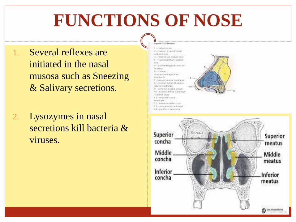
FUNCTIONS OF NOSE
1. Several reflexes are
initiated in the nasal
musosa such as Sneezing
& Salivary secretions.
2. Lysozymes in nasal
secretions kill bacteria &
viruses.

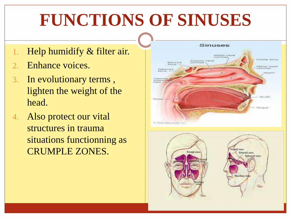
FUNCTIONS OF SINUSES
1. Help humidify & filter air.
2. Enhance voices.
3. In evolutionary terms ,
lighten the weight of the
head.
4. Also protect our vital
structures in trauma
situations functionning as
CRUMPLE ZONES.


FUNCTIONS OF PHARYNX
1. Helps to provide immunity
& formation of Antibodies.
2. Protects the lower
respiratory tract.
3. Forms Plasma Cells &
lymphocytes.
4. Acts as a warning to the
body against infectious
agents in air & food.
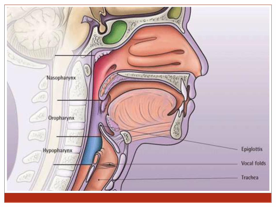


FUNCTIONS OF LARYNX
1. Acts as a Respiratory passage
& air flow regulator.
2. Helps in phonation & speech.
3. Protects the lower respiratory
passage.
4. Fixation of chest as in
climbing & digging.
5. Initiates cough reflex.
6. Helps in promoting venous
return.


FUNCTIONAL ANATOMY-1
TRACHEA :-
Adult- 18mm in dia
11cm in length
Lined with Columnar Ciliated Epithelium.
Divided at the level of Carina (T4) into –
1) Left Major Bronchi
2) Right Major Bronchi

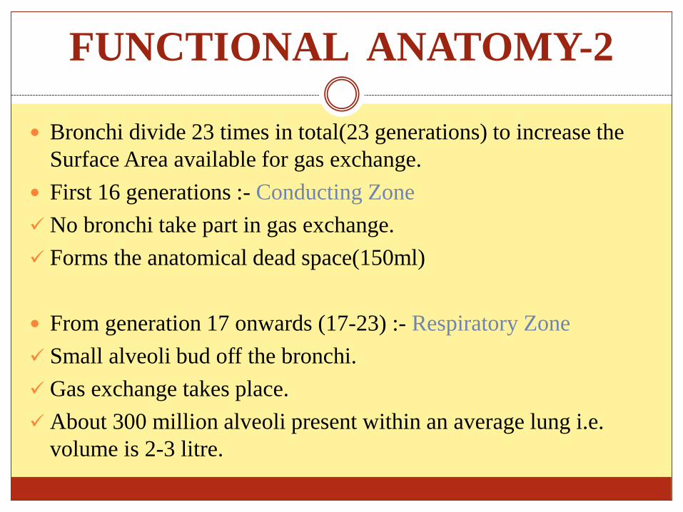
FUNCTIONAL ANATOMY-2
Bronchi divide 23 times in total(23 generations) to increase the
Surface Area available for gas exchange.
First 16 generations :- Conducting Zone
No bronchi take part in gas exchange.
Forms the anatomical dead space(150ml)
From generation 17 onwards (17-23) :- Respiratory Zone
Small alveoli bud off the bronchi.
Gas exchange takes place.
About 300 million alveoli present within an average lung i.e.
volume is 2-3 litre.
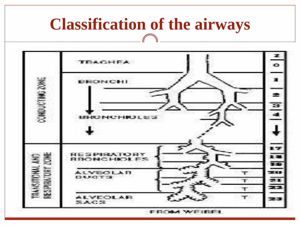
Classification of the airways

CELL TYPE & ITS FUNCTIONS
TYPE I ALVEOLAR CELLS :-
Derived from type II alveolar cells.
Provide a thin layer of Cytoplasm.
Covers 80% of the gas exchange zone.
TYPE II ALVEOLAR CELLS :-
Allow the formation of Surfactant & other enzymes.
TYPE III ALVEOLAR CELLS :-
Main lung defence system.
Alveolar macrophages.

SURFACTANT
Stored in the lamellar bodies of type II Alveolar cells.
Made up of a mixture of phospholipids, plasma proteins, &
Carbohydrate.
An amphipathic molecule with a charged hydrophilic head &
hydrophobic tail.

FUNCTIONS OF SURFACTANT
1. Reduces surface tension within the alveoli which helps
to increase the compliance of the lung.
2. Improves alveolar stability.
3. Keeps alveoli dry by opposing water movement from
the pulmonary interstitium

LUNG VOLUMES-1
Used to give information about diagnosis & progression of disease processes.
A guide to patients’ respiratory reserve.
Measured using basic spirometry at B,T,P,S (Body Temperature and Standard Pressure) using a Wright’s Respirometer.
LUNG VOLUMES(CONT.)-2
Vary with age, sex, height and weight.
Formulated into nomograms.
Residual volume RV - Volume of gas remaining in lungs after a forced expiration (15-20ml/kg)
Expiratory reserve volume ERV – Volume of gas forcefully expired after normal tidal expiration (15ml/kg)
Tidal volume TV – Volume of gas inspired and expired during normal breathing (8-10ml/kg)
LUNG VOLUMES(CONT.)-3
Inspiratory reserve volume IRV – Volume of gas inspired over normal tidal inspiration (45ml/kg)
Any 2 or more volumes added together = a capacity
Total lung capacity – volume of gas in lungs at the end of maximal inspiration (80ml/kg)
Vital Capacity – sum of IRV, TV and ERV (60-70ml/kg)
Functional residual capacity – Sum of ERV and RV (30ml/kg)

Basic spirometry trace of lung volumes
and capacities

FUNCTIONAL RESIDUAL CAPACITY-1
FRC = ERV + RV
The balance between the tendency of the chest wall to spring
outwards & tendency of the lung to collapse.
Is not the same volume all the time.
Can be disrupted by many factors.
FUNCTIONAL RESIDUAL CAPACITY-2
Factors decreasing FRC :-
1) Age
2) Posture – supine position
3) Anaesthesia – muscle relaxants
4) Surgery - Laparoscopic
5) Pulmonary fibrosis
6) Pulmonary oedema
7) Obesity
8) Abdominal Swelling
9) Reduced muscle tone – Reduced diaphragm tone will reduce pull away from the lungs
10) Pregnancy – Increased abdominal pressure.
FUNCTIONAL RESIDUAL CAPACITY-3
Factors increasing FRC :-
Increasing height of patient
Erect position – diaphragm and abdominal organs less able to
encroach upon bases of the lungs
Emphysema – decreased elastic recoil of lung therefore less
tendency of lung to collapse
Asthma – air trapping.

MEASUREMENT OF FRC-1
A spirometer is unable to measure TLC, FRC and RV.
There are 2 methods are used to measure FRC:-
1) Helium dilution
The patient is connected to a spirometer containing a known volume of gas (V1) with a known concentration of helium (C1)
Helium is not metabolised by the body.
The patient begins to breathe normally.
The helium concentration will change as the helium gets diluted in a larger volume of gas due to the patient’s lung volume. (V1 + FRC)
MEASUREMENT OF FRC-2
1) Helium Dilution
The final helium concentration is measured.
The value for FRC can then be derived since 3 of the 4 values are known.
C1 X V1 = C2 x (V1 +V2)
V2 = V1 (C1-C2)/C2
This method measures the volume of gas in the lungs which is participating in gas exchange. It will underestimate the FRC if there are significant areas of gas trapping.
MEASUREMENT OF FRC-3
2) Body Plethysmography
FRC is measured by placing the patient in a closed chamber and
measuring the pressure and volume changes occurring when the
subject makes an inspiratory effort against a closed airway.
Boyle’s gas law (P1V1=P2V2) is applied as ventilation takes
place to derive the FRC.
This technique also takes into account any gas trapped behind
closed airways e.g. in the case of patients with emphysematous
bullae. Helium dilution techniques do not calculate this.

CLOSING CAPACITY(CC)
The volume at which the small airways close during expiration.
Under normal circumstances the FRC is always greater than the
CC however if the FRC was to decrease then this would no
longer be the case and the small airways may close at the end of
normal tidal expiration. This leads to hypoxaemia, atelectasis and
worsening gas exchange due to increasing V/Q mismatch.
Closing capacity increases with age.
Typically closing capacity is equal to FRC at the age of 66 in the
erect position or 44 in the supine position.
PREOXYGENATION-1
The major oxygen store within the body is the Functional
Residual Capacity.
A typical volume for FRC is about 2.2 litres in an average
adult and normally contains 21% oxygen.
Since total body oxygen consumption is about 250mls per
minute this normal store of oxygen will only last just over 1
minute with apnoea.

PREOXYGENATION-2
Preoxygenation is defined as breathing 100% oxygen from a
close fitting mask for 3-5 minutes.
Breathing 100% oxygen for this time will denitrogenate the lungs
and increases the oxygen store to in excess of 1800mls thus
increasing the time to desaturation to about 7-8 minutes assuming
an oxygen consumption of 250mls/min.
PREOXYGENATION-3
FRC is determined by the balance between lung collapse and
chest wall springing outward
FRC affects oxygenation
FRC is the main oxygen store in the body and can be easily and
quickly enriched with oxygen
FRC will change due to a large number of factors.

VENTILATION
Total ventilation (MV) = VT x RR
With each tidal volume about a third the total amount of gas
flowing into the airway and lung does not participate in gas
exchange. This is the physiological dead space.
DEAD SPACE-1
Dead space can be defined as a volume of gas which does not
take part in gas exchange.
Dead space can be classified into 3 types :-
1. Anatomical dead space
This includes any breathing system or airway plus mouth, trachea
and the airways up until the start of the respiratory zone.
The typical volume in an adult is about 150mls.
DEAD SPACE-2
2. Alveolar dead space
This occurs when areas of the lung are being ventilated but not
being perfused and this leads to what is known as V/Q mismatch.
Large increases in alveolar dead space commonly occur in the
following conditions: pneumonia, pulmonary oedema, pulmonary
embolism
3. Physiological dead space
This is a combination of alveolar and anatomical dead space
added together.
Dead space is usually 30% of VT

MEASUREMENT OF DEAD SPACE-1
Fowlers method
This is used to measure anatomical dead space.
A patient takes a breath of 100% oxygen to rid the conducting
zone gases of nitrogen and then exhales through a mouthpiece
capable of analysing nitrogen concentration at the lips.
Initially the exhaled gases contain no nitrogen as this is dead
space gas.
MEASUREMENT OF DEAD SPACE-2
The nitrogen concentration will increase as the alveolar gases are
exhaled.
Nitrogen which is measured following the breath of 100%
oxygen must then have come only from gas exchanging areas of
the lung and not dead space.

MEASUREMENT OF DEAD SPACE-3
Initially expired nitrogen
concentration is plotted
against time as seen in top
graph.
To calculate the anatomical
dead space , expired nitrogen
is plotted against volume.
The upstroke of the curve is
equally divided in half to
give areas A and B. The
anatomical dead space equals
volume 0 up to and including
area B.
BOHR EQUATION-1
Measures physiological dead space.
A complicated equation.
Based upon the fact that all CO2 comes from alveolar gas and the
exhalation of CO2 can therefore be used to measure gas exchange
or lack of gas exchange if there is alveolar dead space (no
perfusion of these alveoli).
For each tidal volume there will be a proportion of dead space
(anatomical) but the amount of gas that is left over should take
part in gas exchange.
BOHR EQUATION-2
In order to derive the equation:-
FACO2- Alveolar CO2
FeCO2-CO2 from mixed expired gases
VT- Tidal volume
VD- Dead space volume (Physiological)
In an adult with normal lungs the value for VD/VT is between
0.20 and 0.35
The expiration of CO2 is calculated by either
VTCO2 = VA x FACO2
or
VTCO2 = VT x FeCO2

BOHR EQUATION-3
As we said above though with each breath there will be a
component of dead space, therefore:
VA = VT – VD
Therefore: (VT – VD) x FACO2 = VT x FeCO2
This can be expressed as:
VD/VT = (FACO2 – FeCO2)/FACO2
There is not really a truly ideal value for FACO2 that can be used
in the equation and therefore arterial pCO2 is used. This is
substituted into the equation giving:
VD/VT = (PaCO2 – PeCO2)/PaCO2
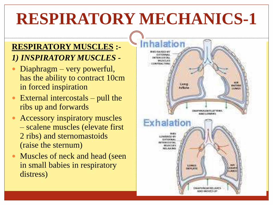
RESPIRATORY MECHANICS-1
RESPIRATORY MUSCLES :-
1) INSPIRATORY MUSCLES -
Diaphragm – very powerful, has the ability to contract 10cm in forced inspiration
External intercostals – pull the ribs up and forwards
Accessory inspiratory muscles – scalene muscles (elevate first 2 ribs) and sternomastoids(raise the sternum)
Muscles of neck and head (seen in small babies in respiratory distress)

RESPIRATORY MECHANICS-2
2) EXPIRATORY MUSCLES –
Expiration is usually passive and relies on the elastic recoil of the
lungs and the chest wall.
Under anaesthesia or extreme exercise,expiration may become
active due to the activation of abdominal muscles.
Muscles have their use in forced expiration.
Abdominal wall muscles – rectus abdominus, internal and
external oblique
Internal intercostal muscles – pull ribs down and inwards

COMPLIANCE-1
Compliance is defined as the volume change per unit pressure
change.
Usually expressed in mls/cmH2O
Compliance = ΔV/ΔP
Classified into chest wall, lung or total lung compliance.
(Distensibility)
Can be measured by inserting an oesophageal probe into a co-
operative patient.
The patient inhales and exhales to a set volume. At each volume
the intrapleural pressure is estimated using the oesophageal
probe.

COMPLIANCE-2
A pressure volume curve can then be plotted.
If during the measurement process no gas flow occurs at each set volume then this is static compliance. (Gas flow ceases and equilibration occurs).
If gas flow continues throughout measurement then this is dynamic compliance.
COMPLIANCE-3
Using a spirometer,certain fixed volumes can easily be measured
e.g. TLC, RV and FRC.
The pressure along the x axis is often plotted as the
transpulmonary pressure (Alveolar pressure – Intrapleural
pressure)
Initially,as can be seen from the above curve at lower lung
volumes,the compliance of the lung is poor and greater pressure
change is required to cause a change in volume. This occurs if the
lungs become collapsed for a period of time.
COMPLIANCE-4
At FRC,compliance is optimal since the elastic recoil of the lung
towards collapse is balanced by the tendency of the chest wall to
spring outwards.
At higher lung volumes,the compliance of the lung again
becomes less as the lung becomes stiffer.
COMPLIANCE-5
Expiration is deemed a passive process due to the elastic recoil of
the lung; because of this,the inspiratory curve is not identical to
the expiratory curve on a correctly drawn compliance curve. This
is known as hysteresis.
Compliance increases in old age and emphysema as elastic lung
tissue is destroyed.
Decreased in pulmonary fibrosis, pulmonary oedema, atelectasis
and in the extremes of lung volume.

WORK OF BREATHING
The energy required for the
work of breathing is mainly
used in the process of
inspiration as energy is
required to overcome airway
resistance, the elastic recoil
of the tissues and the chest
wall and tissue resistance.
The energy stored within the
elastic tissues is used to
provide for expiration.
CLINICAL POINTS
The ETT in an adult should lie 1-2cm superior to the carina
On an X-ray the carina is the point at which the trachea can be seen dividing into the right and left bronchi – around T4
The right major bronchus divides from the trachea at a much less acute angle than the left making it more prone to endobronchialintubation
The right upper lobe bronchus arises only a few centimeters from the carina therefore for one lung ventilation a left sided double lumen tube is favoured to avoid the risks of right upper lobe collapse with a right sided double lumen tube.
CLINICAL POINTS
The FRC is reduced by anaesthesia and therefore hypoxia is
common in a patient with a decrease in FRC.
The application of PEEP enables the FRC to remain greater then
CC and improves oxygenation
PEEP maintains the lungs on the steep part of the compliance
curve which lessens collapse at the bases of the lungs.
Preoxygenation should take place for 3-5 minutes or 4 vital
capacity breaths.
CLINICAL POINTS
Preoxygenation is essential in patients likely to have a decreased
FRC e.g. pregnant, obese.
Adequacy of preoxygenation can be assessed by monitoring end
tidal oxygen – aim for ETO2> 90%.
Patients using their accessory muscles may indicate increased
work of breathing.
PEEP can help to maintain the lung at FRC.
If used correctly CPAP can reduce the work of breathing by
increasing FRC.

SUMMARY & CONCLUSION
The conducting zone begins at the mouth and continues until the
16th generation.This zone transports gas but plays no part in gas
exchange.
The respiratory zone continues from the 17th until the 23rd
generation and is the region where gas exchange occurs.
Spirometry can be used to provide values for all basic lung
volumes except TLC, RV and FRC.

SUMMARY & CONCLUSION
Approximately 1/3 of VT is dead space
There are 3 types of dead space: Anatomical, Physiological and
Alveolar
Physiological dead space is a combination of alveolar and
anatomical dead space
Anatomical dead space is about 150mls in an average adult and is
measured by fowlers method
Physiological dead space is measured using the Bohr equation

SUMMARY & CONCLUSION
Minimise dead space by using the correct sized equipment e.g
HME, breathing circuits.
Consider the application of PEEP to prevent atelectasis and V/Q
mismatch.
Expiration is normally a passive process.
Compliance is the change in volume per unit change in pressure.
Compliance can be dynamic or static depending on whether the
gas flow is continuing or allowed to equilibriate during pressure
measurements.

SUMMARY & CONCLUSION
A spirometer cannot measure TLC,FRC or RV.
Compliance of the lungs is poor at very low or very high lung
volumes.
Compliance is optimal at or just above FRC.

.
THANK YOU


