
Jour
nal o
f Cel
l Sci
ence
RESEARCH ARTICLE
Notch directly regulates the cell morphogenesis genes Reck, talinand trio in adult muscle progenitors
Guillaume Pezeron*, Kat Millen, Hadi Boukhatmi and Sarah Bray`
ABSTRACT
There is growing evidence that activation of the Notch pathway can
result in consequences on cell morphogenesis and behaviour, both
during embryonic development and cancer progression. In general,
Notch is proposed to coordinate these processes by regulating
expression of key transcription factors. However, many Notch-
regulated genes identified in genome-wide studies are involved in
fundamental aspects of cell behaviour, suggesting a more direct
influence on cellular properties. By testing the functions of 25 such
genes we confirmed that 12 are required in developing adult
muscles, consistent with roles downstream of Notch. Focusing on
three, Reck, rhea/talin and trio, we verify their expression in adult
muscle progenitors and identify Notch-regulated enhancers in each.
Full activity of these enhancers requires functional binding sites for
Su(H), the DNA-binding transcription factor in the Notch pathway,
validating their direct regulation. Thus, besides its well-known roles
in regulating the expression of cell-fate-determining transcription
factors, Notch signalling also has the potential to directly affect cell
morphology and behaviour by modulating expression of genes such
as Reck, rhea/talin and trio. This sheds new light on the functional
outputs of Notch activation in morphogenetic processes.
KEY WORDS: Notch, Reck, Talin, Trio, Gene regulation,
Myogenesis, Drosophila
INTRODUCTIONNotch signalling is a local cell communication mechanism highlyconserved throughout the animal kingdom. It is implicated in avariety of developmental and physiological processes and
aberrant Notch activity is linked to many different diseases,including cancers and neurodegenerative disorders (Bolos et al.,2007; Louvi and Artavanis-Tsakonas, 2012). The Notch family of
receptors and the Notch ligands, Delta and Serrate (Jagged invertebrates), are cell surface type I transmembrane proteins.Upon ligand binding, Notch receptors undergo two proteolytic
cleavages that lead to the release of the Notch intracellulardomain (NICD). NICD is the active form of the receptor and actsin the nucleus as a transcriptional regulator, in cooperation with
the DNA-binding protein CSL [also known as CBF1, Su(H),LAG-1] and its co-activator Mastermind (Bray, 2006). Thus,transduction of Notch signalling is relatively simple and primarily
results in the regulation of target genes. The identification andcharacterization of Notch target genes is therefore crucial to fullyunderstand the function of Notch in developmental processes.
The best characterized target genes of Notch encodetranscription factors of the HES/E(spl) and Hey/Hesr/Herp genefamilies (Iso et al., 2003) but more-recent genome-wide studies
have uncovered a broader spectrum of Notch-regulated genes(Djiane et al., 2012; Hurlbut et al., 2009; Krejcı et al., 2009;Mazzone et al., 2010; Terriente-Felix et al., 2013; Wang et al.,
2011). Among these are genes directly involved in cell shape, cellorganization and cell behaviour, whose functional relevancedownstream of Notch has not been explored, despite the fact that
Notch has been implicated in different morphogenetic processesindependently of cell fate. These include the formationof boundaries between different cell populations, such as during
somitogenesis and in the Drosophila wing imaginal disc (Becamand Milan, 2008; Major and Irvine, 2006), cell migration(Schober et al., 2005; Wang et al., 2007) and axon guidance[although the latter may involve a non canonical pathway (Le
Gall et al., 2008)]. In the majority of contexts, this regulation ofmorphogenesis involves a transcriptional hierarchy, whereactivation of Notch results in the expression of a key
transcription factor that in turn coordinates the cell behaviours(Niessen et al., 2008; Saad et al., 2010; Schober et al., 2005;Wang et al., 2007). However, it remains plausible that, in some
tissues, Notch activity might have a more direct role in co-ordinating the genes that implement cell shape changes, althoughthere is as yet little evidence to support this.
One context where Notch is required for regulating thebehaviour of a specified group of cells is in the adult muscleprogenitors (AMPs) in Drosophila. As in mammals, Notch
activity is required to prevent premature differentiation of theAMPs, which are specified in the embryo and ultimately give riseto the adult muscles of the fly. (Anant et al., 1998; Delfini et al.,
2000; Hirsinger et al., 2001). During larval stages, theseprogenitors proliferate to expand the pool of myoblasts andremain associated with imaginal discs (Bate et al., 1991). Then, at
the beginning of metamorphosis the AMPs detach from theepithelium of the imaginal discs and migrate as a ‘swarm’ ofassociated cells (Roy and VijayRaghavan, 1998). At the target
sites, myoblasts fuse with templates formed either from foundermyoblasts or, in a few cases, from persistent larval muscles. Indoing so, the fusing cells contribute to the differentiating muscleso that it achieves the appropriate size and structure. (Fernandes
et al., 1991). Thus, adult myogenesis is a complex morphogeneticprocess, involving proliferation, migration, cell fusion anddifferentiation. As Notch signalling is active both in the
proliferating AMPs, where it inhibits differentiation, and in thesemi-differentiated migrating myoblasts (Bernard et al., 2006;Gildor et al., 2012), it could regulate many aspects of this
morphogenetic process. Indeed, a significant fraction of the genes
Department of Physiology Development and Neuroscience, University ofCambridge, Downing Street, Cambridge CB2 3DY, UK.*Present address: CNRS UMR 7622, UPMC Universite Pierre et Marie Curie,9 Quai Saint Bernard, Boite 24, F-75005, Paris, France.
`Author for correspondence ([email protected])
Received 22 February 2014; Accepted 18 August 2014
� 2014. Published by The Company of Biologists Ltd | Journal of Cell Science (2014) 127, 4634–4644 doi:10.1242/jcs.151787
4634

Jour
nal o
f Cel
l Sci
ence
directly regulated by Notch in DmD8 cells encode cytoskeletalregulatory proteins. DmD8 cells are related to AMPs and in
particular express the transcription factor Twist, which has beenshown to function as a cooperating transcriptional activator formany Notch target genes (Bernard et al., 2010; Krejcı et al.,2009). This suggests that Notch could have a direct role in co-
ordinating genes that control cell behaviours in this setting.Among 25 putative Notch-regulated genes encoding cell-
morphogenesis-related proteins that were identified through
genome-wide studies (Djiane et al., 2012; Krejci and Bray,2007; Terriente-Felix et al., 2013), we found that 12 are essentialfor generating adult flies capable of flight, consistent with
appropriate functions in muscle formation. Focusing on three,Reck, rhea/talin (hereafter referred to as talin) and trio, wedemonstrate that they are expressed in adult muscle progenitors
and exhibit Notch regulation. Thus, these data support the modelthat Notch activity has the potential to directly regulate genes thatcoordinate cell morphology, in addition to its more widelyaccepted role in regulating such characteristics through cell-fate-
determining transcription factors.
RESULTSIdentification of Notch target genes involved inadult myogenesisWe first selected a set of genes with known or inferred function in
the control of cell shape, organization and behaviour fromgenome-wide datasets documenting genes associated with Su(H)-bound regions in response to increased Notch signalling in
cultured cells and in wing imaginal discs (Djiane et al., 2012;Krejcı and Bray, 2007; Terriente-Felix et al., 2013). Such genesencode different types of proteins including Rho or Rac GTPaseexchange factors (e.g. trio), cytoskeleton-binding proteins, a
Netrin receptor implicated in cell migration and axon guidance(unc-5; Keleman and Dickson, 2001) and the matrixmetalloproteinase inhibitor Reck, an inhibitor of cell invasion
in cancer (Takahashi et al., 1998) (Table 1). To test their functionin adult myogenesis, a morphogenetic process where Notchsignalling is implicated in both cell fate and cell migration (Anant
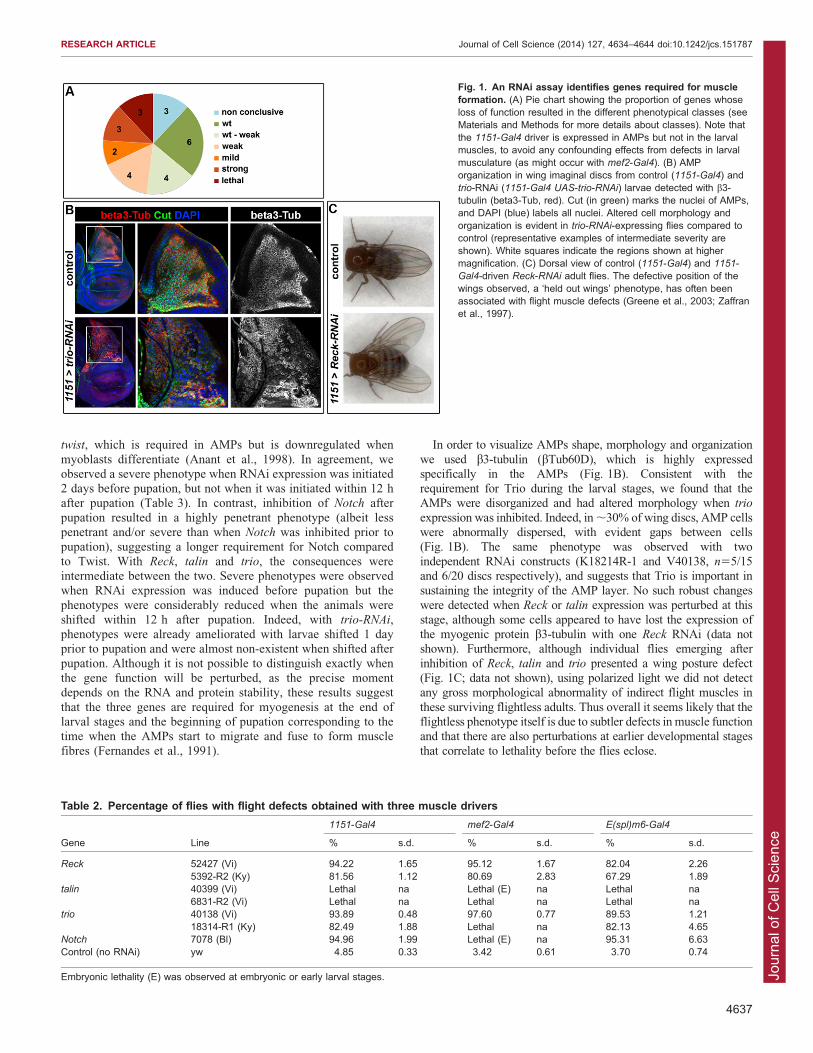
et al., 1998; Gildor et al., 2012), expression of hairpin RNAstargeting the individual genes was directed to the AMPs (using1151-Gal4 as a driver). Progeny from these crosses were assayedfor their ability to fly, as an indication of muscle formation and/or
function, and all genes were categorized according to thepercentage of flight-deficient individuals observed for each line(Fig. 1A; Table 1; supplementary material Table S1). This
approach had the advantage that gene expression was knockeddown at all post-embryonic stages of AMP development, fromtheir proliferation in the larvae through to the forming muscles in
pupae, but was not disrupted in the larval muscles nor in theirembryonic progenitors (Anant et al., 1998). Finally, to avoid falsepositives from off-target effects or from side effects of RNA
interference (RNAi) insertions, where possible we used linesfrom different sources that contained different types of RNAiconstructs.
In most cases (18 out of 25) the results were consistent between
different RNAi lines targeting the same gene (supplementarymaterial Table S1). For example, lines targeting Reck and trio allinduced a high percentage of flightless flies. However, four genes
(ena, GEFmeso, singed and CG6891) were classified as ‘wt–weak’ since the results were inconclusive due to variabilitybetween lines, although overall there was little indication of any
requirement in myogenesis. Furthermore, for three genes (Klar,
wb and Cdep) the results were contradictory because one RNAiline induced a severe phenotype, whereas other lines targeting the
same gene had no or very little effect. Such differences could bedue to differences in the depletion efficiencies or to the inhibitionof off target genes, but we have not pursued those genes further.
In total, knockdown of 12 genes gave consistent phenotypes
suggesting that they are required for myogenesis (Fig. 1A;Table 1; supplementary material Table S1). Three of these(chic, talin and sls) gave lethality at the pharate stage when
inhibited. This is consistent with a deficiency in the adultmuscles, and is consistent with previous data showing that talin
and sls have roles in myogenesis (Brown et al., 2002; Burkart
et al., 2007). Furthermore, in a few instances the adults werefound half emerged from the pupal case and, very occasionally aviable flightless fly eclosed. This suggests that the observed
lethality was, at least in part, due to the inability to emerge fromthe puparium rather than to a major developmental defect. Inagreement, one of the chic lines gave weaker phenotypes,generating adult flies of which 50% were flightless.
Inhibition of nine other genes, including corn and unc-5,resulted in flight defects with variable penetrance. In all cases theproportion of the progeny that were flightless varied, ranging
from completely flightless flies to weak fliers. These appeared torepresent a continuum in the severity of the same phenotype,because flightless flies were only observed when the progeny had
a high percentage of flight defects, whereas weak fliers wereoften observed along with normal fliers. Nevertheless theseincluded good candidates to mediate effects from Notch. For
example both corn and unc-5 have been shown to be expressed inAMPs, and unc-5 contains a Notch-responsive enhancer (Krejcıet al., 2009).
A previous genome-wide RNAi screen to identify genes with
function in muscle formation (Schnorrer et al., 2010) differedfrom our assay by using mef2-Gal4 as the driver, which isexpressed in the muscle lineage from embryonic stages onwards.
Despite this difference, the results are broadly consistent(Table 1). Only two (NijA and cher) of the ten genes with noor little effects in our assay were found to give a muscle
phenotype with the more widespread depletion used in theSchnorrer et al. study. Likewise, among the six genes with astrong phenotype in our analysis, five had been found to berequired for correct muscle formation in the previous screen
(Schnorrer et al., 2010). Notably, three genes (chic, talin and sls)whose depletion induced lethality at pharate stage in our assay,gave embryonic or early larval lethality when inhibited from
embryonic stage onwards so their role in later development wasnot considered in the previous study.
Reck, talin and trio are required in AMP lineagesThree genes with a penetrant phenotype obtained with all testedRNAi lines, Reck, talin and trio, were selected for further analysis
because they had been shown to be associated with Su(H)-boundregions in response to increased Notch signalling in the AMP-related DmD8 cells (Table 1). In addition, they are well-conserved between Drosophila and vertebrates and have been
previously linked to muscle formation: Reck, a metalloproteinaseinhibitor, is expressed in developing muscles in the mouse whereits expression is regulated by myogenic factors (Echizenya et al.,
2005); Talin, a key component of integrin-mediated cell adhesionis involved at different stages of myogenesis in Drosophila
(Brown et al., 2002); and Trio, a GTPase exchange factor (GEF)
has a role in muscle formation in mammals, where it activates
RESEARCH ARTICLE Journal of Cell Science (2014) 127, 4634–4644 doi:10.1242/jcs.151787
4635
Jour
nal o
f Cel
l Sci
ence
Rac1 to promote myoblast fusion (Charrasse et al., 2007; O’Brienet al., 2000).
Because 1151-Gal4 expression is not specifically restricted toAMPs [for example expression occurs in the salivary gland andsome neural cells (Anant et al., 1998)], it was possible that the
observed phenotypes were due to Reck, talin and trio inhibition intissues other than AMPs. We thus first confirmed the requirementof these genes during adult muscle formation by using two otherdrivers, E(spl)m6-Gal4 and mef2-Gal4, to direct RNAi expression
in AMPs (supplementary material Fig. S1). Although smalldifferences were observed in the penetrance obtained with thethree drivers (likely due to different expression levels and
timing), overall the phenotypes observed for all three genes werevery similar (Table 2). As expected, one line targeting talin
induced lethality at embryonic or early larval stage when driven
by mef2-Gal4. Given that mef-2-Gal4 is also expressed inembryonic and larvae muscles, these results are consistent with
the requirement for talin during embryogenesis (Brown et al.,2002; Schnorrer et al., 2010). Similar lethal phenotypes wereobtained when Notch-RNAi was expressed under the same
conditions. Thus, these results confirmed that Reck, talin andtrio are required in the myogenic lineages.
Next, we sought to determine at what stage the three genes arerequired. To do so, we combined UAS-RNAi lines targeting Reck,
talin and trio with the 1151-Gal4 driver in the presence of athermo-sensitive derivative of Gal80 (Gal80ts), the Gal4 inhibitor(McGuire et al., 2003). RNAi expression was then induced at
different stages by incubating larvae at temperatures whereGal80ts was inactivated. As a control, the experiment was firstperformed with an RNAi line targeting the transcription factor
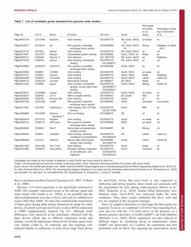
Table 1. List of candidate genes selected from genome wide studies
FBgn ID CG Id Name Function GO term Su(H)
Phenotypeaccordingto ourassay
Phenotype accord-ing to Schnorreret al.
FBgn0001316 CG17046 klarsicht Actin binding GO:0003779 D8, Su(H), NICD,Kc
Uncertain wt
FBgn0024277 CG18214 trio Rho guanine nucleotideexchange factor activity
GO:0005089 D8, Su(H), NICD,Kc
Strong Flightless or lethal(AP)
FBgn0014133 CG1822 bifocal Actin binding GO:0003779 D8, Su(H), NICD wt ndFBgn0011661 CG10701 Moesin Cytoskeletal protein binding GO:0008092 Su(H), NICD, Kc Weak wtFBgn0036101 CG6449 Ninjurin A Cell adhesion GO: 0007155 D8, Su(H), NICD wt FlightlessFBgn0013726 CG8705 peanut Actin binding, microtubule
bindingGO:0003779,
GO:0008017D8, Su(H), NICD wt nd
FBgn0035802 CG33275 CG33275 Rho guanine nucleotideexchange factor activity
GO:0005089 Su(H), NICD wt wt
FBgn0030955 CG6891 CG6891 Actin binding GO:0003779 Su(H), NICD wt–weak ndFBgn0014141 CG3937 cheerio Actin binding GO:0003779 Su(H), NICD Weak FlightlessFBgn0000308 CG9553 chickadee Actin binding GO:0003779 Su(H), NICD Lethal Lethal (E)FBgn0259173 CG42278 cornetto Microtubule binding GO:0008017 D8, NICD Weak wtFBgn0011225 CG5695 jaguar Actin binding, microtubule
binding, myosin light chainbinding
GO:0003779,GO:0008017,GO:0032027
Su(H), NICD Strong Lethal (P)
FBgn0003447 CG32858 singed Actin binding GO:0003779 Su(H), NICD wt–weak wtFBgn0034013 CG8166 unc-5 Netrin receptor activity GO:0005042 D8, Su(H) Mild wtFBgn0000083 CG5730 Annexin B9 Actin binding GO:0003779 Su(H) wt wtFBgn0051536 CG31536 Cdepa Rho guanine nucleotide
exchange factor activityGO:0005089 Su(H) Uncertain Locomotion
FBgn0011202 CG1768 diaphanous Actin binding, Rho GTPasebinding
GO:0003779,GO:0017048
Su(H) Mild wt
FBgn0260866 CG12489 defense
repressor 1
Zinc ion binding GO:0008270 D8 wt wt
FBgn0000578 CG15112 enabled Actin binding GO:0003779 Su(H) wt–weak wtFBgn0050115 CG30115 GEFmeso Rho guanyl-nucleotide
exchange factor activityGO:0005089 NICD wt–weak nd
FBgn0036463 CG5392 Recka Serine-type endopeptidaseinhibitor activity
GO:0004867 D8 Strong nd
FBgn0260442 CG6831 rhea/talin Actin binding, structuralconstituent of cytoskeleton
GO:0003779,GO:0005200
D8 Lethal Lethal (L)
FBgn0086906 CG1915 sallimus Myosin light chain kinaseactivity, actin binding
GO:0004687,GO:0003779
Su(H) Lethal Lethal (E)
FBgn0051352 CG31352 Unc-115a Actin binding GO:0003779 Su(H) Weak wtFBgn0261563 CG42677 wing blister Receptor binding, regulation
of cell adhesion, cellmigration
GO:0005102,GO:0030155,GO:0030334
Su(H) Uncertain wt
Candidates are sorted by the number of datasets in which Su(H) was found bound to their loci.aCdep, Chondrocyte-derived ezrin-like domain containing protein; Reck, Reversion-inducing-cysteine-rich protein with kazal motifs.
Su(H) column: D8, DmD8 cells (Krejcı et al., 2009); Su(H) and NICD, wing imaginal discs overexpressing Su(H) and NICD respectively (Djiane et al., 2012); Kc,Kc cells (Terriente-Felix et al., 2013). The phenotypical category obtained in our assay and the phenotype observed by Schnorrer et al. (Schnorrer et al., 2010)are indicated. wt, wild type; nd, not determined; AP, Adult-pharate, E, embryonic, L, Larval, P: pharate.
RESEARCH ARTICLE Journal of Cell Science (2014) 127, 4634–4644 doi:10.1242/jcs.151787
4636
Jour
nal o
f Cel
l Sci
ence
twist, which is required in AMPs but is downregulated when
myoblasts differentiate (Anant et al., 1998). In agreement, weobserved a severe phenotype when RNAi expression was initiated2 days before pupation, but not when it was initiated within 12 hafter pupation (Table 3). In contrast, inhibition of Notch after
pupation resulted in a highly penetrant phenotype (albeit lesspenetrant and/or severe than when Notch was inhibited prior topupation), suggesting a longer requirement for Notch compared
to Twist. With Reck, talin and trio, the consequences wereintermediate between the two. Severe phenotypes were observedwhen RNAi expression was induced before pupation but the
phenotypes were considerably reduced when the animals wereshifted within 12 h after pupation. Indeed, with trio-RNAi,phenotypes were already ameliorated with larvae shifted 1 dayprior to pupation and were almost non-existent when shifted after
pupation. Although it is not possible to distinguish exactly whenthe gene function will be perturbed, as the precise momentdepends on the RNA and protein stability, these results suggest
that the three genes are required for myogenesis at the end oflarval stages and the beginning of pupation corresponding to thetime when the AMPs start to migrate and fuse to form muscle
fibres (Fernandes et al., 1991).
In order to visualize AMPs shape, morphology and organization
we used b3-tubulin (bTub60D), which is highly expressedspecifically in the AMPs (Fig. 1B). Consistent with therequirement for Trio during the larval stages, we found that theAMPs were disorganized and had altered morphology when trio
expression was inhibited. Indeed, in ,30% of wing discs, AMP cellswere abnormally dispersed, with evident gaps between cells(Fig. 1B). The same phenotype was observed with two
independent RNAi constructs (K18214R-1 and V40138, n55/15and 6/20 discs respectively), and suggests that Trio is important insustaining the integrity of the AMP layer. No such robust changes
were detected when Reck or talin expression was perturbed at thisstage, although some cells appeared to have lost the expression ofthe myogenic protein b3-tubulin with one Reck RNAi (data notshown). Furthermore, although individual flies emerging after
inhibition of Reck, talin and trio presented a wing posture defect(Fig. 1C; data not shown), using polarized light we did not detectany gross morphological abnormality of indirect flight muscles in
these surviving flightless adults. Thus overall it seems likely that theflightless phenotype itself is due to subtler defects in muscle functionand that there are also perturbations at earlier developmental stages
that correlate to lethality before the flies eclose.
Fig. 1. An RNAi assay identifies genes required for muscleformation. (A) Pie chart showing the proportion of genes whoseloss of function resulted in the different phenotypical classes (seeMaterials and Methods for more details about classes). Note thatthe 1151-Gal4 driver is expressed in AMPs but not in the larvalmuscles, to avoid any confounding effects from defects in larvalmusculature (as might occur with mef2-Gal4). (B) AMPorganization in wing imaginal discs from control (1151-Gal4) andtrio-RNAi (1151-Gal4 UAS-trio-RNAi) larvae detected with b3-tubulin (beta3-Tub, red). Cut (in green) marks the nuclei of AMPs,and DAPI (blue) labels all nuclei. Altered cell morphology andorganization is evident in trio-RNAi-expressing flies compared tocontrol (representative examples of intermediate severity areshown). White squares indicate the regions shown at highermagnification. (C) Dorsal view of control (1151-Gal4) and 1151-
Gal4-driven Reck-RNAi adult flies. The defective position of thewings observed, a ‘held out wings’ phenotype, has often beenassociated with flight muscle defects (Greene et al., 2003; Zaffranet al., 1997).
Table 2. Percentage of flies with flight defects obtained with three muscle drivers
1151-Gal4 mef2-Gal4 E(spl)m6-Gal4
Gene Line % s.d. % s.d. % s.d.
Reck 52427 (Vi) 94.22 1.65 95.12 1.67 82.04 2.265392-R2 (Ky) 81.56 1.12 80.69 2.83 67.29 1.89
talin 40399 (Vi) Lethal na Lethal (E) na Lethal na6831-R2 (Vi) Lethal na Lethal na Lethal na
trio 40138 (Vi) 93.89 0.48 97.60 0.77 89.53 1.2118314-R1 (Ky) 82.49 1.88 Lethal na 82.13 4.65
Notch 7078 (Bl) 94.96 1.99 Lethal (E) na 95.31 6.63Control (no RNAi) yw 4.85 0.33 3.42 0.61 3.70 0.74
Embryonic lethality (E) was observed at embryonic or early larval stages.
RESEARCH ARTICLE Journal of Cell Science (2014) 127, 4634–4644 doi:10.1242/jcs.151787
4637

Jour
nal o
f Cel
l Sci
ence
Reck, talin and trio are expressed in AMP cellsGiven the implication that Reck, talin and trio are required in
AMPs for normal development, we assessed whether they areexpressed in these cells, as predicted. Although their overallpatterns differed, all three genes were expressed in AMPs.
Expression of Reck was largely restricted to the AMPs(Fig. 2A,B; supplementary material Fig. S2, for comparison,see expression of the AMP markers Cut and Mef2 in Fig. 2D,H)
where it was present at all stages examined, including young thirdinstar (L3) larvae (Fig. 2A) and early pupae (Fig. 2B).Interestingly the expression levels of Reck seemed to correlatewith the AMP maturation process, as higher expression levels
were detected at the onset of pupation. In addition, Reck was alsodetected in the peripodial margin, possibly reflecting a function indisc eversion (Srivastava et al., 2007). In contrast, Talin was
broadly expressed throughout the wing disc, with a fairlyubiquitous pattern, as described previously (Brown et al.,2002). However, there was also a clear accumulation in the
AMPs, making it plausible that there could be specific regulationof talin in these cells (Fig. 2C,D; supplementary material Fig.S2). Similarly, Trio protein appeared to be expressed widely
throughout the wing disc, including in the AMPs (Fig. 2E;supplementary material Fig. S2). Furthermore, in situ
hybridizations suggested some differential regulation of trio,with higher expression in the notal region and AMPs (Fig. 2F).
Similar enrichment in the AMPs was detected using a trio
‘enhancer trap’ line [corresponding to a P-element insertion at thetranscription start of the longest transcript of trio (Bateman et al.,
2000)] and this high level of LacZ reporter expression wasfound to colocalize with the AMP marker Mef2 (Fig. 2G,H;
supplementary material Fig. S2). Thus, Reck, talin and trio are allexpressed in AMPs, consistent with their proposed function inadult myogenesis.
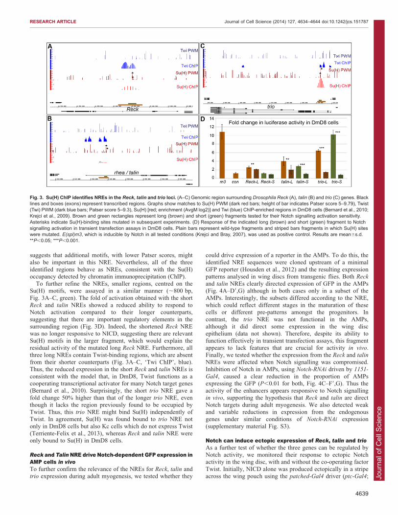
Notch regulated enhancers from Reck, talin and trioReck, talin and trio have previously been identified in genome-
wide studies as putative direct targets regulated by Notchsignalling in the muscle lineage (Krejci et al., 2009). To verifythis, we first tested whether the regions occupied by Su(H) inthese three loci can function as Notch-responsive enhancers
(NREs). DNA fragments corresponding to these Su(H)-boundregions (Fig. 3A–C, brown) were cloned upstream of a minimalluciferase reporter and their ability to respond to the activated
form of Notch (NICD) was analysed in co-transfection assaysusing the DmD8, Twist-expressing, cell line. All three enhancersgave increased luciferase expression in the presence of NICD in
DmD8 cells, and this response was compromised when theSu(H)-binding motifs were mutated [Fig. 3D; P,0.001 for trio
and P,0.05 for talin and Reck – in each case the two best matches
to the Su(H) position weight matrix (PWM) were mutated]. Of thethree, the NRE from the trio locus gave the highest fold change inreporter expression in the presence of NICD (6.5 for the trio NREversus 4.3 and 2.6 for the talin and Reck NRE, respectively) and
also gave the most dramatic reduction when the Su(H) motifs weremutated. In contrast, the consequences on the Reck NRE frommutating two Su(H) motifs was modest, although significant. This
Table 3. Percentage of flight defects (or lethality) resulting from the induction of RNAi transcription at different stages
L3a (3 dpf) L3b (4 dpf) P1–P3 Always at 18˚C
Gene Line % s.d. % s.d. % s.d. % s.d.
Reck 52427 (Vi) 100 na 100 na 34.25 4.93 19.71 2.42talin 40399 (Vi) lethal 100 na 100 na 57.75 7.37 0 na
flightless 0 0 42.25 4.94 1.85trio 40138 (Vi) 100 na 64.55 16.86 19.46 5.76 20.38 0.53Notch 7078 (Bl) 100 na 100 na 86.3 1.32 20.73 1.54twist See Wong et al., 2008. 100 na nd nd 4.26 0.25 4.13 0.3control (no RNAi) yw 3.3 1.22 6.64 1.08 2.89 1.14 2.85 1.51
Larvae combining the indicated UAS-RNAi, 1151-Gal4 and tub-Gal80ts were raised at 18˚C and shifted to 29˚C (allowing RNAi expression) at the indicatedstage. The observed percentage (%) of adults with flight defect (or lethality) and s.d. are indicated. dpf, days post fertilization; L3a, Early third-instar larval stage;L3b, late third-instar larval stage (wandering larvae); P1–P3, Pupal stages 1 to 3 (0–6 h after puparium formation).
Fig. 2. Reck, talin and trio areexpressed in AMP cells. Expressionprofiles of Reck, talin and trio in the wingimaginal disc show that all three genesare expressed in AMPs. (A,B) Reck in
situ hybridization (ish) in young L3 larvae(A) and P1 pupae (B). (C,D) talinexpression profile revealed byimmunostaining and co-stained with theAMP marker Cut (maximum projectionsof z-stacks from confocal acquisitions arepresented; D is shown at twice themagnification of C). (E–H) trio expressionprofile shown by immunostaining(E, Maximum projections), In situ
hybridization (F) and an enhancer-trapreporter line (G) also co-stained with theAMP marker mef2 (H) (H is shown attwice the magnification of G).
RESEARCH ARTICLE Journal of Cell Science (2014) 127, 4634–4644 doi:10.1242/jcs.151787
4638
Jour
nal o
f Cel
l Sci
ence
suggests that additional motifs, with lower Patser scores, might
also be important in this NRE. Nevertheless, all of the threeidentified regions behave as NREs, consistent with the Su(H)occupancy detected by chromatin immunoprecipitation (ChIP).
To further refine the NREs, smaller regions, centred on theSu(H) motifs, were assayed in a similar manner (,800 bp,Fig. 3A–C, green). The fold of activation obtained with the short
Reck and talin NREs showed a reduced ability to respond toNotch activation compared to their longer counterparts,suggesting that there are important regulatory elements in the
surrounding region (Fig. 3D). Indeed, the shortened Reck NREwas no longer responsive to NICD, suggesting there are relevantSu(H) motifs in the larger fragment, which would explain theresidual activity of the mutated long Reck NRE. Furthermore, all
three long NREs contain Twist-binding regions, which are absentfrom their shorter counterparts (Fig. 3A–C, ‘Twi ChIP’, blue).Thus, the reduced expression in the short Reck and talin NREs is
consistent with the model that, in DmD8, Twist functions as acooperating transcriptional activator for many Notch target genes(Bernard et al., 2010). Surprisingly, the short trio NRE gave a
fold change 50% higher than that of the longer trio NRE, eventhough it lacks the region previously found to be occupied byTwist. Thus, this trio NRE might bind Su(H) independently ofTwist. In agreement, Su(H) was found bound to trio NRE not
only in DmD8 cells but also Kc cells which do not express Twist(Terriente-Felix et al., 2013), whereas Reck and talin NRE wereonly bound to Su(H) in DmD8 cells.
Reck and TalinNRE drive Notch-dependent GFP expression inAMP cells in vivoTo further confirm the relevance of the NREs for Reck, talin andtrio expression during adult myogenesis, we tested whether they
could drive expression of a reporter in the AMPs. To do this, the
identified NRE sequences were cloned upstream of a minimalGFP reporter (Housden et al., 2012) and the resulting expressionpatterns analysed in wing discs from transgenic flies. Both Reck
and talin NREs clearly directed expression of GFP in the AMPs(Fig. 4A–D9,G) although in both cases only in a subset of theAMPs. Interestingly, the subsets differed according to the NRE,
which could reflect different stages in the maturation of thesecells or different pre-patterns amongst the progenitors. Incontrast, the trio NRE was not functional in the AMPs,
although it did direct some expression in the wing discepithelium (data not shown). Therefore, despite its ability tofunction effectively in transient transfection assays, this fragmentappears to lack features that are crucial for activity in vivo.
Finally, we tested whether the expression from the Reck and talin
NREs were affected when Notch signalling was compromised.Inhibition of Notch in AMPs, using Notch-RNAi driven by 1151-
Gal4, caused a clear reduction in the proportion of AMPsexpressing the GFP (P,0.01 for both, Fig. 4C–F9,G). Thus theactivity of the enhancers appears responsive to Notch signalling
in vivo, supporting the hypothesis that Reck and talin are directNotch targets during adult myogenesis. We also detected weakand variable reductions in expression from the endogenousgenes under similar conditions of Notch-RNAi expression
(supplementary material Fig. S3).
Notch can induce ectopic expression of Reck, talin and trioAs a further test of whether the three genes can be regulated byNotch activity, we monitored their response to ectopic Notchactivity in the wing disc, with and without the co-operating factor
Twist. Initially, NICD alone was produced ectopically in a stripeacross the wing pouch using the patched-Gal4 driver (ptc-Gal4;
Fig. 3. Su(H) ChIP identifies NREs in the Reck, talin and trio loci. (A–C) Genomic region surrounding Drosophila Reck (A), talin (B) and trio (C) genes. Blacklines and boxes (exons) represent transcribed regions. Graphs show matches to Su(H) PWM (dark red bars; height of bar indicates Patser score 5–9.79), Twist(Twi) PWM (dark blue bars; Patser score 5–9.3), Su(H) [red; enrichment (AvgM log2)] and Twi (blue) ChIP-enriched regions in DmD8 cells (Bernard et al., 2010;Krejci et al., 2009). Brown and green rectangles represent long (brown) and short (green) fragments tested for their Notch signalling activation sensitivity.Asterisks indicate Su(H)-binding sites mutated in subsequent experiments. (D) Response of the indicated long (brown) and short (green) fragment to Notchsignalling activation in transient transfection assays in DmD8 cells. Plain bars represent wild-type fragments and striped bars fragments in which Su(H) siteswere mutated. E(spl)m3, which is inducible by Notch in all tested conditions (Krejci and Bray, 2007), was used as positive control. Results are mean6s.d.**P,0.05; ***P,0.001.
RESEARCH ARTICLE Journal of Cell Science (2014) 127, 4634–4644 doi:10.1242/jcs.151787
4639

Jour
nal o
f Cel
l Sci
ence
Fig. 5A–F). However, none of the genes showed any markedchange in expression under these conditions. Subsequently,because many AMP Notch targets require the transcription
factor Twist to be induced by Notch (Bernard et al., 2010), wetested the effect of co-expressing NICD and Twist together and ofexpressing Twist alone. This combination induced ectopic
expression of all three genes within the ptc expression domain(Fig. 5J–L), whereas none was clearly upregulated when Twistwas expressed alone (Fig. 5G–I). This was even the case for Trio,
suggesting that Twist does cooperate with Notch at this target,despite the fact that the NRE binds Su(H) even in cells that lackTwist. Taken together, these results suggest that Reck, talin andtrio are regulated by Notch in vivo when Twist is present in the
same cells. Although this regulation is likely direct, based on theresults from the reporters, it remains possible that there are alsoindirect mechanisms involved.
DISCUSSIONNotch signalling is widely implicated in the control of cell fate
during development but also has been shown to influence cellarchitecture and behaviour in different morphogenetic processes.In most cases, Notch is proposed to coordinate cellmorphogenesis by regulating the expression of key transcription
factors, rather than by directly regulating the effector genes thatimplement the cell behaviours (Niessen et al., 2008; Saad et al.,2010; Schober et al., 2005; Wang et al., 2007). One well-
characterized example is epithelial-to-mesenchymal transition(EMT), a process that can be triggered by Notch signalling[sometimes in combination with other pathways such as TGFb(Espinoza and Miele, 2013; Wang et al., 2010)] through itsregulation of the key transcription factors Snail and Slug (Niessen
et al., 2008; Saad et al., 2010). Our results suggest, however, thatgenes involved in implementing cell morphology are also directlyregulated by Notch. Building on previous genome-wide analyses
of Notch-regulated genes, which revealed a wide spectrum offunctional targets, we have found that Reck, talin and trio all havesome characteristics of direct Notch targets in the muscle
progenitors. Genome-wide analysis of CSL (also known asRBPJ) binding in mouse and human T-lymphoblastic leukaemiacells also identified several genes implicated in cell architecture
regulation, although those differ from the genes analysed here(Wang et al., 2011). Direct control of genes with roles in co-ordinating cell morphology and behaviour might thus be a generalfeature of Notch activity in different morphogenetic processes.
Two of the three genes, trio and talin, are very widelyexpressed. Thus, a large proportion of their expression likelyoccurs independently of Notch. However, our identification of
Su(H)-responsive enhancers associated with each of these genessuggests that Notch activity can modulate their expression atspecific stages and/or in specific cell types. This therefore
highlights the existence of different categories of Notch-regulatedgenes. Most commonly the focus is on cell-fate-determininggenes, which are specifically switched on in a cell only whenNotch activity is present. The regulation of trio and talin suggests
that Notch activity also augments the expression of genes that arealready transcribed, modifying their expression rather thaninitiating it de novo. Such subtle changes of gene expression
would not have been uncovered by conventional approaches,demonstrating the utility of genome-wide studies in uncoveringthe full spectrum of target genes. In the AMPs, trio is important
for sustaining the normal cell morphology, and might do so byregulating the interaction of AMPs with their niche (Lin et al.,
Fig. 4. Reck and talin NREs drive Notch-dependent GFP expression in AMPs in vivo. (A–B9) Reck (A, a higher magnification is shown in A9) and talin
(B, and higher magnification in B9) long NREs (L-NRE) drive GFP expression in a subset of AMPs as shown by GFP colocalizations with the AMP marker Cut.White squares in A and B indicate regions shown at higher magnification in A9 and B9. Single optical sections from confocal acquisitions are presented.(C–F9) Expression from reporters Reck-L-NRE-GFP (C,C9,E,E9) and talin-L-NRE-GFP (D,D9,F,F9) in wild-type (C–D9) and Notch-depleted conditions (E–F9). Cutexpression was used to mark the AMPs. Green dotted lines in C9,D9,E9 and F9 outline Cut- and GFP-expressing cells. Maximum projections of z-stacks fromconfocal acquisitions are presented. (G) Boxplot representing the percentage of AMPs (Cut-expressing cells) expressing GFP from the Reck-L-NRE-GFP andtalin-L-NRE-GFP reporters in wild-type (CTRL) and Notch-depleted (N-RNAi) conditions. The percentage of AMPs expressing GFP was estimated by manuallymeasuring areas occupied by cells expressing Cut or Cut and GFP. The box represents the interquartile range, the middle line the median, and the whiskersshow 6 1.56 the interquartile range. ***P,0.001.
RESEARCH ARTICLE Journal of Cell Science (2014) 127, 4634–4644 doi:10.1242/jcs.151787
4640

Jour
nal o
f Cel
l Sci
ence
2013) or their transition to a migrating population at the onset of
pupation (Kashef et al., 2009; Moore et al., 2013). However, todistinguish the contribution that Notch regulation makes to trio
and talin functions it will ultimately be necessary to eliminate the
NREs from the endogenous genes.In contrast, Reck exhibits a much more restricted expression
pattern, being specifically upregulated in late-stage AMPs whereNotch is active. The identified NRE directs expression in these
cells, consistent with Reck expression being controlled by Notchactivity in AMPs. Intriguingly, in some mammalian cells (e.g.cortical progenitors,) expression of RECK has been found to
activate Notch signalling by directly inhibiting ADAM10-dependent processing of Notch ligands (Muraguchi et al.,2007). As the swarming myoblasts appear to undergo bi-
directional Notch signalling, it is possible that Reck could beinvolved in subtle fine-tuning of signalling between cells.However, so far there is no evidence to indicate that Reck can
inhibit Kuzbanian, the Drosophila Adam10 homologue. InsteadReck function has been linked to inhibition of the matrix
metalloproteinase MMP1, through its ability, in conjunction with
another metalloproteinase inhibitor, to suppress invasion oftumours that had upregulated MMP1 (Srivastava et al., 2007).Similar characteristics are well documented in mammals, where
RECK functions as a tumour suppressor by inhibiting migration,invasion, and angiogenesis (Meng et al., 2008; Nagini, 2012;Noda et al., 2003; Takahashi et al., 1998). Although we have notbeen able to detect any gross defects in the organization of
indirect flight muscles, suggesting that AMP migration is notseverely affected when Reck is knocked-down, as the RNA isonly upregulated in late stage AMPs it is more likely to be
involved in positively promoting myoblast migration, similar toits role in zebrafish neural crest cells (Prendergast et al., 2012),than in suppressing migration.
Besides the three genes whose regulation we have analysedin some details, at least nine others appear to be required inadult myogenesis, based on the phenotypes seen when their
expression in AMPs is ablated. Two of these, corn and unc-5,have been previously shown to be expressed in AMPs. For unc-5,
Fig. 5. Notch can induce ectopic expression of Reck, talin and trio in the presence of Twist. (A–L) ptc-Gal4; Tub-Gal80ts was used to drive expression ofNICD and/or Twist (Twi) in the wing pouch. The expression profile of Reck (A,D,G,J, in situ hybridization), talin (B,E,H,K, immunostaining) and trio (C,F,I,L,immunostaining) in a wild-type wing pouch (A–C), in NICD-expressing discs (D–F), Twist-expressing discs (G–I) and NICD- plus Twist-expressing discs (J–L).Note the ectopic expression of Reck (J, black arrows) and the upregulation of talin and trio (K,L, white arrows) induced by Notch in the presence of Twist. Cistaining was used to indicate the limit between anterior and posterior domains, along which ptc-Gal4 is expressed (indicated with orange lines).
RESEARCH ARTICLE Journal of Cell Science (2014) 127, 4634–4644 doi:10.1242/jcs.151787
4641

Jour
nal o
f Cel
l Sci
ence
the Su(H)-bound region has been tested and shown to function asan NRE in the AMPs. Thus, the evidence suggests that other
putative targets will be regulated by Notch activity and willcontribute to the functional output in regulating the AMPs cellbehaviours. The model that emerges is that Notch activity notonly regulates transcription factors important in conferring cell
fate identify but also directly affects the expression levels ofgenes encoding proteins that implement cell fates, such as thosewith roles in regulating cellular architectures and behaviours.
MATERIALS AND METHODSDrosophila stocks and geneticsFly stocks used for RNAi experiments are from BDSC (Bloomington,
Indiana, IN), DRGC (Kyoto, Japan) or VDRC (Vienna, Austria).
Individual line numbers are indicated in supplementary material Table
S1. We also used the UAS-twi-RNAi2x line to target twist expression
(Wong et al., 2008). Gene ablation was obtained by crossing UAS-RNAi
lines with the following drivers: 1151-Gal4 (Anant et al., 1998), mef2-
Gal4 (Ranganayakulu et al., 1996) and E(Spl)m6-Gal4 (a gift from Alexis
Lalouette, Universite Paris-Denis Diderot, France), combined with an
UAS-Dcr-2 (BDSC, Bl24650; Dietzl et al., 2007) to enhance the RNAi
effect. Crosses were culture at 25 C and progeny was assayed for their
ability to fly. In order to limit RNAi expression to a defined period of
larval and pupal development, the 1151-Gal4 was combined with a tub-
Gal80ts (McGuire et al., 2003). Crosses were cultured at 19 C and
individually staged larvae or pupae (see Table 3) were shifted to 29 C
(the non-permissive temperature for Gal80ts). Adults were then assayed
for their ability to fly.
To assay AMP cell morphology following knockdown of Reck, talin
and trio, wing imaginal discs from wandering larvae were dissected and
stained for b3-tubulin expression. For the phenotype observed with trio,
discs were scored on the basis of whether the AMP cells were abnormally
dispersed, with gaps evident.
For Notch and Twist gain-of-function experiments the patched[559.1]-
Gal4 driver combined with tub-Gal80ts (ptc-Gal4-Gal80ts) was used to
drive UAS-Ni79.2 (NICD) and/or UAS-Twist expression (Baylies and Bate,
1996). Crosses were cultured at 19 C for 7 days, then shifted to 29 C for
48 h before dissection and staining.
The trio-LacZ reporter line (BL 8594) was previously described
(Bateman et al., 2000).
Flight assayFor each RNAi line tested, at least 40 adult flies aged 2–8 days were
assayed for their ability to fly. For this, flies were dump dropped from
their vials at ,50 cm from the bench and numbers of flies that fell on the
bench were scored (i.e. flies that could not fly away). This test was
repeated twice from independent crosses and the results were averaged.
Depending on the percentage of flies with flight defect, each gene was
categorized as wild-type (‘wt’; less than 5%), ‘weak’ (between 5% and
33%), ‘mild’ (between 33% and 66%) and ‘strong’ (more than 66%).
When lines targeting the same gene were in different categories, the gene
was assigned to the strongest category if all lines were in categories not
different by more than one degree (e.g. strong and mild, or mild and
weak), except with wt and weak in which case the gene was assigned to
the wt–weak category. Finally, genes with lines giving very different
results (e.g. weak and strong) were classified as ‘uncertain’.
Muscle preparationAdult flies were fixed in 4% paraformaldehyde overnight. Thoraces were
cut sagittally, mounted in glycerol and viewed under polarized light.
Luciferase experiments and GFP reportersFor luciferase assays, putative NRE fragments from Reck, talin and trio
were amplified from Drosophila genomic DNA using primers containing
restriction enzyme sequences and cloned into a luciferase vector
containing a minimal promoter from the hsp70 gene (pGL3::Min).
Genome release 5 coordinates of the cloned fragments were Reck, chr3L:
15000573–15003206 (long) and 15001662–15002553 (short); talin
chr3L: 8542368–8545156 (long) and 8543963–8544835 (short); trio
chr3L: 1030426–1033023 (long) and 1030865–1031821 (short). Matches
to Su(H) motifs were identified using Patser (Hertz and Stormo, 1999)
with the position weight matrix described previously (Krejcı et al., 2009).
Mutated Su(H) binding motifs were: in Reck-NRE,
CATGGGAA.CATtGttA (at position 15002089) and GTCACACG.
GaaAaACG (15002161); in talin-NRE, CATGGGAA.CATtGttA
(8544448) and TGGGAGAA.TGGtAttA (8544370); in trio-NRE,
TTCCCACG.TaaGaACG (1031441) and GTCCCACA.GaaCaACA
(1031363). Cell culture conditions and transfections were as described
previously (Nagel et al., 2005; Narasimha et al., 2008). At least three
biological replicates were performed in all experiments. Significance of
differences in luciferase measurements was assayed with unpaired, two-
tailed Welch’s t-test using R software (R Core Team, 2013).
To produce GFP reporters, long NREs (as defined above) were cloned
in the pGreenRabbit vector (pGR) (Housden et al., 2012). Flies carrying
the pGR transgenes were generated by Phi-C31-mediated site-directed
integration on the 86Fb platform. To test whether GFP expression was
dependent on Notch signalling, reporters were combined with 1151-Gal4
and UAS-Notch-RNAi. Crosses were cultured at 25 C for 4 days, then
shifted to 29 C for 48 h before dissection of wandering third-instar larvae
and staining. In all, 12–20 wing imaginal discs obtained from
independent crosses were imaged. Areas occupied by cells expressing
Cut (an AMP marker) or expressing Cut and GFP were measured using
ImageJ (Rasband, 1997-2012). Significance of differences in
measurements was assayed with unpaired, two-tailed Welch’s t-test
using R software.
Immunostaining and in situ hybridizationAntibody staining of wing imaginal discs was performed according to
standard protocol. The following antibodies from DSHB (Developmental
Studies Hybridoma Bank, Iow City, Io, USA) were used: rat anti-ECad
(DCAD2, 1:20), rat anti-Ci (2A1, 1:20), mouse anti-Cut (2B10. 1:20),
mouse anti b-gal (40-1A, 1:20) and mouse anti-Trio (9.4A, 1:20)
antibodies. We also used rabbit anti-GFP (1:500, Life Technologies.
Carlsbad, CA, USA), rabbit anti-Talin [1:500, a gift from Nick Brown
(Brown et al., 2002)], rabbit anti-Mef2 [1:1000, gift of Bruce Paterson
(Lilly et al., 1995)] and anti-b3-tubulin [1:5000, gift from Renate
Renkawitz-Pohl (Rudolf et al., 2012)] antibodies. Samples were imaged
using a Nikon Eclipse C1 confocal miscroscope. Images were processed
with ImageJ and assembled with Adobe Photoshop.
Reck and trio expression were analysed by in situ hybridization with
RNA probes synthesised from PCR amplified DNA fragments (,1.2 kb)
corresponding to Reck exon 7 and 8, and trio exon 4 and 5 of the longest
isoform. In both cases, the two probes gave the same pattern. In situ
hybridization was performed according to standard protocols. Fluorescent
in situ hybridization was performed using Tyramide Signal Amplification
(Perkin-Elmer, Waltham, MA). Standard colorimetric staining was
imaged using a Zeiss Axiophot miscroscope. Fluorescent samples were
analysed as above.
Quantitative RT-PCRWing imaginal discs from third-instar control (1151-Gal4) and Notch-
depleted (1151.N-RNAi) larvae were dissected (20 discs for each
genotype). Dorsal halves (corresponding to the notum, where the AMPs
are located) were separated from the wing pouch and used for RNA
extraction using TRIzol (Life Technologies). Genomic DNA was
eliminated using an Ambion DNA-free kit. cDNA was synthesized
using random hexamers (Promega. Madisson, WI) and M-MLV reverse
transcriptase (Promega). cDNA levels were quantified by real-time (RT)-
PCR using QuantiTec Sybr Green PCR mix (Qiagen, Valencia, CA) and
the AbiPrism machine. The calibration curve was constructed from serial
dilutions of genomic DNA, and values for all genes were normalized to
the levels of Elongation factor 2 (Ef2). The following primers were used.
Ef2, Fwd 59-GCCGATCTGCGCTCTAATAC-39 and Rev 59-ACGAGT-
ATCCTGGACGATGG-39 (within exon 5); Notch, Fwd 59-TGCGATGT-
TCAGACGATTTC-39 and Rev CGTATCCCTGGGAGCAGTAG-39
RESEARCH ARTICLE Journal of Cell Science (2014) 127, 4634–4644 doi:10.1242/jcs.151787
4642
Jour
nal o
f Cel
l Sci
ence
(within exon 5); Reck, Fwd 59-TGGACCAAAACTCGACACTG-39 and
Rev 59-TACTCCTAGGCGGACAATGC-39 (within exon 8); talin, Fwd
59-CAGCAGCAGTGAACTTGGAG-39 and Rev 59-CTGGGTCATCG-
AGGTGAGTC-39 (within exon 15); and trio Fwd 59-ACCCATG-
AAAAGGACGTGAC-39, Rev 59-CTCTCCTGCTGATCCCTCTG-39
(within exon 4 of the longest isoform).
AcknowledgementsWe are grateful to Alexis Lalouette (Institut Jaques Monod, Universite Paris-DenisDiderot, France) for the E(spl)m6-Gal4 line, to Renate Renkawitz-Pohl (Philipps-University Marburg, Germany) for the anti-b3-tubulin antibody, to Nick Brown(Gurdon Institute, University of Cambridge, UK) for anti-Talin antibody and toBruce Paterson (Center for Cancer Research, National Cancer Institute,Bethesda, MD) for anti-Mef2 antibody. We also acknowledge the BloomingtonStock Center (BL), Vienna Drosophila RNAi Center (VDRC), The Kyoto StockCenter (DGRC) and The Developmental Studies Hybridoma Bank (DSHB) forproviding Drosophila strains and antibodies. We thank members of the Braylaboratory for valuable discussions.
Competing interestsThe authors declare no competing interests.
Author contributionsG.P., H.B. and S.B. conceived and designed the experiments; G.P., H.B. and K.M.performed the experiments; G.P., H.B. and S.B. analysed the data; G.P. and K.M.contributed reagents, materials and/or analysis tools; G.P. and S.B. wrote thepaper.
FundingThis work was supported by a programme grant from the Medical ResearchCouncil, UK [grant number G0800034 to S.J.B.]; by fellowships to G.P. fromFondation pour la Recherche Medical and from Marie Curie (Intra EuropeanFellowship) [grant number PIEF-GA-2009-236426]; and by an EMBO Long TermFellowship to H.B. [grant number ALTF 325-2013]. Deposited in PMC for releaseafter 6 months.
Supplementary materialSupplementary material available online athttp://jcs.biologists.org/lookup/suppl/doi:10.1242/jcs.151787/-/DC1
ReferencesAnant, S., Roy, S. and VijayRaghavan, K. (1998). Twist and Notch negativelyregulate adult muscle differentiation in Drosophila. Development 125, 1361-1369.
Bate, M., Rushton, E. and Currie, D. A. (1991). Cells with persistent twistexpression are the embryonic precursors of adult muscles in Drosophila.Development 113, 79-89.
Bateman, J., Shu, H. and Van Vactor, D. (2000). The guanine nucleotideexchange factor trio mediates axonal development in the Drosophila embryo.Neuron 26, 93-106.
Baylies, M. K. and Bate, M. (1996). twist: a myogenic switch in Drosophila.Science 272, 1481-1484.
Becam, I. and Milan, M. (2008). A permissive role of Notch in maintaining the DVaffinity boundary of the Drosophila wing. Dev. Biol. 322, 190-198.
Bernard, F., Dutriaux, A., Silber, J. and Lalouette, A. (2006). Notch pathwayrepression by vestigial is required to promote indirect flight muscle differentiationin Drosophila melanogaster. Dev. Biol. 295, 164-177.
Bernard, F., Krejci, A., Housden, B., Adryan, B. and Bray, S. J. (2010).Specificity of Notch pathway activation: twist controls the transcriptional outputin adult muscle progenitors. Development 137, 2633-2642.
Bolos, V., Grego-Bessa, J. and de la Pompa, J. L. (2007). Notch signaling indevelopment and cancer. Endocr. Rev. 28, 339-363.
Bray, S. J. (2006). Notch signalling: a simple pathway becomes complex. Nat.Rev. Mol. Cell Biol. 7, 678-689.
Brown, N. H., Gregory, S. L., Rickoll, W. L., Fessler, L. I., Prout, M., White,R. A. H. and Fristrom, J. W. (2002). Talin is essential for integrin function inDrosophila. Dev. Cell 3, 569-579.
Burkart, C., Qiu, F., Brendel, S., Benes, V., Haag, P., Labeit, S., Leonard, K.and Bullard, B. (2007). Modular proteins from the Drosophila sallimus (sls)gene and their expression in muscles with different extensibility. J. Mol. Biol.367, 953-969.
Charrasse, S., Comunale, F., Fortier, M., Portales-Casamar, E., Debant, A. andGauthier-Rouviere, C. (2007). M-cadherin activates Rac1 GTPase through theRho-GEF trio during myoblast fusion. Mol. Biol. Cell 18, 1734-1743.
Delfini, M. C., Hirsinger, E., Pourquie, O. and Duprez, D. (2000). Delta 1-activated notch inhibits muscle differentiation without affecting Myf5 and Pax3expression in chick limb myogenesis. Development 127, 5213-5224.
Dietzl, G., Chen, D., Schnorrer, F., Su, K.-C., Barinova, Y., Fellner, M., Gasser,B., Kinsey, K., Oppel, S., Scheiblauer, S. et al. (2007). A genome-wide
transgenic RNAi library for conditional gene inactivation in Drosophila. Nature448, 151-156.
Djiane, A., Krejci, A., Bernard, F., Fexova, S., Millen, K. and Bray, S. J. (2012).Dissecting the mechanisms of Notch induced hyperplasia. EMBO J. 32, 60-71.
Echizenya, M., Kondo, S., Takahashi, R., Oh, J., Kawashima, S., Kitayama, H.,Takahashi, C. and Noda, M. (2005). The membrane-anchored MMP-regulatorRECK is a target of myogenic regulatory factors. Oncogene 24, 5850-5857.
Espinoza, I. and Miele, L. (2013). Deadly crosstalk: Notch signaling at theintersection of EMT and cancer stem cells. Cancer Lett. 341, 41-45.
Fernandes, J., Bate, M. and Vijayraghavan, K. (1991). Development of theindirect flight muscles of Drosophila. Development 113, 67-77.
Gildor, B., Schejter, E. D. and Shilo, B.-Z. (2012). Bidirectional Notch activationrepresses fusion competence in swarming adult Drosophila myoblasts.Development 139, 4040-4050.
Greene, J. C., Whitworth, A. J., Kuo, I., Andrews, L. A., Feany, M. B. andPallanck, L. J. (2003). Mitochondrial pathology and apoptotic muscledegeneration in Drosophila parkin mutants. Proc. Natl. Acad. Sci. USA 100,4078-4083.
Hertz, G. Z. and Stormo, G. D. (1999). Identifying DNA and protein patterns withstatistically significant alignments ofmultiple sequences.Bioinformatics 15, 563-577.
Hirsinger, E., Malapert, P., Dubrulle, J., Delfini, M. C., Duprez, D., Henrique, D.,Ish-Horowicz, D. and Pourquie, O. (2001). Notch signalling acts inpostmitotic avian myogenic cells to control MyoD activation. Development128, 107-116.
Housden, B. E., Millen, K. and Bray, S. J. (2012). Drosophila reporter vectorscompatible with WC31 integrase transgenesis techniques and their use togenerate new notch reporter fly lines. G3 (Bethesda) 2, 79-82.
Hurlbut, G. D., Kankel, M. W. and Artavanis-Tsakonas, S. (2009). Nodal pointsand complexity of Notch-Ras signal integration. Proc. Natl. Acad. Sci. USA 106,2218-2223.
Iso, T., Kedes, L. and Hamamori, Y. (2003). HES and HERP families: multipleeffectors of the Notch signaling pathway. J. Cell. Physiol. 194, 237-255.
Kashef, J., Kohler, A., Kuriyama, S., Alfandari, D., Mayor, R. and Wedlich, D.(2009). Cadherin-11 regulates protrusive activity in Xenopus cranial neural crestcells upstream of Trio and the small GTPases. Genes Dev. 23, 1393-1398.
Keleman, K. and Dickson, B. J. (2001). Short- and long-range repulsion by theDrosophila Unc5 netrin receptor. Neuron 32, 605-617.
Krejcı, A. and Bray, S. (2007). Notch activation stimulates transient and selectivebinding of Su(H)/CSL to target enhancers. Genes Dev. 21, 1322-1327.
Krejcı, A., Bernard, F., Housden, B. E., Collins, S. and Bray, S. J. (2009). Directresponse to Notch activation: signaling crosstalk and incoherent logic. Sci.Signal. 2, ra1.
Le Gall, M., De Mattei, C. and Giniger, E. (2008). Molecular separation of twosignaling pathways for the receptor, Notch. Dev. Biol. 313, 556-567.
Lilly, B., Zhao, B., Ranganayakulu, G., Paterson, B. M., Schulz, R. A. andOlson, E. N. (1995). Requirement of MADS domain transcription factor D-MEF2for muscle formation in Drosophila. Science 267, 688-693.
Lin, G., Zhang, X., Ren, J., Pang, Z., Wang, C., Xu, N. and Xi, R. (2013). Integrinsignaling is required for maintenance and proliferation of intestinal stem cells inDrosophila. Dev. Biol. 377, 177-187.
Louvi, A. and Artavanis-Tsakonas, S. (2012). Notch and disease: a growingfield. Semin. Cell Dev. Biol. 23, 473-480.
Major, R. J. and Irvine, K. D. (2006). Localization and requirement for Myosin II atthe dorsal-ventral compartment boundary of the Drosophila wing. Dev. Dyn.235, 3051-3058.
Mazzone, M., Selfors, L. M., Albeck, J., Overholtzer, M., Sale, S., Carroll, D. L.,Pandya, D., Lu, Y., Mills, G. B., Aster, J. C. et al. (2010). Dose-dependentinduction of distinct phenotypic responses to Notch pathway activation inmammary epithelial cells. Proc. Natl. Acad. Sci. USA 107, 5012-5017.
McGuire, S. E., Le, P. T., Osborn, A. J., Matsumoto, K. and Davis, R. L. (2003).Spatiotemporal rescue of memory dysfunction in Drosophila. Science 302,1765-1768.
Meng, N., Li, Y., Zhang, H. and Sun, X.-F. (2008). RECK, a novel matrixmetalloproteinase regulator. Histol. Histopathol. 23, 1003-1010.
Moore, R., Theveneau, E., Pozzi, S., Alexandre, P., Richardson, J., Merks, A.,Parsons, M., Kashef, J., Linker, C. and Mayor, R. (2013). Par3 controls neuralcrest migration by promoting microtubule catastrophe during contact inhibition oflocomotion. Development 140, 4763-4775.
Muraguchi, T., Takegami, Y., Ohtsuka, T., Kitajima, S., Chandana, E. P. S.,Omura, A., Miki, T., Takahashi, R., Matsumoto, N., Ludwig, A. et al. (2007).RECK modulates Notch signaling during cortical neurogenesis by regulatingADAM10 activity. Nat. Neurosci. 10, 838-845.
Nagel, A. C., Krejci, A., Tenin, G., Bravo-Patino, A., Bray, S., Maier, D. andPreiss, A. (2005). Hairless-mediated repression of notch target genes requiresthe combined activity of Groucho and CtBP corepressors. Mol. Cell. Biol. 25,10433-10441.
Nagini, S. (2012). RECKing MMP: relevance of reversion-inducing cysteine-richprotein with kazal motifs as a prognostic marker and therapeutic target forcancer (a review). Anticancer. Agents Med. Chem. 12, 718-725.
Narasimha, M., Uv, A., Krejci, A., Brown, N. H. and Bray, S. J. (2008). Grainyhead promotes expression of septate junction proteins and influences epithelialmorphogenesis. J. Cell Sci. 121, 747-752.
Niessen, K., Fu, Y., Chang, L., Hoodless, P. A., McFadden, D. and Karsan, A.(2008). Slug is a direct Notch target required for initiation of cardiac cushioncellularization. J. Cell Biol. 182, 315-325.
RESEARCH ARTICLE Journal of Cell Science (2014) 127, 4634–4644 doi:10.1242/jcs.151787
4643
Jour
nal o
f Cel
l Sci
ence
Noda, M., Oh, J., Takahashi, R., Kondo, S., Kitayama, H. and Takahashi, C.(2003). RECK: a novel suppressor of malignancy linking oncogenic signaling toextracellular matrix remodeling. Cancer Metastasis Rev. 22, 167-175.
O’Brien, S. P., Seipel, K., Medley, Q. G., Bronson, R., Segal, R. and Streuli, M.(2000). Skeletal muscle deformity and neuronal disorder in Trio exchangefactor-deficient mouse embryos. Proc. Natl. Acad. Sci. USA 97, 12074-12078.
Prendergast, A., Linbo, T. H., Swarts, T., Ungos, J. M., McGraw, H. F., Krispin,S., Weinstein, B. M. and Raible, D. W. (2012). The metalloproteinase inhibitorReck is essential for zebrafish DRG development. Development 139, 1141-1152.
R Core Team (2013). R: A Language and Environment for Statistical Computing.Vienna, Austria: R Foundation for Statistical Computing.
Ranganayakulu, G., Schulz, R. A. and Olson, E. N. (1996). Wingless signalinginduces nautilus expression in the ventral mesoderm of the Drosophila embryo.Dev. Biol. 176, 143-148.
Rasband, W. S. (1997-2012). ImageJ. Bethesda, MD: US National Institutes ofHealth.
Roy, S. and VijayRaghavan, K. (1998). Patterning muscles using organizers:larval muscle templates and adult myoblasts actively interact to pattern thedorsal longitudinal flight muscles of Drosophila. J. Cell Biol. 141, 1135-1145.
Rudolf, A., Buttgereit, D., Rexer, K. H. and Renkawitz-Pohl, R. (2012).The syncytial visceral and somatic musculature develops independently ofb3-Tubulin during Drosophila embryogenesis, while maternally supplied b1-Tubulin is stable until the early steps of myoblast fusion. Eur. J. Cell Biol. 91,192-203.
Saad, S., Stanners, S. R., Yong, R., Tang, O. and Pollock, C. A. (2010). Notchmediated epithelial to mesenchymal transformation is associated with increasedexpression of the Snail transcription factor. Int. J. Biochem.Cell Biol. 42, 1115-1122.
Schnorrer, F., Schonbauer, C., Langer, C. C. H., Dietzl, G., Novatchkova, M.,Schernhuber, K., Fellner, M., Azaryan, A., Radolf, M., Stark, A. et al. (2010).Systematic genetic analysis of muscle morphogenesis and function inDrosophila. Nature 464, 287-291.
Schober, M., Rebay, I. and Perrimon, N. (2005). Function of the ETStranscription factor Yan in border cell migration. Development 132, 3493-3504.
Srivastava, A., Pastor-Pareja, J. C., Igaki, T., Pagliarini, R. and Xu, T. (2007).Basement membrane remodeling is essential for Drosophila disc eversion andtumor invasion. Proc. Natl. Acad. Sci. USA 104, 2721-2726.
Takahashi, C., Sheng, Z., Horan, T. P., Kitayama, H., Maki, M., Hitomi, K.,Kitaura, Y., Takai, S., Sasahara, R. M., Horimoto, A. et al. (1998). Regulationof matrix metalloproteinase-9 and inhibition of tumor invasion by the membrane-anchored glycoprotein RECK. Proc. Natl. Acad. Sci. USA 95, 13221-13226.
Terriente-Felix, A., Li, J., Collins, S., Mulligan, A., Reekie, I., Bernard, F.,Krejci, A. and Bray, S. (2013). Notch cooperates with Lozenge/Runx to lockhaemocytes into a differentiation programme. Development 140, 926-937.
Wang, X., Adam, J. C. and Montell, D. (2007). Spatially localized Kuzbanianrequired for specific activation of Notch during border cell migration. Dev. Biol.301, 532-540.
Wang, Z., Li, Y., Kong, D. and Sarkar, F. H. (2010). The role of Notch signalingpathway in epithelial-mesenchymal transition (EMT) during development andtumor aggressiveness. Curr. Drug Targets 11, 745-751.
Wang, H., Zou, J., Zhao, B., Johannsen, E., Ashworth, T., Wong, H., Pear,W. S., Schug, J., Blacklow, S. C., Arnett, K. L. et al. (2011). Genome-wideanalysis reveals conserved and divergent features of Notch1/RBPJ binding inhuman and murine T-lymphoblastic leukemia cells. Proc. Natl. Acad. Sci. USA108, 14908-14913.
Wong, M. C., Castanon, I. and Baylies, M. K. (2008). Daughterless dictates Twistactivity in a context-dependent manner during somatic myogenesis. Dev. Biol.317, 417-429.
Zaffran, S., Astier, M., Gratecos, D. and Semeriva, M. (1997). The held outwings (how) Drosophila gene encodes a putative RNA-binding protein involvedin the control of muscular and cardiac activity. Development 124, 2087-2098.
RESEARCH ARTICLE Journal of Cell Science (2014) 127, 4634–4644 doi:10.1242/jcs.151787
4644


