Pharmaceutical Sciences Asia Pharm Sci Asia 2021; 48(5), 450-460
DOI:10.29090/psa.2021.05.20.102
450
Effect of Tetragonula laeviceps propolis from Thailand on periodontal ligament fibroblast
Mahwash Maalik Baloch1, Dutmanee Seriwatanachai1, Chanpen Chanchoa2, Prakan Thanasrisuebwong3, Sujiwan Suebbuk4, Rudee Surarit1* 1 Department of Oral Biology, Faculty of Dentistry, Mahidol University, Bangkok, Thailand 2 Department of Biology, Faculty of Science, Chulalongkorn University, Bangkok, Thailand
3 Dental Implant Center, Dental Hospital, Faculty of Dentistry, Mahidol University, Bangkok, Thailand 4 Department of Oral medicine and Periodontology, Faculty of Dentistry, Mahidol University, Bangkok, Thailand
1. INTRODUCTION
Periodontal diseases are highly prevalent and
can affect up to 90% of the world population. They
have attracted more interest in recent year, since it is
becoming evident that such infection can influence
systemic health in many ways e.g. heart disease1 (Beck
et al., 2005), stroke2 (Hozomi et al., 2012) and pneumonia
in the elderly3 (Sharma et al., 2011). It is a chronic
infection caused by bacterial infection of the periodon-
tium, resulting in degeneration of tissue. This disease is
characterized by an inflammatory reaction in gingival
tissue and subsequently destroys periodontal structures
including cementum, periodontal ligament and alveolar
bone.
An important aspect of periodontal therapies is
to slow down or to arrest the progression of the disease
by scaling and root planning of the tooth. Medications
for example tetracycline, macrolides, quinolone, metro-
nidazole in combination with tetracycline may also be
used, and sometimes surgery may be needed. Therapeutic
methods may involve tissue engineering to regenerate
the alveolar bone, cementum, periodontal ligament and
finally prevent recurrence of the disease. Periodontal
ligament cells play an important role in regeneration
of the periodontium and of the bone, since they possess
*Corresponding author: *Rudee Surarit [email protected]
Pharmaceutical Sciences Asia © 2021 by Faculty of Pharmacy, Mahidol University, Thailand is licensed under CC BY-NC-ND 4.0. To view a copy of this license, visit https:// www.creativecommons.org/licenses/by-nc-nd/4.0/
ABSTRACT
The aim of this study is to investigate the effect of propolis from Thai Tetragonula laeviceps (Thai
stingless bee) on human periodontal ligament fibroblasts (PDLF). Periodontal ligament fibroblasts obtained from
Science CellTM were cultured in Dulbecco’s Modified Eagles Medium (DMEM) containing 10% fetal bovine
serum. The cytotoxicity of propolis was assayed using 3-(4,5-dimethylthiazol-2-yl)-2,5-diphenyltetrazolium
bromide (MTT) method. The cell proliferation assay was evaluated in the presence and absence of propolis at
different concentrations at day 1, 4, 7, and 10. The horizontal migration assay was performed using scratched
assay in monolayer culture, and vertical migration assay was done in a Boyden chamber, stained with toluidine
blue O then counted. The antioxidant activity of propolis was measured based on the scavenging effect on 2,2-
diphenyl-1-picryl-hydrazyl radical (DPPH). The effect of propolis on bone regeneration was determined using
alkaline phosphatase activity and mineralized nodule formation assay. The results showed that propolis, up to
200 mg/ml, respectively, produced no toxicity to PDLF. It was also found that at low concentration propolis could
promote PDLF proliferation and cell migration. The antioxidant activity of propolis was dose dependent. It was
found that the higher the concentration the higher antioxidant effect. Alkaline phosphatase activity was found to
increase markedly at day 14 then decrease in all groups. It was found that propolis could induce nodule formation.
In conclusion, propolis from T. laeviceps can promote PDLF cell proliferation, migration and differentiation.
Keywords: Tetragonula laeviceps, Propolis, Osteoinduction, Periodontal ligament fibroblast
Research Article
Pharmaceutical Sciences Asia
451
stem cell activities.
Propolis is the generic name for a complex
mixture of resinous substances that bees collect from
plants, and is used to coat the inner walls of the bee hive,
to protect the entrance against intruder, and to inhibit
the growth of fungi and bacteria. Propolis has been used
in dentistry for various purposes and has a promising
role for medicine in the future4 (Taheri et al., 2011).
However, different types of bees produce different
propolis due to both species and environment in which
they live. Tetragonula laeviceps, stingless bee commonly
found in tropical regions, can be commercially
cultivated in an artificial beehive and widely distributed
throughout Thailand. It was reported to possess anti- bacterial and antifungal properties5 (Umthong et al.,
2011) Moreover, it also possessed anti proliferative
effect on cancer cell lines6. Therefore, the aim of this
study is to investigate the effect of T. laeviceps propolis
on fibroblasts in human periodontal ligament in terms
of cytotoxicity, cell proliferation, cell migration and
differentiation.
2. MATERIALS AND METHODS
2.1. Propolis
T. Laeviceps propolis was kindly provided by
Professor Chanpen Chanchao, Department of Biology,
Faculty of Science, Chulalongkorn University, Bangkok,
Thailand. Propolis from T. laeviceps was collected from
hives in a mangosteen (Garcinia mangostana) and
rambutan (Nephelium lappaceum) orchard in Makham
district, Chantaburi province, Thailand in May 2012.
It was wrapped in an aluminum foil and kept in the
dark at -20℃ until used. Crude extract of propolis
was obtained by dissolving 10g of propolis in 10 ml.
of dimethylsulfoxide (DMSO) overnight at room tem- perature, then centrifuging and keeping the clear
supernatant at 4°C, This will be stock solution at the
concentration of 1000 mg/ml and further diluting with
cell culture media to obtain various concentrations of
propolis for use in further studies.
2.2. Cell culture
Human periodontal ligament fibroblasts
(PDLF) were purchased from Science CellTM Research
Laboratories. Cells were cultured in Dulbecco’s Modified
Eagle Medium (DMEM), Gibco™, Invitrogen Corpora- tion with 10% (v/v) fetal bovine serum (FBS), Hyclone®,
Hyclone UK Ltd., UK (complete medium) at 37°C in a
humidified air atmosphere containing 5% (v/v) CO2.
2.3. MTT assay for cytotoxicity
PDLF were added to each well of 96-well plates
(Costar®, Corning, USA) at a concentration of 2x10⁴
cells in 200 µl of DMEM per well. Cells were incubated
at 37˚C in humidified atmosphere of 95% air and 5%
CO2 for 24 hours. Then cells were treated with various
concentrations of propolis for 24 hours. In the negative
control group, the cells were treated with DMEM alone.
The DMSO concentration was kept less than 1% which
showed no toxicity to cells. Cell viability was assessed
by the 3-(4,5-dimethylthiazol-2-yl)-2,5-diphenyltetra- zolium bromide (MTT; Sigma-Aldrich, USA) method7.
(Mosmann, 1983). In brief, cells were washed with 1X
PBS and incubated with 0.5 mg/ml MTT (Sigma-
Aldrich™, Inc., USA) for 2 hours. After that, they were
rinsed again with 1X PBS. The formazan product was
dissolved in DMSO for 30 minutes. The absorbance
was measured at 540 nm using EpochTM microplate
spectrophotometer (Biotek®, USA).
2.4. Cell proliferation Assay
PDLF were seeded in 24-well plates (Costar®,
Corning, USA) at a density of 2x10⁴ cells/well in
humidified atmosphere at 5% CO2 at 37⁰C. Cells were
treated with propolis at concentrations of 200, 100, 10,
1, 0.1 g/ml. The medium was changed every other day
and the concentration of propolis was kept at specified
concentration. Cell viability was assessed by the 3-(4,5-
dimethylthiazol-2-yl)-2,5-diphenyltetrazolium bromide
(MTT) method8 (Jaiswal et al., 1997) at day 1, 4, 7 and
10. In the negative control group, cells were treated
with DMEM alone. In the positive control group, cells
were treated with 10% DMSO. All experiments were
performed in triplicate.
2.5. In vitro wound healing assay
Horizontal migration assay was performed
using a modification of the scratch assay method9
(Liang et al., 2007). Briefly, a confluent monolayer of
PDLF cells were cultured on 24 well plates by seeding
cells at the concentration of 1x10⁵ cells per ml of
DMEM per well. Cells were incubated for 24 hours at
37°C in humidified atmosphere of 95% air and 5% CO2.
After this incubation period, a wound was created in the
cultured cells by scraping cells across the surface of the tissue culture well centrally extending to the edge of the well. The wounded well was rinsed with PBS, and
then treated with propolis at various concentrations.
Cells were further incubated for 18 hours allowing the
remaining cells to migrate into the created wound space.
The migrated cells were fixed with iced cold methanol,
washed once with deionized water and stained with
toluidine blue. Images of cell migration were acquired,
and cell migration was determined by counting number
of cells migrating into the wounded area. The experiment
was performed at least 3 times.
MM. Baloch et al. Pharm Sci Asia 2021; 48(5), 450-460
452
Vertical migration assay was performed using a Boyden chamber according to Chen, 200910. Briefly,
PDLF were cultured in a 24 well plates Boyden chamber
by seeding 1x10⁵ cells in 2 ml of DMEM per well. Cells
were incubated at 37°C in humidified atmosphere of
95% air and 5% CO2. After 24 hours, cells were treated
with propolis at various concentrations and then cells
were further incubated for another 24 hours allowing
the cells to migrate vertically. The migrated cells were
fixed with ice-cold methanol for 2 minutes and washed
with deionized water once then stained with toluidine
blue for 2 minutes followed by washing with deionized
water twice. Cell migration images were acquired. Six
areas were taken for each well. Cell migration was
determined by counting the number of cells which
moved to the bottom side of the filter. The experiment
was performed at least 3 times.
2.6. Mineralized nodule formation and quantification
Three experiments were performed according
to the method by Nohutcu et al., 199711. Cells were
observed daily and media was collected at days 7, 14, 21
and 28. PDLF cells were seeded at the concentration of
2x10³ cells per well in a 12 well plates for 24 hours and
then treated with crude propolis at the concentration of 0.1 µg/ml. Bone inducers (50 µg/ml ascorbic acid, 10
mM β-glycerophosphate and 100 nM dexamethasone)
were used as positive control. In the negative control
group, cells were treated with DMEM alone. Cells were
incubated at 37°C in humidified atmosphere of 95% air
and 5% CO2 with change of medium after every 3rd day. Photographs were taken on days 7, 14, 21 and 28. At day 28, after the nodule formed, the medium was
removed. Cells were then washed with PBS twice. Cells
were fixed with 100% cold methanol for 10 minutes and then rinsed twice with deionized water. Cells were
stained with 1% Alizarin Red S (pH 4.1-4.3) (Sigma-
Aldrich, USA) for 10 minutes, after removing dye, and
rinsed with deionized water and air dried for 1 day. Next
day photographs were taken of the nodules. The number
of nodules were counted and calculated per area.
2.7. Alkaline phosphatase activity assay
On day 7, 14 and 21, cells were washed twice
with PBS and lysed with lysis buffer (400 µl) containing
1 mM Phenylmethanesulfonyl fluoride (PMSF; Sigma-
Aldrich, USA) and Cell Lytic M (Sigma-Aldrich, USA)
for 15 minutes. Then cells were scraped and transferred
to microtubes followed by centrifugation at 14,000 rpm
for 15 minutes at 4˚C. Then the supernatant was
collected and stored at -80˚C until use. 100 µl pNPP (p-
nitrophenyl phosphate) was added to 50 µl sample and
incubated at 37°C for 30 min. Alkaline phosphatase
enzyme activity was calculated from the absorbance of
p-nitrophenol product formed at 405 nm determined
using a microplate reader (Bio-Rad).
2.8. Free radical scavenging assay
Antioxidant activity of T.laeviceps propolis was measured based on the scavenging effect of
propolis on 2,2-diphenyle-1-picryl-hydrazyl radical
(DPPH) (St.Louis, USA). Ascorbic acid was used as the reference antioxidant12 (Yamaguchi et al., 1998). A
solution containing various concentrations of propolis
was used to assess their antioxidant activity. Absolute
ethanol (0.1 ml of absolute ethanol and 3.9 ml of 60 μM
DPPH solution) was used as the negative control and 60 μg/ml ascorbic acid was used as a positive control.
All tubes were incubated at 28°C for 30 minutes, after
which the absorbance was read at 515 nm, against
absolute ethanol as the blank. All experiments were
performed in triplicate at each concentration. Percen-
tage DPPH radical scavenging activity was calculated
by the following equation:
% DPPH radical scavenging activity = {(Ac-
As)/Ac}×100
where Ac is the absorbance of the control, and
As is the absorbance of the test samples or standard.
Then % of inhibition was plotted against concentration,
and the IC50 was determined.
3. RESULTS
3.1. Cell viability
The results showed that propolis from T.
Laeviceps up to 200 µg/ml produced no toxicity to
PDL fibroblasts as shown in Figure 1. T. Laeviceps
propolis at the concentrations of 0.1 µg/ml and 1 µg/ml
could also promote PDLF proliferation as shown in
Figure 2. It was also found that 10% DMSO was toxic
and cell viability was reduced to only 28%.
3.2. In vitro wound healing assay
The cell culture wound-closure and the trans
well migration assays were performed to determine the
migration of cell horizontally and vertically. The results
showed that T. Laeviceps propolis at the concentrations
of 0.1 µg/ml, 1 µg/ml and 10 µg/ml could significantly
promote PDLF migration into scratched area as shown
in Figure 3. The number of migrated cells were counted
per area and showed in Figure 4. The greatest migration
activity was found at the concentration of 10 µg/ml.
Statistical significant difference was found in the
group of 0.1, 1, 10 and 100 µg/ml. In order to confirm
the effect of propolis on PDLF migration the transwell
cell migration assay was performed. The result showed
similar effect to the scratch assay. T. Laeviceps propolis

Pharmaceutical Sciences Asia
453
Figure 1. Percentage of cell viability after treated PDLF with various concentration of T. laeviceps propolis. Each bar represents a mean with
SD (n=6). A significant differences shown as asterisk (*) at p<.05 when compare with control group.
Figure 2. Effect of propolis on PDLF cell proliferation (n=3).
MM. Baloch et al. Pharm Sci Asia 2021; 48(5), 450-460
454
Figure 3. Horizontal migration assay of cell treated with propolis.
Figure 4. Number of cells migrated horizontally after treated with various concentrations of propolis. Each points and bar represents a mean
with SD (n=3). A significant differences shown with asterisk (*) at p<.05 when compare with the control group.
Pharmaceutical Sciences Asia
455
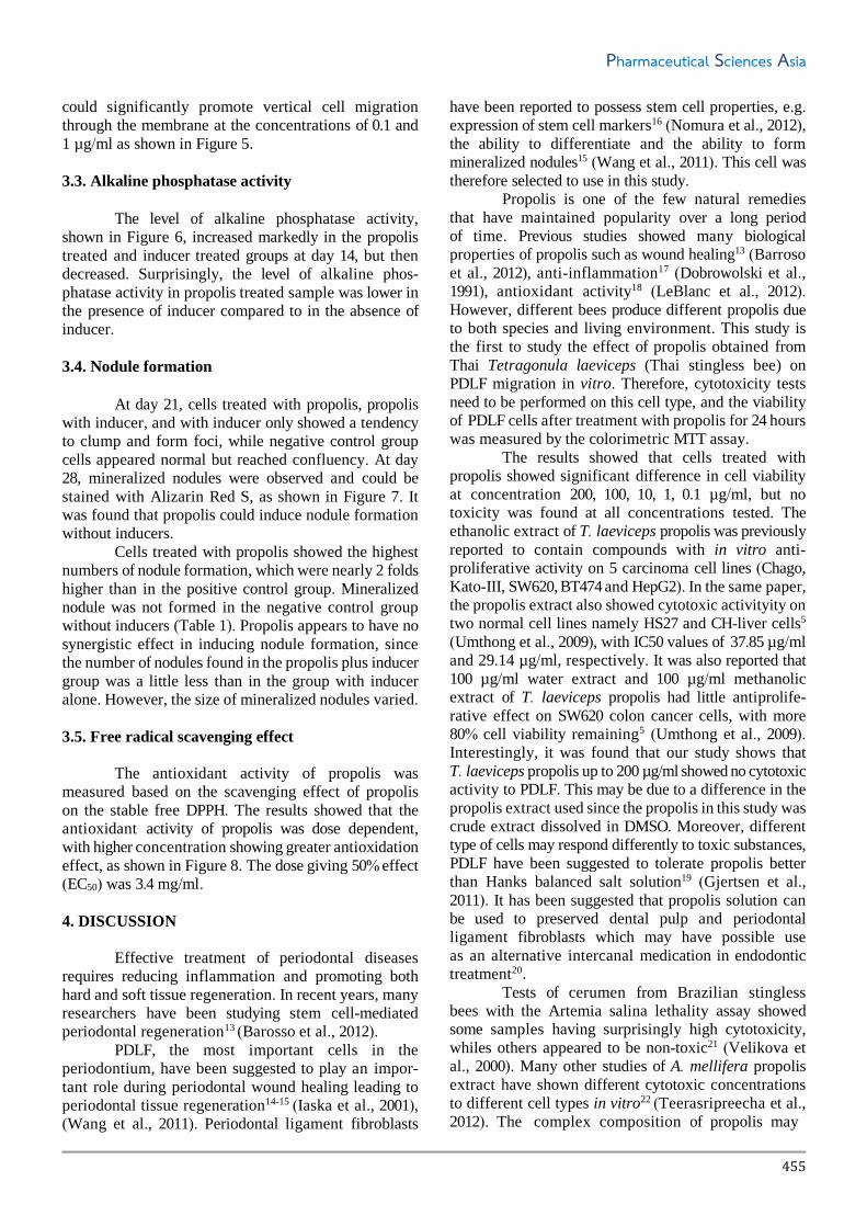
could significantly promote vertical cell migration
through the membrane at the concentrations of 0.1 and
1 µg/ml as shown in Figure 5.
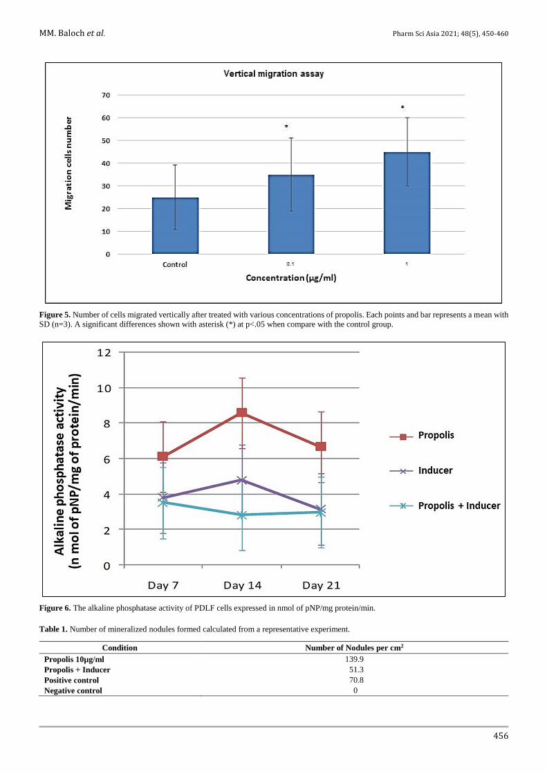
3.3. Alkaline phosphatase activity
The level of alkaline phosphatase activity,
shown in Figure 6, increased markedly in the propolis
treated and inducer treated groups at day 14, but then
decreased. Surprisingly, the level of alkaline phos-
phatase activity in propolis treated sample was lower in
the presence of inducer compared to in the absence of
inducer.
3.4. Nodule formation
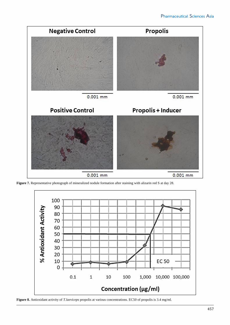
At day 21, cells treated with propolis, propolis
with inducer, and with inducer only showed a tendency
to clump and form foci, while negative control group
cells appeared normal but reached confluency. At day
28, mineralized nodules were observed and could be
stained with Alizarin Red S, as shown in Figure 7. It
was found that propolis could induce nodule formation
without inducers.
Cells treated with propolis showed the highest
numbers of nodule formation, which were nearly 2 folds
higher than in the positive control group. Mineralized
nodule was not formed in the negative control group
without inducers (Table 1). Propolis appears to have no
synergistic effect in inducing nodule formation, since
the number of nodules found in the propolis plus inducer
group was a little less than in the group with inducer
alone. However, the size of mineralized nodules varied.
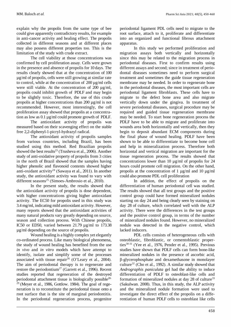
3.5. Free radical scavenging effect
The antioxidant activity of propolis was
measured based on the scavenging effect of propolis
on the stable free DPPH. The results showed that the
antioxidant activity of propolis was dose dependent,
with higher concentration showing greater antioxidation
effect, as shown in Figure 8. The dose giving 50% effect
(EC50) was 3.4 mg/ml.
4. DISCUSSION
Effective treatment of periodontal diseases
requires reducing inflammation and promoting both
hard and soft tissue regeneration. In recent years, many
researchers have been studying stem cell-mediated
periodontal regeneration13 (Barosso et al., 2012).
PDLF, the most important cells in the
periodontium, have been suggested to play an impor- tant role during periodontal wound healing leading to
periodontal tissue regeneration14-15 (Iaska et al., 2001),
(Wang et al., 2011). Periodontal ligament fibroblasts
have been reported to possess stem cell properties, e.g.
expression of stem cell markers16 (Nomura et al., 2012),
the ability to differentiate and the ability to form
mineralized nodules15 (Wang et al., 2011). This cell was
therefore selected to use in this study.
Propolis is one of the few natural remedies
that have maintained popularity over a long period
of time. Previous studies showed many biological
properties of propolis such as wound healing13 (Barroso
et al., 2012), anti-inflammation17 (Dobrowolski et al.,
1991), antioxidant activity18 (LeBlanc et al., 2012).
However, different bees produce different propolis due
to both species and living environment. This study is
the first to study the effect of propolis obtained from
Thai Tetragonula laeviceps (Thai stingless bee) on
PDLF migration in vitro. Therefore, cytotoxicity tests
need to be performed on this cell type, and the viability
of PDLF cells after treatment with propolis for 24 hours
was measured by the colorimetric MTT assay.
The results showed that cells treated with
propolis showed significant difference in cell viability
at concentration 200, 100, 10, 1, 0.1 µg/ml, but no
toxicity was found at all concentrations tested. The
ethanolic extract of T. laeviceps propolis was previously
reported to contain compounds with in vitro anti-
proliferative activity on 5 carcinoma cell lines (Chago,
Kato-III, SW620, BT474 and HepG2). In the same paper,
the propolis extract also showed cytotoxic activityity on
two normal cell lines namely HS27 and CH-liver cells5
(Umthong et al., 2009), with IC50 values of 37.85 µg/ml
and 29.14 µg/ml, respectively. It was also reported that
100 µg/ml water extract and 100 µg/ml methanolic
extract of T. laeviceps propolis had little antiprolife- rative effect on SW620 colon cancer cells, with more
80% cell viability remaining5 (Umthong et al., 2009).
Interestingly, it was found that our study shows that
T. laeviceps propolis up to 200 µg/ml showed no cytotoxic
activity to PDLF. This may be due to a difference in the
propolis extract used since the propolis in this study was
crude extract dissolved in DMSO. Moreover, different
type of cells may respond differently to toxic substances,
PDLF have been suggested to tolerate propolis better
than Hanks balanced salt solution19 (Gjertsen et al.,
2011). It has been suggested that propolis solution can
be used to preserved dental pulp and periodontal
ligament fibroblasts which may have possible use
as an alternative intercanal medication in endodontic
treatment20.
Tests of cerumen from Brazilian stingless bees with the Artemia salina lethality assay showed
some samples having surprisingly high cytotoxicity,
whiles others appeared to be non-toxic21 (Velikova et
al., 2000). Many other studies of A. mellifera propolis
extract have shown different cytotoxic concentrations
to different cell types in vitro22 (Teerasripreecha et al.,
2012). The complex composition of propolis may
MM. Baloch et al. Pharm Sci Asia 2021; 48(5), 450-460
456
Figure 5. Number of cells migrated vertically after treated with various concentrations of propolis. Each points and bar represents a mean with
SD (n=3). A significant differences shown with asterisk (*) at p<.05 when compare with the control group.
Figure 6. The alkaline phosphatase activity of PDLF cells expressed in nmol of pNP/mg protein/min.
Table 1. Number of mineralized nodules formed calculated from a representative experiment.
Condition Number of Nodules per cm2
Propolis 10µg/ml 139.9
Propolis + Inducer 51.3
Positive control 70.8
Negative control 0
Pharmaceutical Sciences Asia
457
Figure 7. Representative photograph of mineralized nodule formation after staining with alizarin red S at day 28.
Figure 8. Antioxidant activity of T.laeviceps propolis at various concentrations. EC50 of propolis is 3.4 mg/ml.
MM. Baloch et al. Pharm Sci Asia 2021; 48(5), 450-460
458
explain why the propolis from the same type of bee
could give apparently contradictory results, for example
in anti-cancer activity and healing effect. The propolis
collected in different seasons and at different places
may also possess different properties too. This is the
limitation of the study with crude extract.
The cell viability at those concentrations was
confirmed by cell proliferation assay. Cells were grown
in the presence and absence of propolis for 10 days. The
results clearly showed that at the concentration of 100
µg/ml of propolis, cells were still growing at similar rate
to control, while at the concentration of 200 µg/ml cells
were still viable. At the concentration of 200 µg/ml,
propolis could inhibit growth of PDLF and may begin
to be slightly toxic. Therefore, the use of this crude
propolis at higher concentrations than 200 µg/ml is not
recommended. However, most interestingly, the cell
proliferation assay showed that propolis at a concentra-
tion as low as 0.1 µg/ml could promote growth of PDLF.
The antioxidant activity of propolis was
measured based on their scavenging effect on the stable
free 2,2-diphenyl-1-picryl-hydrazyl radical.
The antioxidant activity of propolis samples
from various countries, including Brazil, has been
studied using this method. Red Brazilian propolis
showed the best results23 (Trusheva et al., 2006). Another
study of anti-oxidative property of propolis from 3 cities
in the north of Brazil showed that the samples having
higher phenolic and flavonoid contents showed higher
anti-oxidant activity24 (Sawaya et al., 2011). In another
study, the antioxidant activity was found to vary with
different seasons25 (Simoes-Ambrosio et al., 2010).
In the present study, the results showed that
the antioxidant activity of propolis is dose dependent,
with higher concentrations giving higher antioxidant
activity. The EC50 for propolis used in this study was
3.4 mg/ml, indicating mild antioxidant activity. However,
many reports showed that the antioxidant activities of
many natural products vary greatly depending on source,
season and collection process. With Chinese propolis,
IC50 or ED50, varied between 21.79 µg/ml to 173.38
µg/ml depending on the source of propolis.
Wound healing is a highly complex yet elegantly
co-ordinated process. Like many biological phenomena,
the study of wound healing has benefited from the use
in vivo and in vitro models which have attempt to
identify, isolate and simplify some of the processes
associated with tissue repair26 (O’Leary et al., 2004).
The aim of periodontal therapy is to regenerate and
restore the periodontium27 (Garrett et al., 1996). Recent
studies reported that regeneration of the destroyed
periodontal attachment tissue is biologically possible28-
29 (Meyer et al., 1986, Gottlow. 1984). The goal of rege-
neration is to reconstitute the periodontal tissue onto a
root surface that is the site of marginal periodontitis. In the periodontal regeneration process, progenitor
periodontal ligament PDL cells need to migrate to the
root surface, attach to it, proliferate and differentiate
into an organized and functional fibrous attachment
apparatus.
In this study we performed proliferation and
migration assays both vertically and horizontally
since this may be related to the migration process in
periodontal diseases. First to confirm results using
different assays and second; since in treatment of perio-
dontal diseases sometimes need to perform surgical
treatment and sometimes the guide tissue regeneration
membrane may be needed. In order to regenerate bone
in the periodontal diseases, the most important cells are
periodontal ligament fibroblasts. These cells have to
migrate to the defect bone area and also migrate
vertically down under the gingiva. In treatment of
severe periodontal diseases, surgical procedure may be
required and guided tissue regeneration membrane
may be needed. To start bone regeneration process the
PDLF have to be able to migrate and proliferate into
wounds area both horizontally and vertically, then they
begin to deposit abundant ECM components during
the final phase of wound healing. PDLF have been
shown to be able to differentiate to become bone cell
and help in mineralization process. Therefore both
horizontal and vertical migration are important for the
tissue regeneration process. The results showed that
concentrations lower than 10 µg/ml of propolis for 24
hours could promote cell migration. On the other hand,
propolis at the concentration of 1 µg/ml and 10 µg/ml
could also promote PDL cell proliferation
In addition, the effect of propolis on the
differentiation of human periodontal cell was studied.
The results showed that all test groups and the positive
control group could have formed mineralized nodules
starting on day 24 and being clearly seen by staining on
day 28 of culture, which correlated well with the ALP
activity. There were the differences in the test groups
and the positive control group, in terms of the number
of mineralized nodules found. However, no mineralized
nodule was detected in the negative control, which
lacked inducers.
PDL cells consists of heterogeneous cells with
osteoblastic, fibroblastic, or cementoblastic proper-
ties30-31 (Yee et al., 1976, Pender et al., 1995). Previous
studies have shown that PDLF cells can form bone-like
mineralized nodules in the presence of ascorbic acid,
β-glycerophosphate and dexamethasone in monolayer
culture32 (Cho et al., 1992). A similar study showed that
Andrographis paniculata gel had the ability to induce
differentiation of PDLF to osteoblast-like cells and
formation of mineralized nodules at day 28 of culture33
(Sakulwan. 2008). Thus, in this study, the ALP activity
and the mineralized nodule formation were used to
investigate the direct effect of the propolis on a diffe-
rentiation of human PDLF cells to osteoblast like cells
Pharmaceutical Sciences Asia
459
in vitro. ALP is a calcium and phosphate binding protein
and also an enzyme which hydrolyses monophosphate
esters producing a high concentration of phosphates34
(Beertsen et al., 1989) leading to supersaturation and
subsequent precipitation of calcium phosphate salt in
the collagenous substrate35 (Martland et al., 1926). The
activity of the enzyme is considered to be an important
indicator of bone formation and a phenotypic marker of
the osteoblastic cells. ALP appears to be an early marker
of osteoblast differentiation and is expressed at high
levels during the period of extracellular matrix depo-
sition and then down-regulated after the mineralization
stage1 (Beck et al., 1998).
In the present study, the ALP activity
continuously increased, and reaching its peak on day
14 in all groups, and then started to decrease. Cultured
human periosteal-derived cells also showed increase in
ALP expression for up to 2 weeks, with the continuous
decrease thereafter36 (Perk et al., 2007). Therefore, our
results showed a similar pattern of ALP activity in
PDLF cells, but surprisingly, the level of ALP activity
in PDLF also increased in the group lacking inducers.
Moreover, the level of alkaline phosphatase activity in
propolis treated sample was lower in the presence of
inducer compared to in the absence of inducer. This
may due to the antioxidant property of propolis which
may react with ascorbic acid in the osteogenic medium
and resulted in such response. The PDLF may possess
a specific property, since PDLF contains stem cell
property and can differentiate to other cell types such
as osteoblast-like cells. The PDLF cells used in this
study contain more than 99% stem cell property (data
not shown). Thus, ALP activity increases during 14 days
of culture, so the cells are ready to differentiate if the
signal received. However, in the absence of inducer,
cells will not differentiate and continue to grow as
fibroblasts. Therefore, ALP activity may not be a good
candidate for following the mineralization process in
PDLF. However, it is interesting that propolis could
induce ALP activity to increase and remain quite high
after 14 day of culture. In addition, all test groups and
the positive control group showed mineralized nodules
starting on day 24 of culture, and the number and size
of the nodules increase on day 28 of culture. However,
differences in the number of mineralized nodules were
found in the test groups and the positive control, but no
mineralized nodule was detected in negative control
group. Propolis alone could induce PDLF differentia-
tion and mineralization, but use of propolis together
with inducer actually caused reduction in the number
of mineralized nodules, suggesting that these two sub-
stances may have some antagonistic effect on induction.
The mineralized nodules may arise from osteoblast-like
cells formed by PDLF differentiation or result from
mineralization of PDLF itself. Further studies are needed
clarify this, as well as to understand the mechanism of
the induction.
The findings in this study clearly show that
propolis from T. laeviceps could be used to induce bone
formation. This is the first time that such property of
this propolis has been described. The concentration used
for induction is very low at the level of 0.1 µg/ml of
propolis respectively, which is much lower than the
EC50 found the free radical scavenging assay, so it is
unlikely that these two properties are related.
The findings of the present study may help in
the development of new drug to be used for treatment
of periodontal disease, by which bone regeneration
could be induced. With this property of this propolis, it
will be interesting to see whether it may assist in other
bone mineralization or bone tissue damage, such as in
arthritis or other bone diseases. However, further clinical
studies will be required to enable this natural product to
be used in the effective manner.
Moreover, the study about other biological effect
of propolis from T. laeviceps on other cell types may
lead to the development of new regimen for treatment of
other wound type.
5. CONCLUSIONS
Propolis from T. laeviceps can induce prolife-
ration, migration and differentiation of PDLF. Thus,
the results of this study could be used to support the use
of these natural materials in dental practice for the
treatment of periodontal diseases.
6. ACKNOWLEDGEMENT
This project is supported by Higher Education
Commission and Mahidol University under the National
Research Universities Initiative.
Conflict of interest
The authors declare no conflicts of interests.
Funding None to declare.
Ethics approval None to declare.
Article info:
Received June 8, 2020
Received in revised form November 12, 2020
Accepted December 9, 2020
REFERENCES 1. Beck JD, Eke P, Heiss G, Madianos P, Couper D, Lin D, et al.
Periodontal disease and coronary heart disease: a reappraisal of
the exposure. Circulation. 2005;112(1):19-24.
2. Lafon A, Pereira B, Dufour T, Rigouby V, Giroud M, Béjot Y,
et al. Periodontal disease and stroke: a meta-analysis of cohort

MM. Baloch et al. Pharm Sci Asia 2021; 48(5), 450-460
460
studies. Eur J Neurol. 2014;21(9):1155-61.
3. Sharma N, Shamsuddin H. Association between respiratory
disease in hospitalized patients and periodontal disease: a cross-
sectional study. J Periodontol. 2011;82(8):1155-60.
4. Taheri JB, Azimi S, Rafieian N, Zanjani HA. Herbs in dentistry.
Int Dent J. 2011;61(6):287-96.
5. Umthong S, Puthong S, Chanchao C. Trigona laeviceps propolis
from Thailand: antimicrobial, antiproliferative and cytotoxic
activities. Am J Chin Med. 2009;37(5):855-65.
6. Umthong S, Phuwapraisirisan P, Puthong S, Chanchao C. In
vitro antiproliferative activity of partially purified Trigona
laeviceps propolis from Thailand on human cancer cell lines.
BMC Complement Altern Med. 2011;11:37.
7. Mosmann T. Rapid colorimetric assay for cellular growth and
survival: application to proliferation and cytotoxicity assays. J
Immunol Methods. 1983;65(1-2):55-63.
8. Jaiswal N, Haynesworth SE, Caplan AI, Bruder SP. Osteogenic
differentiation of purified, culture-expanded human mesen-
chymal stem cells in vitro. J Cell Biochem. 1997;64(2):295-312.
9. Liang CC, Park AY, Guan JL. In vitro scratch assay: a conve-
nient and inexpensive method for analysis of cell migration in
vitro. Nat Protoc. 2007;2(2):329-33.
10. Chung CA, Chen CY. The effect of cell sedimentation on
measuring chondrocyte population migration using a Boyden
chamber. J Theor Biol. 2009;261(4):610-25.
11. Nohutcu RM, McCauley LK, Koh AJ, Somerman MJ. Expres-
sion of extracellular matrix proteins in human periodontal
ligament cells during mineralization in vitro. J Periodontol.
1997;68(4):320-27.
12. Yamaguchi T, Takamura H, Matoba T, Terao J. HPLC method
for evaluation of the free radical-scavenging activity of foods
by using 1,1-diphenyl-2-picrylhydrazyl. Biosci Biotechnol
Biochem. 1998;62(6):1201-4.
13. Barroso PR, Lopes-Rocha R, Pereira EM, Marinho SA, de
Miranda JL, Lima NL, et al. Effect of propolis on mast cells in
wound healing. Inflammopharmacology. 2012;20(5):289-94.
14. Isaka J, Ohazama A, Kobayashi M, Nagashima C, Takiguchi T,
Kawasaki H, et al. Participation of periodontal ligament cells
with regeneration of alveolar bone. J Periodontol. 2001;72(3): 314-23.
15. Wang L, Shen H, Zheng W, Tang L, Yang Z, Gao Y, et al.
Characterization of stem cells from alveolar periodontal
ligament. Tissue Eng Part A. 2011;17(7-8):1015-26.
16. Nomura Y, Ishikawa M, Yashiro Y, Sanggarnjanavanich S,
Yamaguchi T, Arai C, et al. Human periodontal ligament
fibroblasts are the optimal cell source for induced pluripotent
stem cells. Histochem Cell Biol. 2012;137(6):719-32.
17. Dobrowolski JW, Vohora SB, Sharma K, Shah SA, Naqvi SA,
Dandiya PC. Antibacterial, antifungal, antiamoebic, antiin-
flammatory and antipyretic studies on propolis bee products. J
Ethnopharmacol. 1991;35(1):77-82.
18. LeBlanc L, Paré A, Jean-François J, Hébert M, Surette M,
Touaibia M. Synthesis and Antiradical/Antioxidant Activities
of Caffeic Acid Phenethyl Ester and Its Related Propionic,
Acetic, and Benzoic Acid Analoguesc. Molecules. 2012;17(12):
14637-50.
19. Gjertsen AW, Stothz KA, Neiva KG, Pileggi R. Effect of
propolis on proliferation and apoptosis of periodontal ligament
fibroblasts. Oral Surg Oral Med Oral Pathol Oral Radiol Endod.
2011;112(6):843-8.
20. Al-Shaher A, Wallace J, Agarwal S, Bretz W, Baugh D. Effect
of propolis on human fibroblasts from the pulp and periodontal
ligament. J Endod. 2004;30(5):359-61.
21. Velikova M, Bankova V, Marcucci MC, Tsvetkova I,
Kujumgiev A. Chemical composition and biological activity of
propolis from Brazilian meliponinae. Z Naturforsch C J Biosci.
2000;559-10):785-9.
22. Teerasripreecha D, Phuwapraisirisan P, Puthong S, Kimura K,
Okuyama M, Mori H, et al. In vitro antiproliferative/cytotoxic
activity on cancer cell lines of a cardanol and a cardol enriched
from Thai Apis mellifera propolis. BMC Complement Altern
Med. 2012;12:27.
23. Trusheva B, Popova M, Bankova V, Simova S, Marcucci MC,
Miorin PL, et al. Bioactive constituents of brazilian red propolis.
Evid Based Complement Alternat Med. 2006;3(2):249-54.
24. Sawaya AC, Barbosa da Silva Cunha I, Marcucci MC.
Analytical methods applied to diverse types of Brazilian
propolis. Chem Cent J. 2011;5(1):27.
25. Simoes-Ambrosio LM, Gregorio LE, Sousa JP, Figueiredo-
Rinhel AS, Azzolini AE, Bastos JK, et al. The role of seasonality
on the inhibitory effect of Brazilian green propolis on the
oxidative metabolism of neutrophils. Fitoterapia. 2010;81(8): 1102-8.
26. O'Leary R, Rerek M, Wood EJ. Fucoidan modulates the effect
of transforming growth factor (TGF)-beta1 on fibroblast
proliferation and wound repopulation in in vitro models of
dermal wound repair. Biol Pharm Bull. 2004;27(2):266-70.
27. Garrett S. Periodontal regeneration around natural teeth. Ann
Periodontol. 1996;1(1):621-66.
28. Meyer JR. The regenerative potential of the periodontal
ligament. J Prosthet Dent. 1986;55(2):260-5.
29. Gottlow J, Nyman S, Karring T, Lindhe J. New attachment
formation as the result of controlled tissue regeneration. J Clin
Periodontol. 1984;11(8):494-503.
30. Yee JA, Kimmel DB, Jee WS. Periodontal ligament cell kinetics
following orthodontic tooth movement. Cell Tissue Kinet.
1976;9(3):293-302.
31. Pender N, Heaney TG. Migration and proliferation of progenitor
cells in the connective tissue of rat gingival papilla. J Periodontal
Res. 1995;30(5):312-8.
32. Cho M-I, Matsuda N, Lin W-L, Moshier A, Ramakrishnan PR.
In vitro formation of mineralized nodules by periodontal
ligament cells from the rat. Calcif Tissue Int. 1992;50(5):459-67.
33. Noppamassiri S, Sirirat M, Surarit R, Kasetsuwan J, Rojana-
panthu P. The cytotoxic effect of Andrographis paniculata
extract and Andrographis paniculata gel on human periodontal
ligament cells. Mahidol Dent J. 2009;29(1):37-44.
34. Beertsen W, van den Bos T. Calcification of dentinal collagen
by cultured rabbit periosteum: the role of alkaline phosphatase.
Matrix. 1989;9(2):159-71.
35. Martland M, Robison R. Possible Significance of Hexose-
phosphoric Esters in Ossification: Part VI. Phosphoric Esters in
Blood-Plasma. Biochem J. 1926;20(4):847-55.
36. Park BW, Hah YS, Kim DR, Kim JR, Byun JH. Osteogenic
phenotypes and mineralization of cultured human periosteal-
derived cells. Arch Oral Biol. 2007;52(10):983-9.


