
Hindawi Publishing CorporationJournal of NanomaterialsVolume 2013, Article ID 460518, 6 pageshttp://dx.doi.org/10.1155/2013/460518
Research ArticleBioaccumulation, Subacute Toxicity, and TissueDistribution of Engineered Titanium Dioxide Nanoparticles inGoldfish (Carassius auratus)
Mehmet Ates,1,2 Veysel Demir,1 Ragip Adiguzel,3 and Zikri Arslan2
1 Department of Bioengineering, Tunceli University, Ataturk mahallesi Muhlis Akarsu caddesi, 62000 Tunceli, Turkey2Department of Chemistry and Biochemistry, Jackson State University, P.O. Box 17910, Jackson, MS 39217, USA3Department of Chemical Engineering, Tunceli University, Ataturk mahallesi Muhlis Akarsu caddesi, 62000 Tunceli, Turkey
Correspondence should be addressed to Mehmet Ates; [email protected]
Received 27 June 2013; Revised 22 August 2013; Accepted 2 September 2013
Academic Editor: Xiaoming Li
Copyright © 2013 Mehmet Ates et al. This is an open access article distributed under the Creative Commons Attribution License,which permits unrestricted use, distribution, and reproduction in any medium, provided the original work is properly cited.
The increased use of nanosizedmaterials is likely to result in the release of these particles into the environment. It is, however, unclearif these materials are harmful to aquatic animals. In this study, the sublethal effects of exposure of low and high concentrations oftitanium dioxide nanoparticles (TiO
2NPs) on goldfish (Carassius auratus) were investigated. Accumulation of TiO
2NPs increased
from 42.71 to 110.68 ppb in the intestine and from 4.10 to 9.86 ppb in the gills of the goldfishwith increasing exposure dose from 10 to100mg/L TiO
2NPs. No significant accumulation in the muscle and brain of the fish was detected. Malondialdehyde as a biomarker
of lipid oxidation was detected in the liver of the goldfish. Moreover, TiO2NPs exposure inhibited growth of the goldfish. Although
there was an increase (8.1%) in the body weights of the goldfish for the control group, in the low and high exposure groups 1.8%increase and 19.7% decrease were measured, respectively. The results of this study contribute to the current understanding of thepotential ecotoxicological effects of nanoparticles and highlight the importance of characterization of NPs in understanding theirbehavior, uptake, and effects in aquatic systems and in fish.
1. Introduction
Nanomaterials are used in a wide range of domestic appli-ances and household products, in the manufacture of textilesand electronics, as well as medical products and in biore-mediation technology. There are also concerns about theenvironmental risks of nanotechnology which need to bebalanced against their undoubted benefits to human society[1, 2]. Handy and Shaw [3] reviewed the risks to human healthand identified a number of exposure routes including thedischarge of nanoparticles (NPs) to water and agriculturalland. The chemistry and physical characteristics of the NPsthemselves are key elements in determining their fate andbehavior in aquatic systems.The large surface area, crystallinestructure, and reactivity of some NPs may facilitate transportof these toxic materials in the environment [4]. Certainconditions such as presence of humic and fulvic acids, pH,and specific cation concentrations may favor the stabilization
of NPs in the water column [5].The aquatic species are also atrisk of exposure to the NPs and there is currently little knownabout their uptake, potential toxic effects, and behavior inaquatic systems.
Titanium dioxide (TiO2) NPs have been widely used in
several industries. Nanoparticulate TiO2has been utilized
as an ultraviolet radiation absorber in transparent sunscreenformulations [6] and in specialist photocatalytic coatings forglass [7].The environmental chemistry of TiO
2NPs has been
partly investigated. TiO2NPs can be dispersed in freshwater
by sonication [8], but the primary particles tend to formaggregates of a few 100 nm dimensions, and the aggregatesgradually precipitate from thewater column over a few hours.TiO2has two major crystal structures (rutile and anastase),
and the surface reactivity of the NP is closely defined by thecrystal structure [9].
There is an emerging literature on the effects of NPsfor fish and aquatic invertebrates. NPs have previously been
2 Journal of Nanomaterials
shown to accumulate in cells such as macrophages andhepatocytes [10, 11]. Moreover, they are taken up into aquaticorganisms such as fish, mollusks, crustaceans, and artemia[12–16]. Fish are excellent sentinels of environmental healthas they are sensitive to a wide range of xenobiotic chemicals.Their position in the aquatic food chain means assessmentof the populations, and health of fish can give an indicationof the health of other lower levels of the food chain. Under-standing the effects of NPs on fish is therefore an importantaspect when considering the effects of NPs on the aquaticenvironment as a whole. Potential routes of uptake for NPs infish include absorption via the gill epithelia, via the intestineepithelia as a result of dietary exposure and drinking, or viathe skin [17].
The purpose of this exposure study was to determine sub-acute toxicity, accumulation, and tissue distribution of engi-neered TiO
2NPs in goldfish (Carassius auratus). Due to the
importance of their size and aggregation behavior [18–20],the NPs were characterized by transmission electron micro-scopy (TEM), and the size distribution of NPs was measuredby dynamic light scattering (DSL). Fish tissues were used asin vitromodel to determine the possible uptake of TiO
2NPs
into gill, intestine, muscle, and brain. Total Ti accumulationin each tissue was determined by inductively coupled plasmamass spectrometry (ICP-MS). In addition, MDA was deter-mined as a cause of systemic oxidative stress.
2. Materials and Methods
2.1. Test Organism and Experimental Condition. A group ofhealthy goldfish (Carassius auratus) was purchased from alocal pet shop. The initial body weight and length of the fishwere measured as 4.53 ± 0.06 g and 5.5 ± 0.7 cm, respectively.All fish weremaintained in 30 L glass aquarium supplied via aflow-through system with dechlorinated tap water, enrichedwith oxygen at a temperature of 23 ± 2∘C and pH of 6.8. Fishwere fed daily with commercially available fish feed flakes(TetraFin Goldfish flakes, Germany) at the amount of 0.5% ofmean body weight of the fish. The goldfish were anesthetizedusing 3-aminobenzoic acid ethylester (MS-222; Aqua Life,Syndel Laboratories Ltd., Vancouver, BC, Canada) at a lethaldose for dissection (excess of 200mg/L), and a lower dosewasused for all handling procedures (150mg/L).
2.2. Reagents and Chemicals. TiO2NPs (TiO
210–30 nmNPs,
99.5% pure) were purchased, as uncoated nanomaterials,from Skyspring Nanomaterials Inc., in Houston, TX, USA.TEM image of NPs was spherical with an average particle size(D50) of 10–30 nm and approximate surface area of 50m2/g.
The morphology of the NPs was rutile with pale yellow color,which is the most widely found polymorph of TiO
2in nature
and in high pressure metamorphic rocks. The TiO2NPs
were stored at room temperature in the laboratory until theimplementation of the experimental studies.
Deionized water produced by Barnstead E-pure systemwith the resistivity of 18.0MΩ cm was used to prepare theexposure medium and experimental solutions. Trace metalgrade nitric acid (HNO
3, Fisher Scientific) and hydrofluoric
acid (HF, 99.99%, Sigma Aldrich) used for dissolution of
the goldfish were collected after the exposure to determinethe total uptake levels. Stock titanium standard solution(1000 𝜇gmL−1) was purchased from SCP Science (Cham-plain, NY). Calibration standards for ICP-MS analysis wereprepared within a range from 0 to 500𝜇g L−1 from the stockTi solution in 5% HNO
3. Carbon coated Cu TEM grids (300
mesh) were purchased from Electron Microscopy Sciences(EMS), Hatfield, PA.
2.3. Characterization of NPs. For preparation of exposuremedium, TiO
2NPs were weighed in polypropylene tubes
and dispersed in deionized water. To achieve maximumdispersion, the suspension was homogenized using vortex(Daigger Vortex-Genie 2, Model G560) equipped with atitanium probe. Each suspension was exposed to mixture forabout 2 minutes and then immediately transferred into theexposure glass tanks. The characterizations of the TiO
2NP
suspension were performed using TEM and DLS techniques.Size measurements in dried suspension were made by TEM,while DLS provided the size distribution for the hydratedforms of the NPs. In addition, Zeta potential measurementswere conducted using the DLS instruments to elucidate thesurface charges of the suspensions in the exposure medium.
For TEM measurements, a drop (ca. 8 𝜇L) of solutionwas allowed to dry on a carbon-coated copper grid overnight(CF300Cu). The TEM grids were purchased from ElectronMicroscopy Sciences (EMS). Images were recorded by usingJEOL-1011 TEM instrumentwith 0.2 nm lattice resolution andmagnification power up to 106 under the accelerating voltageof 40 to 100 kV. Captured images were analyzed using ImageJsoftware. For DLS measurement, the protocol followed thestandards of the Nanotechnology Characterization Labora-tory [21]. A stock solution (10mg/100mL) was preparedwith deionized water and diluted to a final concentration of10 𝜇g/mL. Samples were then analyzed with Malvern Zeta-sizer Model Nano ZS according to manufacturer’s protocols.Samples were read in disposable plastic Malvern Cells.
2.4. Experiment Design. NP exposure was conducted toassess acute toxicity and associated behavioral changes ongoldfish by exposing the fish to two different doses, 10 and100mg/L, of the TiO
2NPs, using 5 days static tests according
to OECD 203 testing guidelines [22]. A control group wasalso set up without the test compound. Studies were carriedout in an aquarium (30 L inner volume). The volume for10 L level was marked and filled with freshwater followed byaddition of the NP suspensions prepared as described above.Sight aeration was provided by a line extending to the bottomof the aquarium. Details of the experimental conditions aresummarized in Table 1. Each scheme (control or treatments)was conducted in duplicatewith 5 healthy goldfish. Individuallength and weight of the fish were measured at the beginningand end of the experiment. The data obtained enabledcalculation of the live weight and lengthwise increases andsurvival rates upon completion of the experiment.
2.5. Instrumental Analysis. At the end of the exposure, fishtissues were sampled for instrumental analysis, about 150mgof wet tissue was weighed and digested in Teflon vessels
Journal of Nanomaterials 3
Table 1: Expanded design summary of goldfish (Carassius auratus).
Parameter Control Group A Group BVolume of water inaquarium (L) 10 10 10
TiO2 NPs concentrations(mg/L) 0 10 100
Duration of exposure(day) 5 5 5
Water temperature (∘C) 23 ± 2 23 ± 2 23 ± 2
Oxygen (ppm) 6 ± 1 6 ± 1 6 ± 1
pH value of water(Start–End) 6.30–6.05 6.03–6.15 6.63–6.45
Number of fish withinthe aquarium 5 5 5
Number of replications 2 2 2
in 2mL concentrated HNO3and 0.5mL HF for 2 hours
using a digestion block (DigiPrep MS, SCP Science) at160∘C according to protocols described elsewhere [23]. Atthe end, the contents were visually inspected for completedissolution of TiO
2NPs (e.g., clear solution without any
turbidity) and were diluted to 10mL with deionized water.The sample solutions were analyzed by inductively cou-pled plasma mass spectrometry (ICP-MS) using a Varian820MS ICP-MS instrument (Varian, Australia). ICP-MS isa powerful multielement technique for analysis of fish andother aquatic organisms for toxic metals even at subparts perbillion levels because of its high sensitivity [24]. Titaniumcontent of the solutions was measured to determine theaccumulation pattern of the NPs across the dose of exposure.Total Ti concentration detected was then translated intocorresponding TiO
2NP concentration.
2.6. Oxidative Stress Parameter Analysis. The experiment wasdesigned to allow sublethal physiological effects over theexposure period. The five days exposure time was chosento enable some physiological or biochemical responses tothe test organism. Five fish per treatment were collectedfrom each tank at the end of the experiment for biochemicalanalysis. The extent of lipid peroxidation in the tissues wasdetermined by measuring the quantity of malondialdehyde(MDA) [25]. Quantification of MDA was done followingthe methods described by Maness et al. [26], Esterbauer,and Cheeseman [27]. The method is based on the formationof pink MDA-thiobarbituric acid (TBA) adduct which hasmaximum absorption in acidic solution at 532 nm. Briefly,the liver and muscle tissues were removed separately, imme-diately frozen in liquid nitrogen, and stored at −20∘C untilneeded. The frozen tissues were rinsed in 9-fold chilled100mmol/L, pH 7.8 sodium phosphate buffer solution, andhomogenized by a hand-driven glass homogenizer. Approx-imately 150mg muscle tissue and 10 𝜇L BHT reagent wereimmediately transferred into 500mL cold water in tube andthen the sample was homogenized using sonicator (SonicDismembrator Model 100, Fisher Scientific). The sampleswere sonicated on ice by ultrasounds for 2min at 80% power.
All samples and standards were incubated at 90∘C for onehour, then centrifuged at 12000 rpm for 15minutes to separatethe suspending tissue. The absorbance of the supernatant(reaction mixture) was measured at 532 nm with HP 8452Amodel diode array spectrophotometer. The concentration ofthe MDA formed was calculated based on a standard curvefor the MDA (Sigma Chemical Co.) complex with TBA. Theextent of lipid peroxidation was expressed in nanomoles (ormicromoles) of MDA.
2.7. Statistical Analysis. All experiments were repeated twiceindependently, and datawere recorded as themeanwith stan-dard deviation (SD). One-way analysis of variance (ANOVA)with Tukey’s multiple comparisons was used to detect signif-icant differences among groups. Student’s 𝑡-test was used forpaired comparisons of two groups. In all data analyses, a 𝑃value <0.05 was considered statistically significant.
3. Results and Discussion
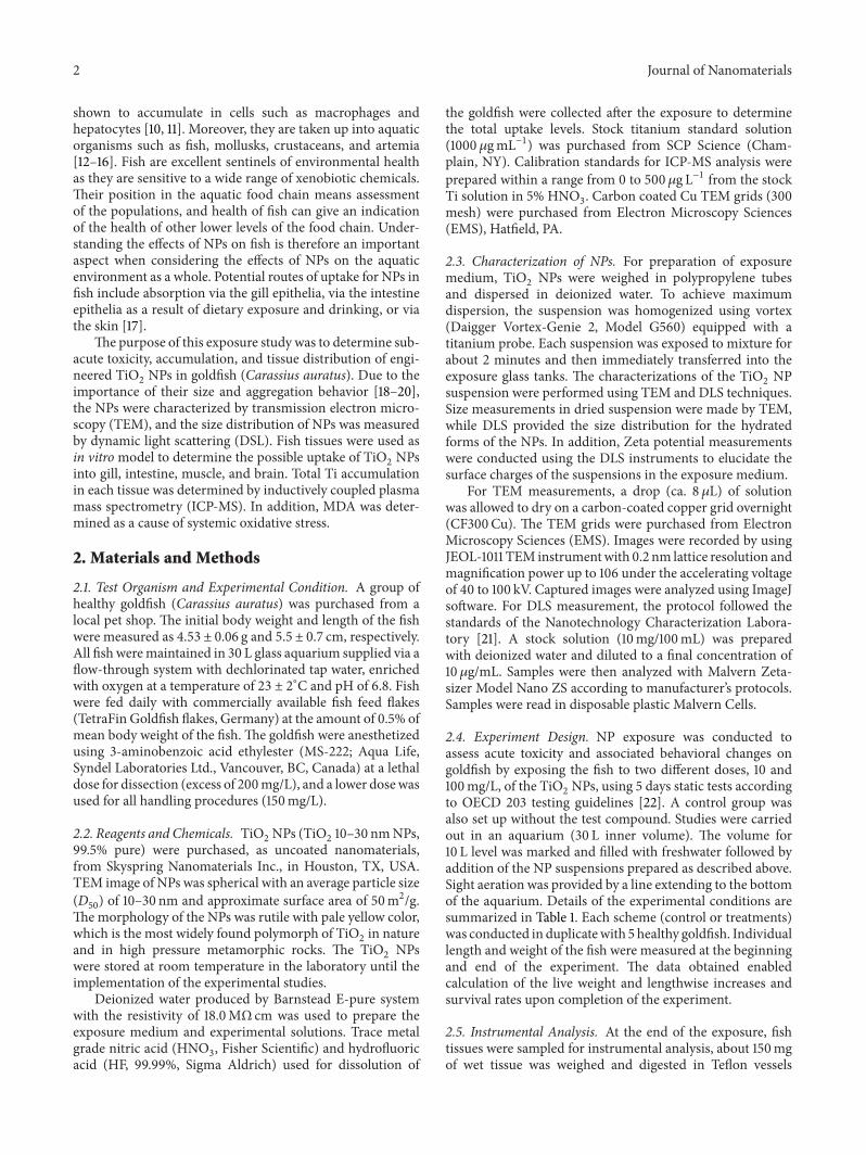
3.1. Characterization of NPs by TEM. TiO2NPs are highly
hydrophobic; therefore they aggregate substantially in aque-ous solutions [28–30].The stability and aggregation behaviorsof NPs within aquatic media are determined by both thephysicochemical properties of the liquid and the charge onthe surface of the NPs. The degree of aggregation of NPshas been shown in some cases to affect toxicity in vitro, andaggregation of the NPs when suspended in water is a knownissue for TiO
2NPs [28, 31]. In this study, the water visi-
bility decreased substantially with increasing concentrationof TiO
2NPs, and at 100 g/L level, the solution was cloudy.
Similar aggregation phenomenon has been reported in manyNP studies including TiO
2where aggregates of NPs can
sink out of the solution very quickly [32]. The TEM imagescollected from the dried suspensions of stock solution andexposure medium are illustrated in Figure 1. The TiO
2NPs
aggregated significantly in water yielding large aggregatesranging from around 100 to as high as 1.0 𝜇m in size.Although aeration assisted in maintaining the homogeneityof the suspensions, aggregation could not be avoided at anyconcentration of TiO
2NPs.
3.2. SizeDistribution and Surface ChargeMeasurement of NPs.Metal oxide particles tend to aggregate to various extents inwater. The size distribution of the NPs is of interest in thisstudy, since TiO
2NPs are highly hydrophobic; the particles
size distribution in water was measured to determine theeffect of the stability. The mean size distribution of TiO
2
NP in water was calculated as 432 ± 32 nm for 10 𝜇g/mLNP suspension. Zeta potentials for the TiO
2NPs in aqueous
suspensions were obtained using the Henry Equation. Aviscosity of 0.8872 cP, a dielectric constant of 78.5, and Henryfunction of 1.330 were used for the calculations. The meanzeta potential was calculated as −31.6±4.5mV at a pH of 7.23in 10 𝜇g/mL aqueous suspensions of NP.
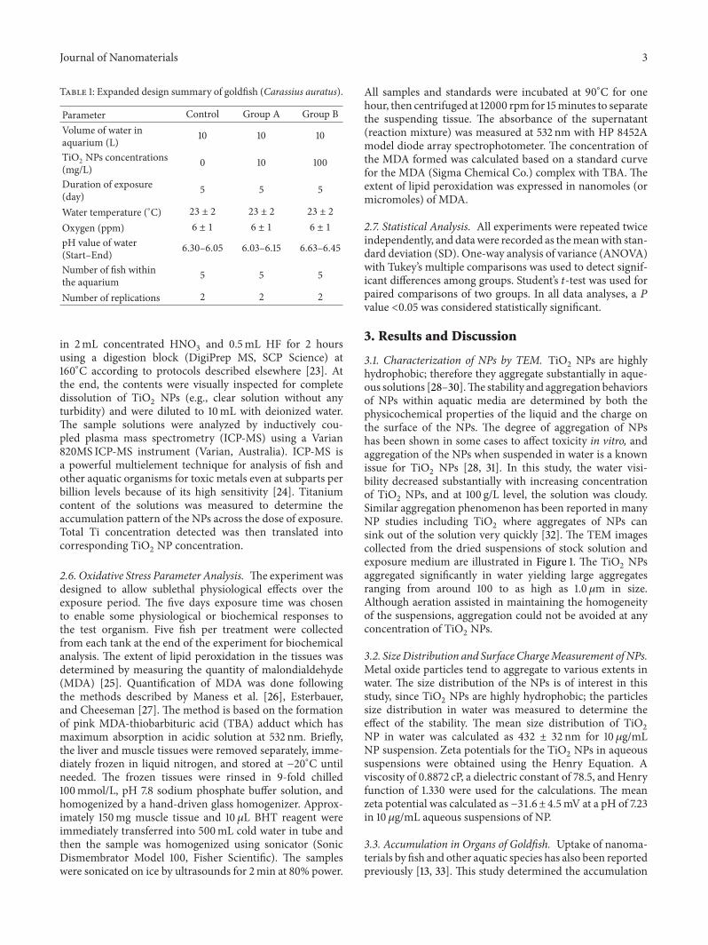
3.3. Accumulation in Organs of Goldfish. Uptake of nanoma-terials by fish and other aquatic species has also been reportedpreviously [13, 33]. This study determined the accumulation
4 Journal of Nanomaterials
(a) (b)Figure 1: TEM images for TiO
2NPs from stock solution (a) and the exposure medium (b).
1
10
100
1000
Control Group A Group B
Ti co
ncen
trat
ion
(ppb
)
Experiment groups
GillIntestine
Muscle
Figure 2: Titanium (Ti) levels in gill, intestine, and muscle of thegoldfish at the end of the exposure experiments (GroupA andGroupB exposed to 10 and 100mg/L TiO
2NPs, resp.).
of the unmodifiedTiO2NPs in fish tissues following exposure
via the water column without the use of a solvent vehicleor prior modification of the NP surface. The chemical fateof the metal oxide NPs in the aquatic environment wasdetermined through a comprehensive evaluation of uptakein fish with full characterization of the NPs in low and highexposure conditions. The gill, intestine, muscle, and brain ofthe goldfish were used as in vitro model to determine thepossible uptake of TiO
2NPs into tissues. For 10mg/L and for
100mg/L TiO2exposure mediums, uptakes of TiO
2NPs in
intestine were measured as 42.71 and 110.68 ppb and in gillsas 4.10 and 9.86 ppb, respectively. ICP-MS analysis showedvery small amount of Ti accumulation in the muscle and noaccumulation in brain tissues of the goldfish (Figure 2). Astudy byMoger et al. [34], however, used coherent anti-StokesRaman Scattering (CARS) to examine the gills of rainbowtrout exposed to 5000𝜇g L−1 TiO
2NPs and confirmed the
presence of small numbers of particle aggregates within thegill tissue.
3.4. Oxidative Stress. Lipid peroxidation generates a groupof products among which are reactive electrophiles such
as epoxides and aldehydes [27, 35, 36]. Malondialdehyde(MDA) is a major product of lipid peroxidation in aqueoussolution. In this study, to elucidate the possible role ofoxidative stress in the effects observed by TiO
2NPs exposure,
MDA content was assayed in liver and muscle of eachexperimental group.The analysis forMDAcontent of goldfishliver showed lipid peroxidation in the controls and exposuregroups. The mean MDA levels in the liver of the fish were4.1 ± 0.5, 7.6 ± 1.1, and 11.3 ± 0.9 nmol/gr for control, andlow and high dose exposure groups, respectively. No MDAlevel was measured in the muscles of the control and theexposure groups. Xiong et al. [29] also studied TiO
2NPs but
on zebrafish and reported similar results as this study thatoxidative effects were more severe in the livers of zebrafishexposed to 50mg/L TiO
2NPs.
Aqueous exposure to low and high dose of TiO2NP
suspension did not cause any fish mortality during theexperimental period (96 hr). Our data is in agreement withthe literature, indicating low acute toxicity of TiO
2NPs
to fish survival. Similarly, Warheit et al. [37] reported thatthe Daphnia magna 48 hr EC50 values and rainbow trout(Oncorhynchus mykiss) 96 hr LC50 values for fine TiO
2
particles and ultrafine TiO2particles based on nominal
concentrations were >100mg/L, and the LC50
for TiO2NPs
was also found to be over 500mg L−1 in fathead minnowPimephales promelas [38]. Furthermore, Zhu et al. [30]showed that exposure to TiO
2NPs at the concentrations up
to 500mg/L for 96 hr did not affect hatching rate and did notcause deformity in embryonic zebrafish.
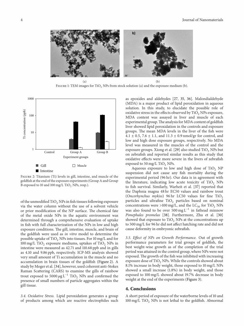
3.5. Effect of NPs on Growth Performance. Out of growthperformance parameters for trial groups of goldfish, thebest weight-wise growth as of the completion of the trialperiod was attained in the control group, where NPs were notexposed.The growth of the fish was inhibited with increasingexposure dose of TiO
2NPs.While the controls showed about
8.1% increase in body weight, those exposed to 10mg/L NPsshowed a small increase (1.8%) in body weight, and thoseexposed to 100mg/L showed about 19.7% decrease in bodyweight at the end of the experiments (Figure 3).
4. Conclusions
A short period of exposure of the waterborne levels of 10 and100mg/L TiO
2NPs is not lethal to the goldfish. Abnormal
Journal of Nanomaterials 5
1.00
2.00
3.00
4.00
5.00
6.00
Control Group A Group B
Wei
ght (
g)
Treatment groups
First weightLast weight
Figure 3: Average live weights (g) for the goldfish at the beginningand at the end of the experiments (Group A and Group B exposedto 10 and 100mg/L TiO
2NPs, resp.).
physiological and behavioral changes of the goldfish occurredunder the higher concentrations during the experimentalperiod. Since TiO
2NPs could cause oxidative stress and the
decreases in the growth rate on fish, the release of TiO2
NPs into the aqueous environment may pose potential risksto aquatic organisms. This needs to be considered againstthe context of a general lack of knowledge of the fate,behavior, and bioavailability of these types of particles innatural systems and suggests a need for longer-term andmore environmentally realistic NP exposure regimes to fullydetermine the transport capabilities of NPs in the aquaticenvironment. Although data on the behavior and effects ofNPs in the environmental food chain would be of primaryimportance for understanding their overall potential hazardfor ecosystems [39], very few studies in fish have examinedthe uptake and partitioning of TiO
2NPs, probably due to
the difficulties involved in measuring low levels of TiO2and
limitations in analytical equipment.
Conflict of Interests
The authors declare that they have no conflict of interests.
Acknowledgments
This project is funded in part by grants from the NationalInstitutes of Health (NIH) through Research Centersin Minority Institutions (RCMI) Program (Grant no.G12RR013459) and the US Department of Defense (DOD)through the Engineer, Research and Development Center(Vicksburg, MS), (Contract no. W912HZ-10-2-0045). Theviews expressed herein are those of the authors and donot necessarily represent the official views of the fundingagencies and any of their subagencies. The authors thankJackson State University, Biostatistical Support Unit, forassistance in statistical analysis.
References
[1] M. Crane and R. D. Handy, “An assessment of regulatory testingstrategies and methods for characterizing the ecotoxicologicalhazards of nanomaterials,” Report for Defra, Department forEnvironment, Food and Rural Affairs, London, UK, 2007.
[2] R. Owen and R. Handy, “Formulating the problems for envi-ronmental risk assessment of nanomaterials,” EnvironmentalScience and Technology, vol. 41, no. 16, pp. 5582–5588, 2007.
[3] R. D. Handy and B. J. Shaw, “Toxic effects of nanoparticles andnanomaterials: implications for public health, risk assessmentand the public perception of nanotechnology,”Health, Risk andSociety, vol. 9, no. 2, pp. 125–144, 2007.
[4] T. Masciangioli andW. X. Zhang, “Environmental technologiesat the nanoscale,” Environmental Science and Technology, vol. 37,no. 5, pp. 102–108, 2003.
[5] M. Baalousha, A. Manciulea, S. Cumberland, K. Kendall, andJ. R. Lead, “Aggregation and surface properties of iron oxidenanoparticles: influence of pH and natural organic matter,”Environmental Toxicology andChemistry, vol. 27, no. 9, pp. 1875–1882, 2008.
[6] G. J. Nohynek, J. Lademann, C. Ribaud, and M. S. Roberts,“Grey Goo on the skin? Nanotechnology, cosmetic and sun-screen safety,” Critical Reviews in Toxicology, vol. 37, no. 3, pp.251–277, 2007.
[7] J. Medina-Valtierra, C. Frausto-Reyes, J. Ramirez-Ortiz, and G.Camarillo-Martınez, “Self-cleaning test of doped TiO
2-coated
glass plates under solar exposure,” Industrial and EngineeringChemistry Research, vol. 48, no. 2, pp. 598–606, 2009.
[8] G. Federici, B. J. Shaw, and R. D. Handy, “Toxicity of titaniumdioxide nanoparticles to rainbow trout (Oncorhynchusmykiss):gill injury, oxidative stress, and other physiological effects,”Aquatic Toxicology, vol. 84, no. 4, pp. 415–430, 2007.
[9] T. Watanabe, A. Nakajima, R. Wang et al., “Photocatalyticactivity and photoinduced hydrophilicity of titanium dioxidecoated glass,” Thin Solid Films, vol. 351, no. 1-2, pp. 260–263,1999.
[10] E.Witasp, N. Kupferschmidt, L. Bengtsson et al., “Efficient inte-rnalization of mesoporous silica particles of different sizesby primary human macrophages without impairment ofmacrophage clearance of apoptotic or antibody-opsonized tar-get cells,” Toxicology and Applied Pharmacology, vol. 239, no. 3,pp. 306–319, 2009.
[11] H. J. Johnston,M. Semmler-Behnke, D.M. Brown,W. Kreyling,L. Tran, and V. Stone, “Evaluating the uptake and intracellularfate of polystyrene nanoparticles by primary and hepatocyte celllines in vitro,” Toxicology and Applied Pharmacology, vol. 242,no. 1, pp. 66–78, 2010.
[12] J. E.Ward andD. J. Kach, “Marine aggregates facilitate ingestionof nanoparticles by suspension-feeding bivalves,”Marine Envi-ronmental Research, vol. 68, no. 3, pp. 137–142, 2009.
[13] S. Kashiwada, “Distribution of nanoparticles in the see-throughmedaka (Oryzias latipes),” Environmental Health Perspectives,vol. 114, no. 11, pp. 1697–1702, 2006.
[14] X. Tao, J. D. Fortner, B. Zhang, Y. He, Y. Chen, and J. B.Hughes, “Effects of aqueous stable fullerene nanocrystals(nC60) on Daphnia magna: evaluation of sub-lethal reproduc-tive responses and accumulation,” Chemosphere, vol. 77, no. 11,pp. 1482–1487, 2009.
6 Journal of Nanomaterials
[15] K. J. Lee, P. D. Nallathamby, L. M. Browning, C. J. Osgood,and X. Nancy Xu, “In vivo imaging of transport and biocom-patibility of single silver nanoparticles in early development ofzebrafish embryos,” ACS Nano, vol. 1, no. 2, pp. 133–143, 2007.
[16] M. Ates, J. Daniels, Z. Arslan, and I. O. Farah, “Effects of aque-ous suspensions of titanium dioxide nanoparticles on Artemiasalina: assessment of nanoparticle aggregation, accumulation,and toxicity,” Environmental Monitoring and Assessment, vol.185, pp. 3339–3348, 2013.
[17] R. D. Handy, F. Von Der Kammer, J. R. Lead, M. Hassellov,R. Owen, and M. Crane, “The ecotoxicology and chemistry ofmanufactured nanoparticles,” Ecotoxicology, vol. 17, no. 4, pp.287–314, 2008.
[18] L. K. Limbach, Y. Li, R. N. Grass et al., “Oxide nanoparticleuptake in human lung fibroblasts: effects of particle size, agglo-meration, and diffusion at low concentrations,” EnvironmentalScience and Technology, vol. 39, no. 23, pp. 9370–9376, 2005.
[19] K. Fujiwara, H. Suematsu, E. Kiyomiya, M. Aoki, M. Sato, andN.Moritoki, “Size-dependent toxicity of silica nano-particles toChlorella kessleri,” Journal of Environmental Science and HealthA, vol. 43, no. 10, pp. 1167–1173, 2008.
[20] C. Carlson, S.M.Hussein, A.M. Schrand et al., “Unique cellularinteraction of silver nanoparticles: size-dependent generation ofreactive oxygen species,” Journal of Physical Chemistry B, vol.112, no. 43, pp. 13608–13619, 2008.
[21] J. D. Clogston and A. K. Patri, “Zeta potential measurement,”Methods in Molecular Biology, vol. 697, pp. 63–70, 2011.
[22] Organisation for Economic Co-operation and Development(OECD), Guideline for the Testing of Chemicals: (Part 203),OECD,Organisation for Economic Co-operation andDevelop-ment, London, UK, 1992.
[23] Z. Arslan, M. Ates, W.McDuffy et al., “Probingmetabolic stabi-lity of CdSe nanoparticles: alkaline extraction of free cadmiumfrom liver and kidney samples of rats exposed to CdSe nanopar-ticles,” Journal of Hazardous Materials, vol. 192, no. 1, pp. 192–199, 2011.
[24] Z. Arslan, “Analysis of fish otoliths by electrothermal vaporiza-tion inductively coupled plasma mass spectrometry: aspects ofprecipitating otolith calcium with hydrofluoric acid for traceelement determination,” Talanta, vol. 65, no. 5, pp. 1326–1334,2005.
[25] I. Erdelmeier, D. Gerard-Monnier, K. Regnard, N.Moze-Henry,J. Yadan, and J. Chaudiere, “Reactions of 1-methyl-2-phe-nylindole with malondialdehyde and 4-hydroxyalkenals. Ana-lytical applications to a colorimetric assay of lipid peroxidation,”Chemical Research in Toxicology, vol. 11, no. 10, pp. 1176–1183,1998.
[26] P. C.Maness, S. Smolinski, D.M. Blake, Z.Huang, E. J.Wolfrum,and W. A. Jacoby, “Bactericidal activity of photocatalytic TiO
2
reaction: toward an understanding of its killing mechanism,”Applied and Environmental Microbiology, vol. 65, no. 9, pp.4094–4098, 1999.
[27] H. Esterbauer and K. H. Cheeseman, “Determination of alde-hydic lipid peroxidation products:malonaldehyde and 4-hydro-xynonenal,”Methods in Enzymology, vol. 186, pp. 407–421, 1990.
[28] L. K. Adams, D. Y. Lyon, and P. J. J. Alvarez, “Comparative eco-toxicity of nanoscale TiO
2, SiO2, and ZnO water suspensions,”
Water Research, vol. 40, no. 19, pp. 3527–3532, 2006.[29] D. Xiong, T. Fang, L. Yu, X. Sima, andW. Zhu, “Effects of nano-
scale TiO2, ZnO and their bulk counterparts on zebrafish: acute
toxicity, oxidative stress and oxidative damage,” Science of theTotal Environment, vol. 409, no. 8, pp. 1444–1452, 2011.
[30] X. Zhu, L. Zhu, Z. Duan, R. Qi, Y. Li, and Y. Lang, “Comparativetoxicity of several metal oxide nanoparticle aqueous suspen-sions to Zebrafish (Danio rerio) early developmental stage,”Journal of Environmental Science and Health A, vol. 43, no. 3,pp. 278–284, 2008.
[31] X. Zhu, Y. Chang, and Y. Chen, “Toxicity and bioaccumulationof TiO
2nanoparticle aggregates in Daphnia magna,” Chemo-
sphere, vol. 78, no. 3, pp. 209–215, 2010.[32] T. H. Chen, C. Y. Lin, and M. C. Tseng, “Behavioral effects
of titanium dioxide nanoparticles on larval zebrafish (Daniorerio),”Marine Pollution Bulletin, vol. 63, no. 5–12, pp. 303–308,2011.
[33] M. N. Moore, “Do nanoparticles present ecotoxicological risksfor the health of the aquatic environment?” Environment Inter-national, vol. 32, no. 8, pp. 967–976, 2006.
[34] J. Moger, B. D. Johnston, and C. R. Tyler, “Imaging metal oxidenanoparticles in biological structures with CARS microscopy,”Optics Express, vol. 16, no. 5, pp. 3408–3419, 2008.
[35] D. R. Janero, “Malondialdehyde and thiobarbituric acid-rea-ctivity as diagnostic indices of lipid peroxidation and peroxida-tive tissue injury,” Free Radical Biology and Medicine, vol. 9, no.6, pp. 515–540, 1990.
[36] L. J. Marnett, “Oxy radicals, lipid peroxidation and DNA dam-age,” Toxicology, vol. 181-182, pp. 219–222, 2002.
[37] D. B. Warheit, R. A. Hoke, C. Finlay, E. M. Donner, K. L. Reed,and C. M. Sayes, “Development of a base set of toxicity testsusing ultrafine TiO
2particles as a component of nanoparticle
risk management,” Toxicology Letters, vol. 171, no. 3, pp. 99–110,2007.
[38] S. Hall, T. Bradley, J. T. Moore, T. Kuykindall, and L. Minella,“Acute and chronic toxicity of nano-scale TiO
2particles to
freshwater fish, cladocerans, and green algae, and effects oforganic and inorganic substrate on TiO
2toxicity,” Nanotoxicol-
ogy, vol. 3, no. 2, pp. 91–97, 2009.[39] A. Kahru, H. C. Dubourguier, I. Blinova, A. Ivask, and K. Kase-
mets, “Biotests and biosensors for ecotoxicology of metal oxidenanoparticles: a minireview,” Sensors, vol. 8, no. 8, pp. 5153–5170, 2008.

Submit your manuscripts athttp://www.hindawi.com
ScientificaHindawi Publishing Corporationhttp://www.hindawi.com Volume 2014
CorrosionInternational Journal of
Hindawi Publishing Corporationhttp://www.hindawi.com Volume 2014
Polymer ScienceInternational Journal of
Hindawi Publishing Corporationhttp://www.hindawi.com Volume 2014
Hindawi Publishing Corporationhttp://www.hindawi.com Volume 2014
CeramicsJournal of
Hindawi Publishing Corporationhttp://www.hindawi.com Volume 2014
CompositesJournal of
NanoparticlesJournal of
Hindawi Publishing Corporationhttp://www.hindawi.com Volume 2014
Hindawi Publishing Corporationhttp://www.hindawi.com Volume 2014
International Journal of
Biomaterials
Hindawi Publishing Corporationhttp://www.hindawi.com Volume 2014
NanoscienceJournal of
TextilesHindawi Publishing Corporation http://www.hindawi.com Volume 2014
Journal of
NanotechnologyHindawi Publishing Corporationhttp://www.hindawi.com Volume 2014
Journal of
CrystallographyJournal of
Hindawi Publishing Corporationhttp://www.hindawi.com Volume 2014
The Scientific World JournalHindawi Publishing Corporation http://www.hindawi.com Volume 2014
Hindawi Publishing Corporationhttp://www.hindawi.com Volume 2014
CoatingsJournal of
Advances in
Materials Science and EngineeringHindawi Publishing Corporationhttp://www.hindawi.com Volume 2014
Smart Materials Research
Hindawi Publishing Corporationhttp://www.hindawi.com Volume 2014
Hindawi Publishing Corporationhttp://www.hindawi.com Volume 2014
MetallurgyJournal of
Hindawi Publishing Corporationhttp://www.hindawi.com Volume 2014
BioMed Research International
MaterialsJournal of
Hindawi Publishing Corporationhttp://www.hindawi.com Volume 2014
Nano
materials
Hindawi Publishing Corporationhttp://www.hindawi.com Volume 2014
Journal ofNanomaterials


