PRACTICAL
GRAM STAINING
م رنا مشعل.م

REAGENTS USED IN GRAM STAIN
1. Crystal Violet
2. Iodine
3. Decolorizer
a. Methanol
b. Acetone
4. Safranine
Gram negative
Gram positive

REAGENTS USED IN GRAM STAIN
1. CRYSTAL VIOLET• Primary stain• Violet colored, stains all micro-org
2. GRAM IODINE• Mordant• Forms Crystal violet iodine complexes
3. DECOLORIZER• Acetone + Methanol• Removes Crystal violet iodine complex
from thin peptidoglycan layers• Dissolves outer layer of Gram negative org

REAGENTS USED IN GRAM STAIN
4. GRAM SAFRANINE
• Counter stain
• Red colored
• Stains thin walled Gram neg org

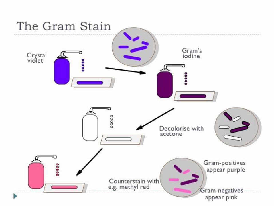
The Gram Stain ProcedureStep 1 - Prepare a Smear
“Bacteria”
Suspend some of the material to be stained in a drop of water on a microscope slide, spread the drop to about the size of a nickel. Fix by Heat or Allow to air dry.
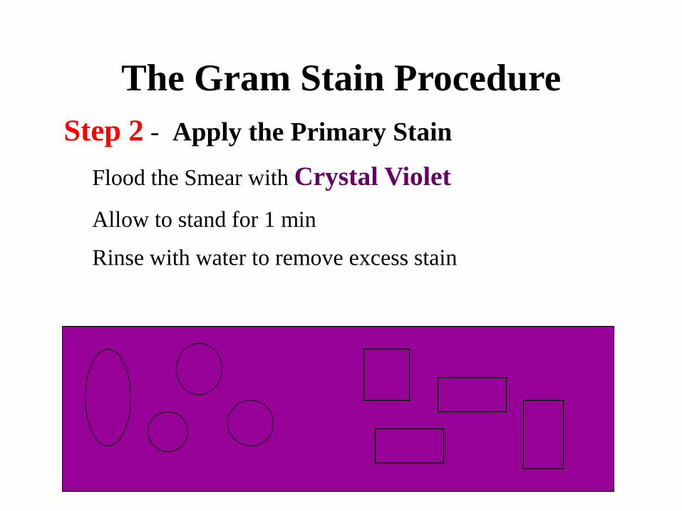
The Gram Stain Procedure
Step 2 - Apply the Primary Stain
Flood the Smear with Crystal Violet
Allow to stand for 1 min
Rinse with water to remove excess stain

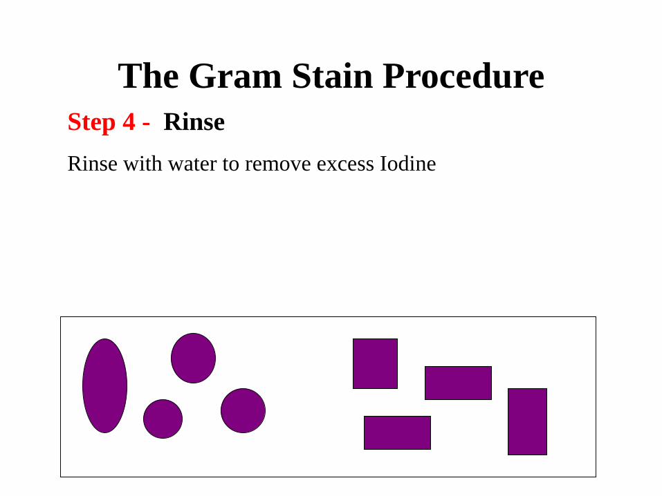
The Gram Stain Procedure
Step 4 - Rinse
Rinse with water to remove excess Iodine

The Gram Stain Procedure
Step 5 - Decolorize
Drip Decolorizer (80% Methanol +20% Acetone)
across the slide about 5 sec
The slide should appear clear

The Gram Stain Procedure
Step 6 - Rinse
Rinse with water to remove excess alcohol
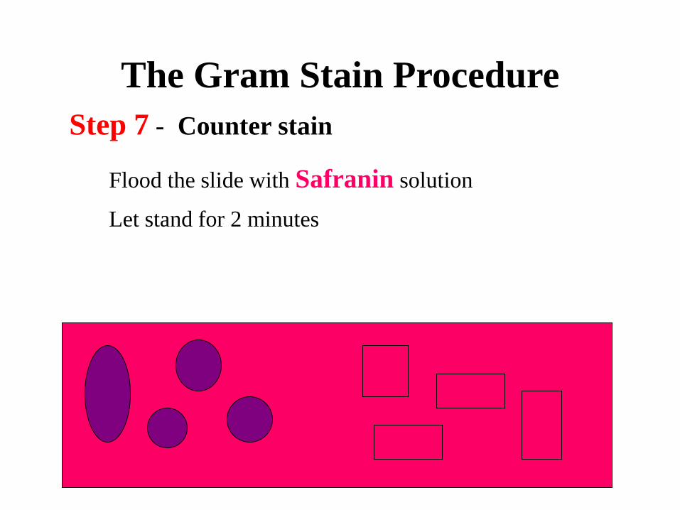
The Gram Stain Procedure
Step 7 - Counter stain
Flood the slide with Safranin solution
Let stand for 2 minutes
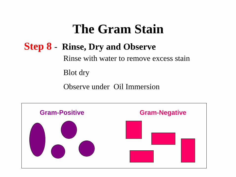
The Gram Stain
Step 8 - Rinse, Dry and Observe
Gram-Positive Gram-Negative
Rinse with water to remove excess stain
Blot dry
Observe under Oil Immersion

CELL WALL OF GRAM POS & NEG

CELL WALL IN GRAM +VE AND GRAM –VE BACTERIA
Cell Wall Structures Gram Positive organisms
Gram Negativeorganisms
Inner cytoplasmic membrane Present Present
Peptidoglycan layer Thick Thin
Teichoic Acid Present Absent
Outer membrane layer Absent Present
Lipid A, LPS , Lipo-protiencomponents
Absent Present
Peri-plasmic space Absent Present