Perioperative Complications and Outcome of Laparoscopic
Cholecystectomy in 20 Dogs
Jacqueline Scott1, Ameet Singh1, Philipp D. Mayhew2, J. Brad Case3, Jeffrey J. Runge4,
Matthieu Gatineau5, and Jessica Kilkenny1
1Department of Clinical Studies, Ontario Veterinary College, University of Guelph, Guelph, Ontario, Canada, 2Department of Surgical and
Radiological Sciences, University of California-Davis, Davis, California, 3Department of Small Animal Clinical Sciences, College of Veterinary
Medicine, University of Florida, Gainesville, Florida, 4Department of Clinical Studies, Matthew J. Ryan Veterinary Hospital at the University of
Pennsylvania, Philadelphia, Pennsylvania, and 5Centre V�et�erinaire DMV, Montreal, Quebec, Canada
Corresponding Author
Ameet Singh
Ontario Veterinary College
University of Guelph
Gordon Street
Guelph, Ontario, Canada
N1G 2W1
Submitted February 2016
Accepted May 2016
DOI:10.1111/vsu.12534
Objective: To report the complications and outcome of dogs undergoing
laparoscopic cholecystectomy for uncomplicated gall bladder disease.
Study Design: Multi-institutional case series.
Animals: Client-owned dogs (n520).
Methods: Medical records of dogs that underwent laparoscopic cholecystectomy
were reviewed and signalment, history, clinical and ultrasound examination findings,
surgical variables, and complications were collated. Laparoscopic cholecystectomy
was performed using a multiport approach. Data were compared between dogs with
successful laparoscopic cholecystectomy and dogs requiring conversion to open
cholecystectomy.
Results: Six dogs (30%) required conversion from laparoscopic to open
cholecystectomy due to inability to ligate the cystic duct (3), evidence of gall
bladder rupture (1), leakage from the cystic duct during dissection (1), and cardiac
arrest (1). Cystic duct dissection was performed in 19 dogs using an articulating
dissector (10), right angle forceps (7), and unrecorded (2). The cystic duct was
ligated in 15 dogs using surgical clips (5), suture (6), or a combination (4). All dogs
were discharged from the hospital and had resolution of clinical signs, although 1
dog developed pancreatitis and 1 dog required revision surgery for bile peritonitis.
There was no significant difference in preoperative blood analysis results, surgical
technique, or duration of hospitalization between dogs undergoing laparoscopic
cholecystectomy and cases converted to open cholecystectomy.
Conclusion: Laparoscopic cholecystectomy can be performed successfully for
uncomplicated gall bladder disease in dogs after careful case selection. The surgeon
considering laparoscopic cholecystectomy should be familiar with a variety
of methods for cystic duct dissection and ligation to avoid difficulties during the
procedure.
Laparoscopic surgery has become popular in veterinary med-
icine1 and is associated with faster return to function,
improved visualization, possible reduction in surgical site
infections, and reduced postoperative pain for selected surgi-
cal procedures in dogs.2–8
In people, laparoscopic cholecystectomy is performed
for symptomatic cholecystolithiasis and is associated with a
significantly shorter hospital stay and shorter convalescence
compared with open cholecystectomy.9–13 Recent reports
cite a conversion rate of 5–10% for multiport laparoscopic
cholecystectomy in people,9–11 most often due to adhesions
obscuring visualization, iatrogenic biliary duct injury, and
hemorrhage.10,13
Laparoscopic cholecystectomy has been described in a
clinical report of uncomplicated gall bladder mucoceles in 6
dogs without evidence of biliary tract rupture or obstruc-
tion.14 All dogs had successful laparoscopic cholecystectomy
without conversion to an open procedure, and all dogs
showed improvement or postoperative resolution of clinical
signs.14
For advanced laparoscopic procedures such as laparo-
scopic cholecystectomy, careful case selection is impor-
tant.14–17 Appropriate case selection for laparoscopic
cholecystectomy in dogs and factors that may indicate the
need for conversion to an open cholecystectomy are not
clearly identified. The objective of this study was to report
the complications and perioperative outcome of dogs under-
going laparoscopic cholecystectomy and describe details of
the laparoscopic procedure, including surgery duration and
conversion rate.
O49Veterinary Surgery 45 (2016) O49–O59VC Copyright 2016 by The American College of Veterinary Surgeons
MATERIALS AND METHODS
Case Selection
Medical records of dogs that underwent laparoscopic chole-
cystectomy for uncomplicated gall bladder disease at the
participating institutions from July 15, 2008 to July 15,
2015 were identified and reviewed. All dogs that underwent
laparoscopic cholecystectomy with or without conversion to
an open cholecystectomy were included. Candidates for lap-
aroscopic cholecystectomy were approximately based upon
previously published recommendations.14 Dogs with clinical
signs, persistent total serum bilirubin concentration eleva-
tions or evidence of gall bladder rupture, abdominal effu-
sion, or extra-hepatic biliary obstruction on ultrasound
examination were not considered candidates for laparo-
scopic cholecystectomy.14
Data retrieved from medical records included age, body
weight, sex, breed, clinical history, current medications, con-
current comorbidities, and physical examination findings.
Complete blood count, serum biochemistry analysis, coagu-
lation parameters, and urinalysis from the perioperative
period were recorded. Results of abdominal ultrasound
examination and results of any additional diagnostic imaging
was recorded and reviewed. Data for surgery included opera-
tive technique, intraoperative findings, surgery and general
anesthesia duration, results of bacterial culture, and histo-
logic findings of gall bladder and liver biopsies. Periopera-
tive complications, medications prescribed at hospital
discharge, recurrence of clinical signs, and duration of post-
operative hospitalization were noted.
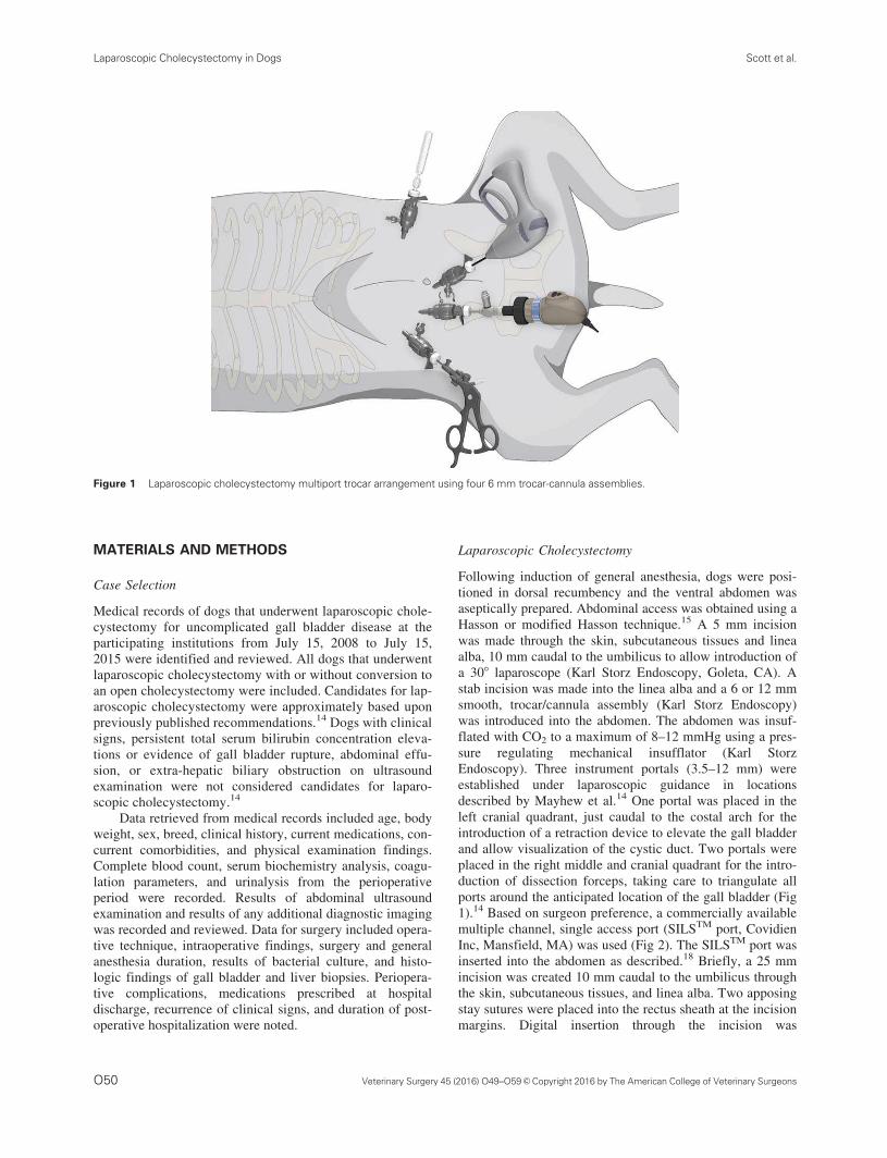
Laparoscopic Cholecystectomy
Following induction of general anesthesia, dogs were posi-
tioned in dorsal recumbency and the ventral abdomen was
aseptically prepared. Abdominal access was obtained using a
Hasson or modified Hasson technique.15 A 5 mm incision
was made through the skin, subcutaneous tissues and linea
alba, 10 mm caudal to the umbilicus to allow introduction of
a 308 laparoscope (Karl Storz Endoscopy, Goleta, CA). A
stab incision was made into the linea alba and a 6 or 12 mm
smooth, trocar/cannula assembly (Karl Storz Endoscopy)
was introduced into the abdomen. The abdomen was insuf-
flated with CO2 to a maximum of 8–12 mmHg using a pres-
sure regulating mechanical insufflator (Karl Storz
Endoscopy). Three instrument portals (3.5–12 mm) were
established under laparoscopic guidance in locations
described by Mayhew et al.14 One portal was placed in the
left cranial quadrant, just caudal to the costal arch for the
introduction of a retraction device to elevate the gall bladder
and allow visualization of the cystic duct. Two portals were
placed in the right middle and cranial quadrant for the intro-
duction of dissection forceps, taking care to triangulate all
ports around the anticipated location of the gall bladder (Fig
1).14 Based on surgeon preference, a commercially available
multiple channel, single access port (SILSTM port, Covidien
Inc, Mansfield, MA) was used (Fig 2). The SILSTM port was
inserted into the abdomen as described.18 Briefly, a 25 mm
incision was created 10 mm caudal to the umbilicus through
the skin, subcutaneous tissues, and linea alba. Two apposing
stay sutures were placed into the rectus sheath at the incision
margins. Digital insertion through the incision was
Figure 1 Laparoscopic cholecystectomy multiport trocar arrangement using four 6 mm trocar-cannula assemblies.
Laparoscopic Cholecystectomy in Dogs Scott et al.
O50 Veterinary Surgery 45 (2016) O49–O59VC Copyright 2016 by The American College of Veterinary Surgeons
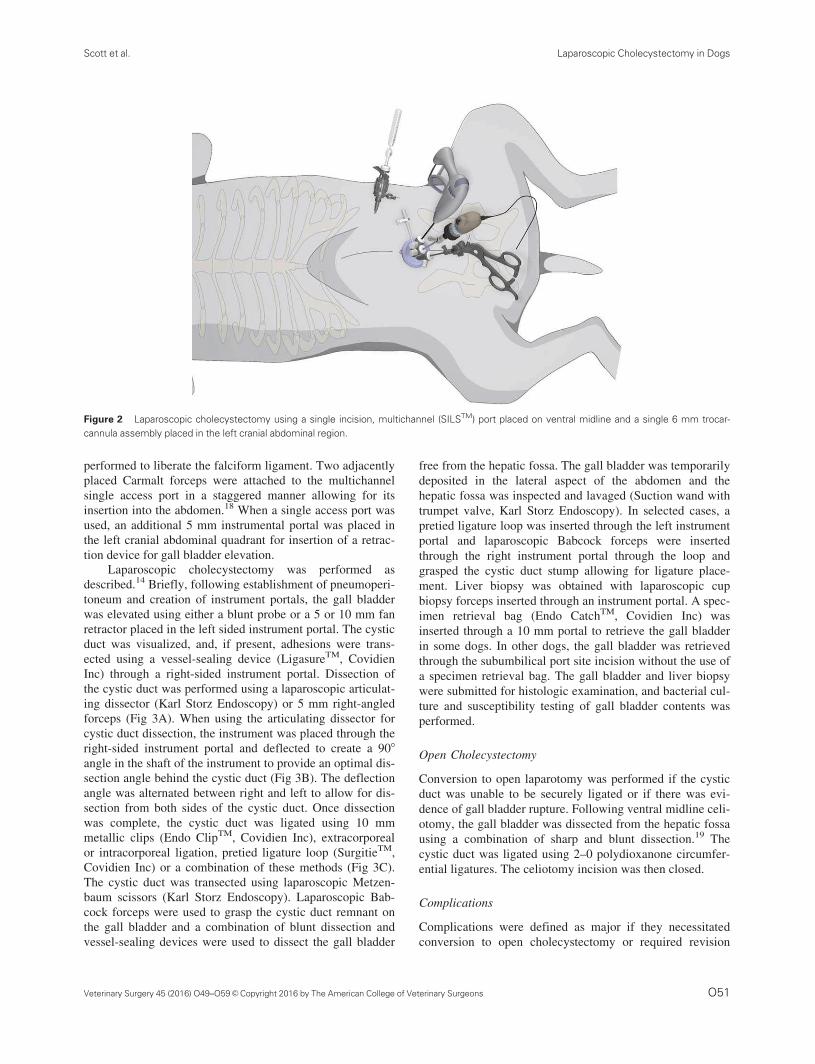
performed to liberate the falciform ligament. Two adjacently
placed Carmalt forceps were attached to the multichannel
single access port in a staggered manner allowing for its
insertion into the abdomen.18 When a single access port was
used, an additional 5 mm instrumental portal was placed in
the left cranial abdominal quadrant for insertion of a retrac-
tion device for gall bladder elevation.
Laparoscopic cholecystectomy was performed as
described.14 Briefly, following establishment of pneumoperi-
toneum and creation of instrument portals, the gall bladder
was elevated using either a blunt probe or a 5 or 10 mm fan
retractor placed in the left sided instrument portal. The cystic
duct was visualized, and, if present, adhesions were trans-
ected using a vessel-sealing device (LigasureTM, Covidien
Inc) through a right-sided instrument portal. Dissection of
the cystic duct was performed using a laparoscopic articulat-
ing dissector (Karl Storz Endoscopy) or 5 mm right-angled
forceps (Fig 3A). When using the articulating dissector for
cystic duct dissection, the instrument was placed through the
right-sided instrument portal and deflected to create a 908
angle in the shaft of the instrument to provide an optimal dis-
section angle behind the cystic duct (Fig 3B). The deflection
angle was alternated between right and left to allow for dis-
section from both sides of the cystic duct. Once dissection
was complete, the cystic duct was ligated using 10 mm
metallic clips (Endo ClipTM, Covidien Inc), extracorporeal
or intracorporeal ligation, pretied ligature loop (SurgitieTM,
Covidien Inc) or a combination of these methods (Fig 3C).
The cystic duct was transected using laparoscopic Metzen-
baum scissors (Karl Storz Endoscopy). Laparoscopic Bab-
cock forceps were used to grasp the cystic duct remnant on
the gall bladder and a combination of blunt dissection and
vessel-sealing devices were used to dissect the gall bladder
free from the hepatic fossa. The gall bladder was temporarily
deposited in the lateral aspect of the abdomen and the
hepatic fossa was inspected and lavaged (Suction wand with
trumpet valve, Karl Storz Endoscopy). In selected cases, a
pretied ligature loop was inserted through the left instrument
portal and laparoscopic Babcock forceps were inserted
through the right instrument portal through the loop and
grasped the cystic duct stump allowing for ligature place-
ment. Liver biopsy was obtained with laparoscopic cup
biopsy forceps inserted through an instrument portal. A spec-
imen retrieval bag (Endo CatchTM, Covidien Inc) was
inserted through a 10 mm portal to retrieve the gall bladder
in some dogs. In other dogs, the gall bladder was retrieved
through the subumbilical port site incision without the use of
a specimen retrieval bag. The gall bladder and liver biopsy
were submitted for histologic examination, and bacterial cul-
ture and susceptibility testing of gall bladder contents was
performed.
Open Cholecystectomy
Conversion to open laparotomy was performed if the cystic
duct was unable to be securely ligated or if there was evi-
dence of gall bladder rupture. Following ventral midline celi-
otomy, the gall bladder was dissected from the hepatic fossa
using a combination of sharp and blunt dissection.19 The
cystic duct was ligated using 2–0 polydioxanone circumfer-
ential ligatures. The celiotomy incision was then closed.
Complications
Complications were defined as major if they necessitated
conversion to open cholecystectomy or required revision
Figure 2 Laparoscopic cholecystectomy using a single incision, multichannel (SILSTM) port placed on ventral midline and a single 6 mm trocar-
cannula assembly placed in the left cranial abdominal region.
Scott et al. Laparoscopic Cholecystectomy in Dogs
O51Veterinary Surgery 45 (2016) O49–O59VC Copyright 2016 by The American College of Veterinary Surgeons

surgery and minor if they were transient with no intervention
required.
Outcome
Follow-up phone conversations and/or clinical examination
with owners determined short-term (<14 days postoperative)
and long-term (>30 days) outcomes. If the dog had died or
was euthanatized, the cause of death was recorded.
Statistical Analysis
Age, body weight, serum total bilirubin concentration,
serum alkaline phosphatase, alanine transaminase, and
Figure 3 Intraoperative images from a 10-year-old, castrated male, Shetland Sheepdog undergoing laparoscopic cholecystectomy. (A) A fan
retractor is used to elevate the gall bladder for visualization of the cystic duct. (B) The cystic duct is dissected with an articulating dissector.
(C) Placement of surgical clips prior to transection.
Laparoscopic Cholecystectomy in Dogs Scott et al.
O52 Veterinary Surgery 45 (2016) O49–O59VC Copyright 2016 by The American College of Veterinary Surgeons

gamma-glutamyl transferase activities, white blood cell count,
platelet count, surgical technique, surgery duration, anesthe-
sia duration, hospitalization duration, and postoperative stay
were categorized by conversion status (laparoscopic chole-
cystectomy vs. conversion to open cholecystectomy). Surgi-
cal technique was described by the total number of ports
placed, the use of SILSTM port compared to multiple single
ports, method of cystic duct dissection (right-angled forceps
and articulating forceps), method of cystic duct ligation
(suture, metallic clips, and combination), and the method of
gall bladder dissection (blunt probe, vessel-sealing device,
and combination). Normality of continuous variables was
confirmed using a Shapiro–Wilk test. Categorical variables
were compared with a Fisher's exact test, and continuous
variables were compared with a Student's t-test for normally
distributed data. Nonparametric data were tested with a Wil-
coxon rank-sum test. P�.05 was considered statistically
significant.
RESULTS
Cases
Twenty dogs were identified in the study period and
included. Breeds included Shetland Sheepdog (8), Mixed-
breed (3), Dachshund (2) and 1 each of a Jack Russell, Pom-
eranian, Cocker Spaniel, Cairn Terrier, West Highland White
Terrier, Bichon Frise, and an Italian Greyhound. The age at
diagnosis ranged from 5 months to 14.7 years (median 10.4).
The body weight ranged from 3.8 to 16.3 kg (median 9.4).
There were 10 spayed females (50%), 9 castrated males
(45%), and 1 intact male (5%). There was no significant dif-
ference in sex (P5.55), age (P5.27), or body weight
(P5.48) between dogs undergoing laparoscopic cholecystec-
tomy vs. cases requiring conversion to open cholecystec-
tomy. Comorbidities included valvular heart disease (7),
hypothyroidism (3), diabetes mellitus (2), hyperadrenocorti-
cism (2), cardiac arrhythmia (1), tracheal collapse (1),
immune-mediated thrombocytopenia (1), immune-mediated
hemolytic anemia (1), seizures (1), and oral melanoma (1).
Preoperative Findings
Fourteen dogs (70%) presented with clinical signs (Table 1).
Six dogs (30%) had evidence of gall bladder disease on
abdominal ultrasound examination that was an unexpected
finding. There was no significant difference in preoperative
serum total bilirubin concentration, serum alkaline phospha-
tase, alanine transaminase and gamma-glutamyl transferase
activities, white blood cell count, or platelet count between
dogs undergoing laparoscopic vs. cases requiring conversion
to open cholecystectomy (Table 2). Coagulation parameters
(prothrombin time, partial thromboplastin time) were
assessed in 6 dogs and were within reference intervals.
Three-view thoracic radiographs were obtained prior to
surgery in 10 dogs and were considered normal, except in 1
dog with mild left atrial enlargement. Abdominal ultrasound
examination was performed for all dogs. There was evidence
of mucocele development with echogenic bile sludge and
stellate bile patterns in 18 dogs.20 Cholelithiasis was present
in 3 dogs but choledocholithiasis was not observed in any
dog. A well circumscribed echogenic mass was seen in the
lumen of the gall bladder in 2 dogs and gall bladder neopla-
sia was suspected.
The gall bladder length and width were reported in the
ultrasound examination for 12 dogs and ranged from 72 3
37 mm to 16 3 14 mm (Table 3). Subjective assessment of
size was reported for 10 dogs and considered severely dis-
tended (1), prominent (1), moderately distended (4), mildly
distended (3), and not dilated (1). Cystic duct diameter was
recorded for 6 dogs, ranging from 3 to 7 mm (median 6).
One dog had multifocal small intestinal muscularis and
Table 1 Number of dogs (%) with clinical signs at presentation in
dogs undergoing laparoscopic cholecystectomy or conversion to open
cholecystectomy
Number of dogs
Clinical
signs
Laparoscopic
n514
Open
n56
Total
n520
None 4 (31%) 2 (29%) 6 (30%)
Vomiting 6 (46%) 4 (57%) 10 (50%)
Anorexia 6 (46%) 3 (43%) 9 (45%)
Lethargy 5 (38%) 2 (29%) 7 (35%)
Polyuria/
polydipsia
1 (8%) 1 (14%) 2 (10%)
Diarrhea 2 (15%) 0 2 (10%)
Abdominal pain 3 (23%) 2 (29%) 5 (25%)
Depression 1 (8%) 1 (14%) 2 (10%)
Panting 1 (8%) 0 1 (5%)
Table 2 Comparison of preoperative serum biochemistry and hematology for dogs undergoing laparoscopic cholecystectomy or conversion
to open cholecystectomy
Parameter (reference interval)
Laparoscopic Median
(range) n516
Converted cases
Mean (range) n56 P-value
Total bilirubin (0–0.2 mg/dL) 0.3 (0.06–1.9) 0.7 (0.06–3.7) 0.79
ALP (23–143 U/L) 357.6 (56–738) 607.6 (128–1095) 0.51
ALT (19–107 U/L) 277.2 (37–1464) 236 (37–788) 0.97
GGT (0–7 U/L) 20.4 (0–125) 18.6 (6–31) 0.22
White blood cell count (4.9–15.4 3 109/L) 10.4 (5.7–19.5) 14.3 (5.5–35.6) 0.32
Platelet count (117–4183 109/L) 411.5 (215–784) 404.7 (231–530) 0.91
ALP, alkaline phosphatase; ALT, alanine transaminase; GGT gamma-glutamyl transferase.
Scott et al. Laparoscopic Cholecystectomy in Dogs
O53Veterinary Surgery 45 (2016) O49–O59VC Copyright 2016 by The American College of Veterinary Surgeons

submucosal thickening. Four dogs had course hepatic paren-
chyma and 2 dogs had mild bilateral adrenomegaly. Two
dogs had mild bilateral chronic renal degenerative changes
and 1 dog had hypoechoic nodules within the spleen.
Laparoscopic Cholecystectomy
Various surgical approaches and instrumentation was used,
according to surgeon preference. All procedures used a multi-
port technique. Procedures used 4 portals14,19 (11), 3 portals
(1), 2 portals (1), and SILSTM port (7). The subumbilical cam-
era port was a 12 mm port (11), 6 mm port (1), or 3.5 mm port
(1). The left instrument port was a 12 mm port (6) or 6 mm
port (14). The right instrument port used a 6 mm port (18) or
3.5 mm port (1). A second instrument portal was placed in the
left cranial quadrant rather than the right cranial quadrant in 1
dog. A 3 port technique, omitting the second right sided
instrument portal, was used in 1 dog and 2 ports were placed
with a 12 mm port as the subumbilical camera port and a
12 mm port as the left instrument port in 1 dog. The most
common port placement combined with the SILSTM port in 6/
7 dogs was to place the SILSTM port in the subumbilical loca-
tion, with the placement of two 6 mm instrument portals para-
median in the right and left cranial abdominal quadrants. In 1/
7 SILSTM port applications, there was only 1 additional instru-
ment port in the left cranial quadrant for placement of the fan
retractor for gall bladder elevation. The use of multiple, single
ports compared to the multi-channel single access port device
did not significantly affect the conversion rate (P51.0), nor
did the total number of ports placed (P5.4).
The gall bladder was elevated towards the ventral body
wall with an instrument placed in the left cranial abdominal
instrument portal in all dogs. Elevation was performed using
a 5 mm fan retractor (13), a 10 mm fan retractor (6), or a
blunt probe (1).
Surgery was converted to open cholecystectomy in 1 dog
after initial exploratory revealed bile leakage and gall bladder
rupture. Cystic duct dissection prior to dissection of the gall
bladder from the hepatic fossa was performed in 18 dogs. In 1
dog, the gall bladder was dissected from the hepatic fossa
prior to cystic duct dissection, which resulted in moderate
hemorrhage and a dependent gall bladder. This obscured visu-
alization of the cystic duct and required conversion to open
cholecystectomy. The method of cystic duct dissection was
not recorded (1), by articulating forceps (10) or 5 mm right-
angled forceps (7). Conversion was required during cystic
duct dissection due to mild iatrogenic biliary leakage or an
inability to safely ligate the cystic duct in 3 dogs. The cystic
duct spread to a width >10 mm, precluding the use of the sur-
gical clips and requiring conversion in 2/3 dogs. Sudden car-
diac arrest necessitating open chest compressions through the
diaphragm required conversion in 1 dog. This dog was suc-
cessfully resuscitated and the cholecystectomy was completed
as an open procedure. The method of cystic duct dissection
was not significantly different between the laparoscopic and
converted cases (P51.0).
Cystic duct ligation during laparoscopic cholecystec-
tomy was successfully performed in the remaining 15 dogs
using metallic clips (9) of which 4 were augmented with
extracorporeal techniques (Roeder knot) or suture alone
(6) with either intracorporeal or extracorporeal techniques
(modified 4S Roeder knot). Two or more ligatures remained
on the proximal end of the duct following transection in all
dogs. There was no significant association between the mate-
rial or method of ligation used between laparoscopic and
converted cases (P51.0).
Table 3 Preoperative appearance and dimensions of the gall bladder and bile ducts on ultrasound examination of dogs undergoing laparoscopic
cholecystectomy or conversion to open cholecystectomy
Gall bladder size Cystic duct size Common bile duct size
Group Descriptive Width 3 length (cm) Wall thickness (cm) Width (cm) Width (cm)
Converted to Open
Cholecystectomy
1 0.6
2 Prominent 0.9
3 Severe distension
4 4.4 3 2.9 0.2 0.3 0.4
5
6 7.2 3 3.7 0.6 2
Laparoscopic 1 Mild distension
2 Mild distension
3 Mild distension 4.1 3 2.5 < 0.7
4 Rounded 6.4 3 3.5 0.3
5 53 3
6 Moderate distension 3.6 3 2 0.2
7 Moderate distension 3.4 3 7.5
8 3.3 3 2.5 Normal
9 Moderate distension 2.6 3 3.3
10 1.6 3 1.4 0.3 0.6 0.2
11 1.3 3 3.7
12 Moderate distension
13 Not dilated
14 4.2 3 3.1 0.6
Laparoscopic Cholecystectomy in Dogs Scott et al.
O54 Veterinary Surgery 45 (2016) O49–O59VC Copyright 2016 by The American College of Veterinary Surgeons
The gall bladder was dissected free from the hepatic
fossa during laparoscopic cholecystectomy in 15 dogs using
a bipolar vessel-sealing device (7), harmonic scalpel (4) or
blunt dissection alone (4). The method of gallbladder dissec-
tion was not significantly different between laparoscopic and
converted cases (P51.0). Intraoperative bleeding was con-
sidered mild (14) and moderate (1) during dissection.
A specimen retrieval bag inserted through the 10 mm
subumbilical port was used in 8 dogs, and not used in 6, with
only mild spillage of bile during extraction in 1/6.
Laparoscopic cholecystectomy was successfully com-
pleted in 14 dogs (70%) and conversion to open cholecystec-
tomy was required in 6 dogs (30%) due to cystic duct
anatomy precluding secure ligation (2), evidence of gall blad-
der rupture (1), leakage from cystic duct during dissection (1),
cardiac arrest (1), and dissection of the gall bladder initially
prior to cystic duct dissection, which obscured visualization of
the cystic duct (1). The anesthesia and surgery duration for
laparoscopic cholecystectomy ranged from 120 to 285
minutes (median 175) and 72 to 180 minutes (median 108),
respectively. There were significantly more intraoperative
complications (yes/no) in the cases converted to open chole-
cystectomy (P5.023). The anesthesia and surgery duration
for the converted open cholecystectomy cases was 135–285
minutes (median 205) and 75–175 minutes (median 103),
respectively. There was no significant difference in anesthesia
or surgery duration between laparoscopic cholecystectomy
and cases requiring conversion to open cholecystectomy.
Histologic and Microbiologic Findings
Histologic examination of the gall bladder was performed in
all dogs and showed evidence of mucosal cystic hyperplasia,
cholecystitis or mucocele formation in 18 dogs with 3/18 hav-
ing concurrent cholelithiasis. The remaining 2 dogs had a pap-
illary adenoma in the gall bladder (1) and mural hemorrhage
and fibrosis consistent with previous rupture or infarction (1).
Both these dogs had successful laparoscopic cholecystectomy.
Liver biopsy was performed in 18 dogs with cholestasis with
biliary hyperplasia, portal hepatitis, and cholangitis (17) and
no abnormalities (1). Bacterial culture of the gall bladder con-
tents resulted in no growth (8), Enterococcus sp. (2), Esche-
richia coli (2), Lactobacillus acidophilus (1), or Clostridium
sp. (1).
Postoperative Complications
One minor and 2 major complications were noted postopera-
tively and there was no significant difference in the frequency
of postoperative complications between the laparoscopic and
converted cases (P51.0). One dog undergoing laparoscopic
cholecystectomy had marked bruising of the ventral abdomen
4 days postoperative, despite normal coagulation parameters
at the time of presentation. No intervention was required and
the bruising resolved by 10 days postoperative. One dog
undergoing open cholecystectomy developed pancreatitis and
ketosis, which resolved with medical management. The third
dog had undergone laparoscopic cholecystectomy and
required surgical revision due to hepatic duct leakage and bile
peritonitis 6 days postoperative. There had been no intraopera-
tive complications noted during the initial procedure in this
dog except for mild bleeding from the quadrate lobe which
resolved without intervention. Two modified Roeder extracor-
poreal knots were used to ligate the cystic duct, prior to trans-
ection between them. An additional ligature was placed on the
cystic duct following transection using a pretied ligature loop
(SurgitieTM). At revision, a partial quadrate liver lobectomy
was performed to resolve a leaking hepatic duct. This dog
recovered without further complication and, at 446 days post-
operative, was clinically normal despite persistent elevations
in serum alanine transaminase activity.
Outcome
Hospitalization duration ranged from 2 to 7 days (median 2)
and all dogs were discharged from hospital. The hospitaliza-
tion duration was not significantly different for dogs under-
going laparoscopic cholecystectomy vs cases requiring
conversion to open cholecystectomy (laparoscopic: range
24–96 hours, median 27 hours, open: range 24–49 hours,
median 27 hours; P5.3). All dogs were discharged with
analgesics (tramadol, buprenorphine and/or meloxicam), and
continued on ursodeoxycholic acid and s-adenosylmethio-
nine/silybin supplements, if previously prescribed. Antibiot-
ics were continued or started in 11 dogs (clindamycin,
amoxicillin/clavulonic acid, enrofloxacin, metronidazole,
and chloramphenicol).
Short-term outcome was available for 19 dogs (14 lapa-
roscopic, 5 open) and long-term outcome for 16 dogs (12
laparoscopic, 4 open) with follow-up ranging from 10 to 900
days (median 268). All dogs with clinical signs attributable
to gall bladder disease had resolution of these signs in 1–14
days, except for the dog that represented 6 days after laparo-
scopic cholecystectomy for acute abdominal pain due to bile
peritonitis. The duration until resolution of clinical signs was
significantly shorter for dogs undergoing laparoscopic chole-
cystectomy vs cases requiring conversion to open cholecys-
tectomy (laparoscopic median 1 days, open median 7 days,
P5.047). Follow-up serum biochemistry at 2 days to 3
months showed persistent elevation of liver enzyme activity
in 12 dogs. Long-term outcomes were reported for 19 dogs
with survival to follow-up in 14 dogs (74%), euthanasia or
death for unrelated disease in 5 dogs (hemoabdomen, urinary
bladder transitional cell carcinoma, renal failure, progressive
endocrine disease, and thrombocytopenia). There was no sig-
nificant difference in survival time for dogs undergoing lapa-
roscopic cholecystectomy vs cases requiring conversion to
open cholecystectomy (P5.37).
DISCUSSION
Laparoscopic cholecystectomy was performed successfully
in 70% of dogs in this cohort using a multi-port technique
and 95% of dogs that were discharged from hospital had
resolution of clinical signs. Only 1 dog required revision
Scott et al. Laparoscopic Cholecystectomy in Dogs
O55Veterinary Surgery 45 (2016) O49–O59VC Copyright 2016 by The American College of Veterinary Surgeons

surgery, in this case for hepatic biliary duct leakage. Previ-
ously described criteria for case selection for laparoscopic
cholecystectomy recommended dogs have no evidence of
biliary obstruction on abdominal ultrasound examination or
on serum total bilirubin concentration.14 The current recom-
mendation for open cholecystectomy is to ensure patency of
the common bile duct with an enterotomy over the major
duodenal papilla and the introduction of a catheter to flush
the common bile duct.19 During laparoscopic cholecystec-
tomy, the patency of the common bile duct cannot be
assessed, although this is done during laparoscopic cholecys-
tectomy in people.34 Thus, it is important for surgeons con-
sidering laparoscopic cholecystectomy in dogs, to be
confident that there is no obstruction of the common bile
duct. Malek et al reported no significant association on sur-
vival for dogs undergoing open cholecystectomy for gall
bladder mucocele where the common bile duct was catheter-
ized to ensure patency compared to those dogs where com-
mon bile duct patency was not assessed.25 However, these
results may be biased based on intraoperative findings.25 In
our study, 6 dogs had elevated serum total bilirubin concen-
tration; however, postoperative complications related to bili-
ary obstruction were not observed. There was no difference
in the serum total bilirubin concentration for dogs under-
going laparoscopic cholecystectomy vs. dogs requiring con-
version to open cholecystectomy. Multiple studies have
failed to find significant differences in serum total bilirubin
concentration, and serum alkaline phosphatase, alanine trans-
aminase and gamma-glutamyl transferase activities, in dogs
undergoing open cholecystectomy, regardless of out-
come.25,46 The findings of previous studies along with the
results of our study suggest that dogs with mild elevations in
serum total bilirubin concentrations may be acceptable can-
didates for laparoscopic cholecystectomy.
Shetland Sheepdogs were overrepresented (8/20) in our
study and the most common clinical signs at presentation for
all dogs was vomiting, lethargy, and reduced food intake, all
findings consistent with previous reports.21,23–26 Comorbid-
ities were noted in the majority of dogs, including hyperadre-
nocorticism, hypothyroidism, and diabetes mellitus. There is
a known association of gall bladder mucoceles with endocri-
nopathies although the pathogenesis remains unclear.24–26
The reports on findings of the abdominal ultrasound
examination were inconsistent, prohibiting clear assessment
of gall bladder size as selection criteria for laparoscopic cho-
lecystectomy. Only 3/6 dogs that were converted to open
cholecystectomy had gall bladder size recorded, all of which
were marked enlarged. In comparison, most dogs undergoing
successful laparoscopic cholecystectomy had mild enlarge-
ment of the gall bladder. Besso et al reported the average
gall bladder volume of dogs with mucoceles was enlarged at
87 mL, compared to the approximated normal gall bladder
volume in a medium sized dog of 15 mL.20 We did not docu-
ment gall bladder volume. Gall bladder wall thickness is not
a good indicator of mucosal hyperplasia or cholecystitis in
dogs,20,26 although in people, gall bladder wall thickness can
be predictive of laparoscopic operability.27,28 Two dogs that
required open cholecystectomy had a gall bladder wall thick-
ness greater than normal (2–3 mm).21,29
Preoperative abdominal ultrasound examination showed
evidence of localized inflammation around the cystic duct in
1 dog. Initial laparoscopic exploration showed this dog had a
gall bladder rupture and required immediate conversion to
open cholecystectomy. Evidence of bile leakage during open
cholecystectomy has no impact on survival and is usually a
localized nonseptic bile peritonitis21,25,30 but correct identifi-
cation preoperatively is important to determine appropriate
laparoscopic cholecystectomy candidates.14 In people, the
presence of pericholecystic fluid and acute cholecystitis is
not a contraindication for laparoscopic cholecystectomy but
is associated with higher conversion rates.9,13 This dog could
have been ruled out as a candidate based on the preoperative
ultrasound findings; however, another dog with evidence of
hyperechoic fat and localized inflammation around the gall
bladder on abdominal ultrasound examination did not have
any evidence of gall bladder rupture or peritonitis and was dis-
charged after laparoscopic cholecystectomy <32 hours post-
operative. These dogs did, however, have different clinical
signs on presentation; the former with vomiting and lethargy
and moderate elevations in serum biochemistry parameters,
the latter without clinical signs and minimal serum biochemis-
try changes. Ultrasound examination is considered a sensitive
and specific tool in detecting bile peritonitis at 85.7 and
100%, respectively,24 but the outcomes of these 2 dogs with
similar findings on ultrasound examination highlight the com-
plexity of case selection for laparoscopic cholecystectomy.
Clinical presentation must be considered in conjunction with
findings on ultrasound examination (gall bladder size, gall
bladder wall thickness, gall bladder wall integrity, and cystic
duct size).
A case series of laparoscopic cholecystectomy in 6 dogs
first described a surgical technique for laparoscopic chole-
cystectomy.14 Variations of this technique were used by dif-
ferent surgeons in our study but no method of approach,
dissection or ligation was associated with conversion to open
cholecystectomy. Single incision laparoscopic cholecystec-
tomy in people is purported to improve cosmesis, reduce
morbidity, and minimize hospital stay.31,32 Gonzalez-Gasch
and Monnet reported on the SILSTM port system for elective
laparoscopic procedures in dogs, citing reduced surgery
duration and complication rate compared to a multi-port
technique.33 Port placement, number and size was not associ-
ated with conversion; however, a 12 mm subumbilical cam-
era portal and 12 mm instrument portal in the left cranial
quadrant appeared beneficial for successful laparoscopic
cholecystectomy. The 12 mm subumbilical portal allows
insertion of a 5 mm laparoscope, an instrument for dissection
if the angle is advantageous and a specimen retrieval bag for
gall bladder extraction through incisional enlargement on
ventral midline. The introduction of a 10 mm fan retractor in
the left paramedian portal for gall bladder elevation subjec-
tively appeared to improve visualization of the cystic duct.
The shift from using a 5 to a 10 mm fan retractor was made
as we gained experience, and this assisted in elevation of
moderately distended gall bladders, without damage to
Laparoscopic Cholecystectomy in Dogs Scott et al.
O56 Veterinary Surgery 45 (2016) O49–O59VC Copyright 2016 by The American College of Veterinary Surgeons

adjacent liver parenchyma. The larger width of the 10 mm
fan blades may create less pressure and tearing of hepatic
parenchyma than the 5 mm blades. The use of a SILSTM port
may minimize surgery duration and morbidity through a
reduction in portal incisions33 while allowing for extraction
of the gall bladder without further extension of an instrument
portal. The SILSTM port was used with a left paramedian
portal in 7 dogs, which the authors believe is essential for
gall bladder elevation.
We believe the use of articulating forceps, initially
designed for use in the SILSTM port to reduce instrument
clashing during cystic duct dissection, improved the angle
for cystic duct dissection compared with straight 5/10 mm
right angle forceps. By deflecting the forceps to 70–908, a
safe angle for dissection around the cranial aspect of the
cystic duct was created and allowed the entire width of the
duct to be encompassed, especially in dogs where the duct
was >5 mm. While the articulating forceps are helpful,
straight right-angled forceps can also be used for safe dissec-
tion of the cystic duct.
Iatrogenic injury of a biliary duct during dissection is
reported in 0.3–2.7% of people undergoing laparoscopic cho-
lecystectomy.10,34 Risk factors include aberrant anatomy,
inflammation, and surgeon inexperience.34 Multiple imaging
modalities are proposed to facilitate dissection including
intraoperative cholangiography, intraoperative laparoscopic
ultrasound examination, and near-infrared fluoroscopic chol-
angiography.10,35 None of these imaging modalities were
used in this study but may be examined in the future.
If biliary duct injury is identified intraoperative, it should
be addressed immediately, which in people involves direct
laparoscopic repair.34 Cystic duct leakage was observed in 1
dog during cystic duct dissection. This leak was mild, identi-
fied quickly and well contained. Although not required, this
could have been addressed during laparoscopy with the place-
ment of ligatures or endoscopic clips.
The occurrence of intraoperative complications was sig-
nificantly more frequent in dogs undergoing open cholecys-
tectomy. This is not unexpected since the reason for open
conversion was a complication during laparoscopy. To deter-
mine an unbiased frequency of complication for laparoscopic
vs. open cholecystectomy, a prospective, randomized trial,
controlling for case complexity, would be required. We had
a 30% conversion rate to open cholecystectomy but believe
this rate could be further reduced with careful case selection
and familiarity with multiple techniques for laparoscopic
cystic duct ligation. We used metallic clips or suture material
to occlude the cystic duct. In people, the use of a self-
locking polymer clip (Hem-O-Lok clipTM, Weck, Research
Triangle Park, NC) is preferred over concern for migration
or dislodgement of the metallic clips.36–38 The polymer clip
has not been evaluated in laparoscopic cholecystectomy in
dogs, but clinical reports have been favourable.39 The use of
a bioabsorbable polymer clip (Laparo clipTM, Covidien Inc)
has also been investigated,36 which may be preferential to
avoid potential nidus stone formation in the cystic duct.40
The best method of cystic duct ligation cannot be discerned
from our study or others due to the lack of randomized
control trials.41 Bile peritonitis did develop 6 days postopera-
tive in 1 dog undergoing laparoscopic cholecystectomy but
was due to inadvertent hepatic duct trauma rather than suture
slippage from the transected cystic duct. While suture or sur-
gical clips appears appropriate for ligation of the cystic duct
during laparoscopic cholecystectomy in dogs, the laparo-
scopic surgeon should be capable of performing other meth-
ods of cystic duct ligation, especially for very dilated cystic
ducts. It is imperative that whatever method of cystic duct
ligation is performed results in complete occlusion of the
entire width of the duct, which is not always possible with
laparoscopic surgical clips.42 Conversion to open cholecys-
tectomy was required in 2 dogs due to spreading of the cystic
duct beyond the capacity of a 10 mm surgical clip. Had intra-
corporeal or extracorporeal suture techniques or a self-
locking polymer clip been used, conversion may have been
avoided. The use of a vessel-sealing device on the ligation
and transection of cystic ducts in healthy canine cadavers is
comparable to 10 mm metallic surgical clips.42 The vessel-
sealing device is a rapid and effective way to ligate and tran-
sect the cystic duct but in vivo investigation is required.
The gall bladder was dissected free from the hepatic
fossa prior to cystic duct isolation and dissection in 1 dog
during laparoscopy. This fundus-first technique is usually
performed in open cholecystectomy19 and is used in people
during laparoscopy where excessive adhesions make access
to the cystic duct difficult.43,44 Differences in the pathology
of gall bladder disease in dogs19,22 compared to people9–12
make this less viable since most dogs present for cholecys-
tectomy of gall bladder mucocele, where the gall bladder is
engorged and heavy with inspissated bile. The weight of the
free gall bladder after dissection, combined with the diffi-
culty of retracting the heavy gall bladder ventrally and the
presence of hemorrhage from the hepatic fossa, make access
to the cystic duct difficult. We recommend a blunt instru-
ment (fan retractor, blunt probe and Cuschieri retractor) be
used for gall bladder elevation when dissecting the cystic
duct because of the fragile gall bladder wall, especially for
gall bladder mucoceles. This is in contrast to the approach in
people where the gall bladder is suspended with a transabdo-
minal suture or grasping instrument.10,32,34
One dog had a cardiac arrest during cystic duct ligation.
This dog was stable preoperative with clinical signs of abdom-
inal pain and mild changes on preoperative serum chemistry
analysis. Some dogs with hepatobiliary disease are poor anes-
thetic candidates due to hypotension associated with vagal
response during gall bladder manipulation, and endotoxemia
secondary to extra-hepatic biliary obstruction.45,46 Subjects
with postoperative hypotension are reported with poorer out-
comes25,30,46 but a recent retrospective study did not find
anesthetic complications in dogs undergoing cholecystectomy
to be any different from dogs undergoing other hepatic sur-
gery.45 Whether the dog in our study had a vagal-induced bra-
dycardic event or an anesthetic-related event is unknown.
The specimen retrieval bag was used in only 8 dogs,
although it is recommended for the extraction of the gall
bladder to minimize spillage of bile, and allow aspiration
of bile contents once partially exteriorized to collapse the
Scott et al. Laparoscopic Cholecystectomy in Dogs
O57Veterinary Surgery 45 (2016) O49–O59VC Copyright 2016 by The American College of Veterinary Surgeons

gall bladder and allow it to be removed through a smaller
incision.14
Extended surgery duration is referenced as a disadvant-
age of some laparoscopic procedures.2,3,5,8 Expertise impacts
surgery duration in laparoscopic cholecystectomy in peo-
ple47,48 but duration is also related to severity of disease.
Duration of surgery in our study was comparable to previous
reports for laparoscopic14 and open cholecystectomy.25
People have a faster convalescence and shorter hospital
stay after laparoscopic cholecystectomy.11,49 The duration of
hospitalization in our study was not different for dogs under-
going laparoscopic cholecystectomy compared to cases that
were converted, although resolution of clinical signs was
shorter for dogs undergoing laparoscopic cholecystectomy.
Interpretation of these results is inconclusive as results may
varying according to disease severity.24,50
Predictive factors for conversion to open cholecystec-
tomy in people include increased white blood cell count,
ultrasound findings of pericholecystic fluid, low serum albu-
min, elevated serum total bilirubin, right cranial abdominal
pain during ultrasound, being male, obesity, comorbidities,
and the presence of a thickened gall bladder wall.13,28,31,49
Since the pathogenesis of disease differs in canines,9–12,19,22
extrapolation of risk factors from people is not possible;
however, careful consideration of preoperative imaging and
the clinical status of the dog is imperative for successful lap-
aroscopic cholecystectomy. We did not find any factors
related to conversion to open cholecystecomy in our study.
Four of 6 conversions in our study were related to techni-
cal issues, lack of confidence performing intracorporeal or
extracorporeal suture techniques when the cystic duct was too
wide for metallic clips, dissection of the gall bladder prior to
the cystic duct impeding visualization, and early conversion
due to biliary leakage. These situations potentially represent a
learning curve in the procedure and it is expected lower con-
version rates for laparoscopic cholecystectomy can be
expected as experience and technical aptitude improve.
This study is limited by possible bias introduced by sur-
geon preferences in case selection and surgical technique.
Laparoscopic cholecystectomy is a relatively new procedure
in veterinary medicine and various centers are developing
and personalizing the technique. As such, we were only able
to examine a small sample size which limits the power of
comparisons.
We had good success with laparoscopic cholecystec-
tomy in a small cohort of dogs. Our results demonstrate that
laparoscopic cholecystectomy can be performed in dogs with
uncomplicated gall bladder disease, with a low complication
rate and an acceptable conversion rate. We expect conver-
sion rates will decrease with gains in clinical experience and
advances in training, as noted in human surgery.10,34 Appro-
priate case selection is imperative to ensure there is no evi-
dence of extra-hepatic biliary obstruction or gall bladder
leakage to limit the risk of conversion. The surgeon consider-
ing laparoscopic cholecystectomy should be familiar with a
variety of methods for cystic duct dissection and ligation to
avoid complications and reduce the rate of conversions.
DISCLOSURE
The authors declare no conflicts of interest related to this
report.
REFERENCES
1. Bleedorn JA, Dykema JL, Hardie RJ: Minimally invasive
surgery in veterinary practice: a 2010 survey of Diplomates and
residents of the American College of Veterinary Surgeons. Vet
Surg 2013;42:635–642
2. Arulpragasm SP, Case JB, Ellison GW: Evaluation of costs and
time required for laparoscopic-assisted versus open cystotomy
for urinary cystolith removal in dogs: 43 cases (2009-2012).
J Am Vet Med Assoc 2013;243:703–708
3. Case JB, Boscan PL, Monnet EL, et al: Comparison of surgical
variables and pain in cats undergoing ovariohysterectomy,
laparoscopic-assisted ovariohysterectomy, and laparoscopic
ovariectomy. J Am Anim Hosp Assoc 2015;51:1–7
4. Culp WTN, Mayhew PD, Brown DC: The effect of
laparoscopic versus open ovariectomy on postsurgical activity
in small dogs. Vet Surg 2009;38:811–817
5. Davidson EB, Moll HD, Payton ME: Comparison of
laparoscopic ovariohysterectomy and ovariohysterectomy in
dogs. Vet Surg 2004;33:62–69
6. Devitt CM, Cox RE, Hailey JJ: Duration, complications, stress,
and pain of open ovariohysterectomy versus a simple method of
laparoscopic-assisted ovariohysterectomy in dogs. J Am Vet
Med Assoc 2005;227:921–927
7. Gower S, Mayhew P: Canine laparoscopic and laparoscopic-
assisted ovariohysterectomy and ovariectomy. Compend Contin
Educ Vet 2008;30:430–440
8. Hancock RB, Lanz OI, Waldron DR, et al: Comparison of
postoperative pain after ovariohysterectomy by harmonic
scalpel-assisted laparoscopy compared with median celiotomy
and ligation in dogs. Vet Surg 2005;34:273–282
9. Comitalo JB. Laparoscopic cholecystectomy and newer
techniques of gallbladder removal. J Soc Laparoendo Surg
2012;16:406–412
10. Narilli P, Filippo A, Campli M, et al: Complications: how to
prevent and manage them, in Agresto F, Campanile F, Vettoretto
N (eds): Laparoscopic cholecystectomy: an evidence-based
guide. New York, NY, Springer, 2014, pp 89–101
11. Keus F, de Jong J, Gooszen HG, et al: Laparoscopic versus
open cholecystectomy for patients with symptomatic
cholecystolithiasis (review). Cochrane Database Syst Rev,
2006;1–157.
12. Urbach DR, Stukel TA: Rate of elective cholecystectomy and
the incidence of severe gallstone disease. Can Med Assoc J
2005;172:1015–1019
13. Rosen M, Brody F, Ponsky J: Predictive factors for conversion
of laparoscopic cholecystectomy. Am J Surg 2002;184:254–258
14. Mayhew PD, Mehler SJ, Radhakrishna A: Laparoscopic
cholecystectomy for management of uncomplicated gall
bladder mucocele in six dogs. Vet Surg 2008;37:625–630
15. Mayhew PD, Culp WTN, Hunt GB, et al: Comparison of
perioperative morbidity and mortality rates in dogs with
Laparoscopic Cholecystectomy in Dogs Scott et al.
O58 Veterinary Surgery 45 (2016) O49–O59VC Copyright 2016 by The American College of Veterinary Surgeons

noninvasive adrenocortical masses undergoing laparoscopic versus
open adrenalectomy. J Am Vet Med Assoc 2014;245:1028–1035
16. Milovancev M, Townsend KL: Current concepts in MIS of the
abdomen. Vet Clin Small Anim 2015;45:507–522
17. Mayhew PD: Advanced laparoscopic procedures (hepatobiliary,
endocrine) in dogs and cats. Vet Clin Small Anim 2009;39:925–
939
18. Case JB, Ellison G: Single incision laparoscopic-assisted
intestinal surgery (SILAIS) in 7 dogs and 1 cat. Vet Surg 2013;
42:629–634
19. Mayhew P, Weisse C: Liver and biliary system, in Tobias K,
Johnston S (eds): Veterinary surgery: small animal surgery. St.
Louis, MO, Elsevier Saunders, 2012, pp 1601–1623
20. Besso JG, Wrigley HR, Gliatto JM, et al: Ultrasonographic
appearance and clinical findings in 14 dogs with gallbladder
mucocele. Vet Radiol Ultrasound 2000;41:261–271
21. Crews LJ, Feeney DA, Jessen CR, et al: Clinical,
ultrasonographic, and laboratory findings associated with
gallbladder disease and rupture in dogs: 45 cases (1997–2007).
J Am Vet Med Assoc 2009;234:359–366
22. Fossum TW, Willard MD: Diseases of the gallbladder and
extrahepatic biliary system, in Ettinger SJ, Feldman EC (eds):
Textbook of veterinary internal medicine. Diseases of the dog
and cat (ed 4). Philadelphia, PA, Saunders, 1995, pp 1393–1398
23. Aguirre AL, Center SA, Randolph JF, et al: Gallbladder disease
in Shetland Sheepdogs: 38 cases (1995–2005). J Am Vet Med
Assoc 2007;231:79–88
24. Pike FS, Berg J, King NW, et al: Gallbladder mucocele in dogs:
30 cases (2000–2002). J Am Vet Med Assoc 2004;224:1615–1622
25. Malek S, Sinclair E, Hosgood E, et al: Clinical findings and
prognostic factors for dogs undergoing cholecystectomy for gall
bladder mucocele. Vet Surg 2013;42:418–426
26. Gookin JL, Correa MT, Peters A, et al: Association of
gallbladder mucocele histologic diagnosis with selected drug
use in dogs: a matched case-control study. J Vet Intern Med
2015;29:1464–1472
27. Chen RC, Liu MH, Tu HY, et al: The value of ultrasound
measurement of gallbladder wall thickness in predicting
laparoscopic operability prior to cholecystectomy. Clin Radiol
1995;50:570–572
28. Raman SR, Moradi D, Samaan BM, et al: The degree of
gallbladder wall thickness and its impact on outcomes after
laparoscopic cholecystectomy. Surg Endosc 2012;26:3174–3179
29. Spaulding KA: Ultrasound corner: gallbladder wall thickness.
Vet Radiol Ultrasound 1993;34:270–272
30. Amsellem PM, Seim HB, MacPhail CM, et al: Long-term
survival and risk factors associated with biliary surgery in dogs:
34 cases (1994–2004). J Am Vet Med Assoc 2006;229:1451–1457
31. Culp BL, Cedillo VE, Arnold DT: Single-incision laparoscopic
cholecystectomy versus traditional four-port cholecystectomy.
Proc Bayl Univ Med Cent 2012;25:319–323
32. Navarra G, Pozza E, Occhionorelli S, et al: One-wound
laparoscopic cholecystectomy. Br J Surg 1997;234:695
33. Gonzalez E, Monnet E: Comparison of single port access versus
multiple port access systems in elective laparoscopy: 98 dogs
(2005–2014). Vet Surg 2015;44:895–899
34. Sherwinter DA, Adler HL, Fink SL, et al: Laparoscopic
cholecystectomy technique: approach considerations,
conventional laparoscopic cholecystectomy, alternative
minimally invasive approaches and complications, in Roberts
KE (ed): Medscape (http://emedicine.medscape.com/article/
1582292-technique) 2015
35. Osayi SN, Wendling MR, Drosdeck JM, et al: Near-infrared
fluorescent cholangiography facilitates identification of biliary
anatomy during laparoscopic cholecystectomy. Surg Endosc
2015;29:368–375
36. Yano H, Okada K, Kinuta M, et al: Efficacy of absorbable clips
compared with metal clips for cystic duct ligation in
laparoscopic cholecystectomy. Surg Today 2003;33:18–23
37. Aminian A, Khorgami Z: Hem-o-Lok clip is safe in minimally
invasive general surgery: a single center experience and review
of data from food and drug administration. J Minim Invasive
Surg Sci 2012;1:52–57
38. Saha SK: Ligating the cystic duct in laparoscopic
cholecystectomy. Am J Surg 2000;179:494–496
39. Mayhew PD, Singh A: Laparoscopic cholecystectomy, in
Fransson BA, Mayhew PD (eds): Small animal laparoscopy and
thoracoscopy. Hoboken, NJ, Wiley-Blackwell, 2015, pp 149–155
40. Kager LM, Ponsioen CY: Unexpected bile duct stones formed
around surgical clips 4 years after laparoscopic
cholecystectomy. Can J Surg 2009;52:114–116
41. Gurusamy KS, Bong JJ, Fusai G, et al: Methods of cystic duct
occlusion during laparoscopic cholecystectomy (review).
Cochrane Database Syst Rev 2010;1–22
42. Marvel S, Monnet E: Use of a vessel sealant device for cystic
duct ligation in the dog. Vet Surg 2014;43:983–987
43. Mahmud S, Masaud M, Canna K, et al: Fundus-first
laparoscopic cholecystectomy: a safe means of reducing the
conversion rate. Surg Endosc 2002;16:581–584
44. Tuveri M, Calo PG, Medas F, et al: Limits and advantages of
fundus-first laparoscopic cholecystectomy: lessons learned.
J Laparoendosc Adv Surg Tech 2008;18:69–75
45. Burns B, Hofmeister E, Brainard B: Anesthetic complications
in dogs undergoing hepatic surgery: cholecystectomy versus
non-cholecystectomy. Vet Anaesth Analg 2014;41:186–190
46. Garcia-Pereira F: Physiology, pathophysiology and anesthetic
management of patients with hepatic disease, in Grimm KA,
Lamont LA, Tranquilli WJ, et al. (eds): Veterinary anesthesia
and analgesia. Hoboken, NJ, Wiley-Blackwell, 2015,
pp 1501–1575
47. Zdichavsky M, Bashin YA, Blumenstock G, et al: Impact of risk
factors for prolonged operative time in laparoscopic
cholecystectomy. Eur J Gastroenterol Hepatol 2012;24:1033–1038
48. Sato N, Yabuki K, Shibao K, et al: Risk factors for a prolonged
operative time in a single-incision laparoscopic
cholecystectomy. HPB (Oxford) 2014;16:177–182
49. Livingston EH, Rege RV: A nationwide study of conversion
from laparoscopic to open cholecystectomy. Am J Surg 2004;
188:205–211
50. Worley DR, Hottinger HA, Lawrence HJ: Surgical management
of gallbladder mucoceles in dogs: 22 cases (1999–2003). J Am
Vet Med Assoc 2004;225:1418–1422
Scott et al. Laparoscopic Cholecystectomy in Dogs
O59Veterinary Surgery 45 (2016) O49–O59VC Copyright 2016 by The American College of Veterinary Surgeons


