10/2/2015
1
Pediatric Musculoskeletal Development & Sports Issues
Tom Bush DNP, FNP‐BC, FAANP
Clinical Associate Professor
University of North Carolina at Chapel Hill
Schools of Nursing and Medicine
Objectives
• Describe conditions that predispose children and adolescents to sports injury
• Identify common injuries in the pediatric population
• Discuss risk factors and prevention strategies for sports injuries in the pediatric population.
Overuse Injuries
• Microtrauma to bone, muscle or tendon from repetitive stress without time to heal– Pain after activity
– Pain during activity without change in performance
– Pain with activity that restricts performance
– Chronic pain at rest
• Year round sports participation– 50% of pediatric sports injuries associated with overuse
• Overtraining leads to burnout

10/2/2015
2
Overuse Injuries
• Injuries more common during peak growth velocity– More likely if underlying biomechanical problem
• Sound training regimen– Maximum of 5 days a week
– At least 1 day off from organized activity
– 2‐3 months off per year
– Cross‐training in off season and with overuse injury
Overuse Injuries
• Preventing burnout
– Age‐appropriate games‐ should be fun
– 1‐2 days off from organized sports each week
– Longer breaks every 2‐3 months
• Cross‐train to maintain conditioning
– Focus on wellness
• Listen to body for cues to slow down or alter training
Overuse Injuries
• Endurance events– Shorter in duration/length
– Careful attention to safety and environmental conditions
• Hyopthermia
• Hyperthermia– Child less able to handle heat stress
– Gradual increase in time/mileage• 10% weekly
10/2/2015
3
Overuse Injuries
• Year‐round training and multiple teams– Focus on one sport or early specialization
• Becoming more common
– One or more teams simultaneously
• Motivation for over involvement– Meeting needs of child or parent?
• College scholarship or Olympic team
– Less than 0.5% of high school athletes make it to professional level
• Athletes who participate in a variety of sports– Have fewer injuries – Play sports longer than those who specialize before puberty
Overuse Injuries
• Guidance– Assess and identify child’s motivation
– 1‐2 days off per week
– 2‐3 months off per year
– Emphasize fun, skill acquisition and safety
– One team per season
• If two, keep training with guidelines above
– Be alert for symptoms of burnout
• Nonspecific complaints, fatigue, poor academic performance
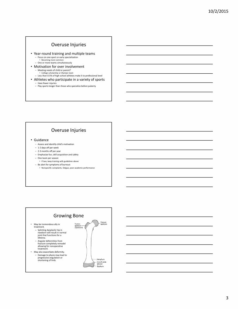
Growing Bone
• May be tremendous ally in treatment.
– Splinting dysplasitc hip in newborn will result in normal joint that functions for a lifetime.
– Angular deformities from fracture completely remodel allowing for nonoperativetreatment.
• May also exacerbate deformity.
– Damage to physis may lead to progressive angulation or shortening of limb.

10/2/2015
4
Sports Injuries
• Traction apophysitis – Overuse injury that occurs in growing child
• Tendon pulls on area of growing bone
• Children and teens seldom get tendonitis
– Repetitive stress and microtrauma• Elbow‐ little leaguer elbow
• Proximal tibia‐ Osgood‐Schlatter disease
• Calcaneous‐ sever disease
– Pelvis
Little Leaguer Elbow
• Traction apophysitis of the medial epicondyle or olecranon– Skeletally immature throwing athlete
• Medial epicondyle or olecranon avulsion– Older child closer to maturity (12‐14)
• Ulnar collateral ligament sprain or tear
• Compression injuries– Osteochondritis dissecans
• 12 and older after capitellum
has ossified
Little Leaguer Elbow
• History– History of overuse
• Number of pitches, innings, year round participation
• Premature use of curveball
• Mechanical symptoms if loose body
• Pain is most common symptom and may be loss of extension in later stages of OCD– Medial in apophysitis, avulsion & UCL injury
– Lateral in OCD and Panner disease
– Posterior in olecranon apophysitis/avulsion
10/2/2015
5
Little Leaguer Elbow
• Radiographs help establish diagnosis– Contralateral views helpful
• MRI may be needed – OCD, Panner disease, UCL injuries
• Treatment– Rest from throwing for 3‐6 months or longer
– PT to maintain strength and restore motion
– Avoid immobilization beyond acute phase
– Surgery for fixation, microfracture or excision of loose body
Cuff & Deltoid Strength
• Patient holds arms out from sides horizontally and tries to lift them
• Normal findings
– Strength should be equal in both arms, and deltoid muscles should be equal in size
• Common abnormalities
– Loss of strength and wasting of the deltoid muscle
Shoulder Range of Motion
• Arms out from sides with elbows bent at 90 degrees; Patient raises hands vertically
• Normal findings– Hands go back equally and at least to vertical position
• Common abnormalities– Loss of external rotation, which may indicate shoulder problem or history of dislocation

10/2/2015
6
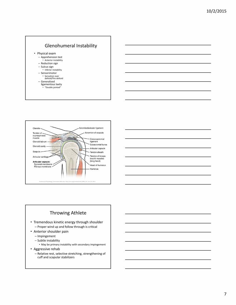
Anatomy & Physiology, ConnexionsWeb site. http://cnx.org/content/col11496/1.6/, Jun 19, 2013.
Age Is Key Variable
• Younger than 30 likely to report symptoms of instability from dislocation/subluxation of glenohumeral joint or AC joint
• Middle‐aged (30‐50) more commonly report impingement. Frozen shoulder may occur in diabetics and thin females in this age group
• Older than 50 more likely to have RCT, DJD or frozen shoulder
Glenohumeral Instability• 50% of all major dislocations
– Anterior 95%• Direct blow to externally rotated, abducted humerus
• Fall on outstretched arm
– Posterior 2‐4%
– Inferior (luxatio erecta) 0.5%
• Age at initial dislocation is prognostic– Recurrence of 55% in those 12‐22 years
– 37% in those 23‐39 years
– 12% at 30‐40 years
10/2/2015
7
Glenohumeral Instability
• Physical exam– Apprehension test
• Anterior instability
– Reduction sign– Sulcus sign
• Inferior instability
– Sensorimotor • Sensation over deltoid/fire deltoid
– Generalized ligamentous laxity
• “Double jointed”
Anatomy & Physiology, ConnexionsWeb site. http://cnx.org/content/col11496/1.6/, Jun 19, 2013.
Throwing Athlete
• Tremendous kinetic energy through shoulder– Proper wind up and follow through is critical
• Anterior shoulder pain– Impingement
– Subtle instability• May be primary instability with secondary impingement
• Aggressive rehab– Relative rest, selective stretching, strengthening of cuff and scapular stabilizers

10/2/2015
8
Glenohumeral Instability
• Most commonly dislocated joint
• Age at initial dislocation is prognostic– Recurrence rates of 55% in 12‐22 years
– 37% in those 23‐29 years
– 12% at 30‐40 years
• Fall on flexed elbow with adducted arm or by direct axial load to externally rotated humerus
• Traumatic dislocation more common in adolescent than in pediatric population– Consider ligamentous laxity if unstable in peds patient
Acromioclavicular Injuries
• AC separation
– Fall onto tip of shoulder (acromion)
– Classified as to degree of separation I‐VI
• Low grade treated with sling
• High grade dislocations may need repair– Obvious deformity and instabiltiy
– Tender over AC joint and pain with adduction
Acromioclavicular Injuries
• Radiographs– AP views of both shoulders
• Stress views may be helpful to differentiate incomplete vs complete disruption
– Low grade separation (subluxation) show little or no displacement
– Grade III and higher injuries show increased distance between acromion and clavicle and between clavicle and coracoid
10/2/2015
9
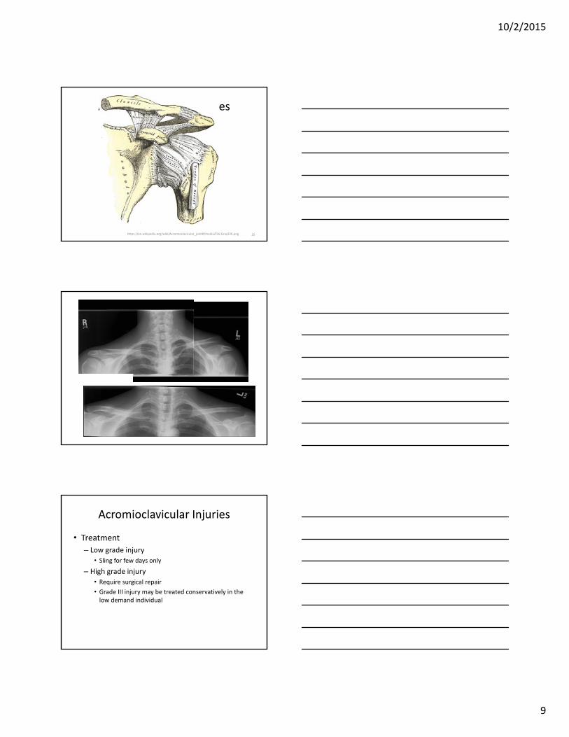
Acromioclavicular Injuries
25https://en.wikipedia.org/wiki/Acromioclavicular_joint#/media/File:Gray326.png
Acromioclavicular Injuries
• Treatment
– Low grade injury
• Sling for few days only
– High grade injury
• Require surgical repair
• Grade III injury may be treated conservatively in the low demand individual
10/2/2015
10
Scoliosis
• Lateral curvature of the spine of > 10°– Small curves are not scoliosis
• Thoracic or lumbar spine (occasionally both)– Associated vertebral rotation with kyphosis or lordosis
• May be congenital– Vertebral anomalies
• Commonly idiopathic • May be secondary to other disorder
• Cerebral palsy• Muscular dystrophy • Myelomeningocele
Idiopathic Scoliosis
Curve size Girls:Boys
6-10° 1:1
11-20° 1.4:1
>21° 5.4:1
• Develops in early adolescence– Male = female in curves < 10°
– Female 7X more likely to have significant, progressive curve requiring treatment
– Progression typically girls at age 10‐16 years
– Not associated with pain• Pain suggests primary condition and requires further evaluation
Scoliosis
• Physical exam– Forward bending test
• Observe from behind• Elevation of rib cage, scapula or paravertebral muscle mass positive finding
– Also assess• Skin• Leg length• Feet alignment• Neuromuscular status
– Beware• Left side thoracic curves have high incidence of spinal cord abnormalities

10/2/2015
11
Scoliosis
By Weiss HR, Goodall D [CC BY 2.0 (http://creativecommons.org/licenses/by/2.0)], via Wikimedia Commons
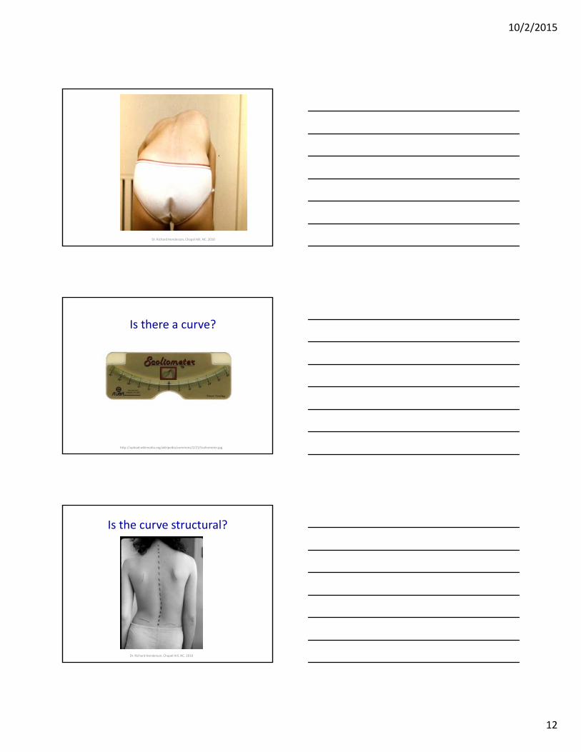
Is there a curve?
Dr. Richard Henderson, Chapel Hill, NC. 2010
Dr. Richard Henderson, Chapel Hill, NC. 2010
10/2/2015
12
Dr. Richard Henderson, Chapel Hill, NC. 2010
Is there a curve?
http://upload.wikimedia.org/wikipedia/commons/2/21/Scoliometer.jpg
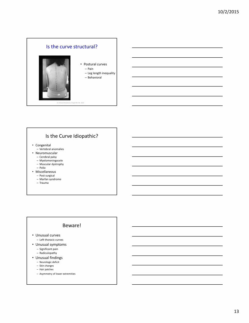
Is the curve structural?
Dr. Richard Henderson, Chapel Hill, NC. 2010
10/2/2015
13
Is the curve structural?
• Postural curves– Pain
– Leg length inequality
– Behavioral
Dr. Richard Henderson, Chapel Hill, NC. 2010
Is the Curve Idiopathic?
• Congenital– Vertebral anomalies
• Neuromuscular– Cerebral palsy– Myelomeningocele– Muscular dystrophy– Polio
• Miscellaneous– Post surgical– Marfan syndrome– Trauma
Beware!
• Unusual curves– Left thoracic curves
• Unusual symptoms– Significant pain
– Radiculopathy
• Unusual findings– Neurologic deficit
– Skin changes
– Hair patches
– Asymmetry of lower extremities

10/2/2015
14
Slipped Capital Femoral Epiphysis
• SCFE– Sudden or gradual displacement of femoral head through
physis.
– Typically during adolescent growth spurt.
• Predisposing factors– Obesity
– Male gender
– Sports
– Femoral retroversion
– Hypothyroidism and growth hormone deficiency
Slipped Capital Femoral Epiphysis
• Mean age at presentation– 12 years for girls (range:10‐14 years)– 13 years for boys (range: 11‐16 years)– Onset before or after typical range is associated with
endocrinopathy.
• Bilateral involvement seen in 40‐50%– Not always affected simultaneously
• May be acute or chronic– Early detection and treatment imperative
Slipped Capital Femoral Epiphysis
• Symptoms– Pain worse with activity
• Localized to anterior thigh or knee.
– May be unable to bear weight
• Exam– Loss of hip internal rotation
• Further reduction of internal rotation with hip flexion.– Loss of internal rotation when hip is flexed to 90°
» Slip is always posterior and often medial
– Loss of abduction and extension
– Affected extremity usually shorter by 1‐3 cm
10/2/2015
15
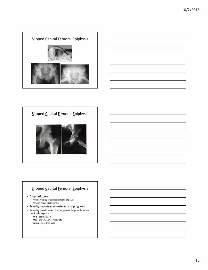
Slipped Capital Femoral Epiphysis
Slipped Capital Femoral Epiphysis
Slipped Capital Femoral Epiphysis
• Diagnostic tests– AP and frog‐leg lateral radiographs of pelvis
– AP view may appear normal
• Severity important in treatment and prognosis
• Severity is estimated by the percentage of femoral neck left exposed: – Mild‐ less than 25%
– Moderate‐ 25‐50% is moderate
– Severe‐more than 50%
10/2/2015
16
Slipped Capital Femoral Epiphysis
• Treatment
– In situ stabilization
• Pin in current position to prevent progression.
– If unstable may require urgent ORIF
– Severe deformity may require realignment osteotomy.
• Chronic painful limp despite treatment
Female Athletes
• Desire to change weight
• Menarche, menstrual regularity and LMP– Eating disorders and amenorrhea
• Osteopenia and osteoporosis
• Sudden cardiac death less likely in females
• Females more likely – Patellofemoral syndrome, foot disorders, stress fractures, ACL rupture
Case #1
• 16 year old female with acute onset knee pain while playing basketball today
• Pain and immediate swelling after coming down from rebound
• Unable to bear weight
• Felt a pop at the time of injury
10/2/2015
17
Reproduced with permission from Griffin LY (ed): Essentials of Musculoskeletal Care, 3rd edition. Rosemont, IL, American Academy of Orthopaedic Surgeons, 2005.
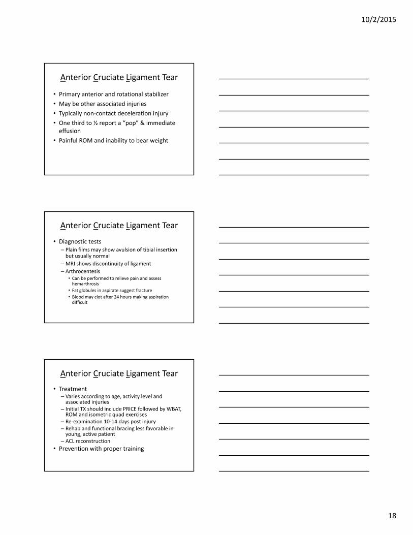
Case #1
• Exam
– Large effusion
– Anterior drawer (knee flexed at 90º) negative
– Anterior translation of tibia in relation to femur
https://en.wikipedia.org/wiki/Lachman_test#/media/File:ACLI_17.jpg
Reproduced with permission from Griffin LY (ed): Essentials of Musculoskeletal Care, 3rd edition. Rosemont, IL, American Academy of Orthopaedic Surgeons, 2005.
10/2/2015
18
Anterior Cruciate Ligament Tear
• Primary anterior and rotational stabilizer
• May be other associated injuries
• Typically non‐contact deceleration injury
• One third to ½ report a “pop” & immediate effusion
• Painful ROM and inability to bear weight
Anterior Cruciate Ligament Tear
• Diagnostic tests– Plain films may show avulsion of tibial insertion but usually normal
– MRI shows discontinuity of ligament
– Arthrocentesis• Can be performed to relieve pain and assess hemarthrosis
• Fat globules in aspirate suggest fracture
• Blood may clot after 24 hours making aspiration difficult
Anterior Cruciate Ligament Tear
• Treatment– Varies according to age, activity level and associated injuries
– Initial TX should include PRICE followed by WBAT, ROM and isometric quad exercises
– Re‐examination 10‐14 days post injury– Rehab and functional bracing less favorable in young, active patient
– ACL reconstruction
• Prevention with proper training
10/2/2015
19
Case #2
• 15 year old male with medial knee pain and occasional clicking
• Most often with “plant and pivot” activity
• Symptoms are intermittent
• Knee “gives out”
• Knee swells after clicking or buckling
• Effusion improves with NSAIDs
Case #2
• Clinical symptoms– Minimal or no trauma
– Gradual onset of effusion and stiffness
– Mechanical symptoms
– Reports of instability
• Exam– Small to moderate effusion
– Medial joint line tenderness
– Pain with full flexion and extension
– Pain and popping on McMurray test
Meniscal Tear
• Treatment– PRICE
– Course of NSAIDs at anti‐inflammatory dose
– Gradual return to activity
– Recurrent catching, popping, locking will likely require surgical debriedment
• May consider injection in older patient
• Imaging– Wt bearing films with notch and sunrise views
– MRI
• Arthroscopy for partial menisectomy vs repair
10/2/2015
20
Case #3
• 12 year old male with knee pain after collision on soccer field
• Knee forced into valgus
• Edema over several hours after the injury
• Pain with weight bearing
• Unable to fully flex knee
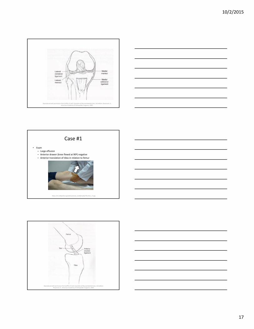
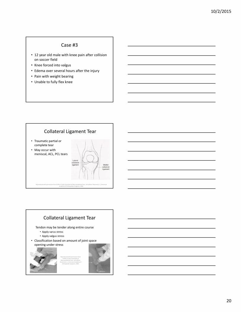
Collateral Ligament Tear
• Traumatic partial or complete tear
• May occur with meniscal, ACL, PCL tears
Reproduced with permission from Griffin LY (ed): Essentials of Musculoskeletal Care, 3rd edition. Rosemont, IL, American Academy of Orthopaedic Surgeons, 2005.
Collateral Ligament Tear
Tendon may be tender along entire course
• Apply varus stress
• Apply valgus stress
• Classification based on amount of joint space opening under stress
Reproduced with permission from Griffin LY (ed): Essentials of
Musculoskeletal Care, 3rd edition. Rosemont, IL, American Academy of
Orthopaedic Surgeons, 2005.

10/2/2015
21
Collateral Ligament Tear
• Treatment – Typically conservative for isolated tears
• Must rule out ACL, PCL and meniscal tears
– PRICE• Hinged brace in higher grade tears
– Weight bearing as tolerated with crutches
– Analgesia with acetaminophen or tramadol• NSAIDS probably OK
– PT includes early ROM, quad strength and gait training
– Surgical repair if other ligaments torn
Patella/quadriceps tendonopathy
• Common in adolescent athlete– Sinding‐Larsen‐Johansson Disease in preadolescents
• Overuse or overload syndrome• Associated with jumping sports• May occur with erratic exercise habits• Weight gain may play role• Anterior knee pain• Pain with sitting, squatting or kneeling• Climbing stairs often increases pain
Patella/quadriceps tendonopathy
• Exam
– Tender at inferior or superior patella pole
– May be mild edema
• No joint effusion
– Fullness of infrapatellar bursa
– AROM is normal but painful
– Quadriceps atrophy if longstanding condition
– Rule out other soft tissue conditions

10/2/2015
22
Patella/quadriceps tendonopathy
• Treatment is primarily symptomatic– Period of rest
• Few days to a few weeks
• Consider brief immobilization
– Analgesia with acetaminophen or tramadol
– PT with focus on ROM, extensor stretching and quadriceps strength
• Ultrasound or iontophoresis may help
– Knee sleeve with patella cutout or patella tendon strap
Sever Disease/Calcaneal Apophysitis
• Repetitive stress and micro trauma
• Posterior heal pain and may have limp
• Apohysis closes
– 9 years in female
– 11 years in male
• Tenderness on heel squeeze
• Radiographs not diagnostic
– Irregularity and sclerosis are normal
Sever Disease/Calcaneal Apophysitis
• Differential Diagnoses
– Achilles tendinopathy
• Can be associated with reactive arthritis or seronegative spondyloarthropathies
– Infection
• Likely unilateral, local swelling, elevated ESR
– Tumor
• Likely unilateral, local swelling, night pain

10/2/2015
23
Sever Disease/Calcaneal Apophysitis
• Treatment
– Activity modification
– Heel lift (short term)
– Achilles stretching
– Ice after activity
– Casting if severe
– Consider infection or neoplastic disease if recalcitrant
Ankle Sprain
• More than 25,000 sprains daily
• Residual symptoms in nearly 40%
• Lateral ligaments most often affected– Inversion injury
– Tibiofibular syndesmosis injury in “high” ankle sprain
• Subtalar joint may also be injured– Interosseous ligament tear
• Medial deltoid injury may also occur– Less common
– Typically associated with eversion injury
Ankle Ligaments
Maughan, M. Ankle sprain. In: UpToDate, Patrice, E. (Ed), UpToDate, Waltham, MA, 2015
10/2/2015
24
Ankle Sprain
• Clinical symptoms– Pain over injured structures
– Swelling
– Loss of function
– May report a “pop” in severe sprain
• Followed by immediate swelling and inability to bear weight
– May report history of previous sprain
Ankle Sprain
• Exam
– Circumferential ecchymosis and swelling
– Tenderness of affected structures
• Palpate medial and lateral malleoli, base of 5th
metatarsal and navicular
– Special tests
• Anterior drawer
Ankle Sprain
• Special tests– Squeeze test
• Compress tibia and fibula at midcalf
– External rotation test• Dorsiflex ankle and externally rotate foot
– Positive test results in pain at distal tibiofibular syndesmosis
• Subtalar joint injury may show tenderness and ecchymosis of medial hindfoot

10/2/2015
25
Ottawa Rules
• Ottawa rules for radiographs
• Tenderness at distal fibula or tibia
• Tenderness at 5th MT base or navicular
• Inability to bear wt. immediately and in clinic
Maughan, M. Ankle sprain. In: UpToDate, Patrice, E. (Ed), UpToDate, Waltham, MA, 2015
Ankle Sprain
• Differential Diagnosis– Fracture of distal fibula, base of 5th metatarsal, medial malleolus, calcaneus, talus (tender over structure, apparent on radiograph)
– Proximal fibula fracture (Maisonneuve‐ proximal fibula, deltoid TTP and positive squeeze test)
– Peroneal tendon tear or subluxation (muscle weakness on eversion, may report repeated popping)
– Osteochondral fracture of talar dome (evident on radiographs, MRI or bone scan)
Ankle Sprain
• Treatment– Analgesia with acetaminophen or tramadol– PRICE with vigorous elevation (toes above nose)– Consider cast or cast boot for 2 weeks if severe– WBAT (crutches as needed for a few days)– Home therapy program
• Range of motion• Stretching exercises after 2 weeks• Strengthening and proprioception exercises
– Stirrup splint for 6 weeks or more– Formal physical therapy!
• Chronic instability common after incomplete rehab
10/2/2015
26
References/Resources
• American Academy of Family Physicians, American Academy of Pediatrics, American College of Sports Medicine. Preparticipation Physical Evaluation, 4th ed, Bernhardt D, Roberts W (Eds), American Academy of Pediatrics, Elk Grove Village, IL 2010.
• Standardized preparticipation athletic evaluation form can be downloaded from the American Academy of Pediatrics http://www.aap.org/en‐us/professional‐resources/practice‐support/Pages/Preparticipation‐Physical‐Evaluation‐Forms.aspx
• Gomez JE, Landry GL, Bernhardt DT. Critical evaluation of the 2‐minute orthopedic screening examination. Am J Dis Child 1993; 147:1109.
• Goldberg, C. (2009). A practical guide to clinical medicine. University of California San Diego. Retrieved from http://meded.ucsd.edu/clinicalmed/joints.htm#MCL
• Griffin LY (ed): Essentials of Musculoskeletal Care, 3rd edition. Rosemont, IL, American Academy of Orthopaedic Surgeons, 2005.
• Maughan, M. Ankle sprain. In: UpToDate, Patrice, E. (Ed), UpToDate, Waltham, MA, 2015


