Noam Ziv
Dept. of Anatomy and Cell Biology
Technion Faculty of Medicine
Haifa, Israel
Microscopy in Cell Biology:Live imaging
Topics:
• Imaging live specimens
• 6D microscopy
• Green fluorescent protein and derivatives
• FRAP
• Photoactivation
• Mouse models
• Tetracysteine-biarsenical tagging
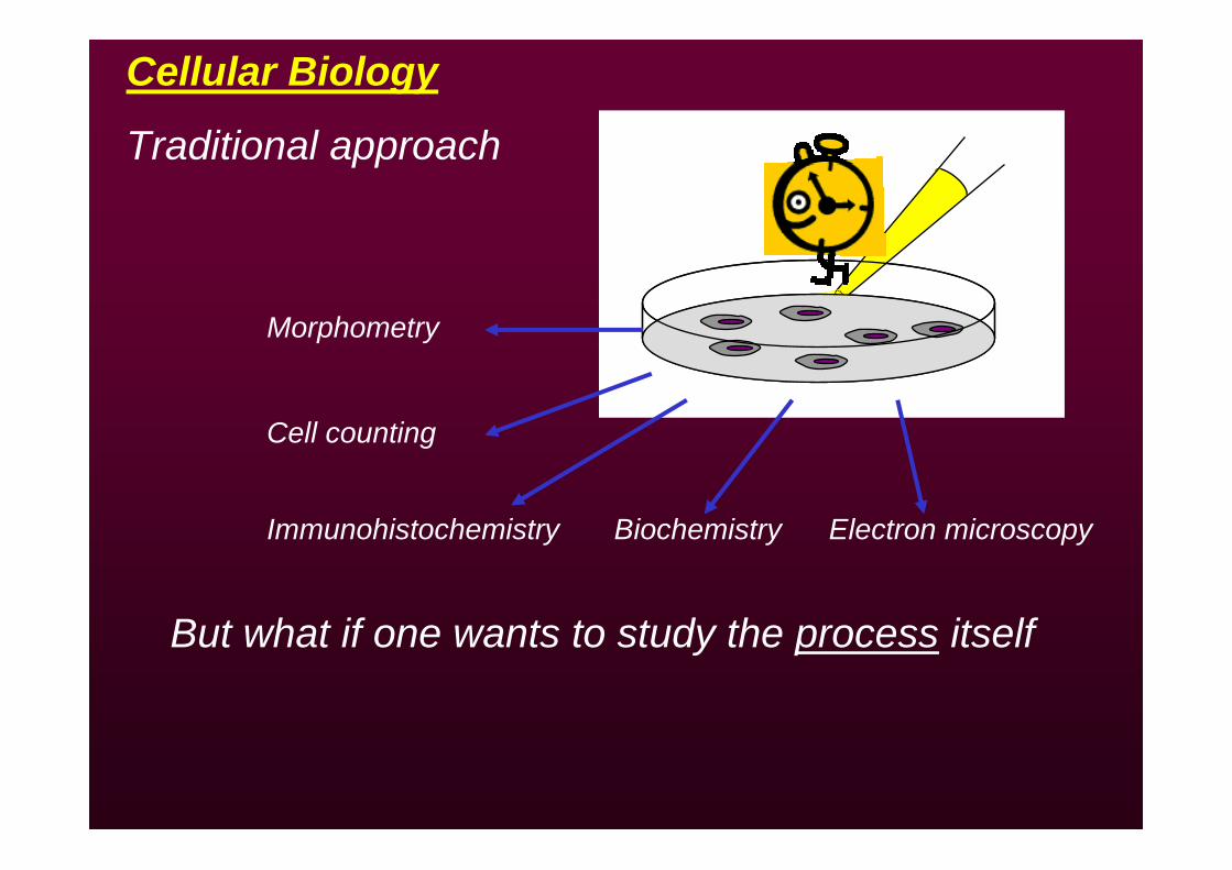
Cellular Biology
Traditional approach
Morphometry
Cell counting
Immunohistochemistry Biochemistry Electron microscopy
But what if one wants to study the process itself
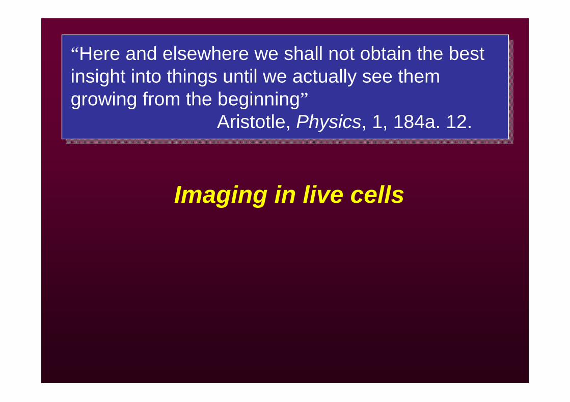
Imaging in live cells
“Here and elsewhere we shall not obtain the best insight into things until we actually see them growing from the beginning”
Aristotle, Physics, 1, 184a. 12.
“Here and elsewhere we shall not obtain the best insight into things until we actually see them growing from the beginning”
Aristotle, Physics, 1, 184a. 12.
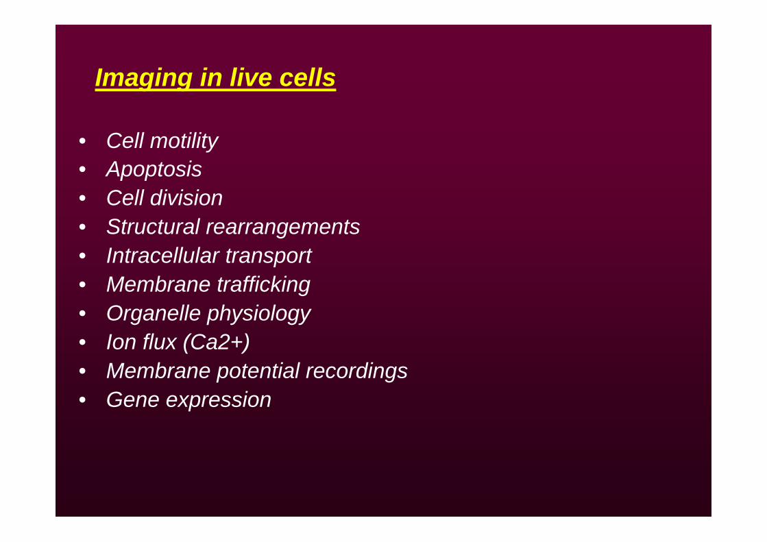
• Cell motility• Apoptosis• Cell division • Structural rearrangements • Intracellular transport• Membrane trafficking• Organelle physiology• Ion flux (Ca2+)• Membrane potential recordings• Gene expression
Imaging in live cells
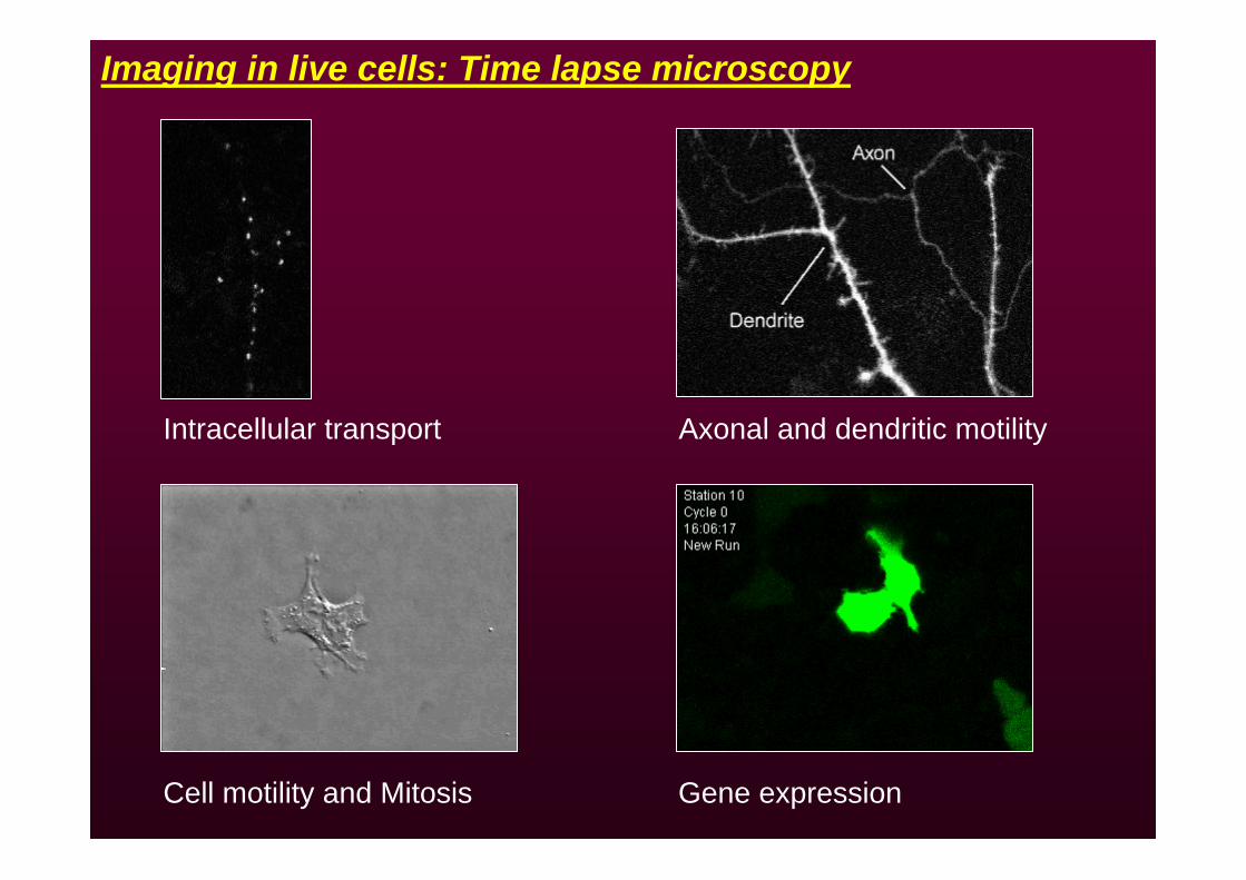
Imaging in live cells: Time lapse microscopy
Intracellular transport Axonal and dendritic motility
Cell motility and Mitosis Gene expression
• Optimal environmental conditions• Phototoxicity • Focus drift• Vital labels
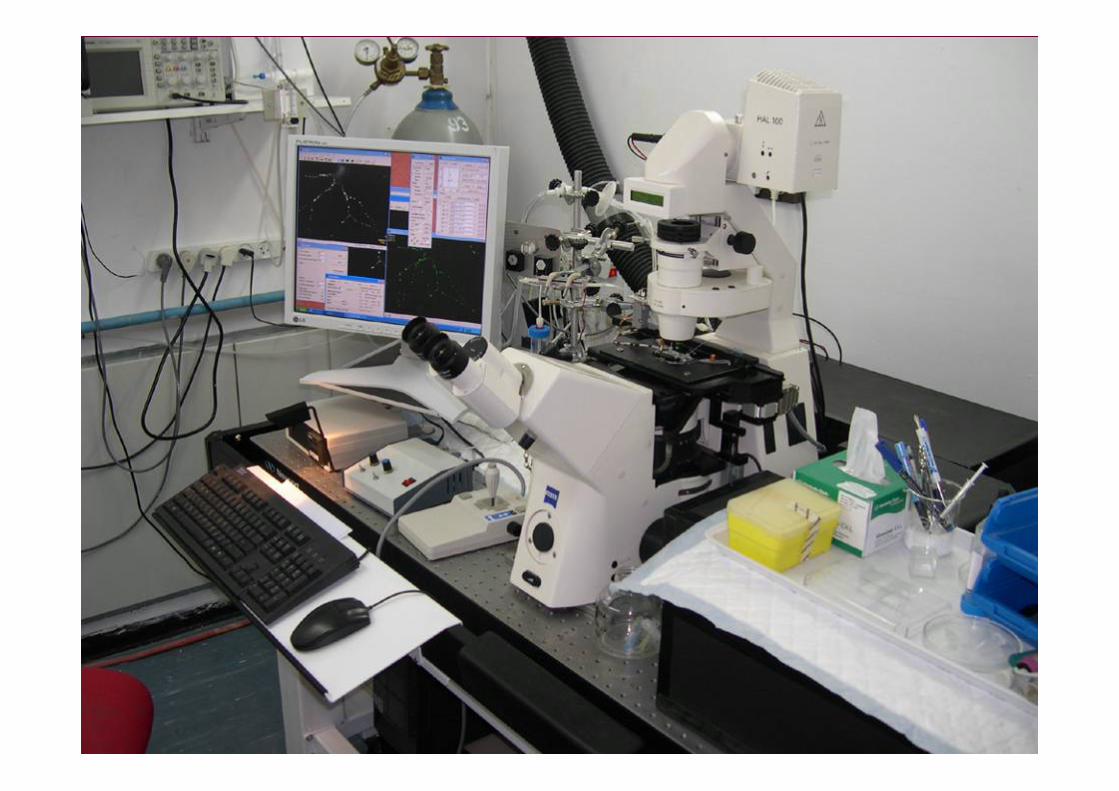
Imaging in live cells: considerations
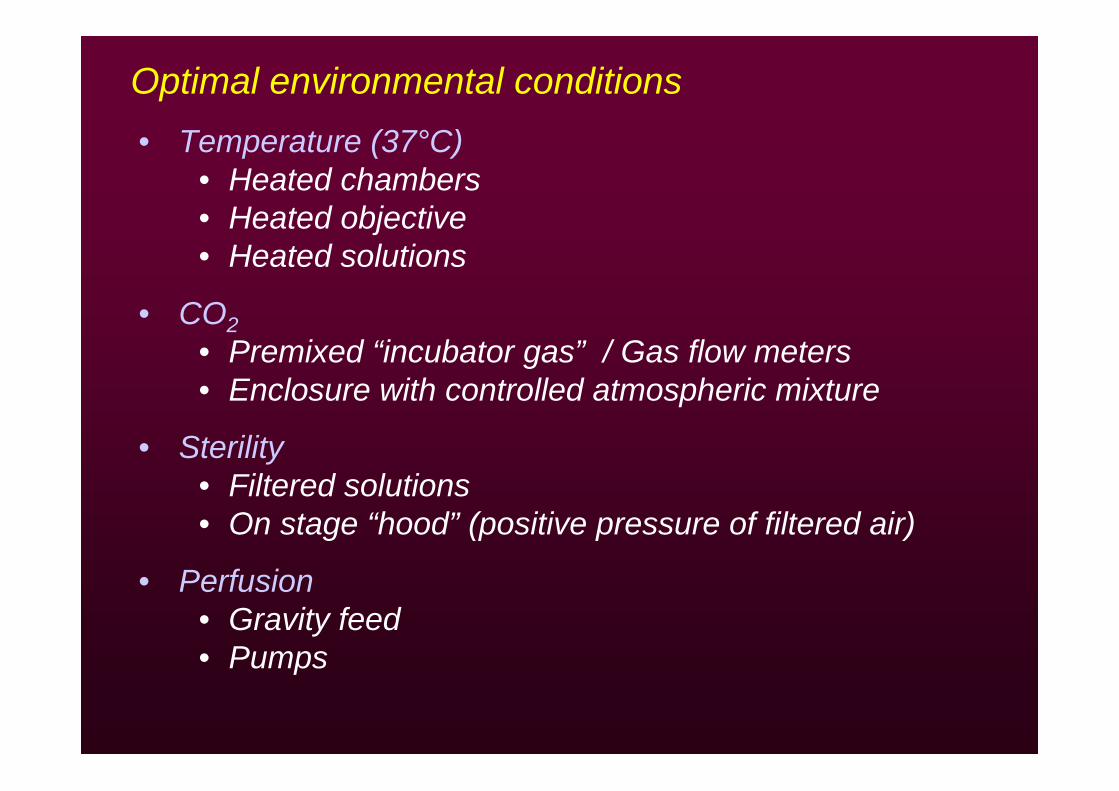
Optimal environmental conditions• Temperature (37°C)
• Heated chambers• Heated objective• Heated solutions
• CO2• Premixed “incubator gas” / Gas flow meters• Enclosure with controlled atmospheric mixture
• Sterility• Filtered solutions• On stage “hood” (positive pressure of filtered air)
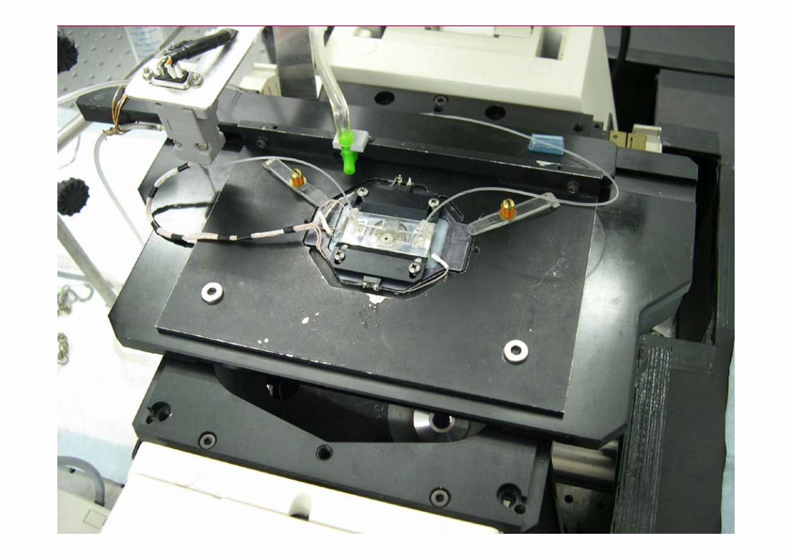
• Perfusion• Gravity feed • Pumps
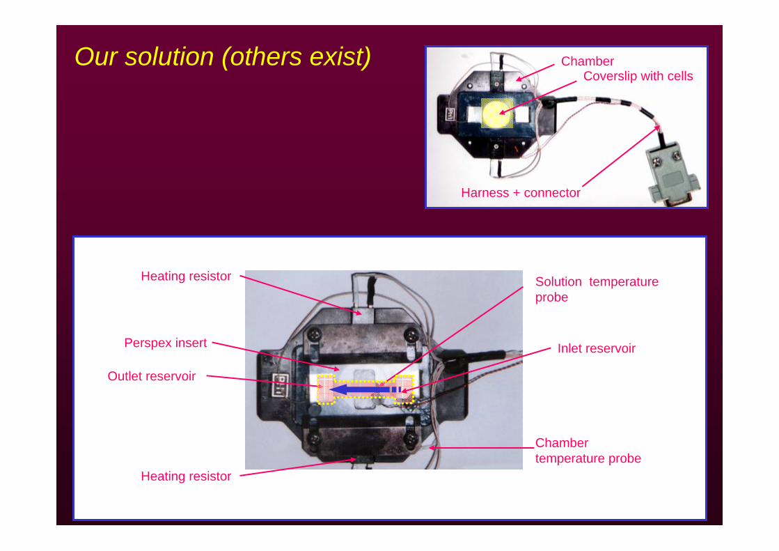
Our solution (others exist)
Heating resistor
ChamberCoverslip with cells
Heating resistor
Chamber temperature probe
Solution temperature probe
Harness + connector
Perspex insert
Outlet reservoir
Inlet reservoir
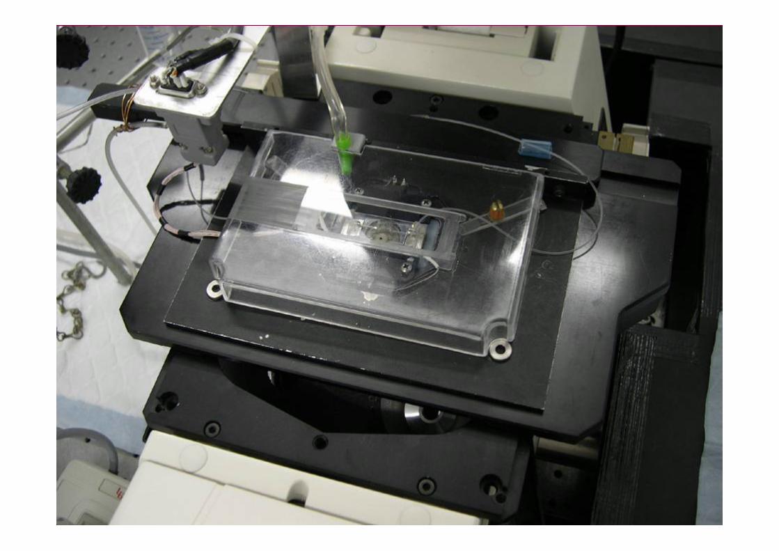
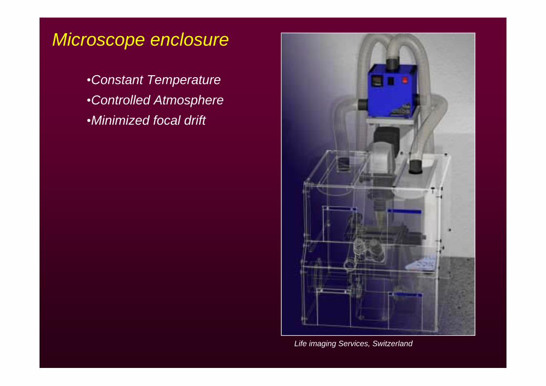
Microscope enclosure
•Constant Temperature•Controlled Atmosphere•Minimized focal drift
Life imaging Services, Switzerland
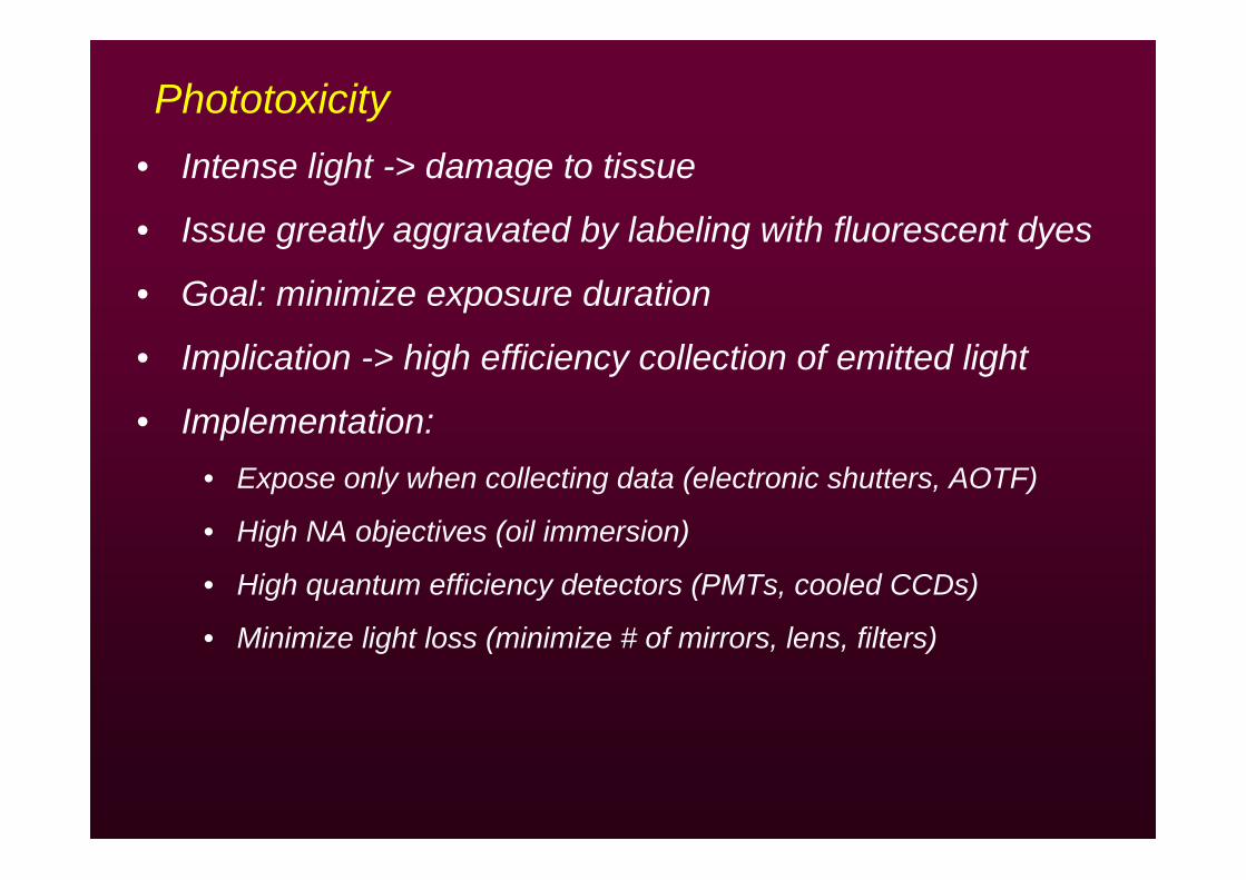
Phototoxicity• Intense light -> damage to tissue
• Issue greatly aggravated by labeling with fluorescent dyes
• Goal: minimize exposure duration
• Implication -> high efficiency collection of emitted light
• Implementation: • Expose only when collecting data (electronic shutters, AOTF)
• High NA objectives (oil immersion)
• High quantum efficiency detectors (PMTs, cooled CCDs)
• Minimize light loss (minimize # of mirrors, lens, filters)
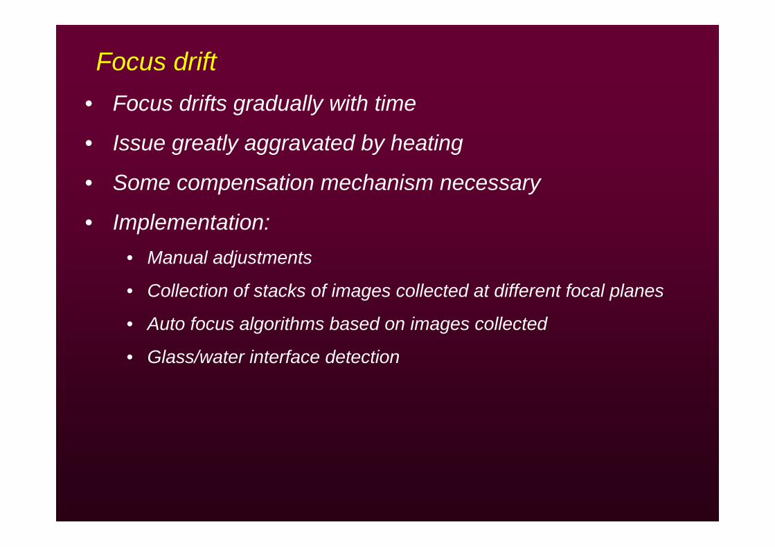
Focus drift• Focus drifts gradually with time
• Issue greatly aggravated by heating
• Some compensation mechanism necessary
• Implementation: • Manual adjustments
• Collection of stacks of images collected at different focal planes
• Auto focus algorithms based on images collected
• Glass/water interface detection
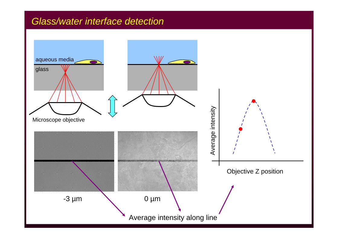
0 µm-3 µm
Average intensity along line
Objective Z position
Ave
rage
inte
nsity
glass
aqueous media
Glass/water interface detection
Microscope objective
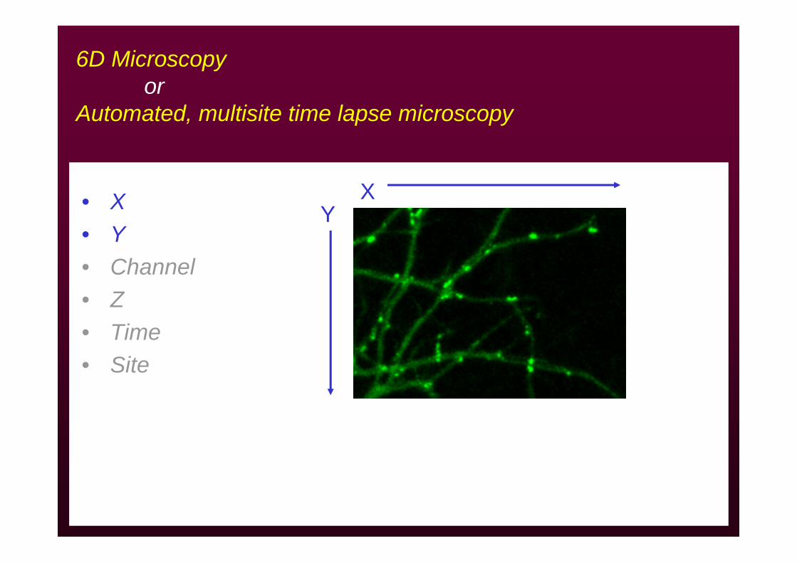
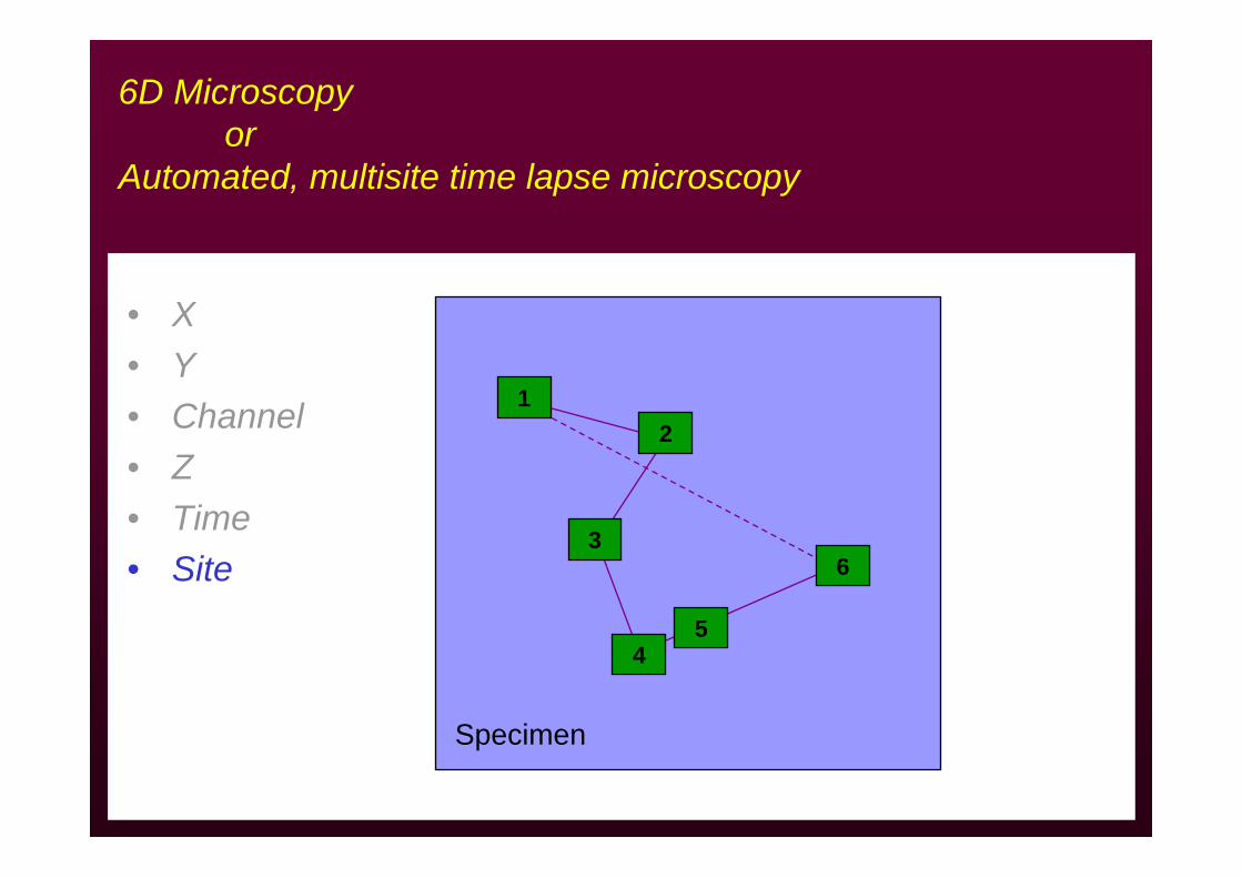
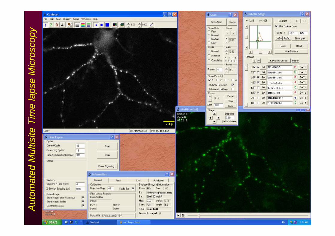
6D Microscopyor
Automated, multisite time lapse microscopy
• X• Y• Channel• Z• Time• Site
XY
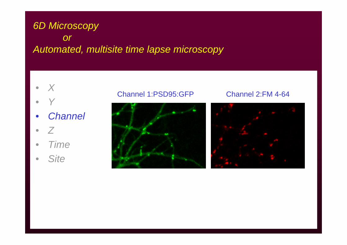
6D Microscopyor
Automated, multisite time lapse microscopy
• X• Y• Channel• Z• Time• Site
Channel 1:PSD95:GFP Channel 2:FM 4-64
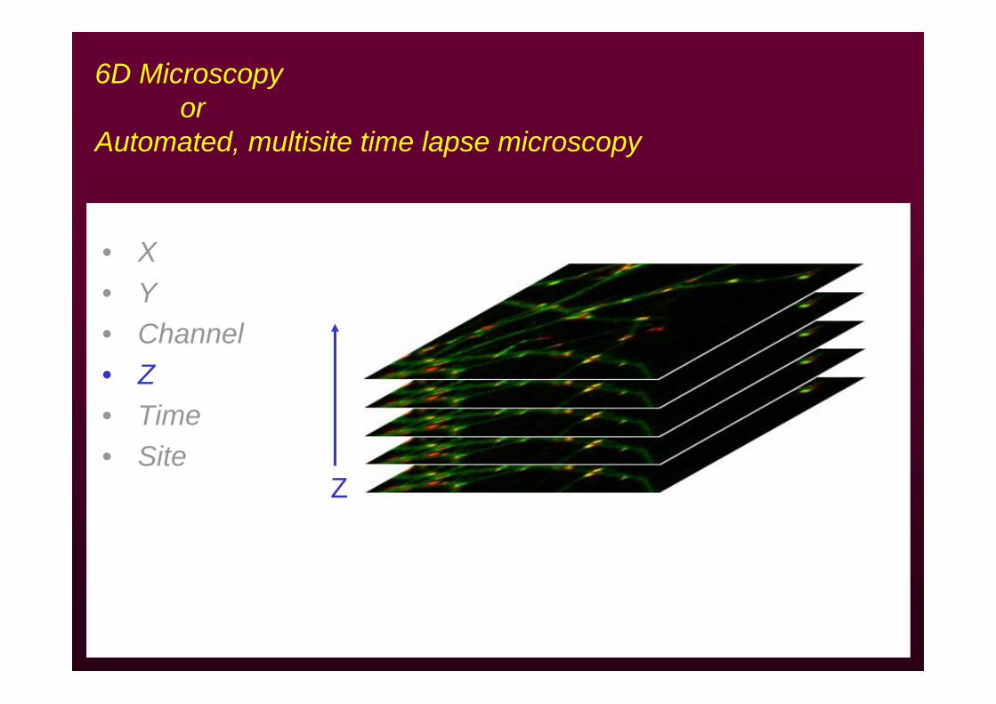
6D Microscopyor
Automated, multisite time lapse microscopy
• X• Y• Channel• Z• Time• Site
Z
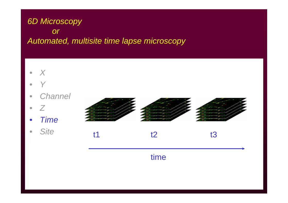
6D Microscopyor
Automated, multisite time lapse microscopy
• X• Y• Channel• Z• Time• Site t1 t2 t3
time
6D Microscopyor
Automated, multisite time lapse microscopy
• X• Y• Channel• Z• Time• Site
Specimen
12
3
45
6
Aut
omat
ed M
ultis
ite T
ime
laps
e M
icro
scop
y
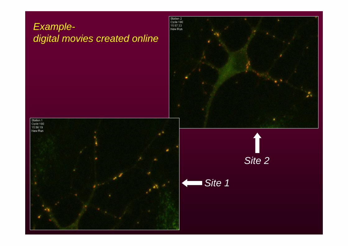
Example-digital movies created online
Site 1
Site 2

Organic compound vital labels
• Intracellular ion concentrations (fura2, fluo3…)• pH (BCECF, SNARF…)• Cell surface (DiI, DiO… )• Membrane potential (Di-4-ANEPPS, RH dyes…)• Organelles (MitoTracker, Rhodamine 123…) • Membrane trafficking (FM 1-43, TMA-DPH…)• Lineage tracers• Enzyme activity• …
(see http://www.probes.com)
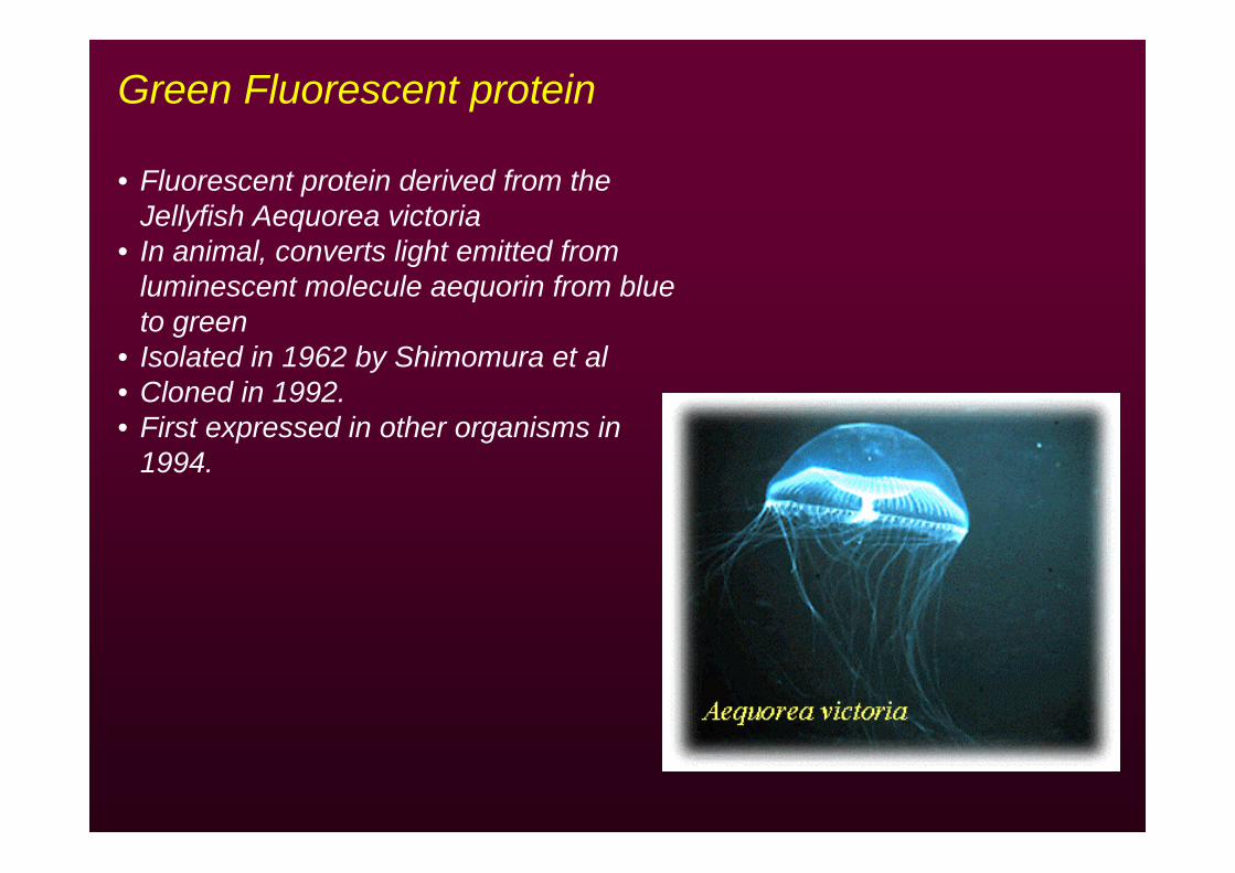
Green Fluorescent protein
• Fluorescent protein derived from the Jellyfish Aequorea victoria
• In animal, converts light emitted from luminescent molecule aequorin from blue to green
• Isolated in 1962 by Shimomura et al• Cloned in 1992.• First expressed in other organisms in
1994.
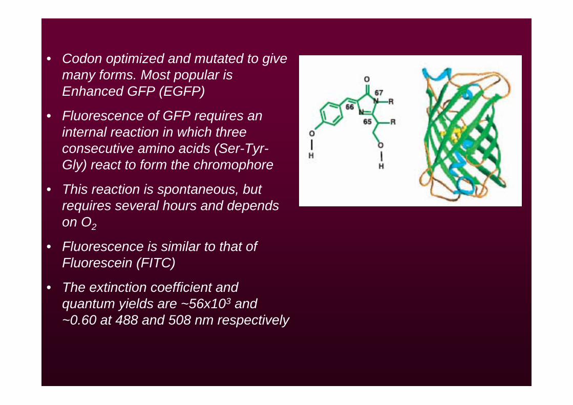
• Codon optimized and mutated to give many forms. Most popular is Enhanced GFP (EGFP)
• Fluorescence of GFP requires an internal reaction in which three consecutive amino acids (Ser-Tyr-Gly) react to form the chromophore
• This reaction is spontaneous, but requires several hours and depends on O2
• Fluorescence is similar to that ofFluorescein (FITC)
• The extinction coefficient and quantum yields are ~56x103 and ~0.60 at 488 and 508 nm respectively
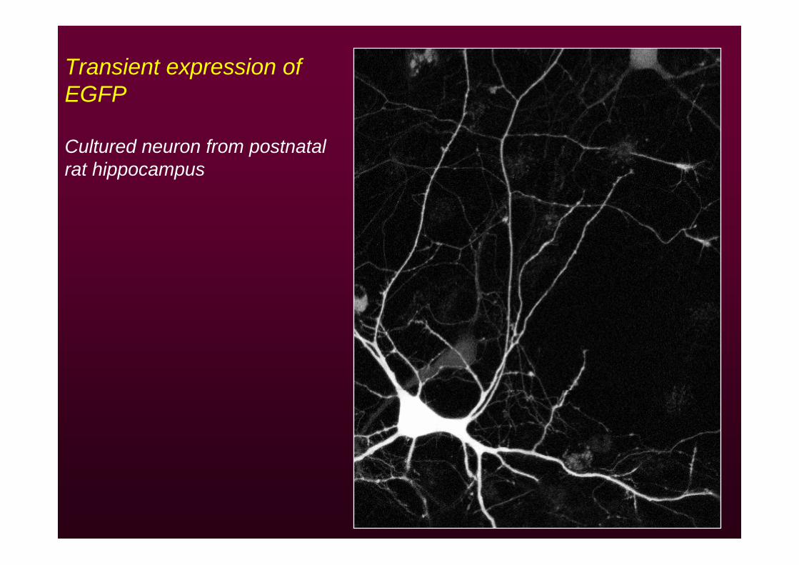
Transient expression of EGFP
Cultured neuron from postnatal rat hippocampus

Molecule of interest
DNAPlasmid
GFP
GFP in cell biology: Chimeric proteins
• Generate a DNA construct coding for a chimera of the protein of interest and GFP
• Insert into mammalian expression vector• Transfect cells (stably or transiently)• Assess functionality of recombinant molecule• Quantify expression levels• Perform experiments
GFP-tagged molecules in cell biology
• Cytoskeleton remodeling• Intracellular transport• Membrane trafficking• Cell adhesion• Spatiotemporal patterns of gene expression• Cell lineage• Apoptosis• Synapse formation and synaptic remodeling
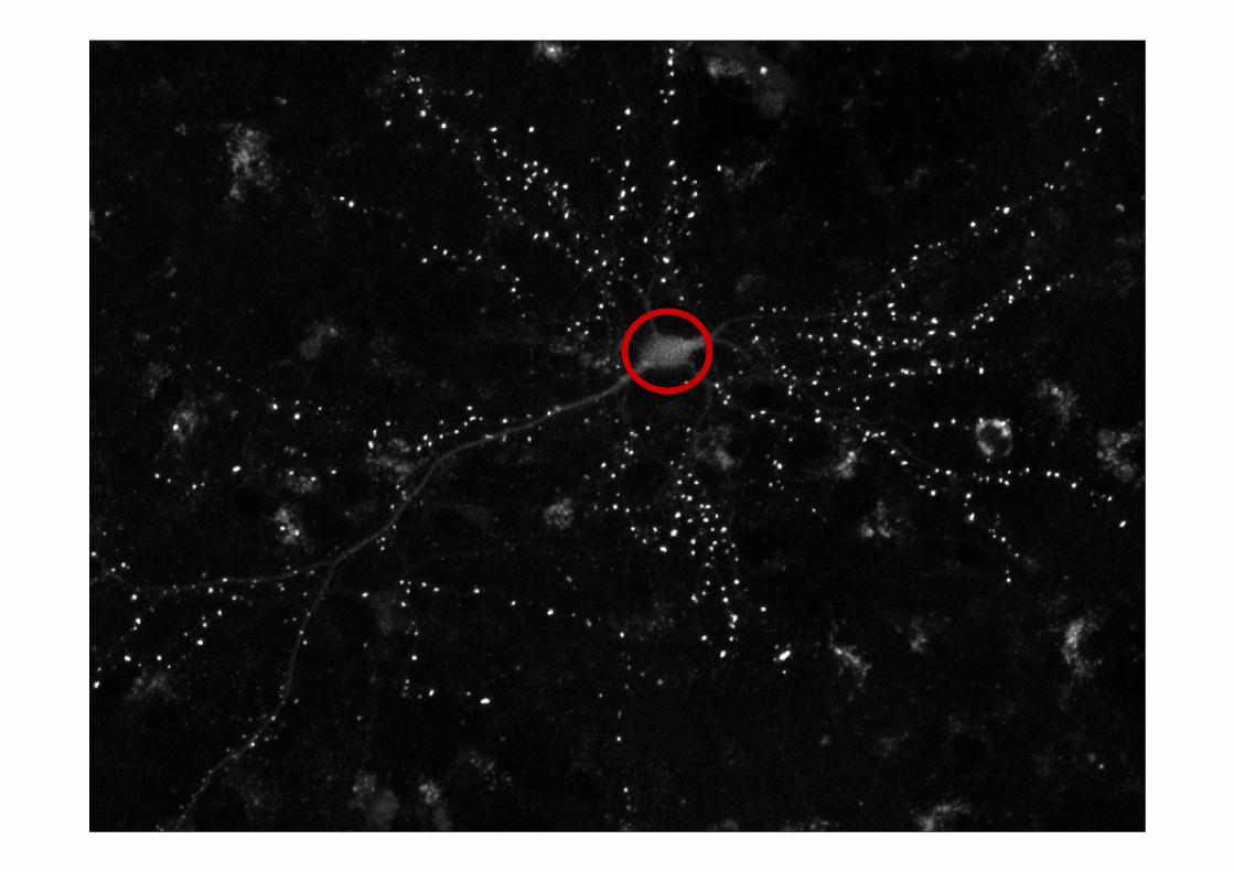
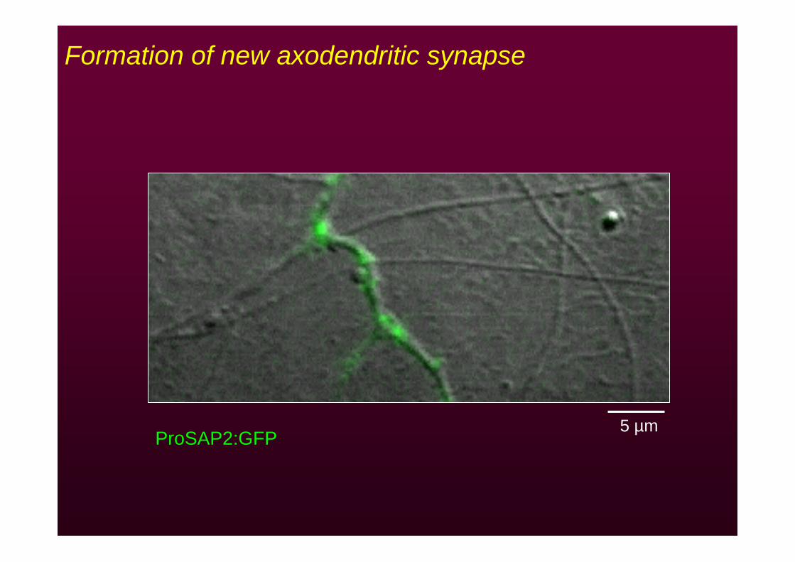
Formation of new axodendritic synapse
ProSAP2:GFP5 µm
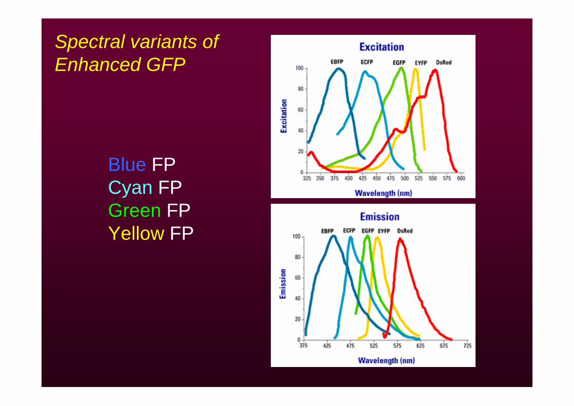
Spectral variants of Enhanced GFP
Blue FPCyan FPGreen FPYellow FP
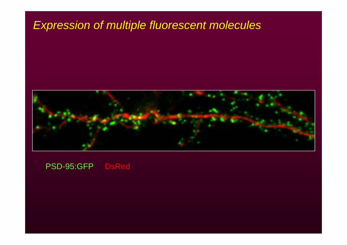
PSD-95:GFP DsRed
Expression of multiple fluorescent molecules
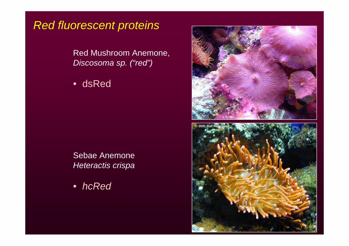
Red fluorescent proteins
Red Mushroom Anemone, Discosoma sp. (“red”)
• dsRed
Sebae AnemoneHeteractis crispa
• hcRed
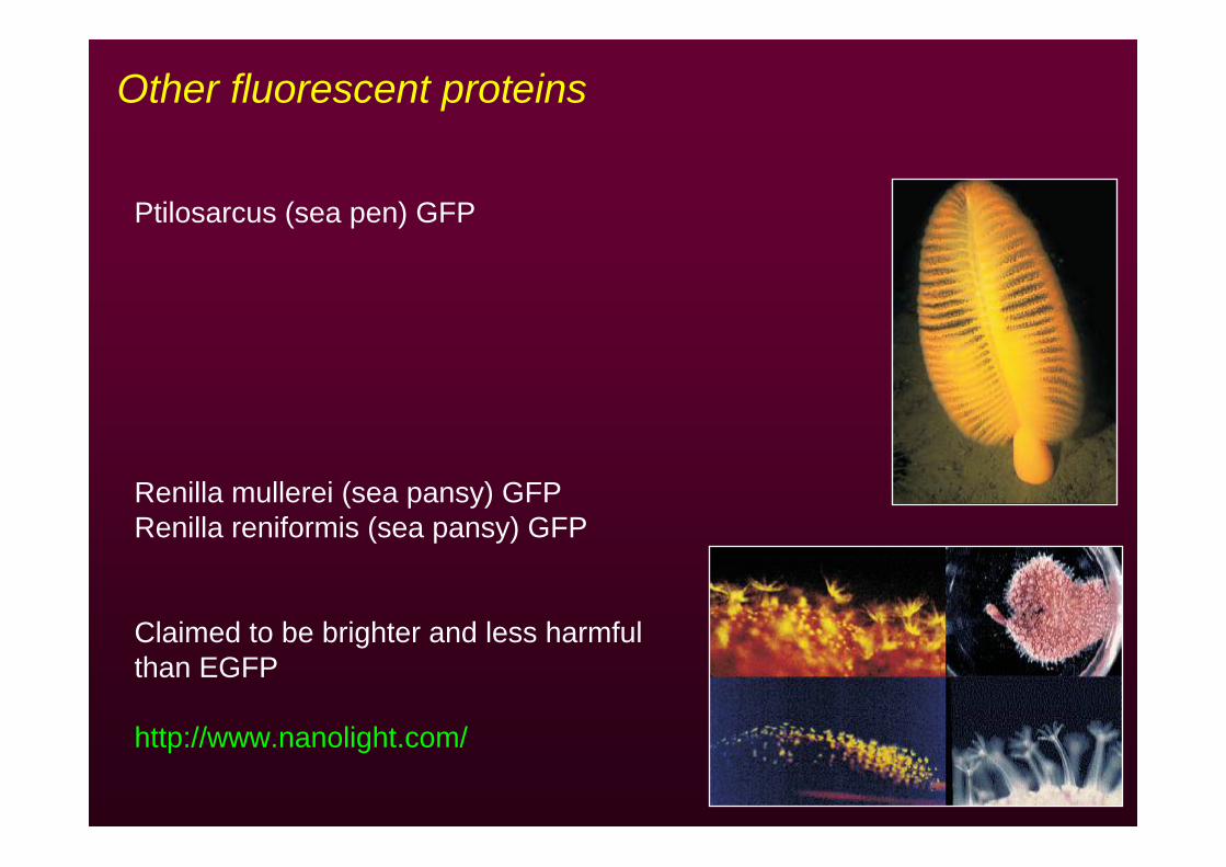
Other fluorescent proteins
Ptilosarcus (sea pen) GFP
Renilla mullerei (sea pansy) GFPRenilla reniformis (sea pansy) GFP
Claimed to be brighter and less harmful than EGFP
http://www.nanolight.com/
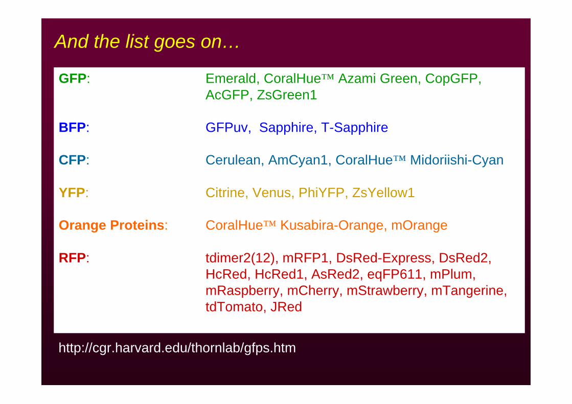
http://cgr.harvard.edu/thornlab/gfps.htm
GFP: Emerald, CoralHue™ Azami Green, CopGFP, AcGFP, ZsGreen1
BFP: GFPuv, Sapphire, T-Sapphire
CFP: Cerulean, AmCyan1, CoralHue™ Midoriishi-Cyan
YFP: Citrine, Venus, PhiYFP, ZsYellow1
Orange Proteins: CoralHue™ Kusabira-Orange, mOrange
RFP: tdimer2(12), mRFP1, DsRed-Express, DsRed2,HcRed, HcRed1, AsRed2, eqFP611, mPlum,mRaspberry, mCherry, mStrawberry, mTangerine,tdTomato, JRed
And the list goes on…
Shaner, Steinbach, Tsien. Nat Methods (2005) 2, 905-909
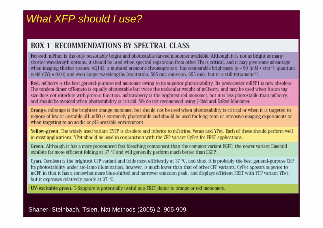
What XFP should I use?
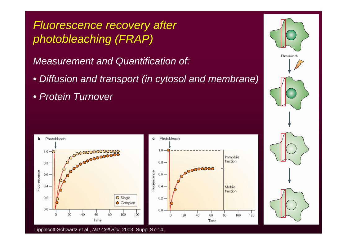
Fluorescence recovery after photobleaching (FRAP)
Measurement and Quantification of:
• Diffusion and transport (in cytosol and membrane)
• Protein Turnover
Lippincott-Schwartz et al., Nat Cell Biol. 2003 Suppl:S7-14.
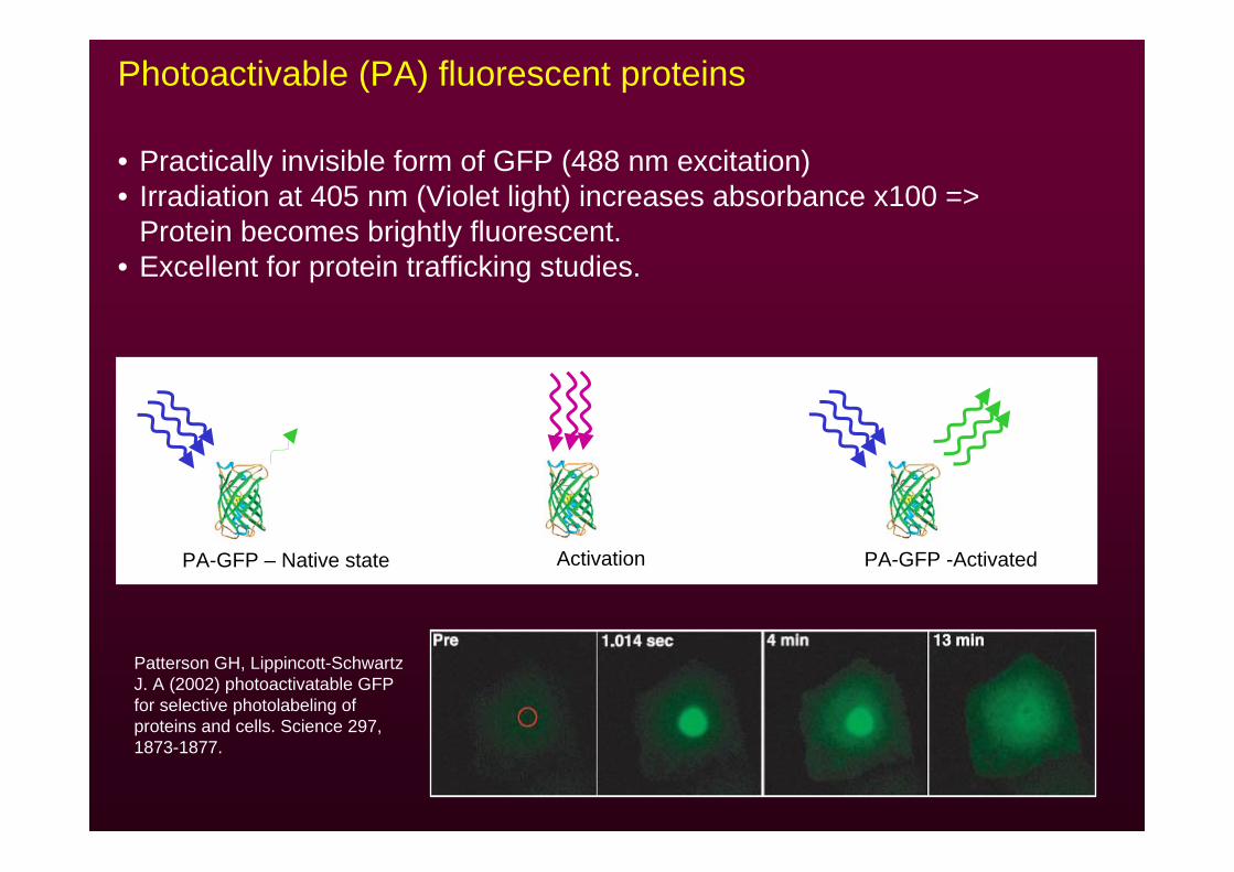
• Practically invisible form of GFP (488 nm excitation)• Irradiation at 405 nm (Violet light) increases absorbance x100 =>
Protein becomes brightly fluorescent.• Excellent for protein trafficking studies.
Photoactivable (PA) fluorescent proteins
Patterson GH, Lippincott-Schwartz J. A (2002) photoactivatable GFP for selective photolabeling of proteins and cells. Science 297, 1873-1877.
PA-GFP – Native state Activation PA-GFP -Activated
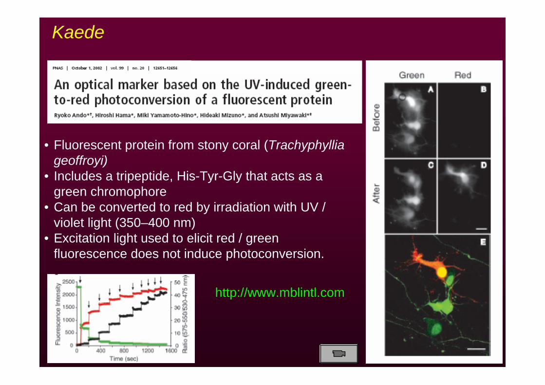
• Fluorescent protein from stony coral (Trachyphyllia geoffroyi)
• Includes a tripeptide, His-Tyr-Gly that acts as a green chromophore
• Can be converted to red by irradiation with UV / violet light (350–400 nm)
• Excitation light used to elicit red / green fluorescence does not induce photoconversion.
Kaede
http://www.mblintl.com
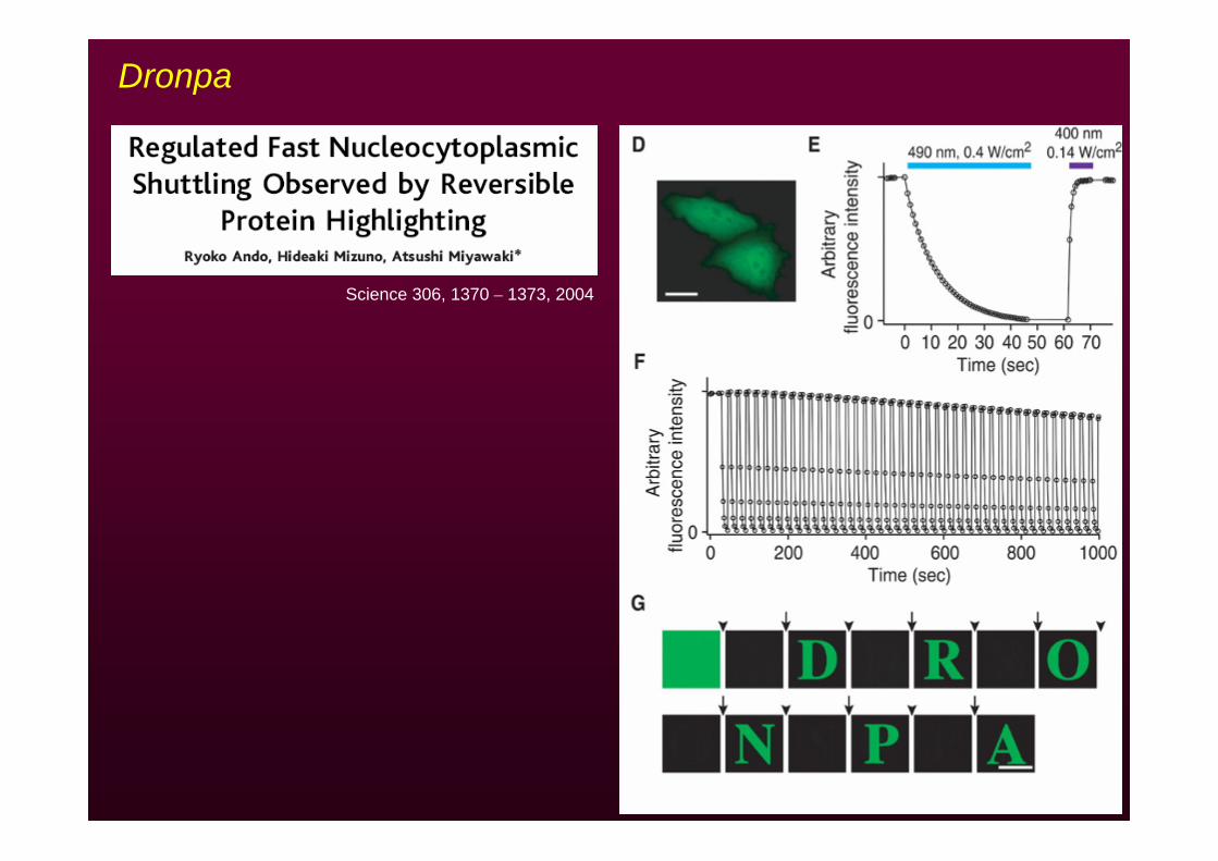
Dronpa
Science 306, 1370 – 1373, 2004
Before Photoactivation (-5 min)
PA + 3 min
PA + 1:15 hours
PA + 7:45 hours
0
1
2
3
4
5
0 2 4 6 8
F/Fo
0
1
2
3
4
5
6
0 2 4 6 8
F/Fo
25±10µm (n=17)50±10µm (n=25)75±10µm (n=14)105±10µm (n=10)
Time (hours)
SomaSoma
PSDs along dendritesPSDs along dendrites
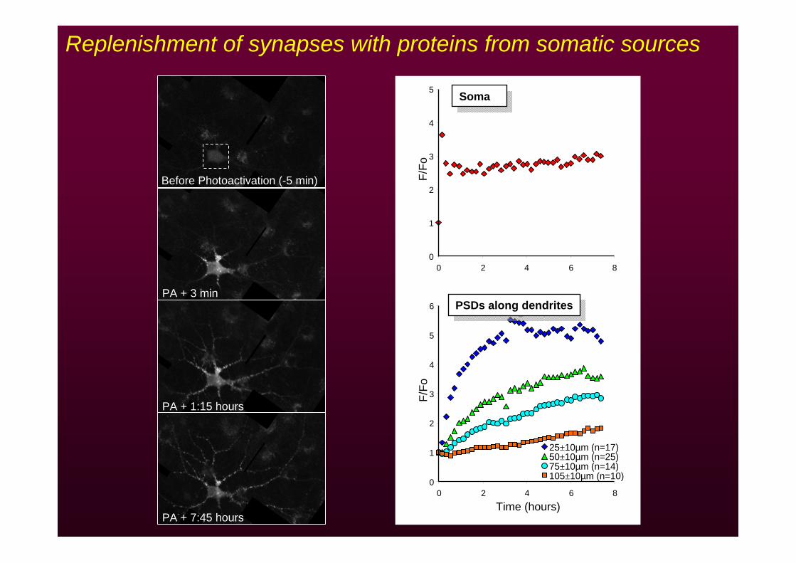
Replenishment of synapses with proteins from somatic sources
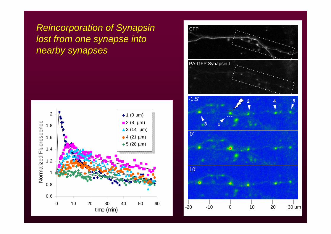
0 10 20 30 µm-10-20
3
2 4 5
1
CFP
PA-GFP:Synapsin I
-1.5’
0’
10’
0.6
0.8
1
1.2
1.4
1.6
1.8
2
0 10 20 30 40 50 60time (min)
Nor
mal
ized
Flu
ores
cenc
e
1 (0 µm)
2 (8 µm)3 (14 µm)
4 (21 µm)5 (28 µm)
Reincorporation of Synapsinlost from one synapse into nearby synapses
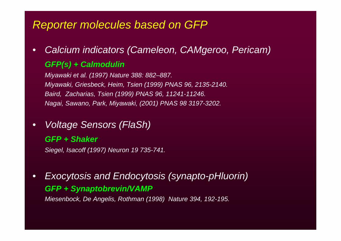
Reporter molecules based on GFP
• Calcium indicators (Cameleon, CAMgeroo, Pericam)GFP(s) + CalmodulinMiyawaki et al. (1997) Nature 388: 882–887.Miyawaki, Griesbeck, Heim, Tsien (1999) PNAS 96, 2135-2140.Baird, Zacharias, Tsien (1999) PNAS 96, 11241-11246.Nagai, Sawano, Park, Miyawaki, (2001) PNAS 98 3197-3202.
• Voltage Sensors (FlaSh)GFP + ShakerSiegel, Isacoff (1997) Neuron 19 735-741.
• Exocytosis and Endocytosis (synapto-pHluorin)GFP + Synaptobrevin/VAMPMiesenbock, De Angelis, Rothman (1998) Nature 394, 192-195.
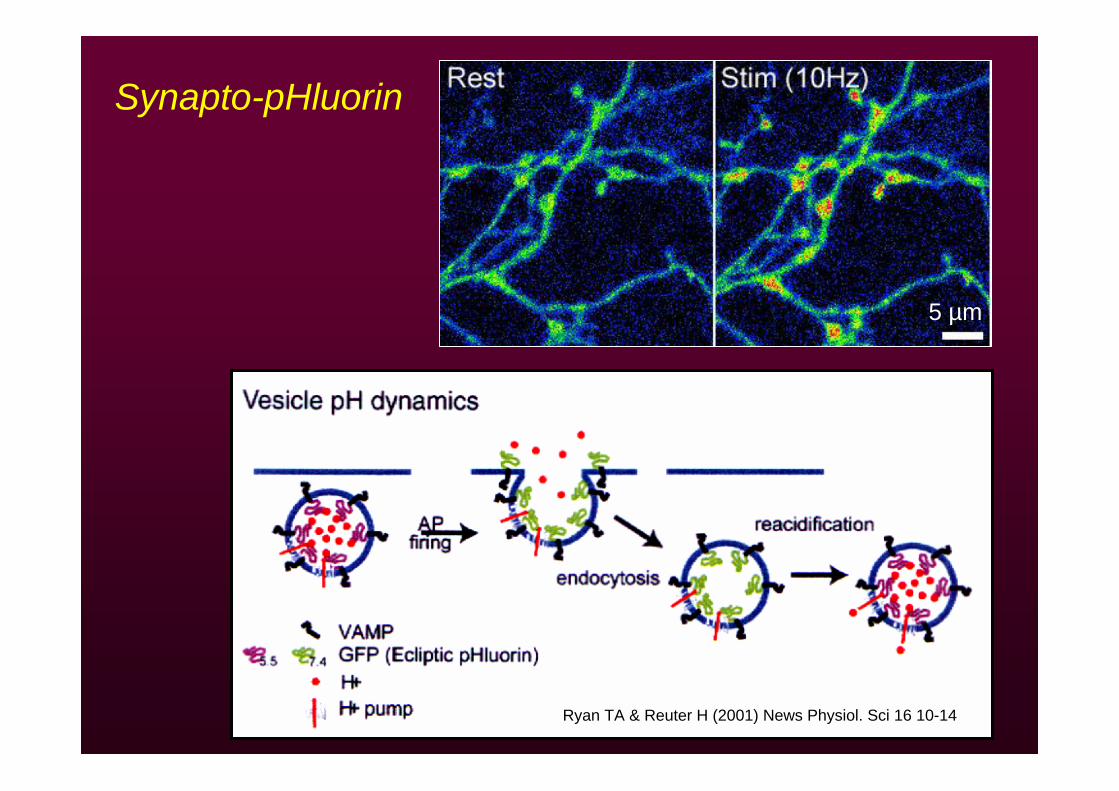
Ryan TA & Reuter H (2001) News Physiol. Sci 16 10-14
Synapto-pHluorin
5 µm
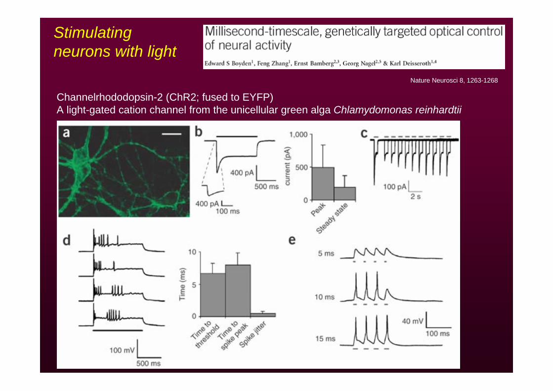
Nature Neurosci 8, 1263-1268
Channelrhododopsin-2 (ChR2; fused to EYFP)A light-gated cation channel from the unicellular green alga Chlamydomonas reinhardtii
Stimulating neurons with light
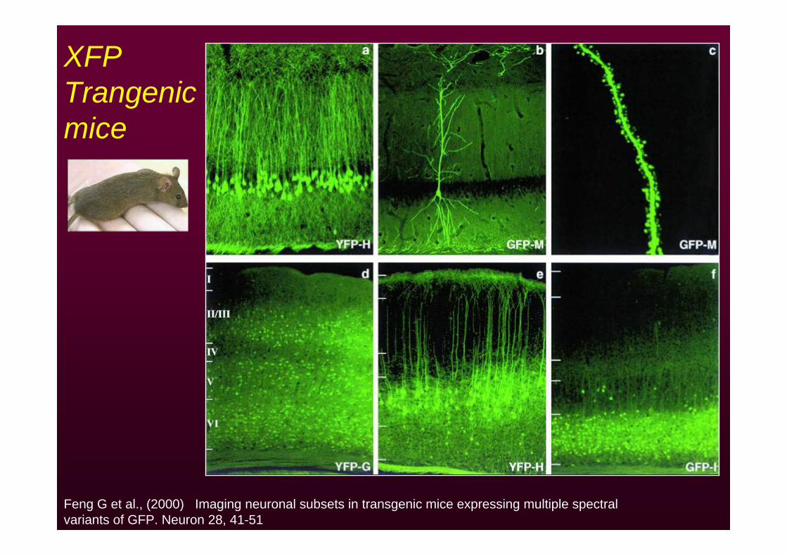
Feng G et al., (2000) Imaging neuronal subsets in transgenic mice expressing multiple spectral variants of GFP. Neuron 28, 41-51
XFP Trangenicmice
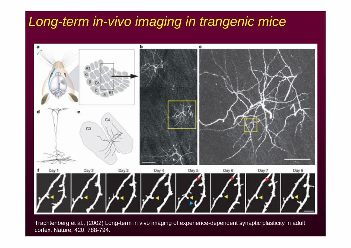
Trachtenberg et al., (2002) Long-term in vivo imaging of experience-dependent synaptic plasticity in adult cortex. Nature, 420, 788-794.
Long-term in-vivo imaging in trangenic mice
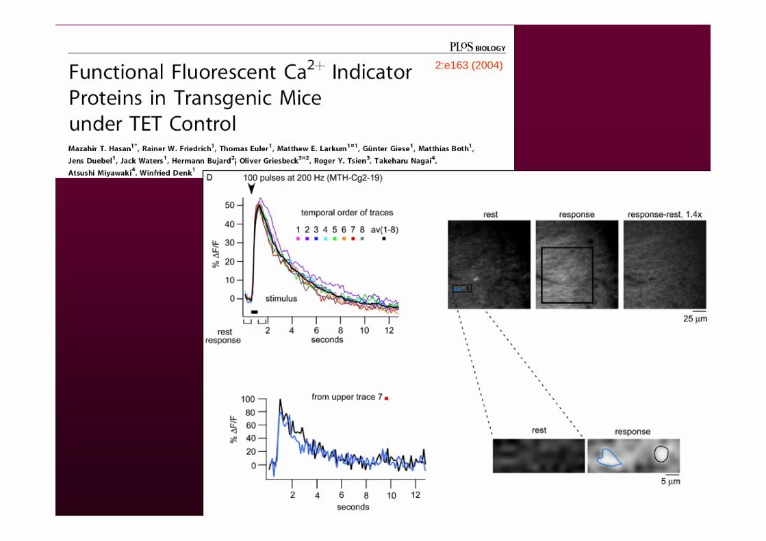
2:e163 (2004)

Knock-in miceHomozygous for Munc-13-1:YFP
Presynaptic protein Munc-13-1 tagged with YFP at original locus in genome.
Mice prepared by Stefan Kalla& Nils Brose, MPI Goettingen
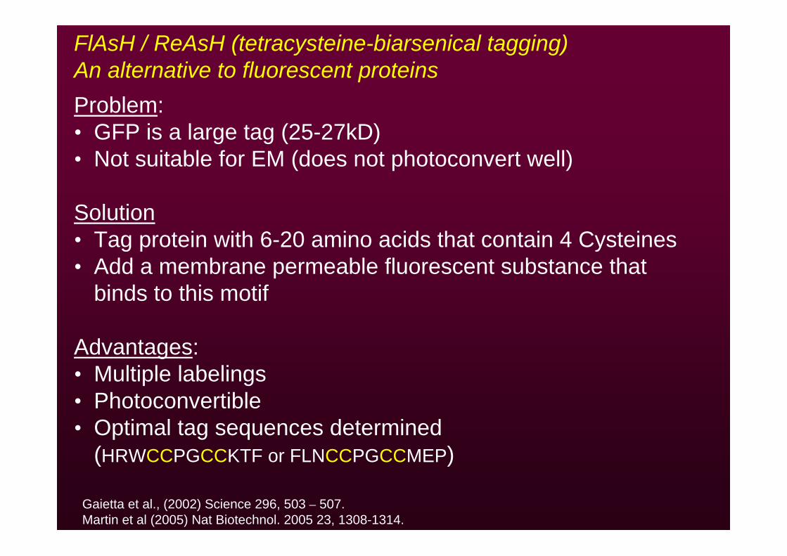
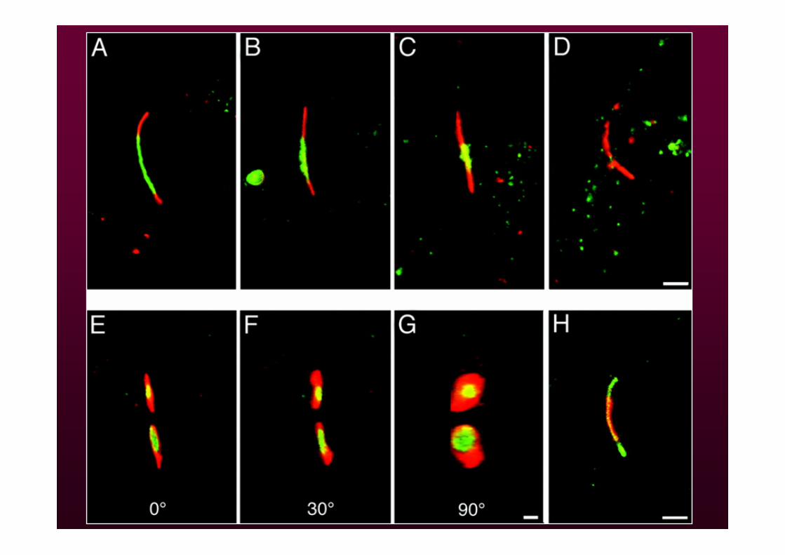
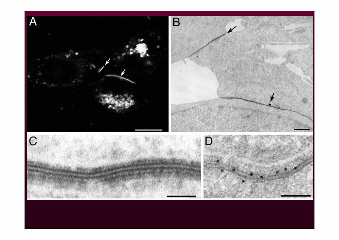
FlAsH / ReAsH (tetracysteine-biarsenical tagging)An alternative to fluorescent proteinsProblem:• GFP is a large tag (25-27kD)• Not suitable for EM (does not photoconvert well)
Solution• Tag protein with 6-20 amino acids that contain 4 Cysteines• Add a membrane permeable fluorescent substance that
binds to this motif
Advantages:• Multiple labelings• Photoconvertible• Optimal tag sequences determined
(HRWCCPGCCKTF or FLNCCPGCCMEP)
Gaietta et al., (2002) Science 296, 503 – 507. Martin et al (2005) Nat Biotechnol. 2005 23, 1308-1314.
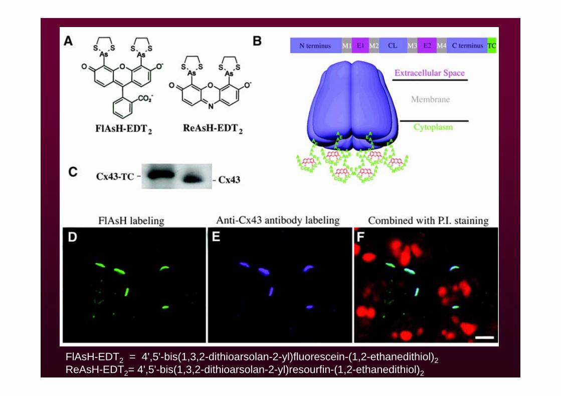
FlAsH-EDT2 = 4',5'-bis(1,3,2-dithioarsolan-2-yl)fluorescein-(1,2-ethanedithiol)2ReAsH-EDT2= 4',5'-bis(1,3,2-dithioarsolan-2-yl)resourfin-(1,2-ethanedithiol)2
Fluorescent proteins: Considerations and potential pitfalls
• Excitation and emission filters – optimize for best S/N• Phototoxicity – less severe but significant• pH sensitivity
• Expression levels• Interference with physiological function of original molecule• Multimerization • Toxicity
Summary
• Live imaging microscopy – a very powerful approach for understanding processes
• Much attention must be paid to maintaining cell viability• Many vital probes currently available• GFP and derivatives – extraordinary tool for studying
molecular and cellular physiology• 6D microscopy provides a means to collect sufficient and
satisfactory data• FRAP – quantitative tool for evaluating molecular dynamics• PA - quantitative and qualitative tool for evaluating
molecular dynamics trafficking and transport• Additional genetically encoded probes appearing daily• Bright future for live imaging!
Ziv lab, Technion, IsraelZiv lab, Technion, IsraelAmirAmir MinerbiMinerbiShlomo TsurielShlomo TsurielRan Ran GevaGevaMichal Michal SternSternRoni KahanaRoni KahanaLarissa Larissa GoldfeldGoldfeldVladimir Vladimir LyakhovLyakhov
CollaborationsCollaborationsCraig Garner, StanfordCraig Garner, StanfordEckart GundelfingerEckart Gundelfinger, , IfNIfN, , MagdeburgMagdeburgThomas Thomas DresbachDresbach, U. Heidelberg, U. HeidelbergNilsNils Brose, MPIBrose, MPI GoettingenGoettingenTobias Tobias BoeckersBoeckers, U. , U. UlmUlm
Tal Tal BreslerBreslerMika ShapiraMika ShapiraHagit Vardinon-FriedmanYaron Ramati

http://www.microscopy.fsu.edu


