
18. 5. 2017
1
PATHOPHYSIOLOGY OF SKELETAL SYSTEM
2016/2017
Eva Lovásová
Department of Pathological Physiology
Faculty of Medicine, P. J. Šafárik University, Košice
DEFINITION OF BONE TISSUE
� Bone tissue forms most of the skeleton, the framework that supports and protects vital organs, bone marrow, acts as mineral reservoir for calcium, has function in acid-base homeostasis and, of course, allows movement.
� Bones are characterized their rigidity,hardness, and power of regeneration and repair.
� Strong but light weight, bone is a dynamic, ever changing tissue. Throughout life, it is continually being broken down and formed.

18. 5. 2017
2
FUNCTIONS OF BONE TISSUE
� Supports the muscles, teeth
� Protects the brain, spinal cord, heart, lungs and other inner organs
� Allows movement
� Maintains ion balance
� Helps with maintaining of acid-base balance
� Bone marrow produces blood cells
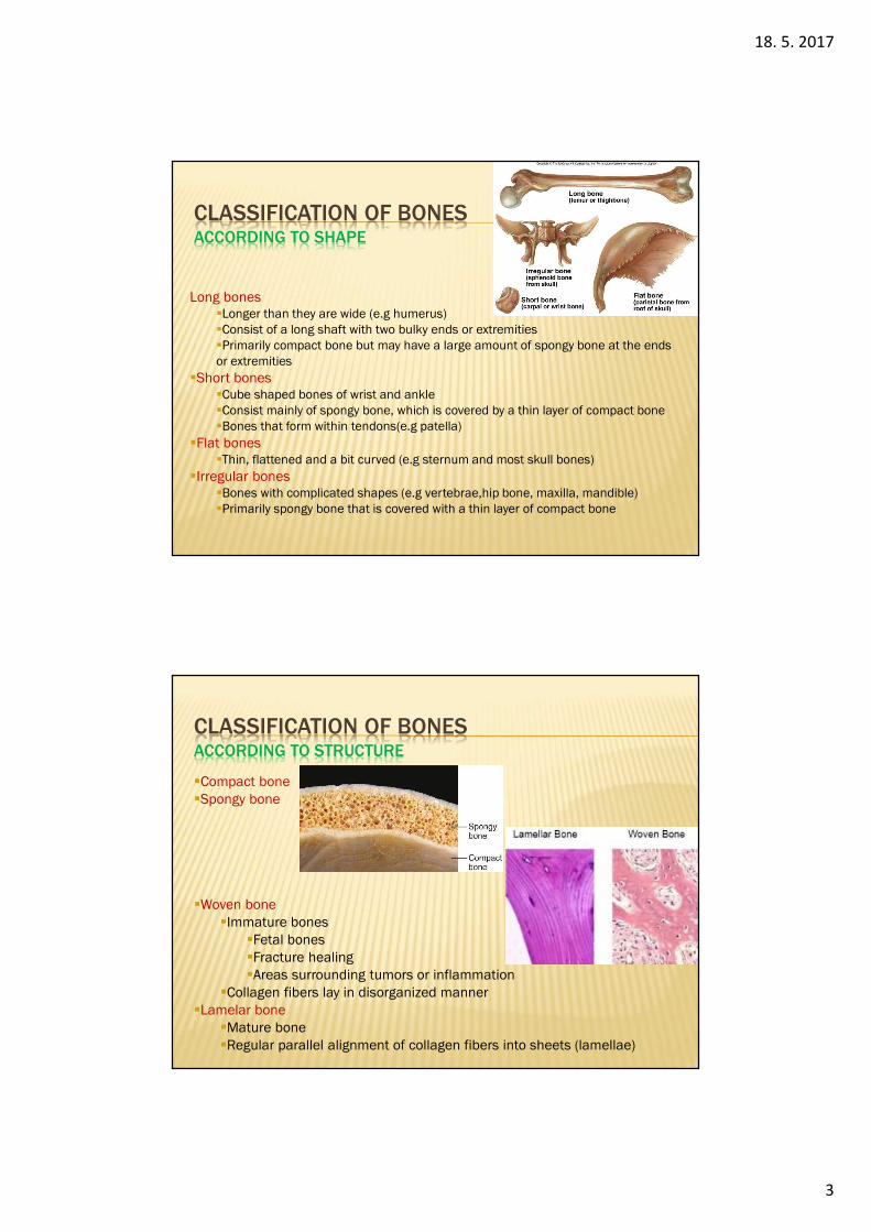
CLASSIFICATION OF BONES
ACCORDING TO POSITION
� Axial skeleton�Skull, vertebral column, ribs, sternum
� Appendicular skeleton�Skeleton of limbs
18. 5. 2017
3
CLASSIFICATION OF BONESACCORDING TO SHAPE
Long bones�Longer than they are wide (e.g humerus) �Consist of a long shaft with two bulky ends or extremities �Primarily compact bone but may have a large amount of spongy bone at the ends or extremities
�Short bones�Cube shaped bones of wrist and ankle �Consist mainly of spongy bone, which is covered by a thin layer of compact bone�Bones that form within tendons(e.g patella)
�Flat bones�Thin, flattened and a bit curved (e.g sternum and most skull bones)
�Irregular bones�Bones with complicated shapes (e.g vertebrae,hip bone, maxilla, mandible)�Primarily spongy bone that is covered with a thin layer of compact bone
CLASSIFICATION OF BONESACCORDING TO STRUCTURE
�Compact bone�Spongy bone
�Woven bone�Immature bones
�Fetal bones�Fracture healing�Areas surrounding tumors or inflammation
�Collagen fibers lay in disorganized manner�Lamelar bone
�Mature bone�Regular parallel alignment of collagen fibers into sheets (lamellae)

18. 5. 2017
4
ANATOMY OF BONES
Long bone
� Diaphysis
� Epiphysis – proximal, distal
� Metaphysis
� Articular cartilage
� Periosteum
� Endosteum
� Medullary cavity
ANATOMY OF BONES
Flat bone

18. 5. 2017
5
BONE MATRIX
�Inorganic components (cca 70 %)�Hydroxyapatite Ca10(PO4)6(OH)2 (99 %)�Other - CaHPO4, MgHPO4, CaCO3
�Organic components (cca 25 %)�Collagen 1 (90 – 95 %)�Osteocalcin (protein)�Osteonectin�Proteoglycans�Glykoproteins�Sialoproteins�Lipids
�Water (cca 5 %)
BONE CELLS
Osteoprogenitor cells�Unspecialized cells derived from mesenchyme�Undergo mitosis and develop into osteoblasts�They are found in the periosteum, endosteum and in canals that contains blood vessels
Osteoblasts�Bone building cells that secrete bone matrix�They lost the ability to divide by mitosis�Secrete collagen and other organic components needed to build bone tissue�The differentiation of osteoprogenitor cells into osteoblasts is accelererated by skeletal growth factors�Functions
�Role in formation of bone matrix�Role in calcification (through the alkaline phosphatase enzymes) �Synthesis of proteins

18. 5. 2017
6
BONE CELLS
Osteocytes�Maintenance of bone�Small flattened and rounded cells embedded in bone lacunae�Derived from mature osteoblasts�Functions
�Help to maintain the bone as living tissue because of there metabolic activity�Maintain the exchange of calcium between the bone and ECF.
Osteoclasts�Bone resorption�Giant phagocytic mutinuecleated cells found in the lacunae of bone matrix, derived from hemopoietic stem cells via monocytes�Functions
�Responsible for bone resorption during bone remodelling�Synthesis and release of lysosomal enzymes necessary for bone resorption
OSTEOGENESIS (BONE GROWTH)
Ossification (osteogenesis) - process of formation of new bone by osteoblasts.
Types of osteogenesis1. Intramembranous ossification
�Laying down of bone into the primitive connective tissue (mesenchyme) resulting in the formation of bones (skull, clavicle, mandible).�Healing process of fractures
2. Endochondral ossification�Cartilage acts as a precursor (e.g., femur, tibia, humerus, radius)�Growing of the length of long bones�Fracture healing.
Steps of osteogenesis1. Synthesis of extracellular organic matrix (osteoid)2. Matrix mineralization leading to the formation of bone3. Remodeling of bone by the process of resorption and reformation

18. 5. 2017
7
ENDOCHONDRALOSSIFICATION
Steps
1. Development of cartilage model2. Growth of cartilage model3. Development of the primary ossification center
�fetal development of bones�a few short bones begin their primary ossification after birth
4. Development of the secondary ossification center�after birth�forms the epiphyses of long bones and the extremities of irregular and flat bones
5. Formation of articular cartilage and epiphyseal plate�after reaching of skeletal maturity (14–18 years of age), all of thecartilage is replaced by bone, fusing the diaphysis and both epiphyses together (epiphyseal closure).
BONE REMODELING
�a lifelong process - old bone is removed from the skeleton (bone resorption), and new bone is added (bone formation)
Function�maintaining of normal function, structure and mineral homeostasis of bone�healing of injuries like fractures but also microdamage which occurs during normal activity
The average lifespan of each remodeled unit in humans is 2–8 months.In the young skeleton, the amount of resorbed bone is proportional to the newly formed- balanced process.Up to the third decade – positive balance.In the third decade - bone mass is at its maximum, and this is maintained with small variations until the age of 50.After the age 50 resorption predominates and the bone mass begins to decrease. Bone remodeling increases in perimenopausal and postmenopausal women.

18. 5. 2017
8
REMODELING UNIT
OsteoclastsOsteoclastsOsteoclastsOsteoclasts�resorbing of bone�derived from mononuclear precursor cells Bone resorption depends on osteoclast secretion of hydrogen ions and cathepsin K enzyme. H+ ions acidify the resorption compartmentto dissolve the mineral component of bone matrix. Cathepsin K digests the proteinaceous matrix, which is mostly composed of type I collagen.
OsteoblastsOsteoblastsOsteoblastsOsteoblasts�bone formation�stimulated by growth hormone, thyroid hormones, estrogens, androgens
REMODELING UNIT
RANKRANKRANKRANKThe cell surface receptor RANK (receptor activator of NFkB) activate osteoclast precursor cells to develop into fully differentiated osteoclasts when RANK is activated by its RANK ligand (RANKL).RANKL is produced mainly by marrow stromal cells and osteoblasts.
OsteoprotegerinOsteoprotegerinOsteoprotegerinOsteoprotegerinOsteoprotegerin (OPG), also known as osteoclast inhibiting factor (OCIF) or osteoclast binding factor (OBF), is a key factor inhibiting the differentiationand activation of osteoclasts, Osteoprotegerin inhibits the binding of RANK to RANKL and inhibits the activation of osteoclasts.
Abnormalties in the balance of OPGL/RANK/OPG system lead to the increasedbone resorption that underlies the bone damage of postmenopausal osteoporosis, Paget’s disease, bone loss in metastatic cancers, and rheumatoid arthritis.

18. 5. 2017
9
REMODELING PHASES
1. Quiescent Phase
bone is at rest2. Activation Phase
activation of the bone surface to resorptionňactivation of osteoclast precursors - differentiation, migration, and fusion ofthe large multinucleated osteoclasts. These cells attach to the mineralized bone surface and initiate resorption by the secretion hydrogen ions andcathepsin K, which degrade bone matrix.3. Resorption Phase
the osteoclasts dissolve the mineral matrix 4. Reversal Phase
bone resorption transitions to bone formation5. Formation Phase
osteoclasts have resorbed a cavity of bone, they detach from the bone surface and are replaced by the osteoblast lineage which in turn initiate bone formation6. Mineralization Phase

18. 5. 2017
10
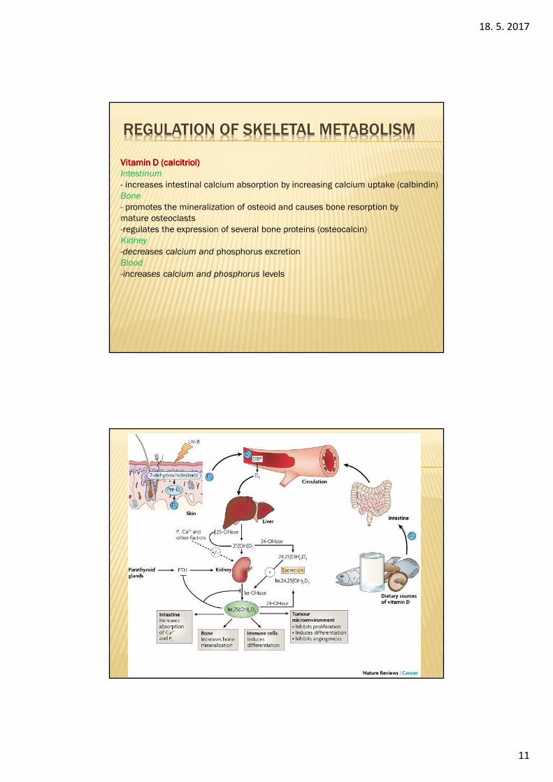
REGULATION OF SKELETAL METABOLISM
Parathyroid HormoneParathyroid HormoneParathyroid HormoneParathyroid Hormone-Peptide hormone (84 amino acids), produced by parathyroid glands-regulates serum calcium and phosphorus concentrations through its receptor-mediated, combined actions on bone, intestine, and kidney- high levels of PTH increase osteoclastic bone resorption- low levels increase osteoblastic bone formation- PTH receptor mainly at the osteoblasts
CalcitoninCalcitoninCalcitoninCalcitonin-Peptide hormone (32 amino acids) produced by parafolicular C-cells of thyroidgland-inhibit osteoclast-mediated bone resorption
18. 5. 2017
11
REGULATION OF SKELETAL METABOLISM
VitaminVitaminVitaminVitamin D (D (D (D (calcitriolcalcitriolcalcitriolcalcitriol))))Intestinum
- increases intestinal calcium absorption by increasing calcium uptake (calbindin)Bone
- promotes the mineralization of osteoid and causes bone resorption bymature osteoclasts-regulates the expression of several bone proteins (osteocalcin)Kidney
-decreases calcium and phosphorus excretionBlood
-increases calcium and phosphorus levels

18. 5. 2017
12
REGULATION OF SKELETAL METABOLISMOther Other Other Other hormonshormonshormonshormonsParathyroid hormone-related protein (PTHrP)
� Function as parathyroid hormone, produced by cancers but also physiologicallyAndrogens
� Anabolic effect through the stimulation of the osteoblast receptors� In childhood activate growth factors – increase bone density� On the end of puberty activate epiphyseal closure
Estrogens� After puberty - epiphyseal closure, but stimulate bone remodeling – after
menopause deficit causes osteoporosis Glucocorticoids
� Bone cell diferentiation during developmentInsulin
� Stimulates matrix synthesis together with IGF-1Growth hormone
� Stimulates osteoblasts activityThyroid hormones
� Stimulate growth resorption but also formation, stimulate the synthesis of osteoid matrix and its mineralization
Growth factors and cytokins (IGF-1)
REGULATION OF SKELETAL METABOLISM
Decrease bone resorption– Calcitonin– EstrogensIncrease bone resorption– PTH/PTHrP– Glucocorticoids– Thyroid hormones– High-dose vitamin DIncrease bone formation– Growth hormone– Vitamin D metabolites– Androgens– Insulin– Low-dose PTH/PTHrP– ProgestogensDecrease bone formation– Glucocorticoids
18. 5. 2017
13
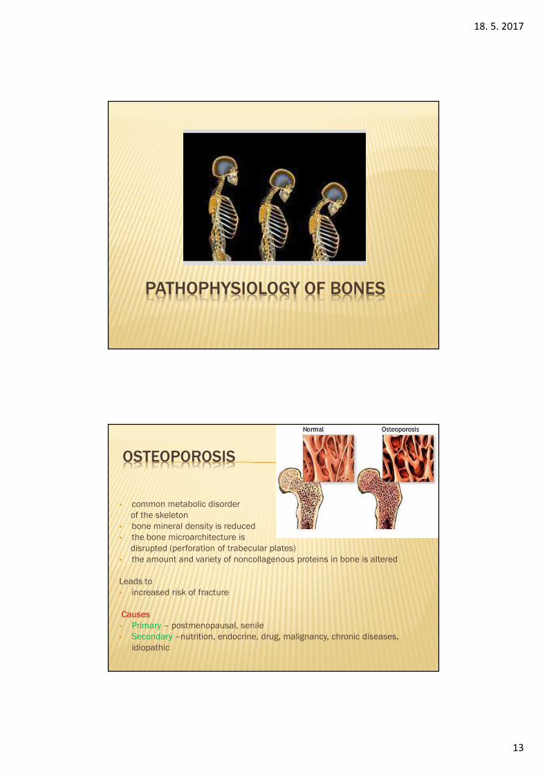
PATHOPHYSIOLOGY OF BONES
OSTEOPOROSIS
� common metabolic disorderof the skeleton
� bone mineral density is reduced� the bone microarchitecture is
disrupted (perforation of trabecular plates)� the amount and variety of noncollagenous proteins in bone is altered
Leads to� increased risk of fracture
Causes� Primary – postmenopausal, senile� Secondary –nutrition, endocrine, drug, malignancy, chronic diseases,
idiopathic

18. 5. 2017
14
OSTEOPOROSIS
Risk factors� Age� Female mainly postmenopausal� Family history� Sedentary lifestyle� Defficiency of calcium or vitamin D� High protein, alcohol, caffeine intake� Smoking� Hyperparathyroidism� Other diseases (diabetes mellitus, celiac disease)� Drugs (Al-containing antacids)
Mechanisms� ↓ estrogens →↑cytokines →↑ RANKL →↑ activity of osteoclasts
OSTEOPOROSIS
Clinical signs� „silent killer of bones“ – long time without signs, pain� decrease of height� fractures of bones – hip, humerus,...� wedging and collapse of vertebrae� kyphosis� hump

18. 5. 2017
15
OSTEOMALACIA
� inadequate mineralization of bones
Causes� deficiency of vit. D – ↓ absorption of calcium from the intestine� phosphate deficiency due to renal diseases or ↓ of intestine absorption
Clinical signs� pain, tenderness and fracture of bones
RICKETS
� inadequate mineralization of bones in children whose epiphyses have not yet fused
Causes� deficiency of vit. D� Vit. D resistant rickets
� phosphate deficiency � mutation of vit. D receptor
Clinical signs� deformation, widening of the bones

18. 5. 2017
16
PAGET´S DISEASE
� Osteitis deformans
� disorder of bone remodeling
� the osteoclasts become abnormally activated, and produce a bizarre and irregular pattern of resorption, to which there is usually an intense osteoblastic response with irregular new bone formation often in the form of woven bone
� increased bone density, but because of the irregular architecture, bone strength is decreased and pathologic fractures may occur.
Causes� hereditary?� environmental factors – virus infection
Cinical signs� enlargement and deformation of bones – face, long bones� CVS disorders� teeth loosing� Pain� Hearing loss
RHEUMATOID ARTHRITIS
� Chronic autoimmune systemic disease
� Synovial inflammation and destruction of the bone architecture
� Women vs. Men 3 : 1
Causes� Autoimmune
� Seropositive form– rheumatoid factor (antibody)� Seronegative form
� T-cells, B-cells, antibodies, macrofages, cytokines (TNFα, IL-1), � Genetic predisposition – HLA-DR4-Dw4 alleles� Production of cytokins – activate osteoclasts� Trigger factor – infection (virus, bacteria, fungi)� Chronic parodontitis is risk factor

18. 5. 2017
17
RHEUMATOID ARTHRITIS
Clinical signs� Joints
� Attacks of symmetric joint pain and morning stiffness� Inflammation and later fibrosis cause movement
limitation� Later deformation, dislocation of joints
� Extra-articular sign� Fatigue, weakness, anorexia, weight loss� Rheumatoid nodules� Fibrosis of lungs� Atherosclerosis
OSTEOARTHRITIS� Degenerative changes of joints
Causes� Unknown� Genetic predisposition� Environmental factors� Obesity� Mechanical stress
Mechanisms� Inflammation produces cytokines that activate production of proteases –
damage of cartilage
Clinical signs� Pain� Difficulty to initiate movement� Joint crepitation

18. 5. 2017
18
GOUT (ARTHRITIS URICA)
� cause - ???� congenital - ↑ production of uric acid - enzyme defects� ↓ excretion of uric acid - defect of renal transporters� acquired - ↑ production of uric acid - obesity, alcohol � high intake of food containing � purins, leukaemia, haemolysis, � cytostatic drugs, Gierke’s disease� ↓ excretion of uric acid - kidney disease� drugs
Clinical Clinical Clinical Clinical symptomssymptomssymptomssymptoms� hyperuricemia� arthritis - acute arthritis� chronic arthritis - tophi� chronic uric acid intestitial nephropathy� uric acid nephrolithiasis (kidney stones
RARE SKELETAL DISEASES
18. 5. 2017
19
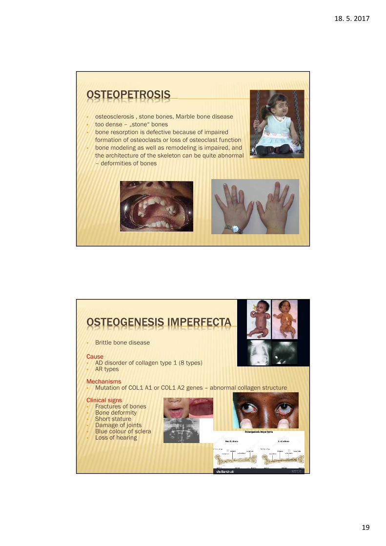
OSTEOPETROSIS
� osteosclerosis , stone bones, Marble bone disease� too dense – „stone“ bones� bone resorption is defective because of impaired
formation of osteoclasts or loss of osteoclast function� bone modeling as well as remodeling is impaired, and
the architecture of the skeleton can be quite abnormal– deformities of bones
OSTEOGENESIS IMPERFECTA
� Brittle bone disease
Cause� AD disorder of collagen type 1 (8 types)� AR types
Mechanisms� Mutation of COL1 A1 or COL1 A2 genes – abnormal collagen structure
Clinical signs� Fractures of bones� Bone deformity� Short stature� Damage of joints� Blue colour of sclera� Loss of hearing

18. 5. 2017
20
EHLERS-DANLOS SYNDROME
� a group of genetic connective tissue disorders
Cause� AD or AR inherited mutation of one of group of
genes – result: defect in synthesis or function of collagen or proteins that interact with collagen
� Affected genes: COL1A1, COL1A2, COL3A1, COL5A1, COL5A2, and TNXB, ADAMTS2, PLOD1...
Clinical signs
� Hyper-flexible joints, luxation and dislocation of joints, swan neck deformity of the fingers, deformities
� osteoarthritis
� Hyperelastic skin, fragile skin, bruising
� Valvular disorders, aneurysm, varices
MARFAN SYNDROME� a genetic connective tissue disorder
Cause� AD inherited mutation in the FBN1 gene on
chromosome 15, which encodes fibrillin-1, a glycoprotein component of the extracellular matrix.
Clinical signs
� Tall, long limbs, long fingers – arachnodactyliy
� Increased joints flexibility
� Scoliosis, lordosis
� Lens dislocation – fibrillin is one protein of apparatus that fix sclera in position
� Valvular disorders, aneurysm, varices
Abraham Lincoln, Nicolo Paganini

18. 5. 2017
21
LITERATURE SOURCES
� Kini U., Nandeesh B.N.: Physiology of Bone Formation, Remodeling, and Metabolism. In: Fogelman I. et al. (eds.) Radionuclide and Hybrid Bone Imaging, Springer-Verlag Berlin Heidelberg, 2012, p. 29-57. DOI 10.1007/078-3-642-02400-9_2.
� Porth C.M.: Unit 12 Musculosceletal system In: Essentials of Pathophysiology, 3rd Edition, Wolters Kluwer Health/Lippincott Williams & Wilkins, 2011, p. 1081- 1158, ISBN 13: 9781451103182/ISBN 10: 1451103182


