
J Clin Exp Dent. 2016;8(3):e327-36. Pathogenesis and clinicohistopathological caractheristics of melanoacanthoma: a systematic review
e327
Journal section: Oral Medicine and Pathology Publication Types: Review
Pathogenesis and clinicohistopathological caractheristics of melanoacanthoma: A systematic review
Elena Cantudo-Sanagustín 1, Aída Gutiérrez-Corrales 1, Manuel Vigo-Martínez 2, María-Ángeles Serrera-Figallo 1, Daniel Torres-Lagares 3, José-Luis Gutiérrez-Pérez 3
1 Master in Oral Surgery2 Medical Doctor. Diplomate in Dental Surgery. Lecture in Oral Medicine. University of Seville3 Professor of Oral Surgery. Co-Head of Master in Oral Surgery. University of Seville
Correspondence:Facultad de Odontología de SevillaC/ Avicena s/n 41009Sevilla, [email protected]
Received: 22/11/2015Accepted: 08/01/2016
Abstract Introduction: The melanoacanthoma is a rare benign pigmented tumor, characterized by a fast radial growth and clinical behavior similar to melanoma. Color changes in oral mucosa and dermis are consequence of increased melanocyte activity as response to an irritant factor. There is a vast phenotypic variety. It is difficult to distinguish between a benign pigmented lesion and a melanoma at its early stage. Due to its clinical relevance is crucial to diagnose possible malignancy of the lesions.Objectives: The aim of this article is to conduct a systematic review of all published articles, as well as update and evaluate etiologic factors and clinicopathological features.Material and Methods: We carried out a search in the Medline database (PubMed) using the key words “oral me-lanoacanthoma” AND “oral melanoacanthosis” AND “oral melanoepithelioma”. Inclusion criteria were all publis-hed articles since its discovery. Demographic data, histological features and immunohistochemical findings were extracted from the full articles.Results: A total of 56 articles were analysed. 114 injuries drawn from these articles were studied, a total of 115 injuries with our contribution case. The 74.78% of authors claim a reactive pathogenesis. The average age of lesión appearance is 34.79 years, with an age range of 5-87 years. There is a predominance of the female sex in solitary phenotype 3: 2 and a ratio of women to men 5: 3 if it is multifocal phenotype. Bilateral phenotype is slight higher in women of 2: 1.Conclusions: Histopathological analysis of the lesión is vital to diagnose malignancy. Therefore, any heteroge-neous, pigmented lesion with irregular edges, raised surface, fast growth and abrupt appearance should be biopsied. More emphasis on the potential irritants should also be put to improve the quality of life of our patients and to reduce morbidity of melanoacanthoma, as well as, several similar clinical behavior disease.
Key words: Melanoacanthoma, oral cáncer, diagnosis.
doi:10.4317/jced.52860http://dx.doi.org/10.4317/jced.52860
Article Number: 52860 http://www.medicinaoral.com/odo/indice.htm© Medicina Oral S. L. C.I.F. B 96689336 - eISSN: 1989-5488eMail: [email protected] in:
PubmedPubmed Central® (PMC)ScopusDOI® System
Cantudo-Sanagustín E, Gutiérrez-Corrales A, Vigo-Martínez M, Serre-ra-Figallo MA, Torres-Lagares D, Gutiérrez-Pérez JL. Pathogenesis and clinicohistopathological caractheristics of melanoacanthoma: A system-atic review. J Clin Exp Dent. 2016;8(3):e327-36.http://www.medicinaoral.com/odo/volumenes/v8i3/jcedv8i3p327.pdf

J Clin Exp Dent. 2016;8(3):e327-36. Pathogenesis and clinicohistopathological caractheristics of melanoacanthoma: a systematic review
e328
IntroductionMelanoacanthoma was first described by Bloch in 1926 as melanoepitheliomoa. In 1960, Mishima and Pinkus introduced the term melanoacanthoma to clarify the term melanoepithelioma type 1 and 2 previously described by Bloch in 1927 (1). The term melanoacanthoma corres-ponds to Bloch`s melanoepithelioma type 1. First lesion in the oral mucosa was presented by Tomey and Dorey in the Maxilofacial and Oral Pathology Congress of the American Academy, in 1978. According to this revision, Schneider et al. described their first case in 1981 (2).Since then, solitary and, less frequent, multiple lesions have been described in the oral mucosa with a total num-ber of 115 cases to the date in our search.Melanoacanthoma is a rare benign mixed epitelial tu-mor, characterized by the mucocutanean pigmentation with dendritics melanocytes dispersed among the epi-thelium with acanthosis areas, espongiosis on melanyne presency. The presence of inflammatory infiltration of linfocitic and eosinophils is a common find (3,4).The high of incidence is between the third or fourth de-cade, it shows higher prevalence in black race and women although some cases were reported in Caucasia race.Lesions may occur as isolated or multiple, plained or raised, with well defined or diffuse edges and the co-lor ranges from dark brown to black. There have been described multiple cases and others with bilateral lesion (Fig. 1). Melanoacanthoma lesions ca be asymptomatic or develop with pain, burning or itching. Its etiology is related to irritative or traumatic factors (5).
Fig. 1. Histological images of the pyogenic granuloma showing an appearance similar to granulation tissue. The histological type of the pyogenic granuloma is non-lobular capillary hemangioma. Arrow heads label blood vessels surrounded by connective tissue.
Cutaneous melanoacanthoma are more likely to appear in head, neck and chest and less frequently in the eyelids or lips. Intraoral lesions are usually asymptomatic and preferentially located in the buccal mucosa (47.54%), palate (18.03%), lips (11.47%) and gum (5.6%) (6).While cutaneous melanoacanthoma never dissapear,
oral melanoacanthoma can regress after the elimination of irritating factor or after being biopsied. Cutaneous variant occurs mainly in fair-skinned adults while the oral melanoacanthoma has a predilection for blacks and younger patients (7).Radial growth it is a high potential pathognomonic sign, it can mask a subyacent melanoma (3).These characteristics have been studied with electronic microscopy, and several tests had been used like: in-munoprecipitation test with the aim of analize patient´s serum to search antibody antimelanoma, inmunofluores-cence to look for present anthygens on melanoma lesion: inmunohistochemical studies have demostrated melano-citic reactivity of the melanocytes which reside on the basal, parabasal and cellular espinous strate for the mar-quer HMB-45; the protein S-100 serves as marquer of the presence of melanocitics dendritics cells, very useful for its diagnosis confirmation, as the marquer Melanin- A also is used with this purpose (7).Quirurgical exéresis shows a great ratio of success wi-thout recurrences (Fig. 2). It offers the advantage of preserve the borders of the lesion for a histhopathologic analisis. Sometimes, even after the incisional biopsy, an involution of the lesion is observed with high frequency. Other ways of treatment are laser ablation with Argon, crioterapy, curettage and the topic application of Flo-urouracil 5% (8).The aim of the present article is to make a systematic review of all the published cases, as to actualize and evaluate ethiologic factors and it clinicopathologic cha-racteristics.
Fig. 2. Same lesion after a follow up of 10 years. It keeps stable.
Material and MethodsA systematic, computerized database search was con-ducted using the National Center for Biotechnology In-formation (NCBI) to search MEDLINE (Pubmed). The search was conducted using the following MeSHterms:” “oral melanoacanthoma” AND “oral melanoacanthosis” AND “oral melanoepithelioma”.

J Clin Exp Dent. 2016;8(3):e327-36. Pathogenesis and clinicohistopathological caractheristics of melanoacanthoma: a systematic review
e329
For the initial selection, we selected all articles published since melanoacanthoma. Demographic data, histological characterestics and immunohistochemical findings were taken from the full text. From the literature a total of 59 articles, in relation with melanoacanthoma, were obtai-ned, three of those were exclude after complete reading. We evaluated 56 articles. A total of 115 patients, inclu-ding the case presented by our team, were diagnosed.Figure 3 describes, in a flow diagram, search phases of our systematic review.
Included
Elegibility
Screening
Identification Records idientified through database searching(n=59)
Records after duplicates removed (n=59)
Records screened (n=59)
Records excluded (n=3)
Full text articles assessed foreligibility (n=56)
Full text articles excluded with reason (n=0)
Studies included in qualitativesynthesis (n=56)
Studies included in quantitativesynthesis (n=0)
Fig. 3. Prisma Flow Diagram: different stages of the search in a sys-tematic review.
ResultsThe review of the literature shows that oral melanoacan-thoma affects patients aged between 5 and 87 years, with a mean age of 34.79 years. Ther is higher prevalence in women, 54.4% versus 38.4% in men.The ratio female-male is 3: 2. There is a predominance of the female sex in solitary phenotype 3: 2 and when the multifocal phenotype is the ratio female-male is 5: 3. In the bilateral phenotype is slight higher in women, 2: 1.The solitary phenotype appears more frequently (18.26% of cases) than the multifocal phenotype (13.91% of cases).The locations from highest to lowest frequency are buc-cal mucosa 33.9%, 13.04% palate, 5.22% lips, 13.91% alveolar mucosa including retromolar área and lingual mucosa, 3.48% tongue, 4.35% back, 3.48% abdomen, 3.48% scapula, 3.48% ear, 3.48% eyelid, 2.6% leg, 2.6% buttock, 1.74% neck, 1.74% floors mouth, 1.74% nose, 1.74% armpit, chest 0.87%, 0.87% vermilion lipstick, hip 0.87%, 0.87% base of the penis, arms 0.87% 0.87% temporal region, 0.87% submental region, 0.87% scro-tum groin area, 0.87% preauricular area ,0.87% shoulder and 0.87% forehead.The most frequent presentation is blackish brown in
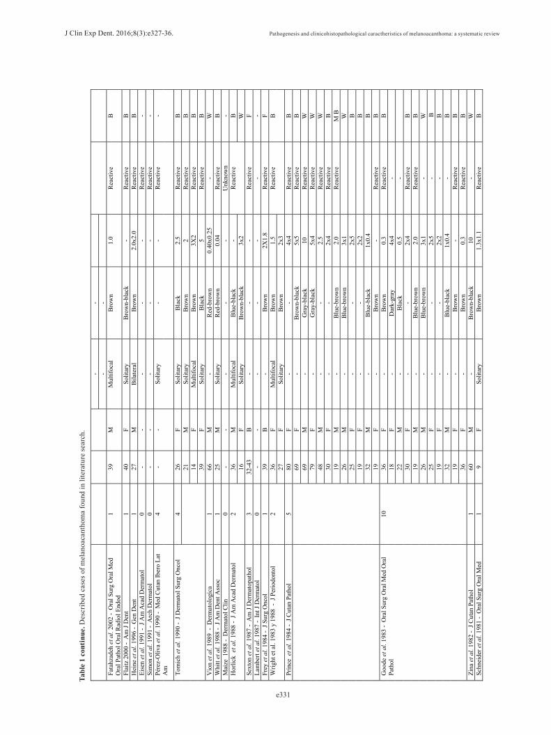
40.8% of cases, followed by bluish black 3.2%, 1.6% reddish brown and grey 0.8%.It has a predilection for black race (37.39%) followed by caucasian (19.13%), latin American (3.47%) and Asians (3.47%). 77.42% of the authors assert that the etiology is related to irritative factors.Table 1, table 1 continue, table 1 continue-1, shows all cases submitted for analysis and the results of our sys-tematic review.
DiscussionThe variability of phenotypic expression justifies the con-troversy of the classification of this entity. There are di-fferent phenotypes with multiple expressions: cutaneous or oral melanoacanthoma, painful or asymptomatic, fast growing or stable, multiple or solitary melanoacantho-mas; as well as different locations and histopathologic features.Zemtsov et al. consider that oral melanoacanthoma is a tumor wrongly named and defined it as an unusual pro-liferation of dendritic melanocytes mucositis in the epi-dermis (9). Horlick, propose the term mucosal melanotic macula for the reactive type of this lesion (10).The etiology is still unclear, but most of the authors as-sociate it with a continuing traumatic process that stimu-late melanocytic activity (3,5,11,12).Most of the injuries are related to trauma and its appea-rance varies from weeks to months. They even dissapear after eliminating irritants or biopsy. This fact makes the reactive etiology stronger.The reactive etiology of melanoacanthoma and other pig-mented lesions may be associated with chronic contact with petroleum derivatives, such as sodium lauryl sul-fate, nitropheno, phentolphthalein, clorophenol, phen-ylenediamine sulfate, cocamidopropyl betaine or amine fluoride. These components are found in toothpastes and mouthwashes and they act as irritants causing morpho-logical changes. Pathology 100% of lesions supports this, and it is related to oral and cutaneous melanoma phenotype (13-15).Most of the the authors observe the ocurrence of the-se lesions in trauma areas of bruxism patients, lesions matching the occlusal plane or very prominent cusps of molars and premolars. Likewise, lesions have also been described in patients with a recent dental restoration, which entails a soft tissue trauma during the adaptation period (16,17).Silver amalgam fillings have also been described as etio-logical factors that may cause pigmentation and changes in the epithelium (17). There is much controversy with this restorative material. Many countries, like Germany and the US, have already forbbided its use in dental the-rapy due to its corrosion and risk of toxicity, while other countries like Spain argue that this material has been used for hundred of years without an apparent risk.

J Clin Exp Dent. 2016;8(3):e327-36. Pathogenesis and clinicohistopathological caractheristics of melanoacanthoma: a systematic review
e330
Tabl
e 1.
Des
crib
ed c
ases
of m
elan
oaca
ntho
ma
foun
d in
lite
ratu
re se
arch
.A
UT
HO
R
NU
MB
ER
OF
PAC
IEN
TS
AG
E
SEX
A
PPE
AR
AN
CE
C
OL
OR
SI
ZE (c
m)
ET
IOLO
GY
E
TH
NIC
ITY
Ken
nedy
et a
l. 2
013
- J I
ndia
n So
c Pe
riodo
ntol
1
13
M
Mul
tifoc
al
Bla
ck-B
row
n 4
Rea
ctiv
e B
W
agne
r et a
l. 20
13 -
Gen
Den
t 1
48
F So
litar
y B
row
n 0.
7 R
eact
ive
W
Patn
ayak
et a
l. 20
13 -
J Lab
Phy
sici
ans
2 15
F
Solit
ary
- 1.
5x1.
0 R
eact
ive
- 53
M
-
1x1
Vas
ani e
t al.
2013
- In
dian
Der
mat
ol O
nlin
e J
1 62
M
So
litar
y B
lack
15
x8
Rea
ctiv
e B
B
hatta
char
yya
2013
-To
days
FD
A
1 72
M
M
ultif
ocal
B
lack
-
- B
D
as C
haga
s et a
l. 20
13 -
BM
C R
es N
otes
1
58
M
Mul
tifoc
al
Bla
ck-B
row
n -
Rea
ctiv
e B
R
ohill
aet
al.
2013
- In
t J C
lin P
edia
tr D
ent
1 12
F
Solit
ary
Bla
ck-B
row
n -
Rea
ctiv
e A
A
shok
Gup
ta e
t al.
2012
- J O
ral M
axill
ofac
Pa
thol
1
22
F M
ultif
ocal
B
lack
-
Rea
ctiv
e -
Gon
dak
et a
l. 20
12 -
Med
Ora
l Pat
ol O
ral C
ir B
ucal
1
- -
Solit
ary
- -
Rea
ctiv
e -
Jain
et a
l. 20
11 -
Indi
an J
Der
mat
ol V
ener
eol
Lepr
ol
1 58
F
Mul
tiple
B
lack
3x
3-12
x10
Rea
ctiv
e -
Gal
indo
et a
l. 20
11 -
JAD
A
1 63
F
Mul
tiple
B
row
n-bl
ack
0.03
-0.0
6 R
eact
ive
W
Shan
kar e
t al.
2011
- In
dian
J D
erm
atol
1
65
M
Solit
ary
-gia
nt
Bro
wn-
blac
k 10
x5
Rea
ctiv
e B
Ta
pia
et a
l. 20
11 -
Qui
ntes
senc
e In
t 1
35
F So
litar
y B
row
n 0,
6x0,
3 R
eact
ive
W
Gee
tha
et a
l. 20
11 -
Indi
an J
Der
mat
ol V
ener
eol
Lepr
ol
1 8
M
Bila
tera
l B
row
n-bl
ack
5x3
Rea
ctiv
e W
Ara
va-P
aras
tatid
is e
t al.
2011
- JA
DA
1
32
F So
litar
y -
- R
eact
ive
W
Bro
oks e
t al.
2010
- Pe
diat
ric D
erm
atol
o 1
17
M
Solit
ary
- -
Rea
ctiv
e W
M
aroc
chio
et a
l. 20
09 -
J Ora
l Sci
1
74
F M
ultif
ocal
B
row
n -
Rea
ctiv
e B
Laks
hmin
aray
anan
et a
l. 20
09 -
Med
Cas
e R
ep
1 24
F
B
row
n 0.
25
Rea
ctiv
e A
B
rook
s et a
l. 20
09 -
J Per
iodo
ntol
1
60
F M
ultif
ocal
B
row
n 0,
1-0,
4 R
eact
ive
W
Bro
oks e
t al.
2008
- G
en D
en
1 47
F
Solit
ary
- 0,
2x0,
3 R
eact
ive
B
Kra
hl e
t al.
2008
- J D
tsch
Der
mat
ol G
es
0 -
- -
- -
Rea
ctiv
e -
Bre
gnie
t al.
2007
- M
ed O
ral P
atol
Ora
l Cir
Buc
al
8 7
M
Solit
ary
Dar
k-br
own
flat
0,3
Rea
ctiv
e W
25
M
-
Dar
k-br
own
flat
1 R
eact
ive
LAM
33
F
- D
ark-
brow
n fla
t 0.
6 R
eact
ive
LAM
40
F
- D
ark-
brow
n fla
t 0.
5 R
eact
ive
LAM
8
M
- D
ark-
brow
n fla
t 0.
3 R
eact
ive
LAM
Y
arom
et a
l. 20
07 -
Int J
Der
mat
ol
1 60
F
Mul
tifoc
al
Bro
wn
- R
eact
ive
W
Ros
iello
et a
l. 20
06 -
Der
mat
ol S
urg
1 38
F
Mul
tifoc
al
Bla
ck-b
row
n 0.
04
- W
A
ndre
ws e
t al.
2005
- A
nn O
tol R
hino
l La
ryng
ol
1 45
M
U
lcer
ated
-
2.5
Rea
ctiv
e B
Con
trera
s et a
l. 20
05 -
Med
Ora
l Pat
ol
1 40
F
- -
- R
eact
ive
W
Kih
icza
k et
al.
2004
-In
t J D
erm
atol
1
45
F -
Bro
wn
2.5x
0.5x
0.8
Rea
ctiv
e -
Buc
hner
et a
l. 20
04 -
J O
ral P
atho
l Med
7
36
F -
Bro
wn
1.0
Rea
ctiv
e B
37
M
-
Bro
wn
0.2
Rea
ctiv
e B
51
M
-
Bla
ck
0.6
Rea
ctiv
e A
24
F
- B
lack
2.
0 R
eact
ive
B
63
M
- -
0.4
Rea
ctiv
e B
52
F
- B
lack
0.
6 R
eact
ive
A
44
F -
Bro
wn
1.0
Rea
ctiv
e B
Fo
rnat
ora
et a
l. 20
03 -
Am
J D
erm
atop
atho
l 10
5-
77
2M
8F
- -
R
eact
ive
7B
3W
Solit
ary
- 0,
2 -
-
J Clin Exp Dent. 2016;8(3):e327-36. Pathogenesis and clinicohistopathological caractheristics of melanoacanthoma: a systematic review
e331
Tabl
e 1
cont
inue
. Des
crib
ed c
ases
of m
elan
oaca
ntho
ma
foun
d in
lite
ratu
re se
arch
.
- -
-
-
Fata
hzad
eh e
t al.
2002
- O
ral S
urg
Ora
l Med
O
ral P
atho
l Ora
l Rad
iol E
ndod
1
39
M
Mul
tifoc
al
Bro
wn
1.0
Rea
ctiv
e B
Flai
tz 2
000
- A
m J
Den
t 1
40
F So
litar
y B
row
n-bl
ack
- R
eact
ive
B
Hei
ne e
t al.
1996
- G
en D
ent
1 27
M
B
ilate
ral
Bro
wn
2.0x
2.0
Rea
ctiv
e B
Ei
sen
et a
l. 19
91 -
J Am
Aca
d D
erm
atol
0
- -
- -
- R
eact
ive
- Si
mon
et a
l. 19
91 -
Arc
h D
erm
atol
0
- -
- -
- R
eact
ive
- Pé
rez-
Oliv
aet
al.
1990
- M
ed C
utan
Iber
o La
t A
m
4 -
- So
litar
y -
- R
eact
ive
-
Tom
ich
et a
l. 19
90 -
J Der
mat
ol S
urg
Onc
ol
4 26
F
Solit
ary
Bla
ck
2.5
Rea
ctiv
e B
21
M
Solit
ary
Bro
wn
2 R
eact
ive
B
14
F M
ultif
ocal
B
row
n 3X
2 R
eact
ive
B
39
F So
litar
y B
lack
5
Rea
ctiv
e B
V
ion
et a
l. 19
89 -
Der
mat
olog
ica
1 66
M
-
Red
-bro
wn
0.40
x0.2
5 -
W
Whi
tt et
al.
1988
- J A
m D
ent A
ssoc
1
25
M
Solit
ary
Red
-bro
wn
0.04
R
eact
ive
B
Mai
ze 1
988
- D
erm
atol
Clin
0
- -
- -
- U
nkno
wn
- H
orlic
k e
t al.
1988
- J A
m A
cad
Der
mat
ol
2 36
M
M
ultif
ocal
B
lue-
blac
k -
Rea
ctiv
e B
16
F
Solit
ary
Bro
wn-
blac
k 3x
2 W
Se
xton
et a
l. 19
87 -
Am
J D
erm
atop
atho
l 3
32-4
3 B
-
- -
Rea
ctiv
e F
Lam
bert
et a
l. 19
87 -
Int J
Der
mat
ol
0 -
- -
- -
- -
Frey
et a
l. 19
84 -
J Sur
g O
ncol
1
39
B
- B
row
n 2X
1.8
Rea
ctiv
e F
Wrig
ht e
t al.
1983
y 1
988
- J P
erio
dont
ol
2 36
F
Mul
tifoc
al
Bro
wn
1.5
Rea
ctiv
e B
27
F
Solit
ary
Bro
wn
2x3
Prin
ceet
al.
1984
- J C
utan
Pat
hol
5 80
F
- -
4x4
Rea
ctiv
e B
69
F
- B
row
n-bl
ack
5x5
Rea
ctiv
e B
69
M
-
Gra
y-bl
ack
10
Rea
ctiv
e W
79
F
- G
ray-
blac
k 5x
4 R
eact
ive
W
48
M
- -
2.5
Rea
ctiv
e W
30
F
- -
2x4
Rea
ctiv
e B
19
M
-
Blu
e-br
own
2.0
Rea
ctiv
e M
B
26
M
- B
lue-
brow
n 3x
1
W
25
F -
- 2x
5
B
19
F -
- 2x
2
B
32
M
- B
lue-
blac
k 1x
0.4
B
19
F
- B
row
n -
Rea
ctiv
e B
G
oode
et a
l. 19
83 -
Ora
l Sur
g O
ral M
ed O
ral
Path
ol
10
36
F -
Bro
wn
0.3
Rea
ctiv
e B
18
F
- D
ark-
gray
4x
4 -
22
M
-
Bla
ck
0.5
-
30
F -
- 2x
4 R
eact
ive
B
19
M
- B
lue-
brow
n 2.
0 R
eact
ive
B
26
M
- B
lue-
brow
n 3x
1 -
W
25
F -
- 2x
5 -
B
19
F -
- 2x
2 -
B
32
M
- B
lue-
blac
k 1x
0.4
- B
19
F
- B
row
n -
Rea
ctiv
e B
36
F
- B
row
n 0.
3 R
eact
ive
B
Zina
et a
l. 19
82 -
J Cut
an P
atho
l 1
60
M
- B
row
n-bl
ack
10
- W
Sc
hnei
der e
t al.
1981
- O
ral S
urg
Ora
l Med
1
9 F
Solit
ary
Bro
wn
1.3x
1.1
Rea
ctiv
e B

J Clin Exp Dent. 2016;8(3):e327-36. Pathogenesis and clinicohistopathological caractheristics of melanoacanthoma: a systematic review
e332
Des
crib
ed c
ases
of m
elan
oaca
ntho
ma
foun
d in
lite
ratu
re se
arch
. F (F
emal
e), M
(Mal
e), W
(Whi
te),
B (B
lack
), A
(Asi
an),
LAM
(Lat
in A
mer
ican
mes
tizo)
, R (R
ecur
renc
e).
Tabl
e 1
cont
inue
-1. D
escr
ibed
cas
es o
f mel
anoa
cant
hom
a fo
und
in li
tera
ture
sear
ch.
Ora
l Pat
hol
Tom
ich
1978
- 32
nd A
nnua
l Mee
ting
of th
e A
mer
ican
Aca
dem
y of
Ora
l Pat
holo
gy
1 39
F
- B
row
n 5
- B
Mat
suok
a et
al.
1979
- A
rch
Der
mat
ol
1 18
F
Mul
tifoc
al
Bla
ck-B
row
n 5-
6 R
eact
ive
B
Schl
appn
er 1
978
- J C
utan
Pat
hol
1 74
M
-
Bro
wn
3x3
- W
D
elac
réta
z 19
75 -
Der
mat
olog
ica
1 87
F
- -
0.09
x0.1
2 R
eact
ive
W
Spot
t et a
l. 19
72 -
Arc
h D
erm
atol
1
40
M
- B
lack
-
Rea
ctiv
e B
Sá
nche
z Y
us e
t al.
1969
- A
ctas
Der
mos
ifilio
gr
8 63
F
- -
- -
- -
M
- -
- -
- 58
M
-
- -
- -
- M
-
- -
- -
58
F -
- -
- -
- M
-
- -
- -
36
M
- -
- -
- -
F -
- -
- -
Mis
him
a et
al.
1960
- A
rch
Der
mat
ol
12
61
F -
- 0.
12x0
.14
Rea
ctiv
e -
46
F -
- 0.
20x0
.20
-
68
F -
- 0.
10x0
.10
-
59
M
- -
0.06
x0.0
6
- 81
M
-
- 0.
10x0
.10
-
77
F -
- 0.
20x0
.20
-
60
F -
- 0.
20x0
.15
-
54
F -
- 0.
13x0
.16
-
62
M
- -
1x1
-
79
M
- -
0.25
x0.2
0
- 63
M
-
- 0.
50x0
.50
-
68
M
- -
0.45
x0.2
8
- 61
M
-
- 0.
20x0
.15
-
Act
ual c
ase
1 53
M
M
ultif
ocal
B
row
n-bl
ack
2.0
Rea
ctiv
e W
Su
mm
ary
115
5-87
(3
4.79
) Fe
mal
e (6
8)
Mal
e (4
7)
Unk
now
n (5
) Pr
opor
tion
7:2
Solit
ary
(11)
=8%
M
ultif
ocal
(6)=
4 8%
U
nkno
wn(
53)=
42,4
%
Bro
wn-
blac
k (5
1)=
44.3
5%
Blu
e-bl
ack
(4)=
3.
48%
R
ed-b
row
n (2
)=1.
6%
Gra
y (2
)=2,
6%
Unk
now
n(56
)=44
.8%
0,04
-8cm
R
eact
ive
(86)
=74,
78
%U
nkno
wn(
29)=
25,2
2%
W(2
2)=1
9,13
%%
B(4
3)=3
7,39
%
LAM
(4)=
3,48
%
A(3
)=2,
60%
U
nkno
wn(
35)=
30,4
3%

J Clin Exp Dent. 2016;8(3):e327-36. Pathogenesis and clinicohistopathological caractheristics of melanoacanthoma: a systematic review
e333
In 2007 Yarom et al. describe as etiologic factors ill-fitting removible prothesis, patients treated for chronic asthma, constant bite of the cheeks, hydrogen peroxide mouthwashes and nonspecific chronic trauma (18,19).There are also described in the literature lesions su-ddenly appeared after implants surgery or associated with ferrous lactate chronic treatment for iron-deficien-cy anemia. Zemtsov et al. proposed to their patients removing too-thpastes and mouthwashes containing hydrogen peroxi-de resulting in the spontaneous resolution of melanoa-canthoma. They show that the most common irritant are mercury and petroleum derivatives as cinnamic alde-hyde, in toothpastes, which may cause allergic contact dermatitis (8).Toothpastes with abrassive components, such as, calcium phosphate or calcium carbonate, act as irritant factors which produce tissue reaction after cronic contact (20).Brooks et al. found that there are alterations of superfi-cial dermis in cutaneous phenotype, but they didn´t ob-served fast growth or spontaneous resolution as it does in oral melanoacanthoma (21,22).Galindo et al., along with other authors, argue that mela-cantoma does not need any specific treatment or follow up because there have not been reported cases of malig-nancy with features of dysplasia or atypia (6).However, we found in the literatura (Zina, in 1982), a case of simple hydroacanthoma with a malignant trans-formation into a porocarcinoma. This extremely rare tumor classification is highly complex due to its histo-pathological similarities with other lesion and degene-rative changes of any lesion with age. The relationship between melanocytes and keratinocytes is very similar to the realtion observed in the melanoacanthoma (23). This rare lesion was named Bort-Jadasshon intraepider-mal epithelioma and it matches with the terms described by Bloch in 1927 and Mishima in 1960 when melanoa-canthoma was called “non-cutaneous benign melanoepi-telioma nevoid” (24,25).Simon believes for there are three variants of melano-cytic seborrheic keratosis: irritant, non-irritating and nested variants. He considers that oral melanoacantho-ma is an irritant seborrheic keratosis and he suggests the term melanoacanthoma should be removed (12).Concerning relations between melanoacanthoma and seborrheic keratosis it should be noted that the only di-fference between the two processes is that in melanoa-canthoma can be found many melanocytes at every level of tumor epidermis, while in seborrheic keratoses, me-lanocytes are not increased and they can only be found in basal area (26). Authors like Sanchez Yus and Simon
Huarte concluded that both tumors are the same enti-ty and they should not be separated. The electron mi-croscope, shows that the distribution and arrangement of melanocytes are notably different in melanoacanto-mas compared to seborrheic keratoses. Melanocytes are small with intense mitotic activity, melanin granules are present in the cytoplasm and several basal keratinocytes proliferate (27).Clinical appearance similar to other pigmented lesions, family history, drug use or systemic drugs, hormones, heavy metals and changes in the morphological pattern are important for the differential diagnosis (11). Some pathological entities described in table 2, table 2 conti-nue should be included.Back to melanoacanthoma histology, this is a pigmen-ted tumor that exhibits great morphological variability. Pathological characteristics show stratified squamous epithelium with proliferation of melanocytes and mela-nin presence in the basal layer and suprabasal layers wi-thout invading the underlying connective tissue, prone to the central keratinization (endoqueratinización). The predominant cell pattern prickly keratinocytes or basal differentiation is present in different areas. Melanocytes have extensive dendritic processes and striking areas of acanthosis (27).The presence of inflammatory infiltrate is found in the great majority of melanoacanthomas. The block in the transference of melanin from melanocytes to keratino-cytes is the nature of this entity. Alteration in the normal pattern and speed differentiation of keratinocytes alters cell characteristic of keratinocytes surface which inhi-bits pigment donation.The etiological hypotheses of reactive origin pigmented lesion is supported by the frequent presence of inflam-matory infiltrate of lymphocytes. Langerhans cells are present in every Malpighian layer except from the basal layer. These cells are related to proliferation control of keratinocytes. Therefore, the stu-dy of Langerhans cells is interesting due to its disposi-tion at every layers (27).
ConclusionsThe histopathological analysis of the melanoacanthoma suspicious lesion is crucial to rule out malignancy, as it may hide a subyacent oral melanoma. Any heterogenous pigmented lesion with irregular borders, raised surface, fast growth and sharp appearance should be biopsied. Since over 75% of reported cases indicate an irritating background, more emphasis must be put on the control of them.This can improve the quality of life of our pa-tients and reduce morbidity of numerous pathologies.

J Clin Exp Dent. 2016;8(3):e327-36. Pathogenesis and clinicohistopathological caractheristics of melanoacanthoma: a systematic review
e334
Characteristics Frequent localitation,higher age
incidence and race
predilection
Clinical characteristics Observations
Intraoral nevus Palate and gums 3rd and 4th
decade
Brown-dark 0.1% of the population
Melanotic macula Increased melanin. Presence of melanocytes
in the basal and parabasal layer with
normal stratified squamous epithelium
Vermilion lower lip, buccal
mucosa and gingiva and
palate
Fisiological pigmentations
Pigmented macules of different sizes and configurations. It
appears since childhood or puberty
Black race Brown-dark Infrequent
Laugier-Hunziker´s sindrome
Acquired benign pathology.
Solitary or multiple macules, lenticular or linear, well-defined or
diffuse margins
Buccal mucosa, lips and
palmoplantar surfaces.
Caucasianwomen.
Second half of the adulthood.
50% of the cases with nail
pigmentation
Brown-grey-dark Infrequent
Peutz-Jegher´s síndrome Autosomal dominant disorder. Hyperchromics
multiple melanotic macules varying in size
and shape.
Buccal mucosa, lips and tongue.
1st decade of life.
60% of patients age of 20
Presence of adenohamartomatous
gastrointestinal polyps. Dark brown
A thorough follow-up of these patients
because of the high risk of malignant
transformation is necessary.
Addison´s disease Endocrine disease with infectious origin,
autoimmune or due to malignant adrenal gland.
Adenocorticotropina overproduction of the
hormone and hyperstimulation of the
melanocytes.
Any region of the oral mucosa
Mucocutaneous hyperpigmentation.
Diffuse / localized pattern. Associated symptoms:
weakness, nausea, vomiting, anorexia, weight loss and
postural hypotension.
It is crucial to diagnose oral lesions, they preced extraoral manifestations.
Vascular pathology Petechiae, ecchymosis, bruises, varicose veins
and hemangiomas
Any region of the oral mucosa
Bluish-red coloration. Pulsatile.
Risk of bleeding.
Osler-Weber-Rendu´s sindrome
Tumor with autosomal dominant hereditary
pattern
Telangiectasia on the lips, tongue and extremities
Reddish-blue spider veins. Oral changes and epistaxis are vital for
early diagnosis. Amalgama tatoo or focal
ArgirosisIt is a iatrogenic injury posterior to soft tissue
implantation of amalgam particles.
Mucosa adjacent fillings
These can be detected in X-rays
Microscopically, the particles are typically
aligned along the collagen fibers and blood vessels, a few
lymphocytes and macrophages they can
also be found
Table 2. Differential diagnosis of various pathologies.

J Clin Exp Dent. 2016;8(3):e327-36. Pathogenesis and clinicohistopathological caractheristics of melanoacanthoma: a systematic review
e335
Melanoma Related to removable prothesis trauma,
irritants such as tobacco and alcohol and poor
oral hygiene.
Maxillarygingiva and the
hard palate mucosa are the locations most
affected African blacks, asians, americans and
hispanics.Tongue, lips or jaw mucosa are other areas of predilection
The peak incidence is in
the seventh decade of life and it is more common in
males.
The clinical features are extremely variable, they can
suddenly appear and fast growth causing ulceration, bleeding and pain (these features are related to the
early stages of oral melanoma), or they can
present macules, plaques, patches or nodules
asymptomatic for years, (in the most advanced) stages.
Primary melanoma of the oral cavity is a rare malignancy (0.2% to
8% of all melanomas in Europe and the United
States). Clinically classified
into four types: nodular melanoma, diffuse surface melanoma, acral lentiginous
melanoma and lentigo maligna melanoma.
Determining the depth of invasion in
millimeters is the most important factor,
especially in cutaneous melanoma prognosis. Most of the cases of
oral melanoma revised were diagnosed when they were in advanced stages so that the ratio
of survival significantly decreases.
Lentigo malign Middle-aged. Peak incidence
65-80 years. Developed in areas of sun
exposure, 86% head and neck.
Dark diamond structures. Slate gray globules points.
Isobara structures. Zig-zag pattern (Brown or
blue-grey dots or blue combined with lines)
The dendritic melanocytes are
cytologically atypical and they are grouped only on the basis of
epithelium. High power of
recurrence. Squamous cell carcinoma
and salivary glands tumors salivary glands
They also contain dendritic melanocytes
Kaposi's sarcoma Characteristic in HIVpatients, but also
found in liver transplant patients, drug injecting
andimmunocompromised
patients
Palate and gingiva
It shows red, blue or purple macula in early stages and
pigmented nodules and plaques in later stages. The
lumps are painful and bleeding.
Histologically, early onset maculas are
composed of lines of endothelial vessels
while, avanced nodular lesions contained
spindle-shaped cells interspersed in the
endothelium. Smoker's Melanosis It is a benign dark
pigmentation in the oral mucosa developed in 25% of the smokers.
Anterior gum and interdental
papilla
Brown-dark It usually disappear when the patient gives
up smoking, persistence make them candidates
for pathological analysis.
Diffuse and multifocal drug-induced melanosis
Drugs that cause oral pigmentation are:
minocycline, antimalarials,
chemotherapy and antiretroviral drugs used in the treatment of HIV
patients.
Hard palate and gingiva
Brown-dark Oral pigmentation dissapears after
removing the drug
Simple pigmented malignant
hydroacanthoma
Malignant tumor often confused with
melanoacanthoma, seborrheic keratoses and
Bowen's disease. Described by Yu-Yun
Lee et al. (38)
Predilection for the limbs of
women.
Brown-dark Extremely rare
Table 2 continue. Differential diagnosis of various pathologies.
J Clin Exp Dent. 2016;8(3):e327-36. Pathogenesis and clinicohistopathological caractheristics of melanoacanthoma: a systematic review
e336
References1. Kennedy Babu SPK, Agila S, Sivaranjani P, Kashyap V. An unusual clinical presentation of gingival melanoacanthoma. J Indian Soc Perio-dontol. 2013;17:657-60.2. Schneider LC, Mesa ML, Haber SM. Melanoacanthoma of the oral mucosa. Oral Surg Oral Med Oral Pathol. 1981;52:284-7.3. Contreras E, Carlos R. Oral melanoacanthosis (melanoachantoma): report of a case and review of the literature. Med Oral Patol Oral Cir Bucal. 2005;10:11-2;9-11.4. Arava-Parastatidis M, Alawi F, Stoopler TE. Multifocal pigmenta-tion of the oral cavity. JADA. 2011;142:53-6.5. Krahl D, Altenburg A, Zouboulis CC. Reactive hyperplasias, pre-cancerous and malignant lesions of the oral mucosa. J Dtsch Dermatol Ges. 2008;6:217-32.6. Galindo P, Padial M, Gómez M, Aneiros J, Mesa F, O’Valle F. Mul-tifocal oral melanoacanthoma and melanotic macula after dental im-plants surgery. JADA. 2011;142:817-24.7. Tapia JL, Quezada D, Gaitan S, Hernández JC, Paez C, Aguirre A. Gingival melanoacanthoma: case report and discussion of its clinical relevance. Quintessence Int. 2011;42:253-8.8. Andrews BT, Trask DK. Oral melanoacanthoma: a case report, a review of the literature, and a new treatment option. Ann Otol Rhinol Laryngol. 2005;114:677-80.9. Zemtsov A, Bergfeld WF. Oral melanoacanthoma with prominent spongiotic intraepithelial vesicles. J Cutan Pathol. 1989;16:365-9.10. Horlick HP, Walther RR, Zegarelli DJ, Silvers DN, Eliezri YD. Mucosal melanotic macule, reactive type: a simulation of melanoma. J Am Acad Dermatol. 1988;19:786-91.11. Gondak RO, da Silva-Jorge R, Jorge J, Lopes MA, Vargas PA. Oral pigmented lesions: Clinicopathologic features and review of the litera-ture. Med Oral Patol Oral Cir Bucal. 2012;17:e919-24.12. Simon P, Requena L, Sánchez-Yus E. How rare is melanoacantho-ma? Arch Dermatol. 1991;127:583-4.13. Lawrence LM, Farquharson A, Brown RS, Vatanka HO. Oral tissue irritants in toothpaste: a case report. J Clin Pediatr Dent. 2013;38:75-8.14. Sajid M, Ilyas M, Basheer C, Tariq M, Daud M, Baig N, et al. Impact of nanoparticles on human and environment: review of toxici-ty factors, exposures, control strategies, and future prospects. Environ Sci Pollut Res Int. 2015;22:4122-43.15. Cvikl B, Lussi A, Gruber R. The in vitro impact of toothpaste ex-tracts on cell viability. Eur J Oral Sci. 2015;123:179-85.16. Whitt JC, Jennings DR, Arendt DM, Vinton JR. Rapidly ex-panding pigmented lesion of the buccal mucosa. J Am Dent Assoc. 1988;117:620-2.17. Sexton FM, Maize JC. Melanotic macules and melanoacanthomas of the lip. A comparative study with census of the basal melanocyte population. Am J Dermatopathol. 1987;9:438-44.18. Yarom N, Hirshberg A, Buchner A. Solitary and multifocal oral melanoacanthoma. Int J Dermatol. 2007;46:1232-6.19. Goode RK, Crawford BE, Callihan MD, Neville BW. Oral mela-noacanthoma. Review of the literature and report of ten cases. Oral Surg Oral Med Oral Pathol. 1983;56:622-8.20. Bouquot JE, Koeppen RG, Haddad Y. Oral and maxillofacial pathology. Case of the month. Toothpaste-induced mucosal etching (TIME). Tex Dent J. 2014;131:574-6,610-2.21. Brooks JK, Sindler AJ, Scheper MA. Oral melanoacanthoma in an adolescent. Pediatric Dermatolo. 2010;27:384-7.22. Brooks JK, Sindler AJ, Papadimitriou JC, Francis LA, Scheper MA. Multifocal melanoacanthoma of the gingiva and hard palate. J Periodontol. 2009;80:527-32.23. Zina AM, Bundino S, Pippione MG. Pigmented hidroacanthoma simplex with porocarcinoma. Light and electron microscopic study of a case. J Cutan Pathol. 1982;9:104-12.24. Schneider LC, Mesa ML, Haber SM. Melanoacanthoma of the oral mucosa. Oral Surg Oral Med Oral Pathol. 1981;52:284-7.25. Mishima Y, Pinkus H. Benign mixed tumor of melanocytes and malpighian cells. Melanoacanthoma: Its relationship to Bloch’s benign non-nevoid melanoepithelioma. Arch Dermatol. 1960;81:539-50.
26. Pérez-Oliva N, Toribio J, Quiñones PA. Histologic aspects of sebo-rrheic keratosis. Med Cutan Ibero Lat Am. 1990;18:70-7.27. Schlappner OL, Rowden G, Philips TM, Rahim Z. Melanoacan-thoma. Ultrastructural and immunological studies. J Cutan Pathol. 1978;5:127-41.
Conflict of InterestThe article have not any conflict of interests.


