
Paracentesis / Thoracentesis An Overview
By Heather Molina, RT(R), PA-C, MMS

Topics To stick or not to stick???? QUIZ (Para)
Ascites overview Pathophysiology Procedural techniques Complications
Pleural effusion overview Procedural techniques Complications
Albumin or not?
Academia vs. Private practices

Stick or Not? 1
A. Yes B. Not without further work up

Stick or Not? 2
A. Yes B. Not without further work up
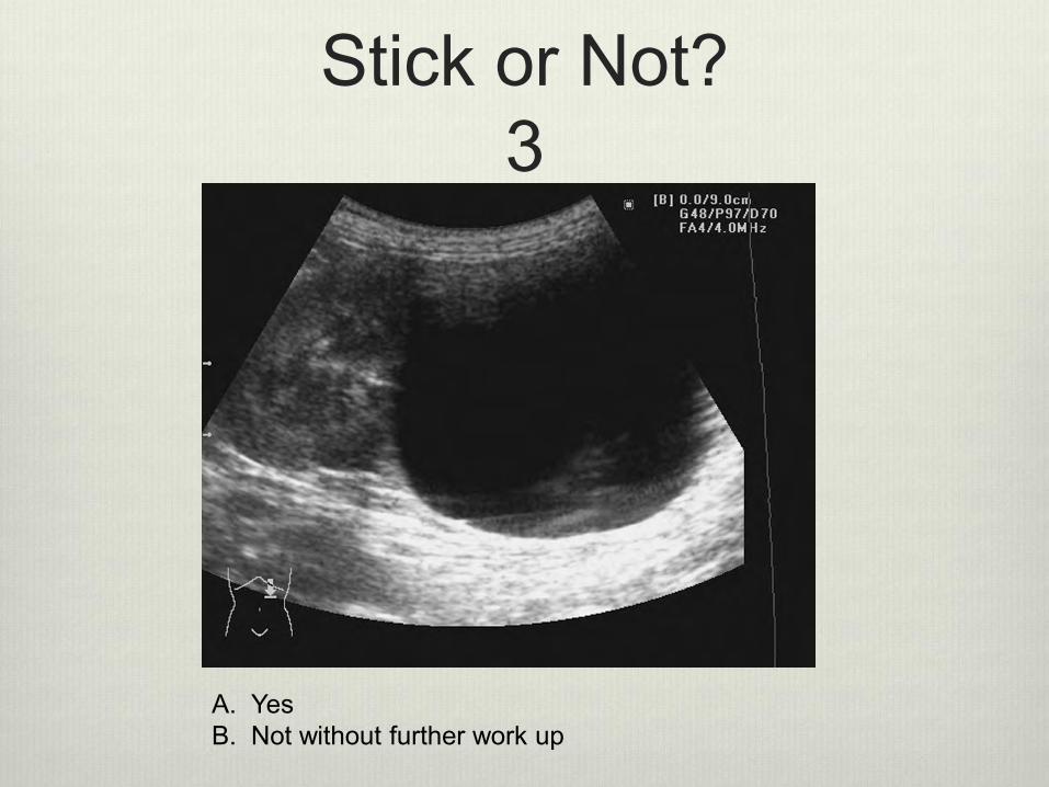
Stick or Not? 3
A. Yes B. Not without further work up
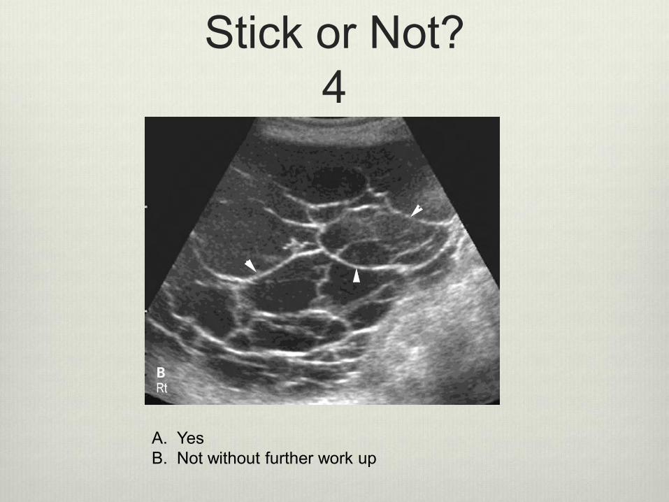
Stick or Not? 4
A. Yes B. Not without further work up
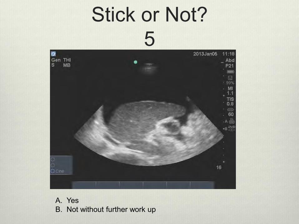
Stick or Not? 5
A. Yes B. Not without further work up

Stick or Not? 6
A. Yes B. Not without further work up
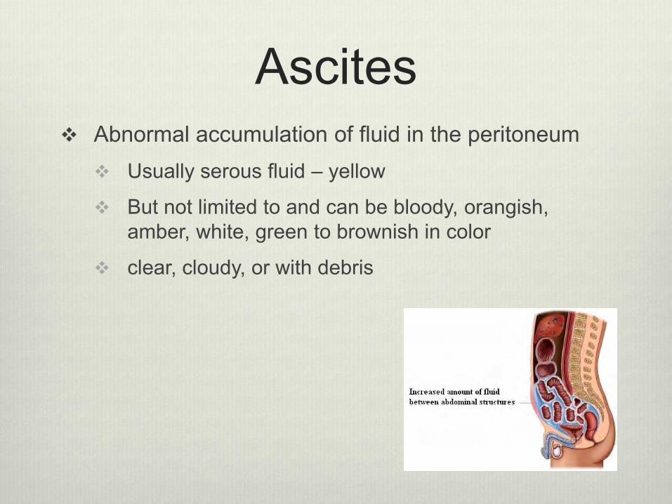
Ascites Abnormal accumulation of fluid in the peritoneum
Usually serous fluid – yellow
But not limited to and can be bloody, orangish, amber, white, green to brownish in color
clear, cloudy, or with debris

Causes of Ascites Protein Loss
Cirrhosis
Tight Junction Dysfunction Malignancy
Infection
Pancreatitis
Down Steam Occlusion Heart failure
Renal failure
Portal occlusion

Causes cont. Oncotic Pressure: Also called colloid osmotic
pressure, is a form of osmotic pressure exerted by proteins in a blood vessel's plasma (blood/liquid) that usually tends to pull water into the circulatory system. It is the opposing force to hydrostatic pressure.
Hydrostatic Pressure: The pressure that the fluid exerts on the walls of its container ie. Down stream occlusion

Pathophysiology of Ascites

Pathophys cont

Pathophys cont.

Contraindications Uncooperative patients
High risk for bleeding
Acute abdomen that requires surgery
Intra-abdominal adhesions
Distended bowel
Abdominal wall cellulitis at site of puncture
Pregnancy (in most cases)

When to do a paracentesis When conservative measures no longer work
Patients with discomfort and difficulty breathing
Concern for infection SBP vs. secondary bacterial peritonitis
To determine etiology of fluid Transudative vs. Exudative

Infection Spontaneous bacterial peritonitis (SBP)
No clear indication for infection
Secondary bacterial peritonitis Frequent paracentesis
Perforated bowel
Abscess Higher Mortality associated
Confirming Infection Symptoms: Fevers, chills, nausea, vomiting,
abdominal pain, general fatigue, encephalopathy
Elevated Neutrophil count of ascitic fluid
Source of infection CT Scan
Surgery
Autopsy

Etiology of Ascitic Fluid
Transudate Exudate
CAUSE Increased hydrostatic pressure, CHF, ESLD, Nephrotic syndrome, SVC syndrome, glomerulonephritis
Inflammation, pneumonia, malignancy, pulmonary TB, pancreatitis, ovarian neoplasm, SLE, RA, drug induced, uremic pleuritis
COLOR Clear, water- like, or pale yellow Cloudy, white, yellow or red
CONSISTENCY Thin and watery, no tissue fragments
Thick and creamy, contains tissue fragments
ODOR None May have an odor
pH Alkaline Acid
SPECIFIC GRAVITY
1.015 or lower 1.018 or higher
PROTEIN CONTENT
Low, less than 3% High, more than 4%
CELL COUNT Low, none or few WBC and RBC High, many WBC and RBC
ENZYME CONTENT
Low High
BACTERIA None May be present
INFLAMMATION None present Associated with inflammation

Complications with para Bleeding
Peritoneal layer
Internal bleeding
Infection
Skin
Fluid
Puncture to liver, bowel or other abdominal structures
Loss of catheter in skin or peritoneum
Post procedural hypotension
Post procedural hyponatremia
Spontaneous hemoperitoneum
Rare complication associated due to mesenteric variceal bleeding after LVP
Hepatorenal syndrome (HRS)

Safe to stick
Image 2 – Showing bowel Image 4 - Loculations
Image 6 – Showing liver

Oops! Should have done further work up.
Image 1 - Carcinomatosis Image 3 – Renal cyst
Image 6 – Ovarian cyst

Hepatorenal Syndrome (HRS)
Precipitating factors of HRS Sepsis
UGI Bleed
Paracentesis
Nephrotoxic drugs Loop diuretics
Antibiotics
NSAIDS

Treatment/ management options for recurrent ascites
Tunneled drainage catheters Pleurx, Aspira, Denver shunt
Usually Hospice patients- (Low protein state)
TIPS- Up stream occlusion
Lymph angio with embolization Not quite as common
Surgery Liver, kidney, heart transplant

Procedural techniques Why most patients complain of pain
Why size matters
Why amount matters
The Z technique
Location/Location/Location
Complaints of pain Typically seen when the area is not numbed up to
the most distal peritoneal layer.
The area being numbed up is not the same area that was re-entered when going accessing the fluid.

Size can matter

Amount removed Hypotension
Hyponatremia
Feeling of illness due to massive fluid shift
Albumin Protein replacement
5-8 g/dl per liter after 5 liters removed
Reduces protein leaking
Helps resolve hypotension, hypovolemia, renal impairment, and hyponatremia

The “Z” technique

Location

Safety Needles
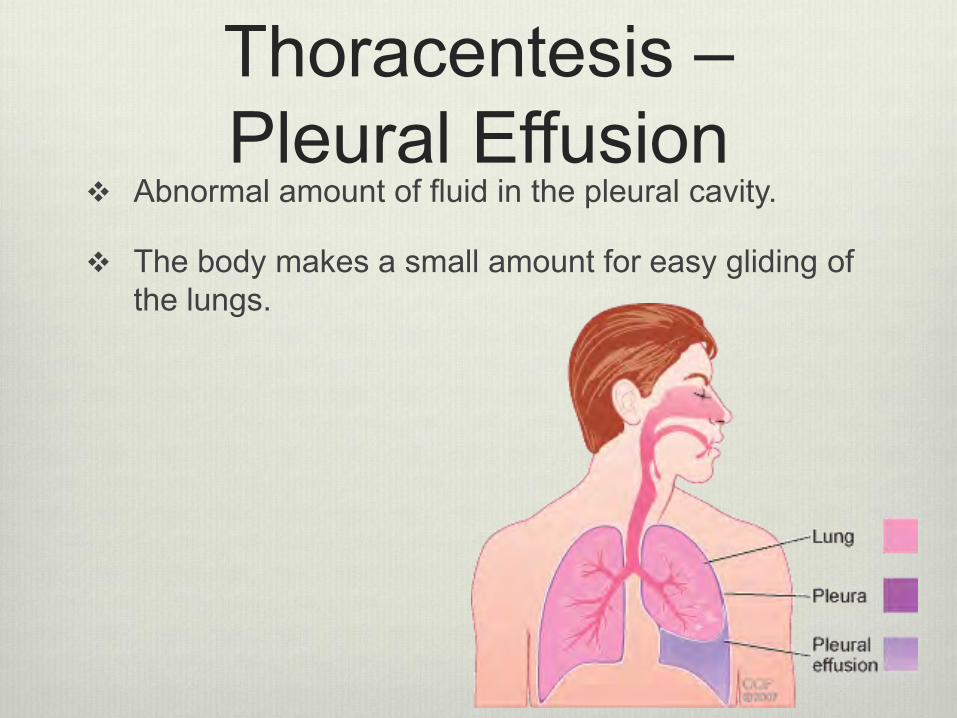
Thoracentesis – Pleural Effusion
Abnormal amount of fluid in the pleural cavity.
The body makes a small amount for easy gliding of the lungs.

Lung Anatomy
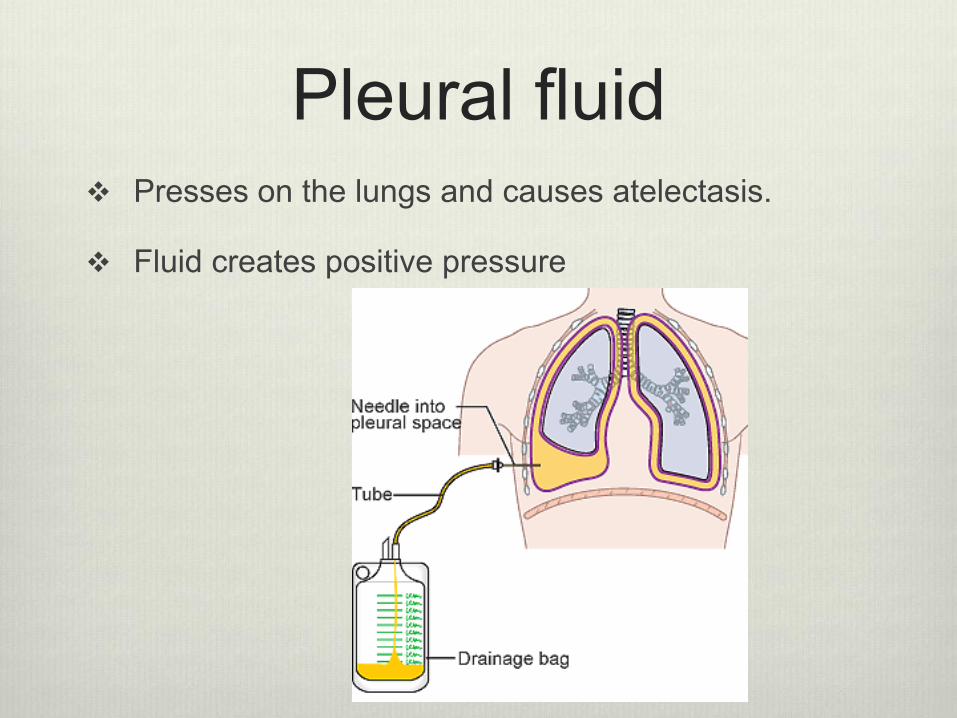
Pleural fluid Presses on the lungs and causes atelectasis.
Fluid creates positive pressure

Indications for Thoracentesis
Symptomatic relief of shortness of breath for large pleural effusions
Diagnostic purposes
Diagnose and treat empyema’s

Pleural Effusions on ultrasound

Pleural Effusion on X Ray and CT

Contraindications of Thoracentesis
High risk of bleeding INR > 1.5
Platelets < 50
Uncooperative patient

Procedural Techniques

Complications of Thoracentesis
Bleeding
Infection
Pneumothorax
Injury to other structures Liver
Spleen
Re-expansion Pulmonary edema

Pneumothorax With image guidance very low chance of hitting lung and
causing PTX
Pneumothorax can be caused by letting air in catheter
Post-op Chest x-ray
Difference between Pneumothorax with air trapped in the pleural space and with “Trapped lung” that can’t expand. Sometimes called “Pneumothorax ex vacuo”

Pulmonary Edema
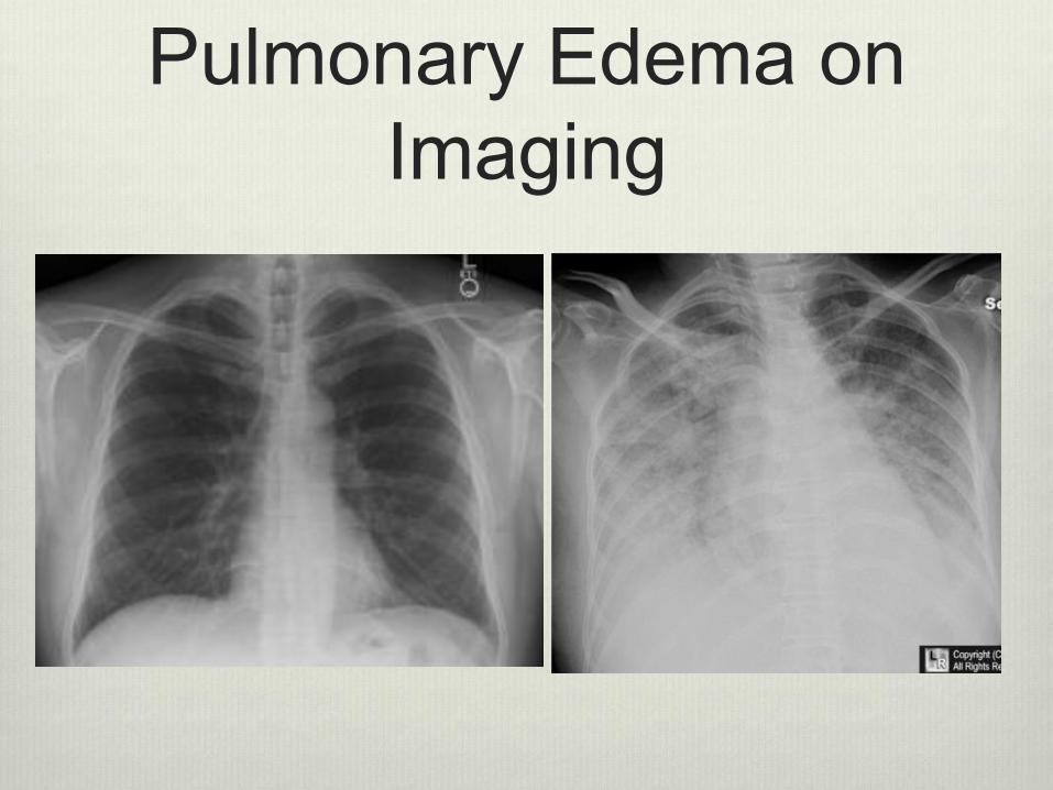
Pulmonary Edema on Imaging
I have liver disease, why is it in my lungs and not my
belly? Hepatic Hydrothorax
When a patient has a recurrent pleural effusion and not ascites
Small microscopic tears in the diaphragm
With breathing this sucks the fluid into the pleural space

Treatment/ Management of Pleural Effusion
Thoracentesis
Chest Tube
Pleurodesis
Pleural Drain Pleurx, Aspira, Denver shunt
Pleural Decortication

Academia vs Private Practice
Reasons for pleurx
Amount of fluid being removed
Amount of albumin to give

References Runyon BA. Low-protein-concentration ascitic fluid is predisposed to spontaneous
bacterial peritonitis. Gastroenterology. 1986 Dec;91(6):1343-6. PubMed PMID: 3770358. http://quizlet.com/19618511/hydrostatic-pressure-vs-osmotic-pressure-flash-cards/
http://faculty.wwu.edu/vawter/PhysicsNet/Topics/Pressure/HydroStatic.html
Malignant ascites: A review of prognostic factors, pathophysiology and therapeutic measures Suma L Sangisetty, Thomas J Miner. World J Gastrointest Surg. 2012 April 27; 4(4): 87–95. Published online 2012 April 27. doi: 10.4240/wjgs.v4.i4.87
http://www.ncbi.nlm.nih.gov/pmc/articles/PMC3020354/
http://www.medscape.com/viewarticle/761213_2
http://www.uptodate.com/contents/thoracentesis-beyond-the-basics
http://www.uptodate.com/contents/hepatic-hydrothorax


