Download - Osteoporosis 131109064516 Phpapp02

OsteoporosisDr. Muhammad Khairussyakirin b Mohd Ali

A skeletal disorder characterised by compromised bone strength predisposing a person to an increased risk of fracture.◦ Bone strength reflects the integration of bone
density and bone quality Bone density (g/cm2 or g/cm3)
determined by peak bone mass and amount of bone loss.
Bone quality architecture, turnover, damage accumulation, and
mineralisation of the bone
DEFINITION

The spine, hips, ribs, and wrists are common areas of bone fractures from osteoporosis.
Classification is based on bone mineral density (BMD).◦ Osteoporosis is defined by BMD of less than –2.5
SD from the young adult mean (T-score)

1

T score: standard deviation of the BMD from the average sex matched 35-year-old
For every 1 decrease in T score, double risk of fracture
1 SD decrease in BMD = 14 year increase in age for predicting hip fracture risk
Regardless of BMD, patients with prior osteoporotic fracture have up to 5 times risk of future fracture!
How to interpret the BMD

Epidemiology 2

In our community, the Chinese had the highest incidence of hip fractures compared to the Malays and Indians.◦ Chinese women accounted for 44.8% of hip
fractures

Bone is continually remodeled throughout our lives . Bone resorption is always followed by bone formation, a phenomenon referred to as coupling
Bone strength is determined by collagenous proteins (tensile strength) and mineralized osteoid (compressive strength). The greater the concentration of calcium, the greater the compressive strength
Normal bone formation and remodeling

Osteoclasts, derived from mesenchymal cells, are responsible for bone resorption, whereas osteoblasts, from hematopoietic precursors, are responsible for bone formation.
The 2 types of cells are dependent on each other for production and linked in the process of bone remodeling

In osteoporosis a reduction in skeletal mass caused by an imbalance between bone resorption and bone formation.
Under physiologic conditions, bone formation and resorption are in a fair balance. A change in either—that is, increased bone resorption or decreased bone formation may result in osteoporosis.

Accelerated bone loss can be affected by hormonal status, as occurs in perimenopausal women
Aging and loss of gonadal function are the 2 most important factors contributing to the development of osteoporosis

Estrogen deficiency accelerates bone loss in postmenopausal women.
Estrogen deficiency can lead to excessive bone resorption accompanied by inadequate bone formation.
Osteoblasts, osteocytes, and osteoclasts all estrogen receptors.
Estrogen deficiency

In the absence of estrogen, T cells promote osteoclast recruitment, and prolonged survival via IL-1, IL-6, and tumor necrosis factor (TNF)–alpha.
With the prolonged osteoclast survival, rate of bone resorption will increase.

In contrast to postmenopausal bone loss, which is associated with excessive osteoclast activity, the bone loss that accompanies aging is associated with a progressive decline in the supply of osteoblasts in proportion to the demand.
After the third decade of life, bone resorption exceeds bone formation
Aging

Calcium and vitamin D help maintain bone homeostasis. Insufficient dietary calcium or impaired intestinal absorption of calcium can lead to secondary hyperparathyroidism. PTH is secreted in response to low serum calcium levels. It increases calcium resorption from bone, decreases renal calcium excretion, and increases renal production of 1,25-dihydroxyvitamin D (1,25[OH]2 D)—an active hormonal form of vitamin D that optimizes calcium and phosphorus absorption
Calcium deficiency

Medications that lead to bone loss (eg, glucocorticoids) can cause osteoporosis. Corticosteroids inhibit osteoblast function and enhance osteoblast apoptosis
Additional factors and conditions

Primary Osteoporosis◦ Postmenopausal osteoporosis.
Accelerated bone loss related to oestrogen deficiency
◦ Age-related osteoporosis. This occurs in both men and women
◦ Idiopathic (rare)
CLASSIFICATION

Secondary osteoporosis

RISK FACTOR

Most patients are asymptomatic and diagnosis is made only after a fracture. Common clinical presentations include:◦ Increasing dorsal kyphosis ◦ Low trauma fracture◦ Loss of height◦ Back pain
Clinical Presentation

a careful history, physical examination and appropriate laboratory investigations, is mandatory
When a patient presents with a low trauma fracture, osteoporosis is a presumptive diagnosis. BMD (DXA) is advised.
If absence of this facility, treatment should still be initiated.
In the absence of a fragility fracture, the gold standard for the diagnosis of osteoporosis remains the measurement of BMD using DXA.
If a BMD measurement is not available, calculating the risk of fractures using Fracture Risk Assessment Tool (FRAX) can help in deciding treatment strategies.
Diagnosis
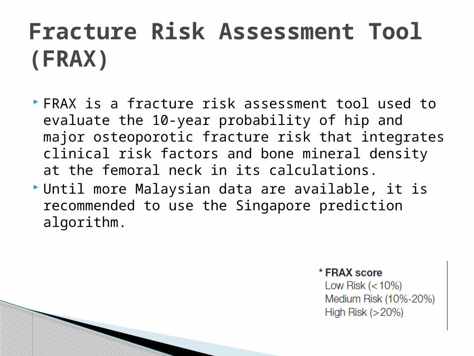
FRAX is a fracture risk assessment tool used to evaluate the 10-year probability of hip and major osteoporotic fracture risk that integrates clinical risk factors and bone mineral density at the femoral neck in its calculations.
Until more Malaysian data are available, it is recommended to use the Singapore prediction algorithm.
Fracture Risk Assessment Tool (FRAX)

A simple clinical screening tool, based on age and weight, Osteoporosis
OSTA was developed for postmenopausal Asian women.
Women in the high risk category and those in the moderate risk category with additional risk factors (e.g. glucocorticoid use, hypogonadism, immobilisation) for osteoporosis should be recommended for DXA
Self-Assessment Tool for Asians (OSTA)

The main aims of investigations are to:1. Confirm the diagnosis of osteoporosis2. Assess fracture risk3. Exclude secondary causes
Investigations

Initial investigations include:1. FBC, ESR2. Bone profile: serum calcium, phosphate, albumin3. Alkaline phosphatase4. Renal function5. Plain X-rays - lateral thoraco-lumbar spine or hip (as indicated)

clinical suspicion of secondary causes:◦ free thyroxine T4 (FT4),◦ thyroid-stimulating hormone (TSH), ◦ testosterone,◦ follicle-stimulating hormone (FSH), ◦ luteinizing hormone (LH), ◦ urine Bence Jones protein,◦ serum protein electrophoresis]

Densitometry ◦ BMD measurement gives an accurate reflection of
bone mass and helps in establishing the diagnosis of osteoporosis
◦ use race-specific reference ranges when available◦ results are reported as
T-scores (comparison with the young adult mean)
The risk of fracture is increased two fold for each SD reduction of T-score in BMD
Z-scores (comparison with the mean of individuals of the same age)
Specific investigations

gold standard for diagnosis measured at the hip and lumbar spine. Prediction of fracture risk is site-specific.
◦ If not available, other skeletal sites can be used to provide an adequate estimation of fracture risk.
Peripheral DXA (phalanges / distal radius / calcaneum)
Dual energy X-ray absorptiometry (DXA)

an alternative technique for measuring bone density in the axial skeleton and vertebral volumetric bone density
The main limitations are the lack of availability in Malaysia and a higher radiation dose compared to DXA
Quantitative computed tomography (QCT)

to identify patients at high risk of future fractures.
They should not be used for the diagnosis of osteoporosis.
used to evaluate treatment efficacy and compliance to therapy
Changes in level of BTM can be seen within 3-6 months after initiation of drug therapy.
Bone Turnover Markers (BTM)


The aim ::to assess the response to treatment.◦ regular clinical assessments◦ peripheral DXA is not recommended ◦ If biochemical markers are available, two separate
baseline measurements of the same marker need to be carried out followed by one repeat measurement 2-3 months after initiating therapy and yearly thereafter, if indicated. These measurements should be taken at the same time of the day to minimise the effect of diurnal variation.
Monitoring of Therapy

NutritionI. Calcium
PREVENTION OF OSTEOPOROSIS AND FALLS
II. Vitamin D◦>50 years old or older, the Malaysian Recommended Nutrient Intake advocates 400 IU of vitamin D per day, but many experts recommend at least 800 to 1000 IU per day

Low body weight and excessive dieting is associated with low bone mineral status and increased fracture risk
Maintenance of a BMI >19 kg/m 2 is recommended for prevention of osteoporosis
III. Body Weight

Patients should be advised to limit their caffeine intake to less than 1 to 2 servings (240 to 360 mls in each serving) of caffeinated drinks per day.
Caffeine intake leads to a slight decrease in intestinal calcium absorption and an increase in urinary calcium excretion
IV. Caffeine intake

Cigarette smoking increases osteoporotic fracture risk and thus should be avoided
Excessive intake of alcohol should be avoided because alcohol has detrimental effects on fracture
V. Smoking and Alcohol intake

Regular physical activity, in particular weight-bearing exercise ◦ (e.g. brisk walking, line dancing) is encouraged in
all age groups in order to maximise peak bone mass, decrease age-related bone loss, maintain muscle strength and balance
The individual’s health status should be taken into consideration when recommending an exercise programme.
• Exercise

o Most osteoporosis-related fractures, especially in the elderly, are a consequence of decreased BMD and falls
• Prevention of falls

I. Hormonal therapy (HT)⁻ HT is an effective treatment for
menopausal symptoms that also offers good protectionfor bone.
₋ Estrogen Therapy (with or without progestin) increases lumbar spine BMD
◦ HT is not recommended in women with breast cancer, coronary heart disease or stroke.
MANAGEMENT OF OSTEOPOROSIS

Bisphosphonates are potent inhibitors of bone resorption.◦ Alendronate, risedronate, ibandronate, zoledronic
acid◦ Two adverse effects have been noted in
bisphosphonate therapy: Atypical femoral shaft fractures Osteonecrosis of the jaw (ONJ) :exposed, non-vital
bone involving maxillofacial structures, with delayed healing despite > 8 weeks of appropriate medical care
ii. Bisphosphonates

CKD Stages 1-3 Patients with CKD stages 1-3 and low T-
scores or low trauma fractures, most likely haveosteoporosis. Bisphosphonates can be usedsafely.
CKD Stages 4-5 Bisphosphonates are not recommended for
patients with an estimated GFR < 30 ml/min
Use of bisphosphonates in renal impairment / Chronic Kidney Disease (CKD)

In established osteoporosis, calcium supplementation alone is not adequate for fracture prevention. However, calcium supplementation is necessary for optimal response to other
treatment modalities
III. Calcium

Activated Vitamin D (calcitriol 0.25 μg bd, alfacalcidol 1 μg od) has been demonstrated to increase BMD in those with established osteoporosis1
IV. Activated Vitamin D

Calcitonin has also been shown to have an analgesic effect for acute pain
in osteoporosis related vertebral fractures Side effects of calcitonin include nausea,
flushing, vomiting and nasal irritation.
V. Calcitonin

1. CPG - Management of Osteoporosis June 2012
2. Harrison's Principles of Internal Medicine, 18th ed
3. www.m.webmd.com/a-to-z-guides/bone-mineral-density-test.
4. www.medscape.com/osteoporosis/overview.
Reference


