Download - Osteoclastic resorption and osteoinduction in the highly purified -tricalcium phosphate implanted i
International Journal of Engineering Research amp Science (IJOER) ISSN [2395-6992] [Vol-2 Issue-3 March- 2016]
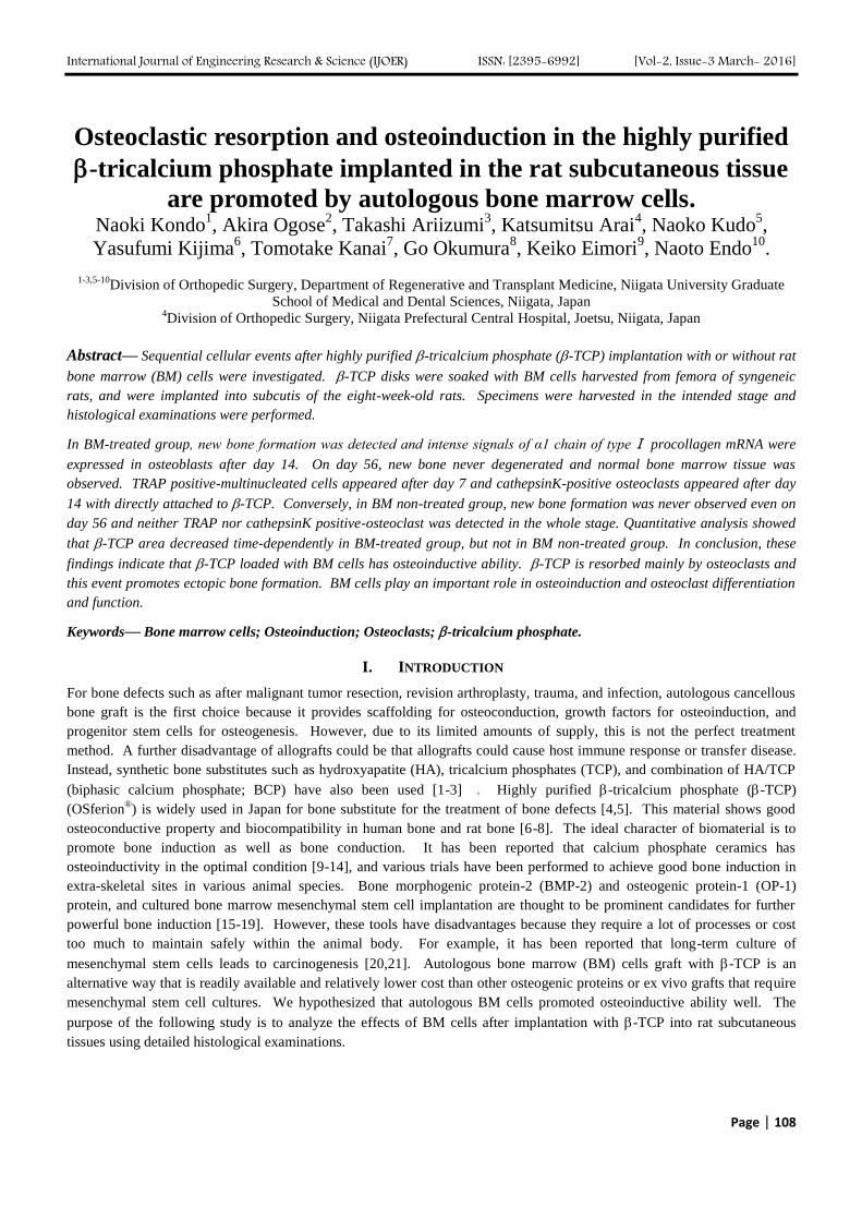
Page | 108
Osteoclastic resorption and osteoinduction in the highly purified
-tricalcium phosphate implanted in the rat subcutaneous tissue
are promoted by autologous bone marrow cells Naoki Kondo
1 Akira Ogose
2 Takashi Ariizumi
3 Katsumitsu Arai
4 Naoko Kudo
5
Yasufumi Kijima6 Tomotake Kanai
7 Go Okumura
8 Keiko Eimori
9 Naoto Endo
10
1-35-10
Division of Orthopedic Surgery Department of Regenerative and Transplant Medicine Niigata University Graduate
School of Medical and Dental Sciences Niigata Japan 4Division of Orthopedic Surgery Niigata Prefectural Central Hospital Joetsu Niigata Japan
Abstractmdash Sequential cellular events after highly purified -tricalcium phosphate (-TCP) implantation with or without rat
bone marrow (BM) cells were investigated -TCP disks were soaked with BM cells harvested from femora of syngeneic
rats and were implanted into subcutis of the eight-week-old rats Specimens were harvested in the intended stage and
histological examinations were performed
In BM-treated group new bone formation was detected and intense signals of α1 chain of typeⅠ procollagen mRNA were
expressed in osteoblasts after day 14 On day 56 new bone never degenerated and normal bone marrow tissue was
observed TRAP positive-multinucleated cells appeared after day 7 and cathepsinK-positive osteoclasts appeared after day
14 with directly attached to -TCP Conversely in BM non-treated group new bone formation was never observed even on
day 56 and neither TRAP nor cathepsinK positive-osteoclast was detected in the whole stage Quantitative analysis showed
that -TCP area decreased time-dependently in BM-treated group but not in BM non-treated group In conclusion these
findings indicate that -TCP loaded with BM cells has osteoinductive ability -TCP is resorbed mainly by osteoclasts and
this event promotes ectopic bone formation BM cells play an important role in osteoinduction and osteoclast differentiation
and function
Keywordsmdash Bone marrow cells Osteoinduction Osteoclasts -tricalcium phosphate
I INTRODUCTION
For bone defects such as after malignant tumor resection revision arthroplasty trauma and infection autologous cancellous
bone graft is the first choice because it provides scaffolding for osteoconduction growth factors for osteoinduction and
progenitor stem cells for osteogenesis However due to its limited amounts of supply this is not the perfect treatment
method A further disadvantage of allografts could be that allografts could cause host immune response or transfer disease
Instead synthetic bone substitutes such as hydroxyapatite (HA) tricalcium phosphates (TCP) and combination of HATCP
(biphasic calcium phosphate BCP) have also been used [1-3] Highly purified -tricalcium phosphate (-TCP)
(OSferionreg) is widely used in Japan for bone substitute for the treatment of bone defects [45] This material shows good
osteoconductive property and biocompatibility in human bone and rat bone [6-8] The ideal character of biomaterial is to
promote bone induction as well as bone conduction It has been reported that calcium phosphate ceramics has
osteoinductivity in the optimal condition [9-14] and various trials have been performed to achieve good bone induction in
extra-skeletal sites in various animal species Bone morphogenic protein-2 (BMP-2) and osteogenic protein-1 (OP-1)
protein and cultured bone marrow mesenchymal stem cell implantation are thought to be prominent candidates for further
powerful bone induction [15-19] However these tools have disadvantages because they require a lot of processes or cost
too much to maintain safely within the animal body For example it has been reported that long-term culture of
mesenchymal stem cells leads to carcinogenesis [2021] Autologous bone marrow (BM) cells graft with -TCP is an
alternative way that is readily available and relatively lower cost than other osteogenic proteins or ex vivo grafts that require
mesenchymal stem cell cultures We hypothesized that autologous BM cells promoted osteoinductive ability well The
purpose of the following study is to analyze the effects of BM cells after implantation with -TCP into rat subcutaneous
tissues using detailed histological examinations
International Journal of Engineering Research amp Science (IJOER) ISSN [2395-6992] [Vol-2 Issue-3 March- 2016]
Page | 109
II MATERIALS AND METHODS
21 Preparation of -TCP
-TCP (OSferion
) was obtained from Olympus Biomaterial Corp (Tokyo Japan) [4] Fine -TCP powder was synthesized
mechanochemically by wet milling CaHPO42H2O and CaCO3 at a molar ratio of 21 were mixed into a slurry with pure
water and beads of zirconia in a pot mill for 24 hrs and then dried at 80C leading to formation of calcium-deficient
hydroxyapatite This crystalline solid was converted to -TCP by calcination at 750C for 1 hr Upon sintering of -TCP
powder at 1050C for 1 h a porous -TCP block was obtained which was then characterized through assessment of the
surface area and pore-size distribution The porosity of the block was 75 and the surface area as measured by the
Brunauer-Emmett-Teller method was 14 m2g [822] The -TCP possessed macropores of 100-400 m and micropores of
less than 5 m Nearly all macropores were interconnected via 100-200 m pores [14] Five-millimeter in diameter and 25
mm in height of -TCP columns were used in this study
22 Animal model and tissue preparation
Thirty female 8-week-old F344Fisher rats were used and randomly divided into the two groups Under general anesthesia
subcutaneous tissue of back in each rat was longitudinally exposed At the same time after euthanized both ends of bilateral
femora of a syngeneic 8-week-old female rat were cut and bone marrow plugs were flushed out using 250 l of phosphate
buffer saline expelled from a syringe through a 23-gauge needle and collected in a sterile 10-cm dish Each 6 -TCP disks
per bone marrow cells obtained from a femur were soaked into extracted BM cells for 30 min in the Petri dish Six -TCP
disks with BM cells per rat were implanted into subcutaneous tissue in back of a rat and defined as BM-treated group As a
control six -TCP disks without BM cells per rat were implanted as well and defined as BM non-treated group After
irrigation with normal saline the wound was closed All animal experiments were conducted according to the ldquoGuideline for
Animal Experimentationrdquo of OLYMPUS CORPORATION
On days 4 7 14 28 56 after the operation the three rats in each group were euthanized and the implanted -TCP disks were
extracted and immersed in 4 paraformaldehyde (PFA) in 01 M phosphate buffer for 5-7 days and then decalcified with
05 M EDTA2Na solution for 2 days at room temperature and dehydrated with a graded series of ethanol treatments prior to
being embedded in paraffin Paraffin sections of 4-m thickness were cut using a microtome (Leica Tokyo Japan) and
stored at 4C for the following histological evaluations
23 TRAP staining
To detect osteoclasts TRAP staining was carried out according to Burstonersquos Azo dye method [23] with some modifications
[24] Briefly a mixture of 3 mg of naphthol AS-BI phosphate (Sigma St Louis MO) 18 mg of red violet LB salt (Sigma
St Louis MO) and 24 mM L(+)-tartaric acid (Wako Osaka Japan) diluted in 01 M sodium acetate buffer (pH 50) were
dropped onto the deparaffinized sections These sections were incubated for 20-30 min at 60C and then counterstained with
hematoxylin
24 Immunohistochemistry of ED1 and cathepsin K
The anti-ED1 monoclonal antibody recognizes a single chain glycoprotein of MW 90000- 110000 that is expressed
predominantly on the lysosomal membrane and at low levels on the cell surface [25] For ED1 immunohistochemistry
antigen retrieval with 02 trypsin at 37C for 20 min was required The tissue sections were treated with 03 hydrogen
peroxide in methanol for 30 min to inhibit endogenous peroxidase and then incubated with 10 goat serum for ED1 and
with 1 bovine serum albumin for cathepsin K for 20 min to reduce nonspecific reactions The sections were then incubated
with anti-ED1 monoclonal antibody diluted 1500 (Beringer Mannheim August Switzerland) for 16h at 4C or with mouse
anti-human cathepsin K antibody (Daiichi Finechemical Takaoka Japan) diluted 1100 for 2h at room temperature and
reacted for 1h with rat MAX-PO (MULTI) secondary antibody (Nichirei Tokyo Japan) without diluted for ED1 or with
horseradish peroxidase-conjugated goat anti-mouse IgG+IgA+IgM antibody (Zymed Laboratories Inc South San Francisco
CA USA) diluted 1100 for cathepsin K at room temperature The peroxidase reaction products were visualized with 3rsquo-
diaminobenzidine tetrahydrochloride (Nichirei Tokyo Japan) Sections were counterstained with hematoxylin
25 In situ hybridization
International Journal of Engineering Research amp Science (IJOER) ISSN [2395-6992] [Vol-2 Issue-3 March- 2016]
Page | 110
To examine the differentiation stages of bone-forming cells deparaffinized serial sections were subjected to mRNA in situ
hybridization as previously described [2627] Plasmid containing 037-kb fragments of mouse α1 chain of type Ⅰ
procollagen (COL1A1) cDNAs was obtained as a gift from the Life Science Research Institute (Asahi-Chemical Industry
Co Shizuoka Japan) After dewaxing in xylene and rehydrating through a series of graded ethanol treatments tissue
sections were treated with 10 gml proteinase K (Roche Diagnostics Mannheim Germany) for 20 min at 37C refixed with
4 PFA solution immersed in 01 M triethanolamine containing 025 acetic acid for 10 min and washed in 01 M
phosphate buffer (pH 74) The samples were then incubated in a hybridization solution [10 mM Tris-HCl (pH 76) 1 mM
EDTA (pH 80) 600 mM NaCl 025 sodium dodecyl sulfate 1 times Denhartrsquos medium 50 (vv) deionized formamide 05
gml probe RNA and 10 dextran sulfate] at 50C in a moist chamber for 16 hrs Negative controls were incubated with
DIG-labeled sense RNA probes After hybridization the slides were washed at 55C with 50 deionized formamide in 2
saline-sodium citrate (SSC) (1 SSC 015 moll NaCl 0015 moll sodium citrate) for 20 min to remove excess riboprobes
Non-specifically hybridized riboprobes were digested with 10 gml of RNase A (Roche Diagnostic) solution at 37C for 30
min The specimens were then washed with 2 times SSC for 15 min and with 02 times SSC for 15 min twice To visualize the
hybridized probe the slides were incubated with alkaline phosphatase-conjugated anti-DIG antibody (Roche Diagnostics) at
room temperature for 60 min after blocking with 15 blocking reagent (Roche Diagnostics) in 100 mM Tris-HCl (pH 75)
for 55 min The specimens were then washed twice with 100 mM Tris-HCl (pH 75) for 15 min and briefly immersed in 100
mM Tris-HCl (pH 95) containing 100 mM NaCl and 50 mM MgCl2 for 5 min The colorimetric reaction was performed
with nitro blue tetrazolium salt and bromo-4-chloro-3-indolyl phosphate solution (Roche Diagnostics) in the dark for 20-120
min and then the reaction was stopped with 10 mM Tris-HCl (pH 76) containing 1 mM EDTA Slides were mounted with
micro cover glass (Matsunami Tokyo Japan) and analyzed under a light microscope with 05 methyl green
counterstaining
26 Semi-quantitative evaluation of the area of -TCP and the ratio of newly formed bone per unit area of -TCP
Specimens were stained with hematoxylin and eosin (HE) The areas of -TCP and newly formed bone were measured using
NIH Image Ver 163 (developed at the US National Institutes of Health and available at httprsbinfonihgovnih-
imagedownloadhtml) About the area of -TCP the specimens on day 4 were inappropriate for the measurements due to
hyperdecalcification and their data were excluded These data were then used to calculate the ratio of newly formed bone per
unit area of -TCP Statistical analyses were performed using StatView software for Windows (Version 50) with a
Bonferroni-Dunn test (either one way ANOVA or a post hoc test) used A value of p lt 005 was considered to indicate a
statistically significant difference
III RESULTS
No newly formed bone was observed on days 4 and 7 in both bone marrow (BM)-treated group and BM non-treated group
On day 4 clotting was observed in the interconnected macropores of -TCP (Fig 1A) On day 7 a large number of
fibroblast-like cells and blood vessels were observed in the interconnected macropores and lots of attached cells appeared
(Fig 1B) After day 14 newly formed bone was detected with attaching to the surface of -TCP in BM-treated group On
day 28 the thickness of newly formed bone was larger than that on day 14 (Figs 1CE) On day 56 most -TCP surface was
covered with newly formed bone and -TCP area seemed to decrease compared to those on days 14 and 28 Furthermore
normal bone marrow tissue was detected in interconnected macropores (Fig1G)
However in the BM non-treated group no new bone formation occurred even in the peripheral region of implanted area on
days 14 28 and 56 although blood vessels were observed in -TCP macropores (Figs1D F and H)
To examine the cellular events of monocyte-macrophage lineage cells we performed TRAP staining immunohistochemistry
of cathepsin K protein which is a specific matrix degradation enzyme synthesized by osteoclasts [28] and ED1 protein
which is detected in the cells of the mononuclear phagocyte system in rats [25]
On day 4 after implantation no TRAP positive cells were detected in both groups (data not shown) On day 7 a few TRAP
positive multinucleated cells were attached to -TCP surface in BM-treated group in the peripheral region (Fig 2A) On
days 14 and 28 abundant TRAP positive-multinucleated cells were detected on the surface of -TCP even in the central
region in BM-treated group HE staining in BM-treated group on day 14 showed that some multinucleated giant cells were
directly attached to -TCP (Fig 2G) However they were sparsely observed around newly formed bone (Figs 2CE)
International Journal of Engineering Research amp Science (IJOER) ISSN [2395-6992] [Vol-2 Issue-3 March- 2016]
Page | 111
FIGURE 1 TIME COURSE AFTER IMPLANTATION
[On days 4 and 7 the new bone could not be detected any implanted area in BM-treated group (A and B) No newly
formed bone was detected either in BM non-treated group
In the BM-treated group ectopic bone formation was detected in the macropores of -TCP with attaching to the
surface of -TCP on day 14 (C)
On day 28 newly formed bone became thicker than that of day 14 and almost all surface of -TCP was covered with
new bone (E) On day 56 in addition to abundant newly formed bone bone marrow tissue was observed in the
macropores of -TCP (G) In contrast in the BM non-treated group newly formed bone could not be detected on
days 14 (D) 28 (F) and 56 (H) though abundant blood vessels were observed BM+ BM-treated group BMndash BM
non-treated group tcp -tricalcium phosphate nb newly formed bone bm bone marrow original magnification A-
H x 40 ]
International Journal of Engineering Research amp Science (IJOER) ISSN [2395-6992] [Vol-2 Issue-3 March- 2016]
Page | 112
In contrast in BM non-treated group no TRAP positive-multinucleated cells were detected even in the peripheral region on
days 7 14 28 (Figs 2BDF) and 56
FIGURE 2 TRAP STAINING AND FINDING OF MULTINUCLEATED GIANT CELLS ON HE STAINING
[On day 7 in BM-treated group a few TRAP-positive multinucleated cells (black arrows) were attached to -TCP in
the periphery of the implanted area (A) On days 14 band 28 in BM-treated group a lot of TRAP-positive
multinucleated cells were directly attached to -TCP (CE) These multinucleated giant cells (black arrow heads)
were confirmed also in HE staining on day 14 in BM-treated group (G) In contrast in BM non-treated group no
TRAP positive cells were detected on days 7 (B) 14 (D) and 28 (F) tcp -tricalcium phosphate st surrounded soft
tissue nb newly formed bone original magnification A-F x 100 Gx 200]
International Journal of Engineering Research amp Science (IJOER) ISSN [2395-6992] [Vol-2 Issue-3 March- 2016]
Page | 113
A lot of cathepsin K positive-osteoclasts were detected after day 14 in BM-treated group and most of them were directly
attached to -TCP or some of them attached to newly formed bone (Figs 3AC) Conversely no cathepsin K positive-
osteoclasts were observed on days 14 (Fig3B) 28 (Fig3D) and 56 in BM non-treated group
FIGURE 3 CATHEPSIN K IMMUNOHISTOCHEMISTY
[In BM-treated group cathepsin K positive osteoclasts were attached to -TCP or newly formed bone on both days 14
and 28 (AC) However in BM non-treated group no cathepsin K positive osteoclasts were observed on days 14 and
28 (BD) tcp -tricalcium phosphate nb newly formed bone original magnification A-D x 100]
On days 4 and 7 abundant ED1 positive-monocytes gathered in -TCP macropores and were attached to -TCP in both BM
treated and non-treated groups with no difference (Figs 4AB)
However ED1 protein immunohistochemical findings were quite different between the two groups after day 14 On day 14
abundant ED1 positive- multinucleated cells were attached to -TCP in BM-treated group (Fig 4C) Furthermore on days
28 and 56 in BM-treated group ED1 positive-multinucleated cells were still attached to -TCP though their number
decreased compared to that on day 14 (Figs 4EG) On the contrary in BM non-treated group no ED1 positive-
multinucleated cells were attached to -TCP on days 14 28 and 56 although ED1 positive-monocytes were sparsely
observed in macropores (Figs 4DFH)
Next to detect bone-forming cells in situ hybridization of COL1A1 mRNA was performed On days 4 and 7 no difference
was detected between both BM-treated and non-treated groups On day 7 COL1A1 mRNA was expressed in mononuclear
cells which were attached -TCP and was expressed in fibroblast-like spindle shaped cells in -TCP macropores in both BM-
treated (Fig 5A) and non-treated (Fig 5B) group On day 14 in BM-treated group osteoblasts lining the newly formed bone
were strongly positive for COL1A1 mRNA and their signals were much stronger than those of the fibroblast-like spindle-
shaped cells on day 7 (Fig 5C) However in BM non-treated group COL1A1 mRNA-positive cells were observed at
surface of -TCP similar to the finding on day 7 and their intensities were much weaker compared to those on day 14 of
BM-treated group (Fig 5D) Same tendency was preserved on days 28 (Figs 5EF) and 56
International Journal of Engineering Research amp Science (IJOER) ISSN [2395-6992] [Vol-2 Issue-3 March- 2016]
Page | 114
FIGURE 4 ED1 IMMUNOHISTOCHEMISTRY
[On days 4 (A) and 7 (B) in BM non-treated group abundant ED1 positive mononuclear cells were attached to -TCP
surface or adjacent to -TCP in the peripheral region On day 14 in BM-treated group lots of ED1 positive
multinucleated cells were attached to -TCP (C) Also on day 28 (E) and 56 (G) in BM-treated group ED1 positive
multinucleated cells were observed However in BM non-treated group on days 14 (D) 28 (F) and 56 (H) no ED1
positive-multinucleated cells were attached to -TCP although ED1 positive-mononuclear cells were detected in the
surface of -TCP or -TCP macropores tcp -tricalcium phosphate nb newly formed bone original magnification
A-H x 100]
International Journal of Engineering Research amp Science (IJOER) ISSN [2395-6992] [Vol-2 Issue-3 March- 2016]
Page | 115
FIGURE 5 IN SITU HYBRIDIZATION OF COL1A1 MRNA
[A On day 7 in BM-treated group COL1A1 mRNA positive mononuclear cells were directly attached -TCP and
fibroblast like spindle shaped cells were expressed in COL1A1 mRNA in -TCP macropores B On day 7 in BM non-
treated group showed similar finding as A C On day 14 in BM-treated group cuboidal shaped COL1A1 mRNA
positive cells lined to newly formed bone and their intensities were much stronger than fibroblast like COL1A1
mRNA positive cells in -TCP macropores D On day 14 in BM non-treated group no cuboidal shaped COL1A1
mRNA positive cells were detected COL1A1 mRNA positive mononuclear cells were still expressed in the surface of
-TCP but their intensities were much weaker than those shown as C E On day 28 in BM-treated group COL1A1
mRNA positive cells lined to the newly formed bone and their shapes were slightly flatter than those shown as C F
On day 28 in BM non-treated group the finding was similar to that shown as D tcp -tricalcium phosphate nb
newly formed bone original magnification A-Fx 100]
International Journal of Engineering Research amp Science (IJOER) ISSN [2395-6992] [Vol-2 Issue-3 March- 2016]
Page | 116
Quantitative analysis demonstrated that the area of -TCP in BM-treated group decreased in a chronological manner and the
area on day 56 was significantly smaller than that on day 7 (plt005) (Fig 6A) However in BM non-treated group the
tendency was not detected (Fig 6B) The ratio of newly formed bone per unit area of -TCP clearly increased from day 14
to day 56 in a time-dependent manner The ratio of newly formed bone per unit area of on days 28 and 56 were significantly
larger than that on day 14 (plt005) (Fig 6C)
(A)
(B)
(C)
FIGURE 6 QUANTITATIVE ANALYSIS
[A The area of -TCP in the BM-treated group The area of -TCP decreased in a time-dependent manner and the
area on day 56 (497163 mm2) was significantly smaller than that on day 7 (697161 mm
2) (plt005) B The area of
-TCP in the BM non-treated group It had no tendency that was shown in A C The ratio of newly formed bone
per unit area of -TCP shown as the mean standard deviation The amount of newly formed bone increased with
time the amounts of newly formed bone per unit area of -TCP on days 28 (021015) and 56 (031014) were
significantly higher than that on day 14 (006006) (plt005) plt005 NS not significance Bar standard
deviation]
IV DISCUSSION
In this study newly formed bone was detected after day 14 in BM-treated group after -TCP implantation in rat
subcutaneous tissue This bone was not formed by endochondral ossification but through intramembranous ossification
since no cartilaginous tissue was observed New bone formation appeared from the peripheral region of implanted site and
gradually extended to the central region This pattern is considered as reasonable since peripheral region is closer from host
International Journal of Engineering Research amp Science (IJOER) ISSN [2395-6992] [Vol-2 Issue-3 March- 2016]
Page | 117
tissue and is vascularized earlier and more efficiently than the central region Some reports have also supported this
interpretation [29-31] However no bone formation was induced in BM non-treated group These findings indicate that BM
cells are very important as a source of osteoinduction in this model
Moreover even on day 56 newly formed bone was preserved without being degenerated and normal bone marrow tissue was
observed suggesting that -TCP loaded with fresh BM cells has a good biocompatibility and osteoinductive ability
The findings of TRAP staining and cathepsin K immunohistochemistry show that BM cells induce the early appearance of
osteoclasts in this model and that osteoclasts resorb -TCP with directly attaching after day 14 Especially TRAP positive-
multinuclear cells were detected on day 7 in BM-treated group though their number was small
This phenomenon is in agreement with those after implantation in rat bone and dog dorsal muscles as we previously reported
[814] As reported by Jarcho [32] bioresorption of calcium phosphate ceramics such as hydroxyapatite (HA) and TCP
consists of solution-mediated processes (the implant dissolves in physiologic solutions) and cell-mediated processes
(phagocytosis) Given that osteoclasts which appeared in the early stage were directly attached to -TCP in BM-treated
group and -TCP area decreased in a time-dependent manner these data strongly support that cell-mediated resorption plays
an important role for bioresorption of -TCP although the involvement of solution-mediated bioresorption should not be
ignored since microporosity possibly could aid in bioresorption by causing microscopic ldquobreak-uprdquo secondary to solution-
mediated resorption [32] and this -TCP has a microporous structure
ED1 immunoreactivities on days 4 and 7 showed that ED1 positive-monocytes gathered adjacent to -TCP or were directly
attached to -TCP and no difference was detected between the two groups These findings suggest that monocytes infiltrate
adjacent to -TCP even in the absence of BM cells After day 14 ED1 positive-multinucleated cells were detected only in
BM-treated group Nagayama et al reported that the intensity of ED1 immunostaining decreased with time after
implantation of -TCP and carbonate apatite into the subcutaneous tissue in back of rats and that it was persisted longer time
in -TCP group [33] This report is very suggestive in that -TCP itself induces macrophages in extra-osseous sites We
indicate that additional BM cells promote much more inductive ability of macrophage around -TCP in this model In
addition osteoclasts belong to monocyte-macrophage lineage cell and monocytes are supposed to fuse and differentiate into
osteoclasts suggesting that BM cells induce differentiation and proper function of osteoclasts In vitro osteoclasts or closely
related to osteoclasts were induced after 3-week-culture of bone marrow cells in the presence of 125-dihydroxyvitamin D3
[34]
About in situ hybridization of COL1A1 mRNA no difference was detected on day 7 between BM-treated and BM non-
treated group However mononuclear cells (osteoblasts) lined to newly formed bone were strongly COL1A1 mRNA
positive on day 14 28 and 56 in BM-treated group which was quite different from those in BM non-treated group These
results indicate that BM cells also induce bone forming ability
Quantitative analyses in this study showed the area of -TCP decreased in a time-dependent manner in BM-treated group
but not in BM non-treated group suggesting that -TCP is continuously resorbed in BM-treated group due to the constant
osteoclast appearance In addition new bone formation consistently increases in BM-treated group suggesting that bone
forming ability is preserved after day 14
Several previous studies reported that fresh bone marrow cells (without cultured) promoted bone induction in extra-osseous
sites after calcium phosphate ceramics implantation such as HA -TCP and biphasic calcium phosphate (HATCP)
[303135] The earliest ectopic bone formation was 3 weeks after implantation of biphasic calcium phosphate (HATCP was
6040) into rat muscle [30] Ectopic bone formation was shown on 1 month and 2 months after implantation of HA and -
TCP with syngeneic rat bone marrow cells into rat subcutis [35] It is noteworthy that ectopic bone formation was detected 2
weeks earlier than these previous reports [303135] after implantation in our study
Cultured bone marrow stromal cells implantation with calcium phosphate ceramics was also reported and bone formation
was detected day 7 days [1929] or 2 weeks [36] after implantation These methods are based on culture for good
differentiation to osteogenic cells using by dexamethasone or -glycerophosphate Given that mesenchymal stem cells
implantation may lead to carcinogenesis [2021] it is indispensable to investigate the safety of mixed implantation combined
calcium phosphate ceramics with mesenchymal stem cells On the other hand there is no report that had adverse effect such
as infection to bone marrow implantation into bone defects or non-union [37]
International Journal of Engineering Research amp Science (IJOER) ISSN [2395-6992] [Vol-2 Issue-3 March- 2016]
Page | 118
Moreover osteoinductive cytokines such as BMP-2 OP-1 and fibroblast growth factor (FGF) have been investigated as
potent promoters of osteoinduction in extra-osseous site [15-1738] In general these cytokines induce ectopic bone
formation in the early stage after implantation with a scaffold However some problems such as immune response
infection and higher cost have been concerned Recently it has been reported that recombinant human BMP-2 which is
widely used for spine surgery has adverse effects such as infection hematoma and excessive edema [3839]
Bone marrow cells loading to -TCP is a very simple and lower cost method to induce ectopic bone formation We consider
that it is reasonable than the calcium phosphate ceramics implantation with the other concomitant use of osteoinductive
cytokines and cultured mesenchymal stromal cells in that it does not cost very much and safer and bone formation was
confirmed as early as on day 14 Erbe et al have shown that bone marrow aspiration and -TCP composite graft is as good
outcome as autograft with bone marrow cells [1] They used ldquoVitossrdquo which is a porous low-density construct prepared by
lightly fusing particles of -TCP is average approximately 1-2 m in diameter and has 90 porosity and interconnected
void space [1]
It remains not elucidated why oseteoclasts proliferated and are differentiated after implantation of -TCP Some possibilities
about the origin of osteoclasts are considered as follows (1) stromal cells in implanted bone marrow induce osteoblasts and
such osteoblasts induce osteoclasts via receptor activator of NF-kB (RANK)-RANK-ligand (RANKL) system and (2)
hematopoietic cells in implanted bone marrow directly induce osteoclasts
Little report was focused on cellular events about the appearance or the function of osteoclast after implantation of -TCP
In our study it is noteworthy that the early (day 7) appearance of osteoclasts prior to new bone formation was confirmed
after implantation of BM cells-loaded -TCP into rat subcutaneous tissue and that osteoclasts play an important role in cell-
mediated bioresorption of -TCP After implantation of -TCP into rabbit bone the peak of osteoclasts was 2 week and it
was earlier than that of osteoblasts Chazono et al called this event as ldquocoupling-like phenomenonrdquo [22] In our previous
study of -TCP implantation in dog dorsal muscles osteoclasts were induced in muscles where these cells never exist and
then new bone formation occurred [14] We indicate that the cellular event shown in our study resembles ldquocoupling-like
phenomenonrdquo and is also very important for osteoinduction Further studies are required for elucidating the mechanism of
macrophage or osteoclast responses to biomaterials
V CONCLUSION
-TCP (OSferion
) is resorbed by osteoclasts and leads to new bone formation when implanted with syngeneic rat bone
marrow cells into subcutaneous tissue These cellular events follow the appearance of a large number of osteoclasts and their
attachment to -TCP The data in this study suggest that -TCP implantation with autogenous bone marrow cells is safe and
not time-consuming method and promotes osteoinduction method compared to that in non BM cells condition
ACKNOWLEDGEMENTS
The authors thank Yoshiaki Tanaka (Division of Orthopedic Surgery Department of Regenerative and Transplant Medicine
Niigata University Graduate School of Medical and Dental Sciences) for their technical assistance
REFERENCES
[1] Erbe EM Marx JG Clineff TD Bellincampi LD Potential of an ultraporous -tricalcium phosphate synthetic cancellous bone void
filler and bone marrow aspirate composite graft Eur Spine J 2001 10 S141-6
[2] Gupta AK Kumar P Keshav K Sigh A Hydroxyapatite crystals as a bone graft substitutes in benign lytic lesions of bone Indian J
Orthop 2015 49 645-55
[3] Brennan MA Renaud A Amiaud J Rojewski MT Schrezenmeier H Heymann D Trichest V Layrolle P Pre-clinical studies of
bone regeneration with human bone marrow stromal cells and biphasic calcium phosphate Stem Cell Res Ther 2014 5 114 doi
101186scrt 504
[4] Ozawa M Tanaka K Morikawa S Chazono M Fujii K Clinical study of the pure -tricalcium phosphate―reports of 167 cases J
East Jpn Orthop Traumatol 2000 12 409-13
[5] Ozawa M Experimental study on bone conductivity and absorbability if the pure -TCP J Jap Soc Biomat 1995 13 17-25
[6] Ogose A Hotta T Hatano H Kawashima H Tokunaga K Endo N Umazu H Histological examination of beta-tricalcium phosphate
graft in human femur J Biomed Mater Res (Appl Biomater) 2002 63 601-4
[7] Ogose A Kondo N Umezu H Hotta T Kawashima H Tokunaga K Ito T Kudo N Hoshino M Gu W Endo N Histological
assessment in grafts of highly purified beta-tricalcium phosphate (OSferion) in human bones Biomaterials 2006 27 1542-9
International Journal of Engineering Research amp Science (IJOER) ISSN [2395-6992] [Vol-2 Issue-3 March- 2016]
Page | 119
[8] Kondo N Ogose A Tokunaga K Ito T Arai K Kudo N Inoue H Irie H Endo N Bone formation and resorption of highly purified
-tricalcium phosphate in the rat femoral condyle Biomaterials 2005 26 5600-8
[9] Urist MR Bone formation by autoinduction Science 1965 159 893-9
[10] Ripamonti U van den Heever B van Wyk J Expression of the osteogenic phenotype in porous hydroxyapatite implanted
extraskeletally in baboons Matrix 1993 13 491-502
[11] Ripamonti U Osteoinduction in porous hydroxyapatite implanted in heterotopic sites of different animal models Biomaterials 1996
17 31-5
[12] Yuan H Yang Z Li Y Zhang X de Bruijn JD de Groot K Osteoinduction by calcium phosphate biomaterials J Mater Sci Mater
Med 1998 9 723-6
[13] Yuan H de Bruijn JD Li Y Feng J Yang Z de Groot K Zhang X Bone formation induced by calcium phosphate ceramics in soft
tissue of dogs a comparative study between porous α-TCP and -TCP J Mater Sci Mater Med 2001 12 7-13
[14] Kondo N Ogose A Tokunaga K Umezu H Arai K Kudo N Hoshino M Inoue H Irie H Kuroda K Mera H Endo N
Osteoinduction with highly purified -tricalcium phosphate in dog dorsal muscles and the proliferation of osteoclasts before
heterotopic bone formation Biomaterials 2006 27 4419-27
[15] Andrades JA Santamaria JA Nimni ME Becerra J Selection and amplification of a bone marrow cell population and its induction to
the chondro-osteogenic lineage by rhOP-1 an in vitro and in vivo study Int J Dev Biol 2001 45 689-93
[16] Kato M Toyoda H Namikawa T Hoshino M Terai H Miyamoto S Takaoka K Optimized use of a biodegradable polymer as a
carrier material for the local delivery of recombinant human bone morphogenic protein-2 (rhBMP-2) Biomaterials 2006 27 2035-
41
[17] Takahashi Y Yamamoto M Tabata Y Enhanced osteoinduction by controlled release of bone morphogenic protein-2 from
biodegradable sponge composed of gelatin and -tricalcium phosphate Biomaterials 2005 26 4856-65
[18] Yamada Y Boo JS Ozawa R Nagasaka T Okazaki Y Hata K Ueda M Bone regeneration following injection of mesenchymal
stem cells and fibrin glue with a biodegradable scaffold J Craniomaxillofac Surg 2003 31 27-33
[19] Boo JS Ymada Y Okazaki Y Hibino Y Okada K Hata K Yoshikawa T Sugiura Y Ueda M Tissue-engineered bone using
mesenchymal stem cells and a biodegradable scaffold J Craniofasc Surg 2002 13 231-9
[20] Tolar J Nauta AJ Osborn MJ Mortari AP McElmurry RT Bell S Xia L Zhou N Riddle M Schroeder TM Westerndorf JJ McIvor
RS Hogendoorn PCW Szuhai K Oseth L Hirsch B Yant SR Kay MA Peister A Prockop DJ Fibbe WE Blazer BR Sarcoma
derived from cultured mesenchymal stem cells Stem cells 2007 25371-9
[21] Rubio D Garcia-Castro J Martin MC de la Fuente R Cigudosa JC Lloyd AC Bernad A Spontaneous human adult stem cell
transformation Cancer Res 2005 65 3035-9
[22] Chazono M Tanaka T Komaki H Fujii K Bone formation and bioresorption after implantation of injectable -tricalcium phosphate
granules-hyaluronate complex in rabbit bone defects J Biomed Mater Res 2004 70A 542-9
[23] Burstone MS Histochemical demonstration of acid phosphatase with naphthol AS-phosphate J Nat Cancer Int 1958 21 523-39
[24] Amizuka N Yamada M Watanabe J Hoshi K Fukushi M Oda K Ikehara Y Ozawa H Morphological examination of bone
synthesis via direct administration of basic fibroblast growth factor into rat bone marrow Microsc Res Tech 1998 41 313-22
[25] Damouiseaux JGMC DӧPP EA Calame W Chao D Macpherson GG Dijkstra CD Rat macrophage lysosomal membrane antigen
recognized by monoclonal antibody ED1 Immunology 1994 83 140-7
[26] Hayami T Endo N Tokunaga K Yamagiwa H Hatano H Uchida M Takahashi HE Spatiotemporal change of rat collagenase
(MMP-13) mRNA expression in the development of rat femoral neck J Bone Miner Metab 2000 18 185-93
[27] Yamagiwa H Tokunaga K Hayami T Hatano H Uchida M Endo N Takahashi HE Expression of metalloproteinase-13
(Collagenase-3) is induced during fracture healing in mice Bone 1999 25 197-203
[28] Troen BR The role of cathepsin K in normal bone resorption Drug News Perspect 2004 17 19-28
[29] Hartman EHM Vehof JWM Spauwen PHM Jansen JA Ectopic bone formation in rats the importance of the carrier Biomaterials
2005 26 1829-35
[30] Ohgushi H Goldberg VM Caplan AI Heterotopic osteogenesis in porous ceramics induced by marrow cells J Orthop Res 1989 7
568-78
[31] Ohgushi H Okumura M Yoshikawa T Inoue K Senpuku N Tamai S Shors EC Bone formation process in porous calcium
carbonate and hydroxyapatite J Biomed Mater Res 1992 26 855-95
[32] Jarcho M Calcium phosphate ceramics as hard tissue prosthetics Clin Orthop Relat Res 1981 157 259-78
[33] Nagayama M Takeuchi H Doi Y Comparison of carbonate apatite and beta-tricalcium phosphate (resorbable calcium phosphates)
implanted subcutaneously into the back of rats Dent Mater J 2006 25 219-25
[34] Takahashi N Kukita T MacDonald BR Bird A Mundy GR McManus LM Miller M Boyde A Jones SJ Roodman GD
Osteoclast-like cells form in long-term human bone marrow but not in peripheral blood cultures J Clin Invest 1989 83 543-50
[35] Ohgushi H Okumura M Tamai S Shors EC Caplan AI Marrow cell induced osteogenesis in porous hydroxyapatite and tricalcium
phosphate a comparative histomorphometric study of ectopic bone formation J Biomed Mater Res 1990 12 1563-7
[36] Dennis JE HaynesworthSE Young RG Caplan AI Osteogenesis in marrow-derived mesenchymal cell porous ceramic composites
transplanted subcutaneously effect of fibronectin and laminin on cell retention and rate of osteogenic expression Cell Transplant
1992 1 23-32
International Journal of Engineering Research amp Science (IJOER) ISSN [2395-6992] [Vol-2 Issue-3 March- 2016]
Page | 120
[37] Goel A Sangwan SS Siwach RC and Ali AM Percutaneous bone marrow grafting for the treatment of tibial non-union Injury
2005 36 203-6
[38] Kubota K Iseki S Kuroda S Oida S Iimura T Duarte WR Ohya K Ishikawa I Kasugai S Synergistic effect of fibroblast growth
factor-4 in ectopic bone formation induced by bone morphogenetic protein-2 Bone 2002 31 465-71
[39] Hansen SM Sasso RC Resorptive response of rhBMP2 simulating infection in an anterior lumbar interbody fusion with a femoral
ring J Spinal Disord Tech 2006 19130-4
[40] Shields LBE Raque GH Glassman SD Campbell M Vitaz T Harpring J Shields CB Adverse effects associated with high-dose
recombinant human bone morphogenetic protein-2 use in anterior cervical spine fusion Spine 2006 31 542-7

International Journal of Engineering Research amp Science (IJOER) ISSN [2395-6992] [Vol-2 Issue-3 March- 2016]
Page | 111
FIGURE 1 TIME COURSE AFTER IMPLANTATION
[On days 4 and 7 the new bone could not be detected any implanted area in BM-treated group (A and B) No newly
formed bone was detected either in BM non-treated group
In the BM-treated group ectopic bone formation was detected in the macropores of -TCP with attaching to the
surface of -TCP on day 14 (C)
On day 28 newly formed bone became thicker than that of day 14 and almost all surface of -TCP was covered with
new bone (E) On day 56 in addition to abundant newly formed bone bone marrow tissue was observed in the
macropores of -TCP (G) In contrast in the BM non-treated group newly formed bone could not be detected on
days 14 (D) 28 (F) and 56 (H) though abundant blood vessels were observed BM+ BM-treated group BMndash BM
non-treated group tcp -tricalcium phosphate nb newly formed bone bm bone marrow original magnification A-
H x 40 ]
International Journal of Engineering Research amp Science (IJOER) ISSN [2395-6992] [Vol-2 Issue-3 March- 2016]
Page | 112
In contrast in BM non-treated group no TRAP positive-multinucleated cells were detected even in the peripheral region on
days 7 14 28 (Figs 2BDF) and 56
FIGURE 2 TRAP STAINING AND FINDING OF MULTINUCLEATED GIANT CELLS ON HE STAINING
[On day 7 in BM-treated group a few TRAP-positive multinucleated cells (black arrows) were attached to -TCP in
the periphery of the implanted area (A) On days 14 band 28 in BM-treated group a lot of TRAP-positive
multinucleated cells were directly attached to -TCP (CE) These multinucleated giant cells (black arrow heads)
were confirmed also in HE staining on day 14 in BM-treated group (G) In contrast in BM non-treated group no
TRAP positive cells were detected on days 7 (B) 14 (D) and 28 (F) tcp -tricalcium phosphate st surrounded soft
tissue nb newly formed bone original magnification A-F x 100 Gx 200]
International Journal of Engineering Research amp Science (IJOER) ISSN [2395-6992] [Vol-2 Issue-3 March- 2016]
Page | 113
A lot of cathepsin K positive-osteoclasts were detected after day 14 in BM-treated group and most of them were directly
attached to -TCP or some of them attached to newly formed bone (Figs 3AC) Conversely no cathepsin K positive-
osteoclasts were observed on days 14 (Fig3B) 28 (Fig3D) and 56 in BM non-treated group
FIGURE 3 CATHEPSIN K IMMUNOHISTOCHEMISTY
[In BM-treated group cathepsin K positive osteoclasts were attached to -TCP or newly formed bone on both days 14
and 28 (AC) However in BM non-treated group no cathepsin K positive osteoclasts were observed on days 14 and
28 (BD) tcp -tricalcium phosphate nb newly formed bone original magnification A-D x 100]
On days 4 and 7 abundant ED1 positive-monocytes gathered in -TCP macropores and were attached to -TCP in both BM
treated and non-treated groups with no difference (Figs 4AB)
However ED1 protein immunohistochemical findings were quite different between the two groups after day 14 On day 14
abundant ED1 positive- multinucleated cells were attached to -TCP in BM-treated group (Fig 4C) Furthermore on days
28 and 56 in BM-treated group ED1 positive-multinucleated cells were still attached to -TCP though their number
decreased compared to that on day 14 (Figs 4EG) On the contrary in BM non-treated group no ED1 positive-
multinucleated cells were attached to -TCP on days 14 28 and 56 although ED1 positive-monocytes were sparsely
observed in macropores (Figs 4DFH)
Next to detect bone-forming cells in situ hybridization of COL1A1 mRNA was performed On days 4 and 7 no difference
was detected between both BM-treated and non-treated groups On day 7 COL1A1 mRNA was expressed in mononuclear
cells which were attached -TCP and was expressed in fibroblast-like spindle shaped cells in -TCP macropores in both BM-
treated (Fig 5A) and non-treated (Fig 5B) group On day 14 in BM-treated group osteoblasts lining the newly formed bone
were strongly positive for COL1A1 mRNA and their signals were much stronger than those of the fibroblast-like spindle-
shaped cells on day 7 (Fig 5C) However in BM non-treated group COL1A1 mRNA-positive cells were observed at
surface of -TCP similar to the finding on day 7 and their intensities were much weaker compared to those on day 14 of
BM-treated group (Fig 5D) Same tendency was preserved on days 28 (Figs 5EF) and 56
International Journal of Engineering Research amp Science (IJOER) ISSN [2395-6992] [Vol-2 Issue-3 March- 2016]
Page | 114
FIGURE 4 ED1 IMMUNOHISTOCHEMISTRY
[On days 4 (A) and 7 (B) in BM non-treated group abundant ED1 positive mononuclear cells were attached to -TCP
surface or adjacent to -TCP in the peripheral region On day 14 in BM-treated group lots of ED1 positive
multinucleated cells were attached to -TCP (C) Also on day 28 (E) and 56 (G) in BM-treated group ED1 positive
multinucleated cells were observed However in BM non-treated group on days 14 (D) 28 (F) and 56 (H) no ED1
positive-multinucleated cells were attached to -TCP although ED1 positive-mononuclear cells were detected in the
surface of -TCP or -TCP macropores tcp -tricalcium phosphate nb newly formed bone original magnification
A-H x 100]
International Journal of Engineering Research amp Science (IJOER) ISSN [2395-6992] [Vol-2 Issue-3 March- 2016]
Page | 115
FIGURE 5 IN SITU HYBRIDIZATION OF COL1A1 MRNA
[A On day 7 in BM-treated group COL1A1 mRNA positive mononuclear cells were directly attached -TCP and
fibroblast like spindle shaped cells were expressed in COL1A1 mRNA in -TCP macropores B On day 7 in BM non-
treated group showed similar finding as A C On day 14 in BM-treated group cuboidal shaped COL1A1 mRNA
positive cells lined to newly formed bone and their intensities were much stronger than fibroblast like COL1A1
mRNA positive cells in -TCP macropores D On day 14 in BM non-treated group no cuboidal shaped COL1A1
mRNA positive cells were detected COL1A1 mRNA positive mononuclear cells were still expressed in the surface of
-TCP but their intensities were much weaker than those shown as C E On day 28 in BM-treated group COL1A1
mRNA positive cells lined to the newly formed bone and their shapes were slightly flatter than those shown as C F
On day 28 in BM non-treated group the finding was similar to that shown as D tcp -tricalcium phosphate nb
newly formed bone original magnification A-Fx 100]
International Journal of Engineering Research amp Science (IJOER) ISSN [2395-6992] [Vol-2 Issue-3 March- 2016]
Page | 116
Quantitative analysis demonstrated that the area of -TCP in BM-treated group decreased in a chronological manner and the
area on day 56 was significantly smaller than that on day 7 (plt005) (Fig 6A) However in BM non-treated group the
tendency was not detected (Fig 6B) The ratio of newly formed bone per unit area of -TCP clearly increased from day 14
to day 56 in a time-dependent manner The ratio of newly formed bone per unit area of on days 28 and 56 were significantly
larger than that on day 14 (plt005) (Fig 6C)
(A)
(B)
(C)
FIGURE 6 QUANTITATIVE ANALYSIS
[A The area of -TCP in the BM-treated group The area of -TCP decreased in a time-dependent manner and the
area on day 56 (497163 mm2) was significantly smaller than that on day 7 (697161 mm
2) (plt005) B The area of
-TCP in the BM non-treated group It had no tendency that was shown in A C The ratio of newly formed bone
per unit area of -TCP shown as the mean standard deviation The amount of newly formed bone increased with
time the amounts of newly formed bone per unit area of -TCP on days 28 (021015) and 56 (031014) were
significantly higher than that on day 14 (006006) (plt005) plt005 NS not significance Bar standard
deviation]
IV DISCUSSION
In this study newly formed bone was detected after day 14 in BM-treated group after -TCP implantation in rat
subcutaneous tissue This bone was not formed by endochondral ossification but through intramembranous ossification
since no cartilaginous tissue was observed New bone formation appeared from the peripheral region of implanted site and
gradually extended to the central region This pattern is considered as reasonable since peripheral region is closer from host
International Journal of Engineering Research amp Science (IJOER) ISSN [2395-6992] [Vol-2 Issue-3 March- 2016]
Page | 117
tissue and is vascularized earlier and more efficiently than the central region Some reports have also supported this
interpretation [29-31] However no bone formation was induced in BM non-treated group These findings indicate that BM
cells are very important as a source of osteoinduction in this model
Moreover even on day 56 newly formed bone was preserved without being degenerated and normal bone marrow tissue was
observed suggesting that -TCP loaded with fresh BM cells has a good biocompatibility and osteoinductive ability
The findings of TRAP staining and cathepsin K immunohistochemistry show that BM cells induce the early appearance of
osteoclasts in this model and that osteoclasts resorb -TCP with directly attaching after day 14 Especially TRAP positive-
multinuclear cells were detected on day 7 in BM-treated group though their number was small
This phenomenon is in agreement with those after implantation in rat bone and dog dorsal muscles as we previously reported
[814] As reported by Jarcho [32] bioresorption of calcium phosphate ceramics such as hydroxyapatite (HA) and TCP
consists of solution-mediated processes (the implant dissolves in physiologic solutions) and cell-mediated processes
(phagocytosis) Given that osteoclasts which appeared in the early stage were directly attached to -TCP in BM-treated
group and -TCP area decreased in a time-dependent manner these data strongly support that cell-mediated resorption plays
an important role for bioresorption of -TCP although the involvement of solution-mediated bioresorption should not be
ignored since microporosity possibly could aid in bioresorption by causing microscopic ldquobreak-uprdquo secondary to solution-
mediated resorption [32] and this -TCP has a microporous structure
ED1 immunoreactivities on days 4 and 7 showed that ED1 positive-monocytes gathered adjacent to -TCP or were directly
attached to -TCP and no difference was detected between the two groups These findings suggest that monocytes infiltrate
adjacent to -TCP even in the absence of BM cells After day 14 ED1 positive-multinucleated cells were detected only in
BM-treated group Nagayama et al reported that the intensity of ED1 immunostaining decreased with time after
implantation of -TCP and carbonate apatite into the subcutaneous tissue in back of rats and that it was persisted longer time
in -TCP group [33] This report is very suggestive in that -TCP itself induces macrophages in extra-osseous sites We
indicate that additional BM cells promote much more inductive ability of macrophage around -TCP in this model In
addition osteoclasts belong to monocyte-macrophage lineage cell and monocytes are supposed to fuse and differentiate into
osteoclasts suggesting that BM cells induce differentiation and proper function of osteoclasts In vitro osteoclasts or closely
related to osteoclasts were induced after 3-week-culture of bone marrow cells in the presence of 125-dihydroxyvitamin D3
[34]
About in situ hybridization of COL1A1 mRNA no difference was detected on day 7 between BM-treated and BM non-
treated group However mononuclear cells (osteoblasts) lined to newly formed bone were strongly COL1A1 mRNA
positive on day 14 28 and 56 in BM-treated group which was quite different from those in BM non-treated group These
results indicate that BM cells also induce bone forming ability
Quantitative analyses in this study showed the area of -TCP decreased in a time-dependent manner in BM-treated group
but not in BM non-treated group suggesting that -TCP is continuously resorbed in BM-treated group due to the constant
osteoclast appearance In addition new bone formation consistently increases in BM-treated group suggesting that bone
forming ability is preserved after day 14
Several previous studies reported that fresh bone marrow cells (without cultured) promoted bone induction in extra-osseous
sites after calcium phosphate ceramics implantation such as HA -TCP and biphasic calcium phosphate (HATCP)
[303135] The earliest ectopic bone formation was 3 weeks after implantation of biphasic calcium phosphate (HATCP was
6040) into rat muscle [30] Ectopic bone formation was shown on 1 month and 2 months after implantation of HA and -
TCP with syngeneic rat bone marrow cells into rat subcutis [35] It is noteworthy that ectopic bone formation was detected 2
weeks earlier than these previous reports [303135] after implantation in our study
Cultured bone marrow stromal cells implantation with calcium phosphate ceramics was also reported and bone formation
was detected day 7 days [1929] or 2 weeks [36] after implantation These methods are based on culture for good
differentiation to osteogenic cells using by dexamethasone or -glycerophosphate Given that mesenchymal stem cells
implantation may lead to carcinogenesis [2021] it is indispensable to investigate the safety of mixed implantation combined
calcium phosphate ceramics with mesenchymal stem cells On the other hand there is no report that had adverse effect such
as infection to bone marrow implantation into bone defects or non-union [37]
International Journal of Engineering Research amp Science (IJOER) ISSN [2395-6992] [Vol-2 Issue-3 March- 2016]
Page | 118
Moreover osteoinductive cytokines such as BMP-2 OP-1 and fibroblast growth factor (FGF) have been investigated as
potent promoters of osteoinduction in extra-osseous site [15-1738] In general these cytokines induce ectopic bone
formation in the early stage after implantation with a scaffold However some problems such as immune response
infection and higher cost have been concerned Recently it has been reported that recombinant human BMP-2 which is
widely used for spine surgery has adverse effects such as infection hematoma and excessive edema [3839]
Bone marrow cells loading to -TCP is a very simple and lower cost method to induce ectopic bone formation We consider
that it is reasonable than the calcium phosphate ceramics implantation with the other concomitant use of osteoinductive
cytokines and cultured mesenchymal stromal cells in that it does not cost very much and safer and bone formation was
confirmed as early as on day 14 Erbe et al have shown that bone marrow aspiration and -TCP composite graft is as good
outcome as autograft with bone marrow cells [1] They used ldquoVitossrdquo which is a porous low-density construct prepared by
lightly fusing particles of -TCP is average approximately 1-2 m in diameter and has 90 porosity and interconnected
void space [1]
It remains not elucidated why oseteoclasts proliferated and are differentiated after implantation of -TCP Some possibilities
about the origin of osteoclasts are considered as follows (1) stromal cells in implanted bone marrow induce osteoblasts and
such osteoblasts induce osteoclasts via receptor activator of NF-kB (RANK)-RANK-ligand (RANKL) system and (2)
hematopoietic cells in implanted bone marrow directly induce osteoclasts
Little report was focused on cellular events about the appearance or the function of osteoclast after implantation of -TCP
In our study it is noteworthy that the early (day 7) appearance of osteoclasts prior to new bone formation was confirmed
after implantation of BM cells-loaded -TCP into rat subcutaneous tissue and that osteoclasts play an important role in cell-
mediated bioresorption of -TCP After implantation of -TCP into rabbit bone the peak of osteoclasts was 2 week and it
was earlier than that of osteoblasts Chazono et al called this event as ldquocoupling-like phenomenonrdquo [22] In our previous
study of -TCP implantation in dog dorsal muscles osteoclasts were induced in muscles where these cells never exist and
then new bone formation occurred [14] We indicate that the cellular event shown in our study resembles ldquocoupling-like
phenomenonrdquo and is also very important for osteoinduction Further studies are required for elucidating the mechanism of
macrophage or osteoclast responses to biomaterials
V CONCLUSION
-TCP (OSferion
) is resorbed by osteoclasts and leads to new bone formation when implanted with syngeneic rat bone
marrow cells into subcutaneous tissue These cellular events follow the appearance of a large number of osteoclasts and their
attachment to -TCP The data in this study suggest that -TCP implantation with autogenous bone marrow cells is safe and
not time-consuming method and promotes osteoinduction method compared to that in non BM cells condition
ACKNOWLEDGEMENTS
The authors thank Yoshiaki Tanaka (Division of Orthopedic Surgery Department of Regenerative and Transplant Medicine
Niigata University Graduate School of Medical and Dental Sciences) for their technical assistance
REFERENCES
[1] Erbe EM Marx JG Clineff TD Bellincampi LD Potential of an ultraporous -tricalcium phosphate synthetic cancellous bone void
filler and bone marrow aspirate composite graft Eur Spine J 2001 10 S141-6
[2] Gupta AK Kumar P Keshav K Sigh A Hydroxyapatite crystals as a bone graft substitutes in benign lytic lesions of bone Indian J
Orthop 2015 49 645-55
[3] Brennan MA Renaud A Amiaud J Rojewski MT Schrezenmeier H Heymann D Trichest V Layrolle P Pre-clinical studies of
bone regeneration with human bone marrow stromal cells and biphasic calcium phosphate Stem Cell Res Ther 2014 5 114 doi
101186scrt 504
[4] Ozawa M Tanaka K Morikawa S Chazono M Fujii K Clinical study of the pure -tricalcium phosphate―reports of 167 cases J
East Jpn Orthop Traumatol 2000 12 409-13
[5] Ozawa M Experimental study on bone conductivity and absorbability if the pure -TCP J Jap Soc Biomat 1995 13 17-25
[6] Ogose A Hotta T Hatano H Kawashima H Tokunaga K Endo N Umazu H Histological examination of beta-tricalcium phosphate
graft in human femur J Biomed Mater Res (Appl Biomater) 2002 63 601-4
[7] Ogose A Kondo N Umezu H Hotta T Kawashima H Tokunaga K Ito T Kudo N Hoshino M Gu W Endo N Histological
assessment in grafts of highly purified beta-tricalcium phosphate (OSferion) in human bones Biomaterials 2006 27 1542-9
International Journal of Engineering Research amp Science (IJOER) ISSN [2395-6992] [Vol-2 Issue-3 March- 2016]
Page | 119
[8] Kondo N Ogose A Tokunaga K Ito T Arai K Kudo N Inoue H Irie H Endo N Bone formation and resorption of highly purified
-tricalcium phosphate in the rat femoral condyle Biomaterials 2005 26 5600-8
[9] Urist MR Bone formation by autoinduction Science 1965 159 893-9
[10] Ripamonti U van den Heever B van Wyk J Expression of the osteogenic phenotype in porous hydroxyapatite implanted
extraskeletally in baboons Matrix 1993 13 491-502
[11] Ripamonti U Osteoinduction in porous hydroxyapatite implanted in heterotopic sites of different animal models Biomaterials 1996
17 31-5
[12] Yuan H Yang Z Li Y Zhang X de Bruijn JD de Groot K Osteoinduction by calcium phosphate biomaterials J Mater Sci Mater
Med 1998 9 723-6
[13] Yuan H de Bruijn JD Li Y Feng J Yang Z de Groot K Zhang X Bone formation induced by calcium phosphate ceramics in soft
tissue of dogs a comparative study between porous α-TCP and -TCP J Mater Sci Mater Med 2001 12 7-13
[14] Kondo N Ogose A Tokunaga K Umezu H Arai K Kudo N Hoshino M Inoue H Irie H Kuroda K Mera H Endo N
Osteoinduction with highly purified -tricalcium phosphate in dog dorsal muscles and the proliferation of osteoclasts before
heterotopic bone formation Biomaterials 2006 27 4419-27
[15] Andrades JA Santamaria JA Nimni ME Becerra J Selection and amplification of a bone marrow cell population and its induction to
the chondro-osteogenic lineage by rhOP-1 an in vitro and in vivo study Int J Dev Biol 2001 45 689-93
[16] Kato M Toyoda H Namikawa T Hoshino M Terai H Miyamoto S Takaoka K Optimized use of a biodegradable polymer as a
carrier material for the local delivery of recombinant human bone morphogenic protein-2 (rhBMP-2) Biomaterials 2006 27 2035-
41
[17] Takahashi Y Yamamoto M Tabata Y Enhanced osteoinduction by controlled release of bone morphogenic protein-2 from
biodegradable sponge composed of gelatin and -tricalcium phosphate Biomaterials 2005 26 4856-65
[18] Yamada Y Boo JS Ozawa R Nagasaka T Okazaki Y Hata K Ueda M Bone regeneration following injection of mesenchymal
stem cells and fibrin glue with a biodegradable scaffold J Craniomaxillofac Surg 2003 31 27-33
[19] Boo JS Ymada Y Okazaki Y Hibino Y Okada K Hata K Yoshikawa T Sugiura Y Ueda M Tissue-engineered bone using
mesenchymal stem cells and a biodegradable scaffold J Craniofasc Surg 2002 13 231-9
[20] Tolar J Nauta AJ Osborn MJ Mortari AP McElmurry RT Bell S Xia L Zhou N Riddle M Schroeder TM Westerndorf JJ McIvor
RS Hogendoorn PCW Szuhai K Oseth L Hirsch B Yant SR Kay MA Peister A Prockop DJ Fibbe WE Blazer BR Sarcoma
derived from cultured mesenchymal stem cells Stem cells 2007 25371-9
[21] Rubio D Garcia-Castro J Martin MC de la Fuente R Cigudosa JC Lloyd AC Bernad A Spontaneous human adult stem cell
transformation Cancer Res 2005 65 3035-9
[22] Chazono M Tanaka T Komaki H Fujii K Bone formation and bioresorption after implantation of injectable -tricalcium phosphate
granules-hyaluronate complex in rabbit bone defects J Biomed Mater Res 2004 70A 542-9
[23] Burstone MS Histochemical demonstration of acid phosphatase with naphthol AS-phosphate J Nat Cancer Int 1958 21 523-39
[24] Amizuka N Yamada M Watanabe J Hoshi K Fukushi M Oda K Ikehara Y Ozawa H Morphological examination of bone
synthesis via direct administration of basic fibroblast growth factor into rat bone marrow Microsc Res Tech 1998 41 313-22
[25] Damouiseaux JGMC DӧPP EA Calame W Chao D Macpherson GG Dijkstra CD Rat macrophage lysosomal membrane antigen
recognized by monoclonal antibody ED1 Immunology 1994 83 140-7
[26] Hayami T Endo N Tokunaga K Yamagiwa H Hatano H Uchida M Takahashi HE Spatiotemporal change of rat collagenase
(MMP-13) mRNA expression in the development of rat femoral neck J Bone Miner Metab 2000 18 185-93
[27] Yamagiwa H Tokunaga K Hayami T Hatano H Uchida M Endo N Takahashi HE Expression of metalloproteinase-13
(Collagenase-3) is induced during fracture healing in mice Bone 1999 25 197-203
[28] Troen BR The role of cathepsin K in normal bone resorption Drug News Perspect 2004 17 19-28
[29] Hartman EHM Vehof JWM Spauwen PHM Jansen JA Ectopic bone formation in rats the importance of the carrier Biomaterials
2005 26 1829-35
[30] Ohgushi H Goldberg VM Caplan AI Heterotopic osteogenesis in porous ceramics induced by marrow cells J Orthop Res 1989 7
568-78
[31] Ohgushi H Okumura M Yoshikawa T Inoue K Senpuku N Tamai S Shors EC Bone formation process in porous calcium
carbonate and hydroxyapatite J Biomed Mater Res 1992 26 855-95
[32] Jarcho M Calcium phosphate ceramics as hard tissue prosthetics Clin Orthop Relat Res 1981 157 259-78
[33] Nagayama M Takeuchi H Doi Y Comparison of carbonate apatite and beta-tricalcium phosphate (resorbable calcium phosphates)
implanted subcutaneously into the back of rats Dent Mater J 2006 25 219-25
[34] Takahashi N Kukita T MacDonald BR Bird A Mundy GR McManus LM Miller M Boyde A Jones SJ Roodman GD
Osteoclast-like cells form in long-term human bone marrow but not in peripheral blood cultures J Clin Invest 1989 83 543-50
[35] Ohgushi H Okumura M Tamai S Shors EC Caplan AI Marrow cell induced osteogenesis in porous hydroxyapatite and tricalcium
phosphate a comparative histomorphometric study of ectopic bone formation J Biomed Mater Res 1990 12 1563-7
[36] Dennis JE HaynesworthSE Young RG Caplan AI Osteogenesis in marrow-derived mesenchymal cell porous ceramic composites
transplanted subcutaneously effect of fibronectin and laminin on cell retention and rate of osteogenic expression Cell Transplant
1992 1 23-32
International Journal of Engineering Research amp Science (IJOER) ISSN [2395-6992] [Vol-2 Issue-3 March- 2016]
Page | 120
[37] Goel A Sangwan SS Siwach RC and Ali AM Percutaneous bone marrow grafting for the treatment of tibial non-union Injury
2005 36 203-6
[38] Kubota K Iseki S Kuroda S Oida S Iimura T Duarte WR Ohya K Ishikawa I Kasugai S Synergistic effect of fibroblast growth
factor-4 in ectopic bone formation induced by bone morphogenetic protein-2 Bone 2002 31 465-71
[39] Hansen SM Sasso RC Resorptive response of rhBMP2 simulating infection in an anterior lumbar interbody fusion with a femoral
ring J Spinal Disord Tech 2006 19130-4
[40] Shields LBE Raque GH Glassman SD Campbell M Vitaz T Harpring J Shields CB Adverse effects associated with high-dose
recombinant human bone morphogenetic protein-2 use in anterior cervical spine fusion Spine 2006 31 542-7
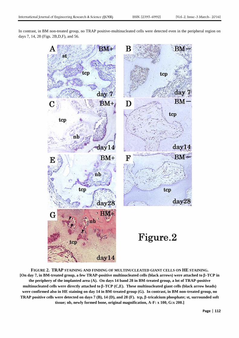
International Journal of Engineering Research amp Science (IJOER) ISSN [2395-6992] [Vol-2 Issue-3 March- 2016]
Page | 112
In contrast in BM non-treated group no TRAP positive-multinucleated cells were detected even in the peripheral region on
days 7 14 28 (Figs 2BDF) and 56
FIGURE 2 TRAP STAINING AND FINDING OF MULTINUCLEATED GIANT CELLS ON HE STAINING
[On day 7 in BM-treated group a few TRAP-positive multinucleated cells (black arrows) were attached to -TCP in
the periphery of the implanted area (A) On days 14 band 28 in BM-treated group a lot of TRAP-positive
multinucleated cells were directly attached to -TCP (CE) These multinucleated giant cells (black arrow heads)
were confirmed also in HE staining on day 14 in BM-treated group (G) In contrast in BM non-treated group no
TRAP positive cells were detected on days 7 (B) 14 (D) and 28 (F) tcp -tricalcium phosphate st surrounded soft
tissue nb newly formed bone original magnification A-F x 100 Gx 200]
International Journal of Engineering Research amp Science (IJOER) ISSN [2395-6992] [Vol-2 Issue-3 March- 2016]
Page | 113
A lot of cathepsin K positive-osteoclasts were detected after day 14 in BM-treated group and most of them were directly
attached to -TCP or some of them attached to newly formed bone (Figs 3AC) Conversely no cathepsin K positive-
osteoclasts were observed on days 14 (Fig3B) 28 (Fig3D) and 56 in BM non-treated group
FIGURE 3 CATHEPSIN K IMMUNOHISTOCHEMISTY
[In BM-treated group cathepsin K positive osteoclasts were attached to -TCP or newly formed bone on both days 14
and 28 (AC) However in BM non-treated group no cathepsin K positive osteoclasts were observed on days 14 and
28 (BD) tcp -tricalcium phosphate nb newly formed bone original magnification A-D x 100]
On days 4 and 7 abundant ED1 positive-monocytes gathered in -TCP macropores and were attached to -TCP in both BM
treated and non-treated groups with no difference (Figs 4AB)
However ED1 protein immunohistochemical findings were quite different between the two groups after day 14 On day 14
abundant ED1 positive- multinucleated cells were attached to -TCP in BM-treated group (Fig 4C) Furthermore on days
28 and 56 in BM-treated group ED1 positive-multinucleated cells were still attached to -TCP though their number
decreased compared to that on day 14 (Figs 4EG) On the contrary in BM non-treated group no ED1 positive-
multinucleated cells were attached to -TCP on days 14 28 and 56 although ED1 positive-monocytes were sparsely
observed in macropores (Figs 4DFH)
Next to detect bone-forming cells in situ hybridization of COL1A1 mRNA was performed On days 4 and 7 no difference
was detected between both BM-treated and non-treated groups On day 7 COL1A1 mRNA was expressed in mononuclear
cells which were attached -TCP and was expressed in fibroblast-like spindle shaped cells in -TCP macropores in both BM-
treated (Fig 5A) and non-treated (Fig 5B) group On day 14 in BM-treated group osteoblasts lining the newly formed bone
were strongly positive for COL1A1 mRNA and their signals were much stronger than those of the fibroblast-like spindle-
shaped cells on day 7 (Fig 5C) However in BM non-treated group COL1A1 mRNA-positive cells were observed at
surface of -TCP similar to the finding on day 7 and their intensities were much weaker compared to those on day 14 of
BM-treated group (Fig 5D) Same tendency was preserved on days 28 (Figs 5EF) and 56
International Journal of Engineering Research amp Science (IJOER) ISSN [2395-6992] [Vol-2 Issue-3 March- 2016]
Page | 114
FIGURE 4 ED1 IMMUNOHISTOCHEMISTRY
[On days 4 (A) and 7 (B) in BM non-treated group abundant ED1 positive mononuclear cells were attached to -TCP
surface or adjacent to -TCP in the peripheral region On day 14 in BM-treated group lots of ED1 positive
multinucleated cells were attached to -TCP (C) Also on day 28 (E) and 56 (G) in BM-treated group ED1 positive
multinucleated cells were observed However in BM non-treated group on days 14 (D) 28 (F) and 56 (H) no ED1
positive-multinucleated cells were attached to -TCP although ED1 positive-mononuclear cells were detected in the
surface of -TCP or -TCP macropores tcp -tricalcium phosphate nb newly formed bone original magnification
A-H x 100]
International Journal of Engineering Research amp Science (IJOER) ISSN [2395-6992] [Vol-2 Issue-3 March- 2016]
Page | 115
FIGURE 5 IN SITU HYBRIDIZATION OF COL1A1 MRNA
[A On day 7 in BM-treated group COL1A1 mRNA positive mononuclear cells were directly attached -TCP and
fibroblast like spindle shaped cells were expressed in COL1A1 mRNA in -TCP macropores B On day 7 in BM non-
treated group showed similar finding as A C On day 14 in BM-treated group cuboidal shaped COL1A1 mRNA
positive cells lined to newly formed bone and their intensities were much stronger than fibroblast like COL1A1
mRNA positive cells in -TCP macropores D On day 14 in BM non-treated group no cuboidal shaped COL1A1
mRNA positive cells were detected COL1A1 mRNA positive mononuclear cells were still expressed in the surface of
-TCP but their intensities were much weaker than those shown as C E On day 28 in BM-treated group COL1A1
mRNA positive cells lined to the newly formed bone and their shapes were slightly flatter than those shown as C F
On day 28 in BM non-treated group the finding was similar to that shown as D tcp -tricalcium phosphate nb
newly formed bone original magnification A-Fx 100]
International Journal of Engineering Research amp Science (IJOER) ISSN [2395-6992] [Vol-2 Issue-3 March- 2016]
Page | 116
Quantitative analysis demonstrated that the area of -TCP in BM-treated group decreased in a chronological manner and the
area on day 56 was significantly smaller than that on day 7 (plt005) (Fig 6A) However in BM non-treated group the
tendency was not detected (Fig 6B) The ratio of newly formed bone per unit area of -TCP clearly increased from day 14
to day 56 in a time-dependent manner The ratio of newly formed bone per unit area of on days 28 and 56 were significantly
larger than that on day 14 (plt005) (Fig 6C)
(A)
(B)
(C)
FIGURE 6 QUANTITATIVE ANALYSIS
[A The area of -TCP in the BM-treated group The area of -TCP decreased in a time-dependent manner and the
area on day 56 (497163 mm2) was significantly smaller than that on day 7 (697161 mm
2) (plt005) B The area of
-TCP in the BM non-treated group It had no tendency that was shown in A C The ratio of newly formed bone
per unit area of -TCP shown as the mean standard deviation The amount of newly formed bone increased with
time the amounts of newly formed bone per unit area of -TCP on days 28 (021015) and 56 (031014) were
significantly higher than that on day 14 (006006) (plt005) plt005 NS not significance Bar standard
deviation]
IV DISCUSSION
In this study newly formed bone was detected after day 14 in BM-treated group after -TCP implantation in rat
subcutaneous tissue This bone was not formed by endochondral ossification but through intramembranous ossification
since no cartilaginous tissue was observed New bone formation appeared from the peripheral region of implanted site and
gradually extended to the central region This pattern is considered as reasonable since peripheral region is closer from host
International Journal of Engineering Research amp Science (IJOER) ISSN [2395-6992] [Vol-2 Issue-3 March- 2016]
Page | 117
tissue and is vascularized earlier and more efficiently than the central region Some reports have also supported this
interpretation [29-31] However no bone formation was induced in BM non-treated group These findings indicate that BM
cells are very important as a source of osteoinduction in this model
Moreover even on day 56 newly formed bone was preserved without being degenerated and normal bone marrow tissue was
observed suggesting that -TCP loaded with fresh BM cells has a good biocompatibility and osteoinductive ability
The findings of TRAP staining and cathepsin K immunohistochemistry show that BM cells induce the early appearance of
osteoclasts in this model and that osteoclasts resorb -TCP with directly attaching after day 14 Especially TRAP positive-
multinuclear cells were detected on day 7 in BM-treated group though their number was small
This phenomenon is in agreement with those after implantation in rat bone and dog dorsal muscles as we previously reported
[814] As reported by Jarcho [32] bioresorption of calcium phosphate ceramics such as hydroxyapatite (HA) and TCP
consists of solution-mediated processes (the implant dissolves in physiologic solutions) and cell-mediated processes
(phagocytosis) Given that osteoclasts which appeared in the early stage were directly attached to -TCP in BM-treated
group and -TCP area decreased in a time-dependent manner these data strongly support that cell-mediated resorption plays
an important role for bioresorption of -TCP although the involvement of solution-mediated bioresorption should not be
ignored since microporosity possibly could aid in bioresorption by causing microscopic ldquobreak-uprdquo secondary to solution-
mediated resorption [32] and this -TCP has a microporous structure
ED1 immunoreactivities on days 4 and 7 showed that ED1 positive-monocytes gathered adjacent to -TCP or were directly
attached to -TCP and no difference was detected between the two groups These findings suggest that monocytes infiltrate
adjacent to -TCP even in the absence of BM cells After day 14 ED1 positive-multinucleated cells were detected only in
BM-treated group Nagayama et al reported that the intensity of ED1 immunostaining decreased with time after
implantation of -TCP and carbonate apatite into the subcutaneous tissue in back of rats and that it was persisted longer time
in -TCP group [33] This report is very suggestive in that -TCP itself induces macrophages in extra-osseous sites We
indicate that additional BM cells promote much more inductive ability of macrophage around -TCP in this model In
addition osteoclasts belong to monocyte-macrophage lineage cell and monocytes are supposed to fuse and differentiate into
osteoclasts suggesting that BM cells induce differentiation and proper function of osteoclasts In vitro osteoclasts or closely
related to osteoclasts were induced after 3-week-culture of bone marrow cells in the presence of 125-dihydroxyvitamin D3
[34]
About in situ hybridization of COL1A1 mRNA no difference was detected on day 7 between BM-treated and BM non-
treated group However mononuclear cells (osteoblasts) lined to newly formed bone were strongly COL1A1 mRNA
positive on day 14 28 and 56 in BM-treated group which was quite different from those in BM non-treated group These
results indicate that BM cells also induce bone forming ability
Quantitative analyses in this study showed the area of -TCP decreased in a time-dependent manner in BM-treated group
but not in BM non-treated group suggesting that -TCP is continuously resorbed in BM-treated group due to the constant
osteoclast appearance In addition new bone formation consistently increases in BM-treated group suggesting that bone
forming ability is preserved after day 14
Several previous studies reported that fresh bone marrow cells (without cultured) promoted bone induction in extra-osseous
sites after calcium phosphate ceramics implantation such as HA -TCP and biphasic calcium phosphate (HATCP)
[303135] The earliest ectopic bone formation was 3 weeks after implantation of biphasic calcium phosphate (HATCP was
6040) into rat muscle [30] Ectopic bone formation was shown on 1 month and 2 months after implantation of HA and -
TCP with syngeneic rat bone marrow cells into rat subcutis [35] It is noteworthy that ectopic bone formation was detected 2
weeks earlier than these previous reports [303135] after implantation in our study
Cultured bone marrow stromal cells implantation with calcium phosphate ceramics was also reported and bone formation
was detected day 7 days [1929] or 2 weeks [36] after implantation These methods are based on culture for good
differentiation to osteogenic cells using by dexamethasone or -glycerophosphate Given that mesenchymal stem cells
implantation may lead to carcinogenesis [2021] it is indispensable to investigate the safety of mixed implantation combined
calcium phosphate ceramics with mesenchymal stem cells On the other hand there is no report that had adverse effect such
as infection to bone marrow implantation into bone defects or non-union [37]
International Journal of Engineering Research amp Science (IJOER) ISSN [2395-6992] [Vol-2 Issue-3 March- 2016]
Page | 118
Moreover osteoinductive cytokines such as BMP-2 OP-1 and fibroblast growth factor (FGF) have been investigated as
potent promoters of osteoinduction in extra-osseous site [15-1738] In general these cytokines induce ectopic bone
formation in the early stage after implantation with a scaffold However some problems such as immune response
infection and higher cost have been concerned Recently it has been reported that recombinant human BMP-2 which is
widely used for spine surgery has adverse effects such as infection hematoma and excessive edema [3839]
Bone marrow cells loading to -TCP is a very simple and lower cost method to induce ectopic bone formation We consider
that it is reasonable than the calcium phosphate ceramics implantation with the other concomitant use of osteoinductive
cytokines and cultured mesenchymal stromal cells in that it does not cost very much and safer and bone formation was
confirmed as early as on day 14 Erbe et al have shown that bone marrow aspiration and -TCP composite graft is as good
outcome as autograft with bone marrow cells [1] They used ldquoVitossrdquo which is a porous low-density construct prepared by
lightly fusing particles of -TCP is average approximately 1-2 m in diameter and has 90 porosity and interconnected
void space [1]
It remains not elucidated why oseteoclasts proliferated and are differentiated after implantation of -TCP Some possibilities
about the origin of osteoclasts are considered as follows (1) stromal cells in implanted bone marrow induce osteoblasts and
such osteoblasts induce osteoclasts via receptor activator of NF-kB (RANK)-RANK-ligand (RANKL) system and (2)
hematopoietic cells in implanted bone marrow directly induce osteoclasts
Little report was focused on cellular events about the appearance or the function of osteoclast after implantation of -TCP
In our study it is noteworthy that the early (day 7) appearance of osteoclasts prior to new bone formation was confirmed
after implantation of BM cells-loaded -TCP into rat subcutaneous tissue and that osteoclasts play an important role in cell-
mediated bioresorption of -TCP After implantation of -TCP into rabbit bone the peak of osteoclasts was 2 week and it
was earlier than that of osteoblasts Chazono et al called this event as ldquocoupling-like phenomenonrdquo [22] In our previous
study of -TCP implantation in dog dorsal muscles osteoclasts were induced in muscles where these cells never exist and
then new bone formation occurred [14] We indicate that the cellular event shown in our study resembles ldquocoupling-like
phenomenonrdquo and is also very important for osteoinduction Further studies are required for elucidating the mechanism of
macrophage or osteoclast responses to biomaterials
V CONCLUSION
-TCP (OSferion
) is resorbed by osteoclasts and leads to new bone formation when implanted with syngeneic rat bone
marrow cells into subcutaneous tissue These cellular events follow the appearance of a large number of osteoclasts and their
attachment to -TCP The data in this study suggest that -TCP implantation with autogenous bone marrow cells is safe and
not time-consuming method and promotes osteoinduction method compared to that in non BM cells condition
ACKNOWLEDGEMENTS
The authors thank Yoshiaki Tanaka (Division of Orthopedic Surgery Department of Regenerative and Transplant Medicine
Niigata University Graduate School of Medical and Dental Sciences) for their technical assistance
REFERENCES
[1] Erbe EM Marx JG Clineff TD Bellincampi LD Potential of an ultraporous -tricalcium phosphate synthetic cancellous bone void
filler and bone marrow aspirate composite graft Eur Spine J 2001 10 S141-6
[2] Gupta AK Kumar P Keshav K Sigh A Hydroxyapatite crystals as a bone graft substitutes in benign lytic lesions of bone Indian J
Orthop 2015 49 645-55
[3] Brennan MA Renaud A Amiaud J Rojewski MT Schrezenmeier H Heymann D Trichest V Layrolle P Pre-clinical studies of
bone regeneration with human bone marrow stromal cells and biphasic calcium phosphate Stem Cell Res Ther 2014 5 114 doi
101186scrt 504
[4] Ozawa M Tanaka K Morikawa S Chazono M Fujii K Clinical study of the pure -tricalcium phosphate―reports of 167 cases J
East Jpn Orthop Traumatol 2000 12 409-13
[5] Ozawa M Experimental study on bone conductivity and absorbability if the pure -TCP J Jap Soc Biomat 1995 13 17-25
[6] Ogose A Hotta T Hatano H Kawashima H Tokunaga K Endo N Umazu H Histological examination of beta-tricalcium phosphate
graft in human femur J Biomed Mater Res (Appl Biomater) 2002 63 601-4
[7] Ogose A Kondo N Umezu H Hotta T Kawashima H Tokunaga K Ito T Kudo N Hoshino M Gu W Endo N Histological
assessment in grafts of highly purified beta-tricalcium phosphate (OSferion) in human bones Biomaterials 2006 27 1542-9
International Journal of Engineering Research amp Science (IJOER) ISSN [2395-6992] [Vol-2 Issue-3 March- 2016]
Page | 119
[8] Kondo N Ogose A Tokunaga K Ito T Arai K Kudo N Inoue H Irie H Endo N Bone formation and resorption of highly purified
-tricalcium phosphate in the rat femoral condyle Biomaterials 2005 26 5600-8
[9] Urist MR Bone formation by autoinduction Science 1965 159 893-9
[10] Ripamonti U van den Heever B van Wyk J Expression of the osteogenic phenotype in porous hydroxyapatite implanted
extraskeletally in baboons Matrix 1993 13 491-502
[11] Ripamonti U Osteoinduction in porous hydroxyapatite implanted in heterotopic sites of different animal models Biomaterials 1996
17 31-5
[12] Yuan H Yang Z Li Y Zhang X de Bruijn JD de Groot K Osteoinduction by calcium phosphate biomaterials J Mater Sci Mater
Med 1998 9 723-6
[13] Yuan H de Bruijn JD Li Y Feng J Yang Z de Groot K Zhang X Bone formation induced by calcium phosphate ceramics in soft
tissue of dogs a comparative study between porous α-TCP and -TCP J Mater Sci Mater Med 2001 12 7-13
[14] Kondo N Ogose A Tokunaga K Umezu H Arai K Kudo N Hoshino M Inoue H Irie H Kuroda K Mera H Endo N
Osteoinduction with highly purified -tricalcium phosphate in dog dorsal muscles and the proliferation of osteoclasts before
heterotopic bone formation Biomaterials 2006 27 4419-27
[15] Andrades JA Santamaria JA Nimni ME Becerra J Selection and amplification of a bone marrow cell population and its induction to
the chondro-osteogenic lineage by rhOP-1 an in vitro and in vivo study Int J Dev Biol 2001 45 689-93
[16] Kato M Toyoda H Namikawa T Hoshino M Terai H Miyamoto S Takaoka K Optimized use of a biodegradable polymer as a
carrier material for the local delivery of recombinant human bone morphogenic protein-2 (rhBMP-2) Biomaterials 2006 27 2035-
41
[17] Takahashi Y Yamamoto M Tabata Y Enhanced osteoinduction by controlled release of bone morphogenic protein-2 from
biodegradable sponge composed of gelatin and -tricalcium phosphate Biomaterials 2005 26 4856-65
[18] Yamada Y Boo JS Ozawa R Nagasaka T Okazaki Y Hata K Ueda M Bone regeneration following injection of mesenchymal
stem cells and fibrin glue with a biodegradable scaffold J Craniomaxillofac Surg 2003 31 27-33
[19] Boo JS Ymada Y Okazaki Y Hibino Y Okada K Hata K Yoshikawa T Sugiura Y Ueda M Tissue-engineered bone using
mesenchymal stem cells and a biodegradable scaffold J Craniofasc Surg 2002 13 231-9
[20] Tolar J Nauta AJ Osborn MJ Mortari AP McElmurry RT Bell S Xia L Zhou N Riddle M Schroeder TM Westerndorf JJ McIvor
RS Hogendoorn PCW Szuhai K Oseth L Hirsch B Yant SR Kay MA Peister A Prockop DJ Fibbe WE Blazer BR Sarcoma
derived from cultured mesenchymal stem cells Stem cells 2007 25371-9
[21] Rubio D Garcia-Castro J Martin MC de la Fuente R Cigudosa JC Lloyd AC Bernad A Spontaneous human adult stem cell
transformation Cancer Res 2005 65 3035-9
[22] Chazono M Tanaka T Komaki H Fujii K Bone formation and bioresorption after implantation of injectable -tricalcium phosphate
granules-hyaluronate complex in rabbit bone defects J Biomed Mater Res 2004 70A 542-9
[23] Burstone MS Histochemical demonstration of acid phosphatase with naphthol AS-phosphate J Nat Cancer Int 1958 21 523-39
[24] Amizuka N Yamada M Watanabe J Hoshi K Fukushi M Oda K Ikehara Y Ozawa H Morphological examination of bone
synthesis via direct administration of basic fibroblast growth factor into rat bone marrow Microsc Res Tech 1998 41 313-22
[25] Damouiseaux JGMC DӧPP EA Calame W Chao D Macpherson GG Dijkstra CD Rat macrophage lysosomal membrane antigen
recognized by monoclonal antibody ED1 Immunology 1994 83 140-7
[26] Hayami T Endo N Tokunaga K Yamagiwa H Hatano H Uchida M Takahashi HE Spatiotemporal change of rat collagenase
(MMP-13) mRNA expression in the development of rat femoral neck J Bone Miner Metab 2000 18 185-93
[27] Yamagiwa H Tokunaga K Hayami T Hatano H Uchida M Endo N Takahashi HE Expression of metalloproteinase-13
(Collagenase-3) is induced during fracture healing in mice Bone 1999 25 197-203
[28] Troen BR The role of cathepsin K in normal bone resorption Drug News Perspect 2004 17 19-28
[29] Hartman EHM Vehof JWM Spauwen PHM Jansen JA Ectopic bone formation in rats the importance of the carrier Biomaterials
2005 26 1829-35
[30] Ohgushi H Goldberg VM Caplan AI Heterotopic osteogenesis in porous ceramics induced by marrow cells J Orthop Res 1989 7
568-78
[31] Ohgushi H Okumura M Yoshikawa T Inoue K Senpuku N Tamai S Shors EC Bone formation process in porous calcium
carbonate and hydroxyapatite J Biomed Mater Res 1992 26 855-95
[32] Jarcho M Calcium phosphate ceramics as hard tissue prosthetics Clin Orthop Relat Res 1981 157 259-78
[33] Nagayama M Takeuchi H Doi Y Comparison of carbonate apatite and beta-tricalcium phosphate (resorbable calcium phosphates)
implanted subcutaneously into the back of rats Dent Mater J 2006 25 219-25
[34] Takahashi N Kukita T MacDonald BR Bird A Mundy GR McManus LM Miller M Boyde A Jones SJ Roodman GD
Osteoclast-like cells form in long-term human bone marrow but not in peripheral blood cultures J Clin Invest 1989 83 543-50
[35] Ohgushi H Okumura M Tamai S Shors EC Caplan AI Marrow cell induced osteogenesis in porous hydroxyapatite and tricalcium
phosphate a comparative histomorphometric study of ectopic bone formation J Biomed Mater Res 1990 12 1563-7
[36] Dennis JE HaynesworthSE Young RG Caplan AI Osteogenesis in marrow-derived mesenchymal cell porous ceramic composites
transplanted subcutaneously effect of fibronectin and laminin on cell retention and rate of osteogenic expression Cell Transplant
1992 1 23-32
International Journal of Engineering Research amp Science (IJOER) ISSN [2395-6992] [Vol-2 Issue-3 March- 2016]
Page | 120
[37] Goel A Sangwan SS Siwach RC and Ali AM Percutaneous bone marrow grafting for the treatment of tibial non-union Injury
2005 36 203-6
[38] Kubota K Iseki S Kuroda S Oida S Iimura T Duarte WR Ohya K Ishikawa I Kasugai S Synergistic effect of fibroblast growth
factor-4 in ectopic bone formation induced by bone morphogenetic protein-2 Bone 2002 31 465-71
[39] Hansen SM Sasso RC Resorptive response of rhBMP2 simulating infection in an anterior lumbar interbody fusion with a femoral
ring J Spinal Disord Tech 2006 19130-4
[40] Shields LBE Raque GH Glassman SD Campbell M Vitaz T Harpring J Shields CB Adverse effects associated with high-dose
recombinant human bone morphogenetic protein-2 use in anterior cervical spine fusion Spine 2006 31 542-7
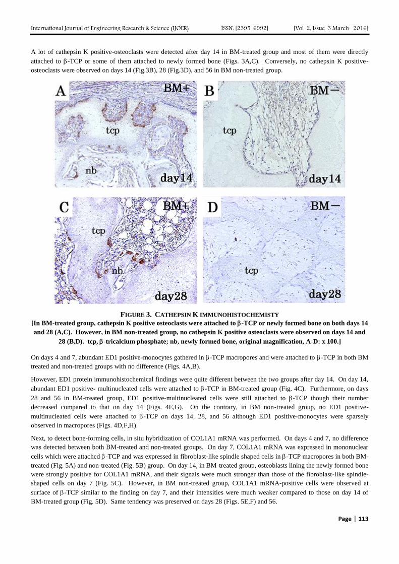
International Journal of Engineering Research amp Science (IJOER) ISSN [2395-6992] [Vol-2 Issue-3 March- 2016]
Page | 115
FIGURE 5 IN SITU HYBRIDIZATION OF COL1A1 MRNA
[A On day 7 in BM-treated group COL1A1 mRNA positive mononuclear cells were directly attached -TCP and
fibroblast like spindle shaped cells were expressed in COL1A1 mRNA in -TCP macropores B On day 7 in BM non-
treated group showed similar finding as A C On day 14 in BM-treated group cuboidal shaped COL1A1 mRNA
positive cells lined to newly formed bone and their intensities were much stronger than fibroblast like COL1A1
mRNA positive cells in -TCP macropores D On day 14 in BM non-treated group no cuboidal shaped COL1A1
mRNA positive cells were detected COL1A1 mRNA positive mononuclear cells were still expressed in the surface of
-TCP but their intensities were much weaker than those shown as C E On day 28 in BM-treated group COL1A1
mRNA positive cells lined to the newly formed bone and their shapes were slightly flatter than those shown as C F
On day 28 in BM non-treated group the finding was similar to that shown as D tcp -tricalcium phosphate nb
newly formed bone original magnification A-Fx 100]
International Journal of Engineering Research amp Science (IJOER) ISSN [2395-6992] [Vol-2 Issue-3 March- 2016]
Page | 116
Quantitative analysis demonstrated that the area of -TCP in BM-treated group decreased in a chronological manner and the
area on day 56 was significantly smaller than that on day 7 (plt005) (Fig 6A) However in BM non-treated group the
tendency was not detected (Fig 6B) The ratio of newly formed bone per unit area of -TCP clearly increased from day 14
to day 56 in a time-dependent manner The ratio of newly formed bone per unit area of on days 28 and 56 were significantly
larger than that on day 14 (plt005) (Fig 6C)
(A)
(B)
(C)
FIGURE 6 QUANTITATIVE ANALYSIS
[A The area of -TCP in the BM-treated group The area of -TCP decreased in a time-dependent manner and the
area on day 56 (497163 mm2) was significantly smaller than that on day 7 (697161 mm
2) (plt005) B The area of
-TCP in the BM non-treated group It had no tendency that was shown in A C The ratio of newly formed bone
per unit area of -TCP shown as the mean standard deviation The amount of newly formed bone increased with
time the amounts of newly formed bone per unit area of -TCP on days 28 (021015) and 56 (031014) were
significantly higher than that on day 14 (006006) (plt005) plt005 NS not significance Bar standard
deviation]
IV DISCUSSION
In this study newly formed bone was detected after day 14 in BM-treated group after -TCP implantation in rat
subcutaneous tissue This bone was not formed by endochondral ossification but through intramembranous ossification
since no cartilaginous tissue was observed New bone formation appeared from the peripheral region of implanted site and
gradually extended to the central region This pattern is considered as reasonable since peripheral region is closer from host
International Journal of Engineering Research amp Science (IJOER) ISSN [2395-6992] [Vol-2 Issue-3 March- 2016]
Page | 117
tissue and is vascularized earlier and more efficiently than the central region Some reports have also supported this
interpretation [29-31] However no bone formation was induced in BM non-treated group These findings indicate that BM
cells are very important as a source of osteoinduction in this model
Moreover even on day 56 newly formed bone was preserved without being degenerated and normal bone marrow tissue was
observed suggesting that -TCP loaded with fresh BM cells has a good biocompatibility and osteoinductive ability
The findings of TRAP staining and cathepsin K immunohistochemistry show that BM cells induce the early appearance of
osteoclasts in this model and that osteoclasts resorb -TCP with directly attaching after day 14 Especially TRAP positive-
multinuclear cells were detected on day 7 in BM-treated group though their number was small
This phenomenon is in agreement with those after implantation in rat bone and dog dorsal muscles as we previously reported
[814] As reported by Jarcho [32] bioresorption of calcium phosphate ceramics such as hydroxyapatite (HA) and TCP
consists of solution-mediated processes (the implant dissolves in physiologic solutions) and cell-mediated processes
(phagocytosis) Given that osteoclasts which appeared in the early stage were directly attached to -TCP in BM-treated
group and -TCP area decreased in a time-dependent manner these data strongly support that cell-mediated resorption plays
an important role for bioresorption of -TCP although the involvement of solution-mediated bioresorption should not be
ignored since microporosity possibly could aid in bioresorption by causing microscopic ldquobreak-uprdquo secondary to solution-
mediated resorption [32] and this -TCP has a microporous structure
ED1 immunoreactivities on days 4 and 7 showed that ED1 positive-monocytes gathered adjacent to -TCP or were directly
attached to -TCP and no difference was detected between the two groups These findings suggest that monocytes infiltrate
adjacent to -TCP even in the absence of BM cells After day 14 ED1 positive-multinucleated cells were detected only in
BM-treated group Nagayama et al reported that the intensity of ED1 immunostaining decreased with time after
implantation of -TCP and carbonate apatite into the subcutaneous tissue in back of rats and that it was persisted longer time
in -TCP group [33] This report is very suggestive in that -TCP itself induces macrophages in extra-osseous sites We
indicate that additional BM cells promote much more inductive ability of macrophage around -TCP in this model In
addition osteoclasts belong to monocyte-macrophage lineage cell and monocytes are supposed to fuse and differentiate into
osteoclasts suggesting that BM cells induce differentiation and proper function of osteoclasts In vitro osteoclasts or closely
related to osteoclasts were induced after 3-week-culture of bone marrow cells in the presence of 125-dihydroxyvitamin D3
[34]
About in situ hybridization of COL1A1 mRNA no difference was detected on day 7 between BM-treated and BM non-
treated group However mononuclear cells (osteoblasts) lined to newly formed bone were strongly COL1A1 mRNA
positive on day 14 28 and 56 in BM-treated group which was quite different from those in BM non-treated group These
results indicate that BM cells also induce bone forming ability
Quantitative analyses in this study showed the area of -TCP decreased in a time-dependent manner in BM-treated group
but not in BM non-treated group suggesting that -TCP is continuously resorbed in BM-treated group due to the constant
osteoclast appearance In addition new bone formation consistently increases in BM-treated group suggesting that bone
forming ability is preserved after day 14
Several previous studies reported that fresh bone marrow cells (without cultured) promoted bone induction in extra-osseous
sites after calcium phosphate ceramics implantation such as HA -TCP and biphasic calcium phosphate (HATCP)
[303135] The earliest ectopic bone formation was 3 weeks after implantation of biphasic calcium phosphate (HATCP was
6040) into rat muscle [30] Ectopic bone formation was shown on 1 month and 2 months after implantation of HA and -
TCP with syngeneic rat bone marrow cells into rat subcutis [35] It is noteworthy that ectopic bone formation was detected 2
weeks earlier than these previous reports [303135] after implantation in our study
Cultured bone marrow stromal cells implantation with calcium phosphate ceramics was also reported and bone formation
was detected day 7 days [1929] or 2 weeks [36] after implantation These methods are based on culture for good
differentiation to osteogenic cells using by dexamethasone or -glycerophosphate Given that mesenchymal stem cells
implantation may lead to carcinogenesis [2021] it is indispensable to investigate the safety of mixed implantation combined
calcium phosphate ceramics with mesenchymal stem cells On the other hand there is no report that had adverse effect such
as infection to bone marrow implantation into bone defects or non-union [37]
International Journal of Engineering Research amp Science (IJOER) ISSN [2395-6992] [Vol-2 Issue-3 March- 2016]
Page | 118
Moreover osteoinductive cytokines such as BMP-2 OP-1 and fibroblast growth factor (FGF) have been investigated as
potent promoters of osteoinduction in extra-osseous site [15-1738] In general these cytokines induce ectopic bone
formation in the early stage after implantation with a scaffold However some problems such as immune response
infection and higher cost have been concerned Recently it has been reported that recombinant human BMP-2 which is
widely used for spine surgery has adverse effects such as infection hematoma and excessive edema [3839]
Bone marrow cells loading to -TCP is a very simple and lower cost method to induce ectopic bone formation We consider
that it is reasonable than the calcium phosphate ceramics implantation with the other concomitant use of osteoinductive
cytokines and cultured mesenchymal stromal cells in that it does not cost very much and safer and bone formation was
confirmed as early as on day 14 Erbe et al have shown that bone marrow aspiration and -TCP composite graft is as good
outcome as autograft with bone marrow cells [1] They used ldquoVitossrdquo which is a porous low-density construct prepared by
lightly fusing particles of -TCP is average approximately 1-2 m in diameter and has 90 porosity and interconnected
void space [1]
It remains not elucidated why oseteoclasts proliferated and are differentiated after implantation of -TCP Some possibilities
about the origin of osteoclasts are considered as follows (1) stromal cells in implanted bone marrow induce osteoblasts and
such osteoblasts induce osteoclasts via receptor activator of NF-kB (RANK)-RANK-ligand (RANKL) system and (2)
hematopoietic cells in implanted bone marrow directly induce osteoclasts
Little report was focused on cellular events about the appearance or the function of osteoclast after implantation of -TCP
In our study it is noteworthy that the early (day 7) appearance of osteoclasts prior to new bone formation was confirmed
after implantation of BM cells-loaded -TCP into rat subcutaneous tissue and that osteoclasts play an important role in cell-
mediated bioresorption of -TCP After implantation of -TCP into rabbit bone the peak of osteoclasts was 2 week and it
was earlier than that of osteoblasts Chazono et al called this event as ldquocoupling-like phenomenonrdquo [22] In our previous
study of -TCP implantation in dog dorsal muscles osteoclasts were induced in muscles where these cells never exist and
then new bone formation occurred [14] We indicate that the cellular event shown in our study resembles ldquocoupling-like
phenomenonrdquo and is also very important for osteoinduction Further studies are required for elucidating the mechanism of
macrophage or osteoclast responses to biomaterials
V CONCLUSION
-TCP (OSferion
) is resorbed by osteoclasts and leads to new bone formation when implanted with syngeneic rat bone
marrow cells into subcutaneous tissue These cellular events follow the appearance of a large number of osteoclasts and their
attachment to -TCP The data in this study suggest that -TCP implantation with autogenous bone marrow cells is safe and
not time-consuming method and promotes osteoinduction method compared to that in non BM cells condition
ACKNOWLEDGEMENTS
The authors thank Yoshiaki Tanaka (Division of Orthopedic Surgery Department of Regenerative and Transplant Medicine
Niigata University Graduate School of Medical and Dental Sciences) for their technical assistance
REFERENCES
[1] Erbe EM Marx JG Clineff TD Bellincampi LD Potential of an ultraporous -tricalcium phosphate synthetic cancellous bone void
filler and bone marrow aspirate composite graft Eur Spine J 2001 10 S141-6
[2] Gupta AK Kumar P Keshav K Sigh A Hydroxyapatite crystals as a bone graft substitutes in benign lytic lesions of bone Indian J
Orthop 2015 49 645-55
[3] Brennan MA Renaud A Amiaud J Rojewski MT Schrezenmeier H Heymann D Trichest V Layrolle P Pre-clinical studies of
bone regeneration with human bone marrow stromal cells and biphasic calcium phosphate Stem Cell Res Ther 2014 5 114 doi
101186scrt 504
[4] Ozawa M Tanaka K Morikawa S Chazono M Fujii K Clinical study of the pure -tricalcium phosphate―reports of 167 cases J
East Jpn Orthop Traumatol 2000 12 409-13
[5] Ozawa M Experimental study on bone conductivity and absorbability if the pure -TCP J Jap Soc Biomat 1995 13 17-25
[6] Ogose A Hotta T Hatano H Kawashima H Tokunaga K Endo N Umazu H Histological examination of beta-tricalcium phosphate
graft in human femur J Biomed Mater Res (Appl Biomater) 2002 63 601-4
[7] Ogose A Kondo N Umezu H Hotta T Kawashima H Tokunaga K Ito T Kudo N Hoshino M Gu W Endo N Histological
assessment in grafts of highly purified beta-tricalcium phosphate (OSferion) in human bones Biomaterials 2006 27 1542-9
International Journal of Engineering Research amp Science (IJOER) ISSN [2395-6992] [Vol-2 Issue-3 March- 2016]
Page | 119
[8] Kondo N Ogose A Tokunaga K Ito T Arai K Kudo N Inoue H Irie H Endo N Bone formation and resorption of highly purified
-tricalcium phosphate in the rat femoral condyle Biomaterials 2005 26 5600-8
[9] Urist MR Bone formation by autoinduction Science 1965 159 893-9
[10] Ripamonti U van den Heever B van Wyk J Expression of the osteogenic phenotype in porous hydroxyapatite implanted
extraskeletally in baboons Matrix 1993 13 491-502
[11] Ripamonti U Osteoinduction in porous hydroxyapatite implanted in heterotopic sites of different animal models Biomaterials 1996
17 31-5
[12] Yuan H Yang Z Li Y Zhang X de Bruijn JD de Groot K Osteoinduction by calcium phosphate biomaterials J Mater Sci Mater
Med 1998 9 723-6
[13] Yuan H de Bruijn JD Li Y Feng J Yang Z de Groot K Zhang X Bone formation induced by calcium phosphate ceramics in soft
tissue of dogs a comparative study between porous α-TCP and -TCP J Mater Sci Mater Med 2001 12 7-13
[14] Kondo N Ogose A Tokunaga K Umezu H Arai K Kudo N Hoshino M Inoue H Irie H Kuroda K Mera H Endo N
Osteoinduction with highly purified -tricalcium phosphate in dog dorsal muscles and the proliferation of osteoclasts before
heterotopic bone formation Biomaterials 2006 27 4419-27
[15] Andrades JA Santamaria JA Nimni ME Becerra J Selection and amplification of a bone marrow cell population and its induction to
the chondro-osteogenic lineage by rhOP-1 an in vitro and in vivo study Int J Dev Biol 2001 45 689-93
[16] Kato M Toyoda H Namikawa T Hoshino M Terai H Miyamoto S Takaoka K Optimized use of a biodegradable polymer as a
carrier material for the local delivery of recombinant human bone morphogenic protein-2 (rhBMP-2) Biomaterials 2006 27 2035-
41
[17] Takahashi Y Yamamoto M Tabata Y Enhanced osteoinduction by controlled release of bone morphogenic protein-2 from
biodegradable sponge composed of gelatin and -tricalcium phosphate Biomaterials 2005 26 4856-65
[18] Yamada Y Boo JS Ozawa R Nagasaka T Okazaki Y Hata K Ueda M Bone regeneration following injection of mesenchymal
stem cells and fibrin glue with a biodegradable scaffold J Craniomaxillofac Surg 2003 31 27-33
[19] Boo JS Ymada Y Okazaki Y Hibino Y Okada K Hata K Yoshikawa T Sugiura Y Ueda M Tissue-engineered bone using
mesenchymal stem cells and a biodegradable scaffold J Craniofasc Surg 2002 13 231-9
[20] Tolar J Nauta AJ Osborn MJ Mortari AP McElmurry RT Bell S Xia L Zhou N Riddle M Schroeder TM Westerndorf JJ McIvor
RS Hogendoorn PCW Szuhai K Oseth L Hirsch B Yant SR Kay MA Peister A Prockop DJ Fibbe WE Blazer BR Sarcoma
derived from cultured mesenchymal stem cells Stem cells 2007 25371-9
[21] Rubio D Garcia-Castro J Martin MC de la Fuente R Cigudosa JC Lloyd AC Bernad A Spontaneous human adult stem cell
transformation Cancer Res 2005 65 3035-9
[22] Chazono M Tanaka T Komaki H Fujii K Bone formation and bioresorption after implantation of injectable -tricalcium phosphate
granules-hyaluronate complex in rabbit bone defects J Biomed Mater Res 2004 70A 542-9
[23] Burstone MS Histochemical demonstration of acid phosphatase with naphthol AS-phosphate J Nat Cancer Int 1958 21 523-39
[24] Amizuka N Yamada M Watanabe J Hoshi K Fukushi M Oda K Ikehara Y Ozawa H Morphological examination of bone
synthesis via direct administration of basic fibroblast growth factor into rat bone marrow Microsc Res Tech 1998 41 313-22
[25] Damouiseaux JGMC DӧPP EA Calame W Chao D Macpherson GG Dijkstra CD Rat macrophage lysosomal membrane antigen
recognized by monoclonal antibody ED1 Immunology 1994 83 140-7
[26] Hayami T Endo N Tokunaga K Yamagiwa H Hatano H Uchida M Takahashi HE Spatiotemporal change of rat collagenase
(MMP-13) mRNA expression in the development of rat femoral neck J Bone Miner Metab 2000 18 185-93
[27] Yamagiwa H Tokunaga K Hayami T Hatano H Uchida M Endo N Takahashi HE Expression of metalloproteinase-13
(Collagenase-3) is induced during fracture healing in mice Bone 1999 25 197-203
[28] Troen BR The role of cathepsin K in normal bone resorption Drug News Perspect 2004 17 19-28
[29] Hartman EHM Vehof JWM Spauwen PHM Jansen JA Ectopic bone formation in rats the importance of the carrier Biomaterials
2005 26 1829-35
[30] Ohgushi H Goldberg VM Caplan AI Heterotopic osteogenesis in porous ceramics induced by marrow cells J Orthop Res 1989 7
568-78
[31] Ohgushi H Okumura M Yoshikawa T Inoue K Senpuku N Tamai S Shors EC Bone formation process in porous calcium
carbonate and hydroxyapatite J Biomed Mater Res 1992 26 855-95
[32] Jarcho M Calcium phosphate ceramics as hard tissue prosthetics Clin Orthop Relat Res 1981 157 259-78
[33] Nagayama M Takeuchi H Doi Y Comparison of carbonate apatite and beta-tricalcium phosphate (resorbable calcium phosphates)
implanted subcutaneously into the back of rats Dent Mater J 2006 25 219-25
[34] Takahashi N Kukita T MacDonald BR Bird A Mundy GR McManus LM Miller M Boyde A Jones SJ Roodman GD
Osteoclast-like cells form in long-term human bone marrow but not in peripheral blood cultures J Clin Invest 1989 83 543-50
[35] Ohgushi H Okumura M Tamai S Shors EC Caplan AI Marrow cell induced osteogenesis in porous hydroxyapatite and tricalcium
phosphate a comparative histomorphometric study of ectopic bone formation J Biomed Mater Res 1990 12 1563-7
[36] Dennis JE HaynesworthSE Young RG Caplan AI Osteogenesis in marrow-derived mesenchymal cell porous ceramic composites
transplanted subcutaneously effect of fibronectin and laminin on cell retention and rate of osteogenic expression Cell Transplant
1992 1 23-32
International Journal of Engineering Research amp Science (IJOER) ISSN [2395-6992] [Vol-2 Issue-3 March- 2016]
Page | 120
[37] Goel A Sangwan SS Siwach RC and Ali AM Percutaneous bone marrow grafting for the treatment of tibial non-union Injury
2005 36 203-6
[38] Kubota K Iseki S Kuroda S Oida S Iimura T Duarte WR Ohya K Ishikawa I Kasugai S Synergistic effect of fibroblast growth
factor-4 in ectopic bone formation induced by bone morphogenetic protein-2 Bone 2002 31 465-71
[39] Hansen SM Sasso RC Resorptive response of rhBMP2 simulating infection in an anterior lumbar interbody fusion with a femoral
ring J Spinal Disord Tech 2006 19130-4
[40] Shields LBE Raque GH Glassman SD Campbell M Vitaz T Harpring J Shields CB Adverse effects associated with high-dose
recombinant human bone morphogenetic protein-2 use in anterior cervical spine fusion Spine 2006 31 542-7
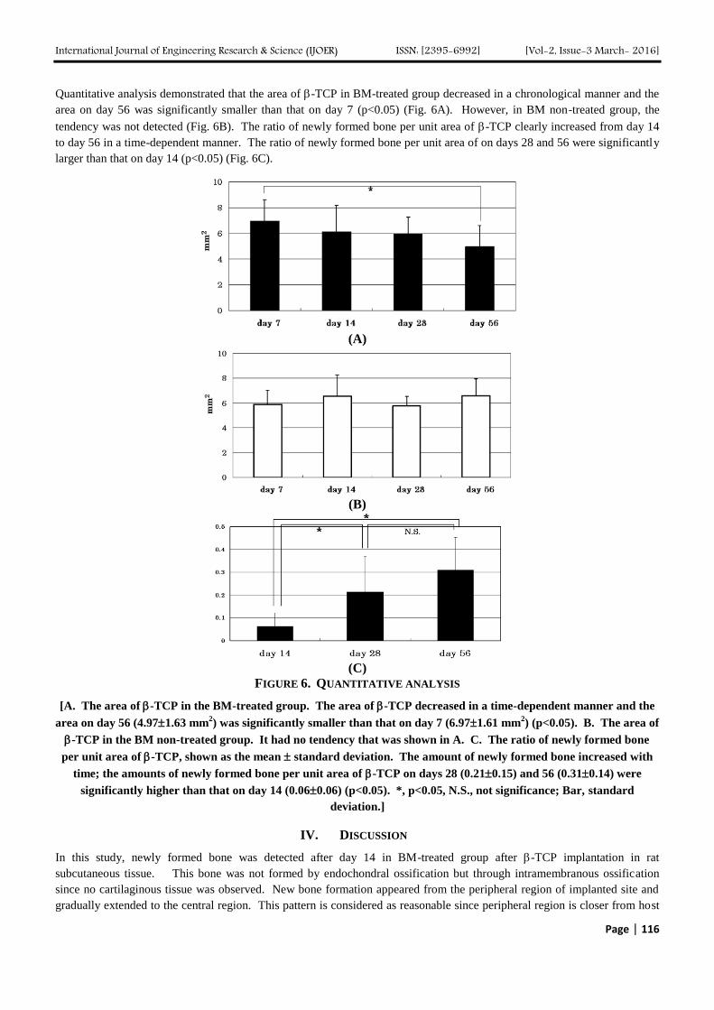
International Journal of Engineering Research amp Science (IJOER) ISSN [2395-6992] [Vol-2 Issue-3 March- 2016]
Page | 120
[37] Goel A Sangwan SS Siwach RC and Ali AM Percutaneous bone marrow grafting for the treatment of tibial non-union Injury
2005 36 203-6
[38] Kubota K Iseki S Kuroda S Oida S Iimura T Duarte WR Ohya K Ishikawa I Kasugai S Synergistic effect of fibroblast growth
factor-4 in ectopic bone formation induced by bone morphogenetic protein-2 Bone 2002 31 465-71
[39] Hansen SM Sasso RC Resorptive response of rhBMP2 simulating infection in an anterior lumbar interbody fusion with a femoral
ring J Spinal Disord Tech 2006 19130-4
[40] Shields LBE Raque GH Glassman SD Campbell M Vitaz T Harpring J Shields CB Adverse effects associated with high-dose
recombinant human bone morphogenetic protein-2 use in anterior cervical spine fusion Spine 2006 31 542-7


