Download - Open Research Online Testing the survival of microfossils in artificial martian sedimentary

Open Research OnlineThe Open University’s repository of research publicationsand other research outputs
Testing the survival of microfossils in artificial martiansedimentary meteorites during entry into Earth’satmosphere: the STONE 6 experimentJournal ItemHow to cite:
Foucher, Frédéric; Westall, Frances; Brandstätter, Franz; Demets, René; Parnell, John; Cockell, Charles S.; M.Edwards, Howell G.; Bény, Jean-Michel and Brack, André (2010). Testing the survival of microfossils in artificialmartian sedimentary meteorites during entry into Earth’s atmosphere: the STONE 6 experiment. Icarus, 207(2) pp.616–630.
For guidance on citations see FAQs.
c© 2010 Elsevier Inc.
Version: Accepted Manuscript
Link(s) to article on publisher’s website:http://dx.doi.org/doi:10.1016/j.icarus.2009.12.014
Copyright and Moral Rights for the articles on this site are retained by the individual authors and/or other copyrightowners. For more information on Open Research Online’s data policy on reuse of materials please consult the policiespage.
oro.open.ac.uk
Accepted Manuscript
Testing the survival of microfossils in artificial martian sedimentary meteorites
during entry into Earth’s atmosphere: the STONE 6 experiment
Frédéric Foucher, Frances Westall, Franz Brandstätter, René Demets, John
Parnell, Charles S. Cockell, Howell G. M. Edwards, Jean-Michel Bény, André
Brack
PII: S0019-1035(09)00503-X
DOI: 10.1016/j.icarus.2009.12.014
Reference: YICAR 9273
To appear in: Icarus
Received Date: 30 July 2009
Revised Date: 3 December 2009
Accepted Date: 4 December 2009
Please cite this article as: Foucher, F., Westall, F., Brandstätter, F., Demets, R., Parnell, J., Cockell, C.S., M. Edwards,
H.G., Bény, J-M., Brack, A., Testing the survival of microfossils in artificial martian sedimentary meteorites during
entry into Earth’s atmosphere: the STONE 6 experiment, Icarus (2009), doi: 10.1016/j.icarus.2009.12.014
This is a PDF file of an unedited manuscript that has been accepted for publication. As a service to our customers
we are providing this early version of the manuscript. The manuscript will undergo copyediting, typesetting, and
review of the resulting proof before it is published in its final form. Please note that during the production process
errors may be discovered which could affect the content, and all legal disclaimers that apply to the journal pertain.

ACCEPTED MANUSCRIPT
Testing the survival of microfossils in artificial martian
sedimentary meteorites during entry into Earth’s
atmosphere: the STONE 6 experiment.
Frederic Fouchera, Frances Westalla, Franz Brandstatterb, Rene Demetsc,John Parnelld, Charles S. Cockelle, Howell G. M. Edwardsf, Jean-Michel
Benyg, Andre Bracka
aCentre de Biophysique Moleculaire, UPR CNRS 4301, Orleans, FrancebNaturhistorisches Museum, Burgring 7, 1010 Wien, Austria
cEuropean Space & Technology Centre (ESTEC), Keplerlaan 1, Postbus 299, 2200 AGNoordwijk, The Netherlands
dSchool of Geosciences, University of Aberdeen, Aberdeen AB24 3UE, United KingdomePlanetary and Space Sciences Research Institute, Centre for Earth, Planetary Space and
Astronomical Research, Open University, Milton Keynes, United KingdomfChemical and Forensic Sciences, School of Life Sciences, University of Bradford,
Bradford BD7 1DP, United KingdomgInstitut des Sciences de la Terre d’Orleans, UMR CNRS 6113, Orleans, France
Abstract
If life ever appeared on Mars, could we find traces of primitive life embedded
in sedimentary meteorites? To answer this question, a 3.5 billion-year-old
volcanic sediment containing microfossils was embedded in the heat shield
of a space capsule in order to test survival of the rock and the microfossils
during entry into the Earth’s atmosphere (the STONE 6 experiment). The
silicified volcanic sediment from the Kitty’s Gap Chert (Pilbara, Australia) is
considered to be an excellent analogue for Noachian-age volcanic sediments.
The microfossils in the chert are also analogues for potential martian life.
Email address: [email protected] (Frederic Foucher)
Preprint submitted to Icarus December 18, 2009
ACCEPTED MANUSCRIPT
An additional goal was to investigate the survival of living microorganisms
(Chroococcidiopsis) protected by a 2 cm thick layer of rock in order to test
whether living endolithic organisms could survive atmospheric entry when
protected by a rocky coating.
Mineralogical alteration of the sediment due to shock heating was mani-
fested by the formation of a fusion crust, cracks in the chert due to prograde
and retrograde changes of α quartz to β quartz, increase in the size of the
fluid inclusions, and dewatering of the hydromuscovite-replaced volcanic pro-
toliths. The carbonaceous microfossils embedded in the chert matrix survived
in the rock away from the fusion crust but there was an increase in the ma-
turity index of the kerogen towards the crust. We conclude that this kind
of sediment can survive atmospheric entry and, if it contains microfossils,
they could also survive. The living microorganisms were, however, com-
pletely carbonised by flame leakage to the back of the sample and therefore
non-viable. However, using an analytical model to estimate the temperature
reached within the sample thickness, we conclude that, even without flame
leakage, the living organisms probably need to be protected by at least 5 cm
of rock in order to be shielded from the intense heat of entry.
Key words: Meteorites, Thermal histories, Astrobiology, Mineralogy, Mars
2

ACCEPTED MANUSCRIPT
1. Introduction
1.1. Research into the oldest traces of life
Research into the origin of life and the most ancient traces of life is ham-
pered by the fact that suitable rocks dating back to the first billion years
of Earth’s history are lacking due to alteration by metamorphism and de-
struction by plate tectonics. The oldest known sedimentary rocks in the Isua
Greenstone Belt and on Akilia island, in south-western Greenland (3.85-
3.7 Ga, Nutman et al., 1997; Blichert-Toft et al., 1999) consist of cherts
(i.e. silicified volcanic silts and sands), banded iron formation (BIF) and
minor strata such as conglomerates (Fedo and Whitehouse, 2002), although
the amphibolite to granulite-grade metamorphism makes interpretation of
the sedimentary protoliths difficult (Mojzsis et al., 1996; Rosing, 1999 van
Zuilen et al. (2002) Westall and Folk (2003). The oldest unambiguous traces
of life occur in 3.5-3.3 Ga-old Early Archean silicified sediments (cherts)
from the Pilbara (Australia) and Barberton (South Africa) greenstone belts.
The cherts contain a variety of traces of life, including colonies of small coc-
coidal chemo(litho)trophic microorganisms on volcanic rock/particle surfaces
(Furnes et al., 2004; Westall et al., 2006b) and anoxygenic photosynthetic
mats formed in sub-aerial shallow water environments (Allwood et al., 2006,
2009; Walsh, 2004; Westall et al., 2006a; Tice and Lowe, 2004). From the
relatively advanced level of evolution of these microorganisms Westall and
Southam (2006) concluded that life had to have appeared much earlier, be-
fore ≈4.0 Ga. The presence of microorganisms in shallow water to littoral
3

ACCEPTED MANUSCRIPT
environments show that the relatively inhospitable conditions on the primi-
tive Earth (high flux of UV radiation, moderate to relatively high seawater
temperatures, high seawater salinity, anoxygenic atmosphere, meteorite bom-
bardment, etc.) did not inhibit life.
The presence of liquid water on Mars during the Noachian period (-4.5
to 3.5 Ga) is now well established (Bibring et al., 2005; Carr, 2006) and the
hydrological system of Noachian Mars as been described by Fasset and Head-
III (2008). Environmental conditions on early Mars during this period were
habitable and, in some respects, similar to those existing on the primitive
Earth (presence of liquid water, moderate to relatively high water temper-
atures in the vicinity of hydrothermal vents and volcanic activity, probably
high salinity, probably slightly acidic pH due to the CO2 atmosphere, high
flux of UV radiation, meteorite bombardment, etc.) (Westall, 2005). Since
Mars had the same ingredients for life (liquid water in contact with mineral
surfaces, carbon and other essential elements, and sources of energy), it is
widely believed that life could have appeared on that planet (Brack, 1996;
McKay, 2008; Sephton and Botta, 2008; Southam et al., 2007; Southam and
Westall, 2007; Walter and des Marais, 1993; Wentworth et al., 2005). Given
the lack of terrestrial rocks old enough to contain traces of the first living
cells, the discovery of traces of primitive life in ancient rocks on Mars would
help understand the origin of life in general (Brack, 1997; Brack et al., 1999;
Westall et al., 2000).
Present and future martian missions are dedicated to studies of the hab-
4
ACCEPTED MANUSCRIPT
itable potential of the martian surface (rocks and regolith), as well as to the
search for past and present traces of life. The lander Phoenix looked for
water on the surface of Mars and tried to determine if life could still exist in
the surface materials (Renno et al., 2009). However, the probability of find-
ing living microorganisms in the uppermost centimetres of regolith is very
small. Nevertheless, one of our goals was to test the hypothesis that liv-
ing endolithic organisms (i.e. microorganisms that live within rocks) could
survive ejection from the host planet, transport through space, and entry
into the atmosphere of another planet, such as Earth. With this objective in
mind, we subjected living endolithic organisms, Chroococcidiopsis, protected
by a 2 cm thick layer of rock to the heat shock of entry into the Earth’s
atmosphere.
Other missions to Mars will investigate the habitability of the surface (the
NASA-led Mars Science Laboratory (MSL), named Curiosity, 2011 mission)
and search for traces of present or past life (the ESA led 2018 ExoMars
mission). The search for traces of life in situ promises to be challenging since
it is already difficult to unambiguously identify traces of fossil life in ≈3.5
Ga-old terrestrial rocks, even with the availability of highly sophisticated
instrumentation capable of high resolution observation and analysis. Thus,
the most promising method of studying martian materials to detect traces of
life will be the international Mars Sample Return mission planned for 2023
(The MEPAG-ND-SAG, 2008).
An alternative method of searching for traces of martian microbial re-
5
ACCEPTED MANUSCRIPT
mains (if life appeared on Mars) is to analyse sedimentary meteorites. This
solution has two advantages: it is cheaper than space exploration and very
accurate analyses can be made in the laboratory. The SNC (Shergottite,
Nakhlite, Chassignite) meteorites form a small group of rare meteorites orig-
inating from Mars. All the known 52 SNC meteorites so far found1 consist
of igneous rocks, mainly basaltic, that are a priori incompatible with the
presence of biogenic remains (except as endoliths). However, sediments de-
posited in bodies of standing water would constitute suitable materials for
hosting traces of life. The STONE 6 experiment was therefore carried out
in order to test the survival of martian analogue sediments and microfossils
embedded in the sediments during entry into the Earth’s atmosphere. The
survival of living endolithic microorganisms was also investigated.
1.2. STONE experiments
STONE 6 is the latest in a series of experiments whose objectives are to
test the effect of entry into the Earth’s atmosphere on different sedimentary
meteorite analogues (Brack et al., 2002). The rock samples are fixed around
the stagnation point of the heat shield of spherical FOTON capsules, 2250
kg in weight and 2.3 m in diameter, used to carry out experiments in lower
Earth orbit.
Previous successful STONE experiments include the STONE 1 experi-
1As the writing of this paper (November 2009) the number of martian me-teorites found is 53, 52 SNC meteorites and ALH84001 (Papike et al., 2009,http://www.imca.cc/mars/martian-meteorites-list.htm).
6

ACCEPTED MANUSCRIPT
ment which tested a carbonate (dolostone, i.e. dolomite, fossil calcareous
shells, quartz and feldspar), and an artificial sediment composed of basalt
(80%) and gypsum (20%), and a reference sample of dolerite (a variety of
basalt) (Brack et al., 2002). Only the dolostone survived. Although it did
not exhibit a fusion crust, its composition changed due to high temperature
processes; the carbonate decomposed into CaO and MgO (periclase). All
the samples in the 2005 STONE 5 experiment, dolerite, carbonate cemented
sandstone, and gneissic impactite, survived atmospheric entry (Brandstatter
et al., 2008; Parnell et al., 2008). A fusion crust formed on the dolerite sug-
gesting that the temperatures attained at the heat shield of the capsule were
comparable to those experienced by meteoroids. This STONE experiment
also tested the survival of living bacterial and fungal spores, as well as dried
vegetative endoliths that were placed in tiny wells drilled into the 1 cm-thick
rock discs. However, the shield constituted by the rocks (1 cm) was too thin
and the microorganisms were carbonized during atmospheric entry (Cock-
ell et al., 2007). Since endolithic photosynthesising microorganisms require
cannot live at great depths within a rock, it is unlikely that they could have
been imported from another planet, therfore Cockell et al. (2007) concluded
that photosynthesis had to have evolved on Earth.
Our experiment, STONE 6, tested the effect of the atmospheric entry
on three samples, the reference sample of dolerite and two sedimentary rocks
samples. The latter included Devonian lake sediments ≈400 My-old from the
Orkney Islands, Scotland and a ≈3.5 billion years old volcanic sandstone, the
7
ACCEPTED MANUSCRIPT
Kitty’s Gap Chert, from the Pilbara region of Australia. This paper concen-
trates on the observations and analysis carried out on the latter sample. [A
second experiment that flew on the same mission, LITHOPANSPERMIA,
consisted of a rock coated with fungal species and is not part of our experi-
ment, de la Torre et al. (2009).]
2. Materials and methods
2.1. The Kitty’s Gap Chert
The 3.446 Ga-old Kitty’s Gap Chert (Fig. 1) forms part of the Coppin
Gap Greenstone Belt, in the Pilbara, north-western Australia. it consists of
layers of silicified volcanic silts and sands deposited in an intertidal environ-
ment (Westall et al., 2006b; de Vries et al., 2006). The volcanic precursors
consisted of K-feldspars, Ti-bearing biotites, volcanic glass shards, amphi-
boles, and ghost spherulites that were replaced by K-phyllosilicates (hydro-
muscovite, KAl2[(OH,F)2|AlSi3O10]-H2O) (Orberger et al., 2006). Early di-
agenetic silicification of the volcaniclastic sediments was due to silica-rich
hydrothermal fluids as well as silica-saturated seawater (van den Boorn et
al., 2007). The rock was subjected to regional low grade metamorphism
(prehnite-pumpellyite grade, Westall et al., 2006b). Westall et al. (2006b)
documented traces of primitive small, anaerobic fossil microorganisms in
these sediments, including silicified biofilms and colonies of coccoidal, rod-
shaped, filamentous microorganisms. Although remnants of photosynthetic
mats (photosynthesis is a relatively advanced metabolism) occur in the sed-
8

ACCEPTED MANUSCRIPT
iments, the majority of the microorganisms were probably lithotrophic (a
very early metabolism), obtaining their carbon and energy from inorganic
sources, in this case the rock/mineral surfaces. The carbonaceous microfos-
sils are well preserved owing to their precocious silicification and the low-
grade metamorphism of the rocks, although the carbon in the microfossils is
thermally mature as is to be expected in rocks of this age and metamorphic
grade.
The shallow water volcanic environmental setting of the Kitty’s Gap
Chert is one that could have existed on Noachian Mars (Westall, 2005; Fasset
and Head-III, 2008; Greenwood and Blake, 2006), as demonstrated by recent
orbital and in situ geochemical studies (Poulet et al., 2005; Bibring et al.,
2005; Squyres et al., 2004; Bishop et al., 2008; Milliken et al., 2008; Squyres
et al., 2008). These sediments were, moreover, deposited in an epoch (or
soon after) when Mars could have hosted life at its surface (Westall, 2005;
Southam and Westall, 2007). The primitive chemo(litho)trophic organisms
that lived on the Early Archaean Earth could have inhabited the Noachian
martian surface. For this reason, the small, primitive cells that characterise
the Kitty’s Gap sediments can be considered as analogies for putative early
martian microorganisms. The sediments with their fossilised microorganisms
therefore constitute useful analogues for martian rocks of Noachian age.
9

ACCEPTED MANUSCRIPT
2.2. Sample preparation before launch
In the previous STONE experiments, the rocks were cut into discs 7 cm
diameter and 1 cm thickness. One of the objectives of the STONE 6 mission
was to test one aspect of the hypothesis of panspermia by evaluating the
survival of microbial spores protected by the thickness of the samples. The
STONE 5 experiment demonstrated that 1 cm of rock (cut as flat discs) was
not sufficient and it was thus decided to increase the thickness. For practical
reasons, the samples being now thicker than the sample holders, the rocks
were milled into a flanged dome shape that was 2 cm thick at its apex in order
to provide more protection for the living microorganisms. The 3.5 Ga-old
Kitty’s Gap Chert was, however, too fractured to be able to be cut into the
required shape and was therefore crushed into ≈3 mm-sized fragments and
mixed with space-qualified cement (Sauereisen Electrotemp cement No.8 )
before being poured into a mould of the required form. The space cement
is a magnesium phosphate (aMgHx(POy)z.bH2O) in a zircon (ZrSiO4) and
silica (SiO2) base. The final shape of the sample after machining before its
fixation on the capsule is shown in Fig. 2. The sample holder consists of an
annular disk of phenolic silica, i.e. the same material used for the ablative
capsule heat shield. The exposed part of the sample was 5 cm in diameter.
The holder was fixed onto the FOTON-M3 capsule near the stagnation point
by carbon-carbon screws.
A live, hydrated culture of the photosynthetic endolithic microorganism,
Chroococcidiopsis, was painted on the back of the rock before it was screwed
10
ACCEPTED MANUSCRIPT
into the sample holder.
2.3. Flight details
The FOTON-M3 capsule was launched by a Soyuz-U launcher rocket
from Baikonur (Kazakhstan) on 14 September 2007. On the 26th September
after 12 days in lower Earth orbit, the velocity of the capsule was slightly
decreased from 7.7 km/sec to 7.6 km/sec for atmospheric entry. The re-entry
module underwent thermal shock as it entered the stratosphere. Mineralogi-
cal changes detailed below show that the temperature on the ablation surface
started from a few oC, increased up to 2000oC at the apex and decreased to
less than 0oC within a few tens of seconds. The speed of the capsule was
reduced by parachute and brake rocket for landing in a field in Kazakhstan.
Two of the three STONE samples survived the experiment, as did the
LITHO- PANSPERMIA sample; the reference dolerite sample was lost. A
photograph of the capsule after landing is shown in Fig. 3. The Kitty’s Gap
Chert sample exhibited a bright white fusion crust across the entire exposed
surface (see insert in Fig. 3). The final thickness of the sample ranged from
9 to 10 mm, corresponding to a loss of ≈50% of the initial thickness by
ablation.
After landing, the samples were carefully and rapidly removed from the
capsule in relatively sterile conditions using glove-covered hands and stored
in a protective holder (Fig. 4) for transport to a clean room at the European
Space & Technology Centre (ESTEC, Noordwijk, The Netherlands), from
11
ACCEPTED MANUSCRIPT
where they were collected for analysis. The transport containers had been
specifically constructed to protect the samples during transport after the dis-
integration of one of the rocks (sandstone) used in the STONE 5 experiment.
2.4. Sample preparation after landing
In order to study changes in the Kitty’s Gap Chert fragments through the
thickness of the reconstituted rock, 30 µm and 500 µm thick polished sections
were prepared (Fig. 5). Some of the sections were etched by hydrofluoric
acid (HF) in order to dissolve the chert and to reveal the embedded organic
matter. The method chosen consisted of etching portions of the 500 µm
thick sections with HF droplets (24% in concentration) deposited for 45 min,
followed by abundant rinsing with milliQ water. Slightly etched surfaces of
30 µm thick thin sections were also observed (HF 2.5% and HF 5% during
30 min).
2.5. Analytical methods
Several methods were used to observe, analyse and characterize the sam-
ple. An optical microscope (Olympus BX51, CBM-Orleans) was used to
make preliminary observations and to localize specific areas of interest. Atomic
force microscopy (AFM, Veeco D3100, CBM-Orleans) and Scanning Electron
Microscopy (FEG SEM, Hitachi S4200, Centre de Microscopie Electronique,
Orleans) were used both on etched and un-etched surfaces to make high mag-
nification observations of specific structures. Elemental analyses were made
with an Energy Dispersive X-ray spectrometer (EDX) attached to the SEM,
12

ACCEPTED MANUSCRIPT
as well a Cameca SX 50 microprobe (BRGM-Orleans). Raman spectroscopy
was made with a Dilor XY800 (ISTO-Orleans), and WITec Alpha 500RA
(CBM-Orleans) to make compositional analysis and to study the changes in
the organic matter and in the minerals with reference to distance from the
fusion crust.
3. Results
3.1. General optical aspect
The postflight rock exhibits a flat bright white vitreous surface, in com-
parison to the matt variegated cream-coloured cupola of the preflown recon-
stituted rock. The sample is 9 to 10 mm thick, whereas the preflown rock
had a maximum thickness of 20 mm at its apex.
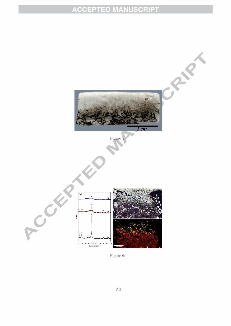
In cross section, the postflight sample can be divided into three zones
parallel to the fusion crust (Fig. 5). The fusion crust itself is ≈0.8 mm
thick and characterised by a bright white colour and glassy reflection. The
lower ≈5 mm of the sample appears blackened to the naked eye although
in thin section it is only the cement that has become black. In between the
fusion crust and the lower blackened layer the sample shows a dull creamy
white colour. In the following, mineralogical and morphological changes in
the flown rock will be described for each constituent with respect to distance
from the exposed surface, labelled x.
13
ACCEPTED MANUSCRIPT
3.2. Rock fragments
3.2.1. Chert matrix
Preflight sample. The original rock consists of volcanic protoliths replaced
by hydromuscovite within a microcrystalline (≤ 1 µm) matrix of α quartz
(quartz) that, in places, has been slightly recrystallised into euhedral crystals
5-10 µm in size. Small (≈1 µm) and irregular-shaped fluid inclusions occur in
the chert matrix of the original rock. Sample preparation for flight produced
no mechanical modifications, such as fracturing (N.B. the original rock is
moderately fractured with the fractures coloured by Fe oxide deposits).
Postflight sample. In the fusion crust (i.e. for x < 0.8 mm), the chert frag-
ments are completely fused to a glassy texture in which no volcanic protoliths
can be distinguished (Fig. 6b). Several bubbles consisting of trapped air (Ra-
man analyses) are also observed (Fig. 6b), the air being incorporated during
rapid cooling of the melted material after atmospheric entry. Broadening of
the 465 cm−1 peak of quartz in the Raman spectrum (Fig. 6a) taken within
the fusion crust indicates that the quartz phase is completely amorphous. As
can be observed in the optical micrograph and the Raman map of the 465
cm−1 quartz peak (Figs. 6b, c), the transition between the fusion crust and
the underlying non-fused sample is relatively sharp with only a narrow band
of mixed amorphous and crystalline quartz between 0.7 mm < x < 0.8 mm
(Fig. 6a). Quartz below 0.8 mm from the sample surface is well preserved
(Fig. 6a) although some fractures traverse the chert fragments (Fig. 7a).
In this zone fractures in the chert matrix also radiate out from embedded
14
ACCEPTED MANUSCRIPT
volcanic protolith particles and are commonly lined with bubbles (Fig. 7b).
There is no change in the size and shape of the fluid inclusions in the chert in
the zone furthest away from the fusion crust but, in the middle of the sample
(between 0.8 mm and 3 mm), the H2O-filled inclusions have rounded shapes
and can be up to 50 µm in size.
3.2.2. Volcanic protoliths
Preflight sample. The silicified hydromuscovite-replaced volcanic protoliths
in the original rock exhibit irregular angular to euhedral shapes, depending
on the original composition of the protolith (feldspar, volcanic glass, py-
roxene; Orberger et al., 2006). Raman spectra of the hydromuscovite are
characterised by a weak but highly fluorescent signal, the weakness of the
signal being due to strong silicification of the original rock.
Ti oxides, anatase and rutile, are associated with the volcanic protoliths
and are common throughout the sample. Anatase is the dominant phase
and occurs in aggregates dispersed throughout the chert fragments, as well
as along the edges of the volcanic protholiths. Rutile occurs as tiny (≈1 µm)
sparse isolated crystals within the anatase, especially inside the protoliths.
Carbon is frequently associated with anatase.
Postflight sample. In the fusion crust (top 0.8 mm) where the chert fragments
are glassy, there is no trace of the protoliths due to complete melting of the
minerals (Fig. 6b). In the middle of the section (between 0.8 mm and 5
mm), the volcanic protoliths have an inhomogeneous dark appearance (Fig.
15
ACCEPTED MANUSCRIPT
8a) compared to their transparent brownish colour at the back of the sample
(Fig. 8b). They are characterised by water-filled spherical inclusions (Fig.
9) although the concentration of the inclusions within the hydromuscovite is
variable owing to local variations in the degree of silica replacement of the
phyllosilicate.
3.3. Cement
Preflight sample. The relatively porous cement has a whitish colour and con-
tains zircon and MgO crystals up to 100 µm in size mixed in a matrix con-
sisting of hydrated magnesium phosphate and silica.
Postflight sample. The most immediately apparent change in the cement is
the dark colouration in the lower ≈5 mm of the flown sample (Fig. 5) and
fusion with the chert fragments in the fusion crust (top 0.8 mm) leading
to the incorporation of Mg in the fused chert (Fig. 10e). As in the chert
fragments, air bubbles occur in the cement in the fusion crust and up to
2 mm away from the sample surface. Fractures occur in the space cement
throughout the thickness of the sample and are common around the chert
particles.
Zircon crystals in the cement are well preserved below the fusion crust
(up to 2 mm from the surface) (Fig. 10d) but adjacent to the fusion crust
(between 0.8 and 2 mm) the mineral occurs in three forms: as well preserved
crystals, as aggregates of tiny (≈1 µm) spherical crystals (Fig. 10c), and as
dendritic zircon crystals (Fig. 10b). The tiny spherical crystals are associ-
16

ACCEPTED MANUSCRIPT
ated with a new mineralogical phase that could not be identified by Raman
spectroscopy (Fig. 10a). Electron microprobe analyses show that it con-
tains zircon and magnesium oxide. The distinct change in the morphology,
and size of the crystals in the fusion crust (Fig. 10), indicates that temper-
atures exceeded the maximum service temperature of the cement (1426oC,
manufacturer’s data).
3.4. Microfossils
Preflight sample. Fossilised microbial cells in the form of colonies of coccoids,
chains of coccoids, filaments and associated EPS were identified by HR-SEM
microscopy in the original rock (Fig. 11a,b) (cf. Westall et al., 2006b). The
fossil cells are individually too small (< 1 µm) to be distinguished in the
fine grained chert matrix by optical microscopy. HR-SEM documented the
close relationship between the fossilised colonies of coccoids and the volcanic
protoliths (Westall et al., 2006b, Fig. 11a,b). Optical microscopy shows that
aggregates of dark spots are commonly associated with the volcanic protoliths
and Raman spectroscopy confirmed their carbonaceous composition (Fig.
12). Orberger et al. (2006) also noted a close association between carbon and
the surfaces of the volcanic protoliths. D and G peak intensities in the Raman
spectra of the spots of carbon in the aggregates indicate that the kerogen has
a maturity that is consistent with the low grade metamorphism experienced
by rocks in the region (prehnite-pumpellyite to lowermost greenschist). We
interpret the aggregates observed by optical microscopy in thin section to be
17
ACCEPTED MANUSCRIPT
the same colonies of coccoids observed by HR-SEM.
Postflight sample. HR-SEM observation of unetched and etched surfaces of
thin sections of the flown samples document the presence of a variety of car-
bonaceous structures including aggregates of tiny carbonaceous spots (Figs.
13, 14), coccoidal structures, filaments and EPS (Figs. 11c,d, 13), interpreted
to be the same types of microfossils described by Westall et al. (2006b) in a
sawn, etched surface of the Kitty’s Gap Chert. The Raman analyses of large
aggregates of spots of carbonaceous matter demonstrated the maturation of
the kerogen towards the fusion crust (see Fig. 15).
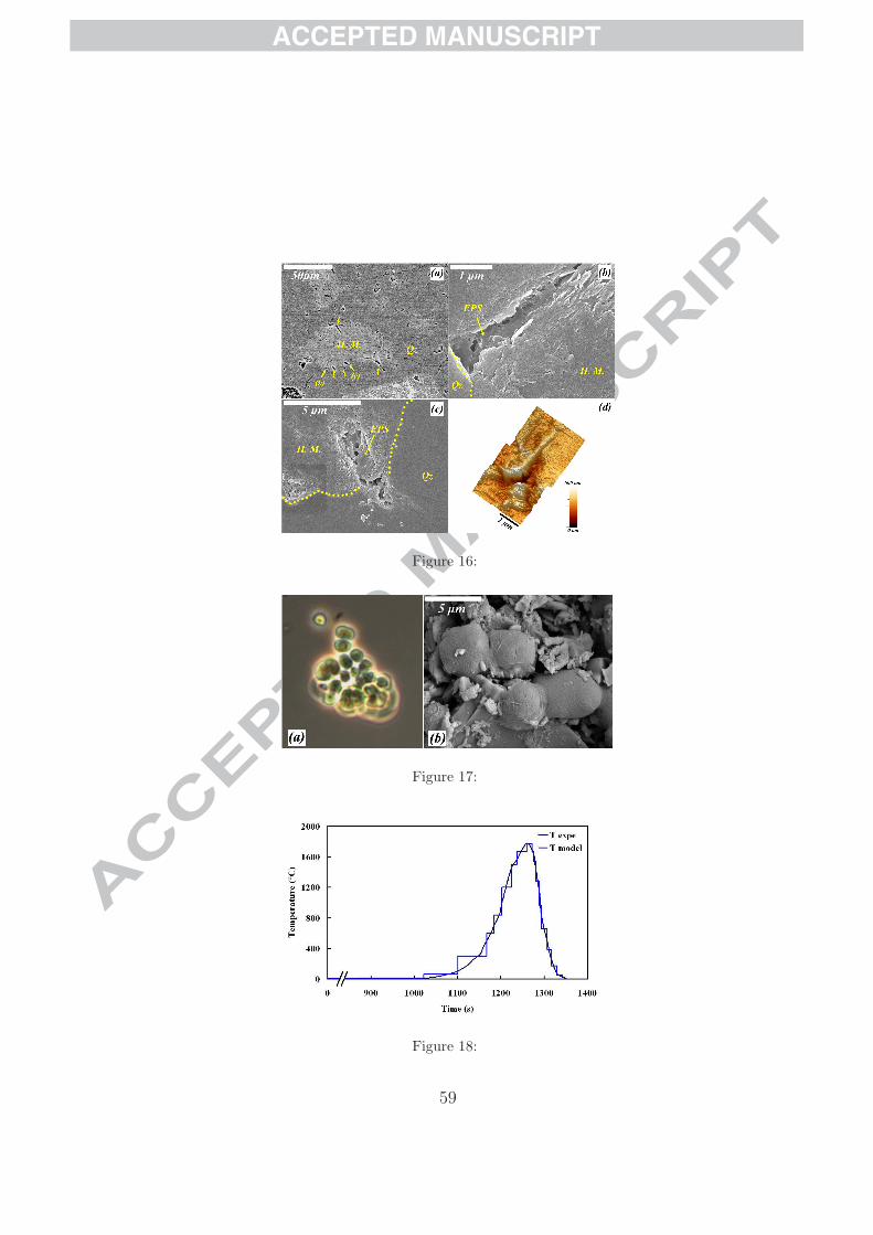
Our observations of thin section surfaces brought to light another type of
carbonaceous structure associated with the volcanic protolith surfaces. Some
of the protoliths exhibit tunnel-like cavities at their edges (Fig. 16a) filled
with smooth, globular or filamentous filmy carbonaceous matter (Fig. 16b-
d), probably representing microbial corrosion pits filled with EPS and the
degradation products of chemolithotrophic microorganisms (cf. Furnes et
al., 2004; Banerjee et al., 2007).
3.5. Modern Chroococcidiopsis biofilm
The back of the sample was inoculated with a live culture of Chroococ-
cidiopsis microorganisms using a brush. These ≈5 µm sized photosynthetic
microorganisms (Fig. 17a) occur naturally as endolithic organisms within
cracks in rock surfaces (Cockell et al., 2007).
No live microorganisms were found on the back of the flown rock. HR-
18

ACCEPTED MANUSCRIPT
SEM revealed carbonaceous oval cell-like structures occurring either singly
(Fig. 17b), or attached to each other in apparent cell division, associated
with a carbonaceous film. These structures have a limited size range from 4-5
µm. Given their carbonaceous composition and morphological similarity with
the original microorganisms, we believe that they represent the carbonised
remains of Chroococcidiopsis. Had the carbonaceous spheroids been produced
simply from the soot of the burnt organisms, they would not exhibit the
regularity of size and shape that is characteristic of these microorganisms.
Raman analysis of the burnt carbon showed D and G band components
characteristic of trigonal and tetrahedral carbon bonding structures, referred
to as the graphite and diamond structural types.
4. Discussion
4.1. Mineralogical changes
Mineralogical changes observed in the flown Kitty’s Gap Chert include (1)
melting of quartz (1723oC, Shchipalov, 2002), hydromuscovite (1320oC) and
cement (1426oC, manufacturer’s data) in the fusion crust, (2) the change in
quartz phase from α quartz into β quartz at 573oC (Mosbah et al., 1997), (3)
an increase in the size of fluid inclusions in the chert, and (4) the devolatiliza-
tion of the hydromuscovite (250oC, Threadgold, 1959). The change in phase
from α to β is accompanied by a corresponding change in crystal lattice size
from (a = b = 4.913 A, c = 5.405 A for the α quartz and a = b = 4.9977 A
and c = 5.4601 A for the β quartz; Mosbah et al., 1997). Rapid quenching
19

ACCEPTED MANUSCRIPT
of the rock brings about a retrograde phase change back to α quartz and
another change in lattice dimension. The dilatation and relaxation of the
lattice are probably the causes of the fractures observed in the quartz.
In order to determine the thermal diffusion experienced during atmo-
spheric entry throughout the Kitty’s Gap Chert sample, we constructed a
new model based on the previous models used by Brandstatter et al. (2008)
and Parnell et al. (2008). The details of our model are explained in Annex 1.
The model was calibrated using the quartz melting temperature of 1723oC
(Fig. 6) which allowed us to determine that the temperature of the exposed
surface was 1775oC. Since the rock particles are embedded in cement, the
thermal diffusivity of the cement was used in the model to estimate the tem-
peratures within the sample. Since diffusivity in the cement is lower than
in the chert, the curves plotted in Fig. 19 correspond to minimal calculated
temperatures. They show that the estimated minimal temperature at the
back of the sample is 630oC.
The white colour of the fusion crust may be related more to the fusion of
the space cement than to that of the embedded chert fragments. The dark
colour of the cement in the lower part of the sample is similar to that noted
by Brandstatter et al. (2008) for the carbonate cement of the sandstone in
the STONE 5 experiment, which was attributed to thermal alteration. As in
the STONE 5 experiment, it appears that flames and heat penetrated behind
the sample holder through gaps between the carbon screws and the siliceous
sample holder. However, the phase changes observed within the sample show
20
ACCEPTED MANUSCRIPT
that alteration due to heat from the trailing side was not a significant con-
tribution. This explains the good correlation between the analytical model
and the observations.
4.2. Survival of microfossils
Figure 11 is a comparison of pre-flight and post-flight microstructures
(from Westall et al., 2006b) showing that both the filamentous and the coc-
coid structures are well preserved in the flown rock. The ratio of the graphite
(G) and disorder (D) peak intensity (G/D) is plotted versus the distance from
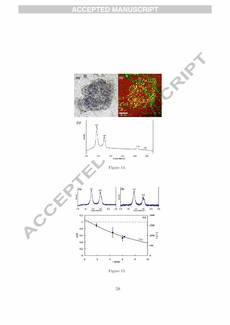
the exposed surface in Fig. 15. The G/D ratio increases with temperature
increase and decreasing distance from the fusion crust, thus demonstrating
thermal maturation of the kerogen during atmospheric entry. Graphitization
occurs typically at a temperature of about 900oC, depending on pressure
conditions Bustin et al. (1995). In conclusion, in spite of the mineralogi-
cal alteration during atmospheric entry and the maturation of the kerogen,
the silicified microorganisms and EPS in the un-melted part of the sample
were physically well preserved. We conclude that microfossils from Mars (if
martian life existed) could be found in martian sedimentary meteorites.
4.3. Panspermia
Despite the fact that flames may have penetrated to the back of the rock
and burnt the Chroococcidiopsis biofilm, mineralogical transformations due
to the transfer of heat from the surface of the rock indicate that temperatures
at the back of the sample were about 630oC (Fig. 20). This is too high
21

ACCEPTED MANUSCRIPT
for survival of living microorganisms. Thus even without flames reaching
the back of the sample, the 2 cm of rock protection would not have been
sufficient to protect endolithic microorganisms. At the surface of the Earth,
phototosynthetic endoliths, such as Chroococcidiopsis, need to have minimum
access to light and therefore never penetrate at depths greater than ≈ 5 mm
into the rock. These results are thus in agreement with the conclusion of
Cockell et al. (2007) that it is unlikely that photosynthetic organisms can
survive transport between planets and, thus, that photosynthesis must have
appeared independently on Earth.
What about chemo(litho)trophic organisms? Lithotrophic microorgan-
isms are not dependent on light, obtaining their carbon and energy from
inorganic sources. They are common colonisers of rocks and have been found
at great depths in deep mines, such as the 3 km deep gold mines of South
Africa (Lin et al., 2006). Knowing that rocks ejected during impact can
originate from some depth in the crust (≤ 100 m, Melosh, 1994, 2003), it is
possible that rocks containing lithotrophic microorganisms in their fractures
could be ejected by impact. Indeed, if life is still present on Mars, it will
occur in protected subsurface environments (McKay et al., 1992). Horneck
et al. (2001) have shown that microbial spores can resist space conditions
when protected by a rock/mineral coating but resistance of the living mi-
croorganisms to atmospheric entry had not been tested until the STONE 5
and 6 experiments.
The heat flux model (Annex 1, Fig. 19) can be used to determine the
22
ACCEPTED MANUSCRIPT
minimal thickness of rock necessary to ensure the survival of living organisms.
Using the thermal diffusivity of the basalt, the most common rock type that
could contain endolithic organisms (κbasalt = 8.10−7 m2.s−1), the maximal
temperature reached within the sample has been plotted in Fig. 20. It is
shown that the thickness needs to be higher than ≈5 cm in order to keep a
maximal temperature at the bottom of the meteorite that does not exceed
113oC, the maximal temperature for (hyperthermophilic) life. However, it
is worth noting that this model is based on the heating profile experienced
by the FOTON capsule. In the case of true meteorites it has been shown
that the velocity is higher in such a way that the temperatures generated by
frictional heating will be higher but will last less time (Parnell et al., 2008).
As a consequence, the temperature reached within the rock will be probably
lower than the estimated one in the STONE 6 experiment. The increase of
temperature at the surface will be associated with higher ablation of matter
but this effect will be counterbalanced by the rotation of the meteorites and
by a decrease in the heating time. The quantity of matter ablated during
this experiment can thus be considered to be relatively similar to that for
true meteorites.
It should be noted that, although the Mars analogue sediment and its
microfossils survived the thermal shock of atmospheric entry this experiment
does not show if they could have survived the mechanical pressures exerted
during the ejection of rocks from Mars. The martian meteorites show moder-
ate levels of shock, typically about 30 GPa (Head et al., 2002). Temperatures
23
ACCEPTED MANUSCRIPT
can be up to 10,000oC near the impact point leading to vaporization of the
rock, but are typically between 500-3000oC for the surrounding rock. 1 com-
pares the compressive strengths of a variety of naturally occurring litholo-
gies. These values provide some indication of the resistance of different rock
types to pressure, thus providing an approximation of their reaction to im-
pact pressure. Chert has a higher compressive strength (¿250 MPa) than
limestone (50-100 MPa) or highly weathered rock (1-5 MPa). Because of
their compactness, the silicified volcanic sediments from the 3.446 Ga-old
Kitty’s Gap better resist these temperatures than less consolidated porous
materials (Stoffler, 1984; French, 1998). Moreover, despite the intense effects
of impacts on the country rock, Melosh (1985) determined that intact rock
fragments can be ejected from planetary surfaces without suffering too much
petrological damage if they were protected by stress wave interference close
to the free surface. Indeed, the existence of intact (but shocked) volcanic
rocks from Mars is testimony to this process. O’Keefe and Ahrens (1985)
calculated that rock fragments about 22 m in diameter could be launched to
escape velocity from a 50 km crater on Mars.
5. Conclusion
The objectives of the STONE 6 experiment were to determine the effects
of thermal alteration during atmospheric entry of martian analogue sediments
and the survival of extant and fossil microorganisms during atmospheric en-
try. The admixture of fragments of ≈3.5 Ga-old, silicified volcanic sediments
24

ACCEPTED MANUSCRIPT
and space cement survived the thermal shock well, forming a white fusion
crust. This contrasts strongly with the black crust typical of basaltic martian
meteorites (although the colour of the fused material in our experiment may
be an artefact of the space cement mixture).
A number of critical mineralogical phase changes and alterations occurred
in the chert fragments at different depths in the thickness of the flown rock,
including melting of the quartz and hydromuscovite, transition of quartz
into quartz and back to quartz, and degassing of OH in the hydromuscovite.
The evolution of the temperature within the sample could be thus modelled,
showing that the maximum temperature reached at the surface of the sample
was 1775oC and at the back of the sample it was ≈630oC.
Microfossils embedded in the chert fragments survived structurally, ex-
cept in the fusion crust and only a slight maturation of the kerogen was
observed. By analogy, it is concluded that microfossils dating back from
the Noachian period could also survive the atmospheric entry and that they
could be found in martian sedimentary meteorites, assuming survival during
the initial impact leading to ejection. Although our model of the thermal
gradient within the sample shows that 2 cm of rock thickness would not
be sufficient to protect live microorganisms from the high temperatures ex-
perienced at the back of the sample, it may still be possible for endolithic
lithotrophs in martian crustal rocks to survive ejection, space transport and
atmospheric entry in larger meteorites.
25
ACCEPTED MANUSCRIPT
Acknowledgments
We thank the European Space Agency for providing the flight opportunity
on the FOTON M3. CNES supported the experiment financially. We thank
the Museum of Natural History of Vienna for preparation of the reconstituted
Kitty’s Gap Chert sample.
Annex 1
Modelling temperature distribution
We constructed an analytical model in order to determine the evolution
of the temperature within the sample during atmospheric entry. Previously
Kimura et al. (2003) and Parnell et al. (2008) used a Heviside step function
(i.e. T (t) = 0 for t < 0, T (t) = Tmax for 0 < t < τ and T (t) = 0 for t > τ)
to model the thermal conditions undergone by meteorites. This model is
associated with very rapid heating, temperature retention for a short period
of time and then very rapid cooling. However, the experimental data of
the heat stream measured on the capsule shows that heating, and thus the
temperature, at the surface of the sample was progressive (Fig. 18). In
order to take this evolution into account as well as possible in the model, the
curve was decomposed into several sections of constant temperature (Fig.
18). Adopting an initial temperature of 0oC, which is approximately the
temperature at the top of the stratosphere (stratopause), the temperature at
26
ACCEPTED MANUSCRIPT
the heating surface at the time t is thus given by:
T (0, t) =
T0 for t ≤ τ0
T1 for τ0 < t ≤ τ1
...
Tn for τn−1 < t ≤ τn
(1)
The global function T (x, t) is thus expressed for each step n as Tn(x, t).
Time evolution of the temperature distribution can be obtained by solving
the following diffusion equation for each step:
∂T
∂t= κ
∂2T
∂x2. (2)
Where κ is the thermal diffusivity of the material. Since the sample is initially
in thermal equilibrium, for the first step the solution is given by:
T0(x, t) = T0. (3)
Mathematical induction can be used to prove that the statement
Tn(x, t) = Tn−1(x, t) + (Tn − Tn−1)
(
1 − erfx
√
4κ(t − τn−1)
)
(4)
holds for all natural numbers n, with erf the error function given by :
erf(z) =2√π
∫ z
0
e−ζ2
dζ. (5)
27

ACCEPTED MANUSCRIPT
thus:
∂ erf(z)
∂z=
2√π
e−z2
. (6)
It is worth noting that for x = 0, the condition Tn(0, t) = Tn (Eq. 1) is
satisfied.
Basis:
In the following, we define:
χn =x
√
4κ(t − τn−1)(7)
and thus:
∂χn
∂x=
1√
4κ(t − τn−1), (8)
∂χn
∂t= − x
2(t − τi−1)√
4κ(t − τn−1). (9)
For n = 1:
T1(χ1) = T0 + (T1 − T0)(1 − erf χ1) (10)
28
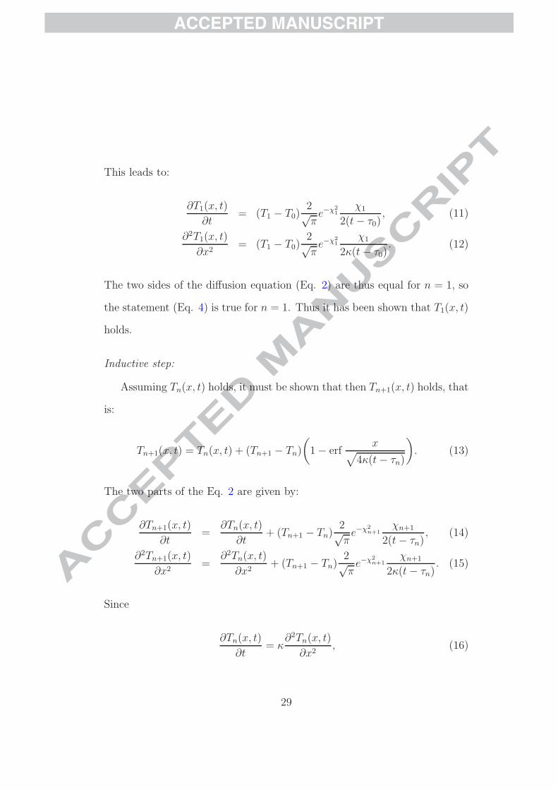
ACCEPTED MANUSCRIPT
This leads to:
∂T1(x, t)
∂t= (T1 − T0)
2√π
e−χ21
χ1
2(t − τ0), (11)
∂2T1(x, t)
∂x2= (T1 − T0)
2√π
e−χ21
χ1
2κ(t − τ0). (12)
The two sides of the diffusion equation (Eq. 2) are thus equal for n = 1, so
the statement (Eq. 4) is true for n = 1. Thus it has been shown that T1(x, t)
holds.
Inductive step:
Assuming Tn(x, t) holds, it must be shown that then Tn+1(x, t) holds, that
is:
Tn+1(x, t) = Tn(x, t) + (Tn+1 − Tn)
(
1 − erfx
√
4κ(t − τn)
)
. (13)
The two parts of the Eq. 2 are given by:
∂Tn+1(x, t)
∂t=
∂Tn(x, t)
∂t+ (Tn+1 − Tn)
2√π
e−χ2n+1
χn+1
2(t − τn), (14)
∂2Tn+1(x, t)
∂x2=
∂2Tn(x, t)
∂x2+ (Tn+1 − Tn)
2√π
e−χ2n+1
χn+1
2κ(t − τn). (15)
Since
∂Tn(x, t)
∂t= κ
∂2Tn(x, t)
∂x2, (16)
29

ACCEPTED MANUSCRIPT
it is deduced that
∂Tn+1(x, t)
∂t= κ
∂2Tn+1(x, t)
∂x2. (17)
therefore Tn+1(x, t) holds.
Since both the basis and the inductive step have been proved, it has now
been proved Eq. 4 is solution for all natural n Q.E.D.
Numerical application
In order to fit the ”stairs” curve used in the model (Fig. 18), a well known
temperature is needed. The observations show that the quartz particles
located at the exposed surface melted in the first ≈0.8 mm (Fig. 6); the
temperature reached at this depth is thus equal to the melting point of quartz,
i.e. 1723oC (Shchipalov, 2002). Using the thermal diffusivity of the quartz
(κQz = 5.10−6 m2.s−1), Eq. (4) leads to the maximum temperature reached at
the surface: 1775oC. This temperature is then used as a reference to estimate
the temperature deeper within the sample
Since the rock particles are embedded in the cement, the thermal dif-
fusivity of the cement (κcement = 6.10−7 m2.s−1) is used to determine the
temperature distribution within the sample. Since the cement is the least
diffusive component, the temperatures obtained in the model therefore cor-
respond to minimal values.
The temperature versus time is plotted for various depths in Fig. 19. The
curves have been smoothed in order to erase the stepped aspect associated
30

ACCEPTED MANUSCRIPT
with the model. The following relevant mineral alteration temperatures have
been added to Fig. 19 in order to compare the model with the experimental
observations:
• T quartzf = 1723oC, the melting point of quartz (Shchipalov, 2002),
• T cement = 1426oC, the maximum service temperature of the cement,
• Tmuscovitef = 1320oC, the melting point of muscovite,
• Tana→rut = 610oC, the phase transition temperature of anatase into
rutile (Jamieson and Olinger, 1969),
• T quartzα→β = 573oC, the phase transition temperature from α quartz into
β quartz (Shchipalov, 2002, Mosbah et al., 1997),
• T hydro = 250oC, the lower temperature of loss of hydroxyl water in
hydromuscovite (Threadgold, 1959).
Thus, according to the model, the quartz melted in the first 0.4 mm, the
cement underwent temperatures exceeding the maximum service temperature
in the first 2 mm and the muscovite melted in the first 3 mm. Throughout
the sample, anatase transforme into rutile, α quartz turned into β quartz and
devolatilization occurred in the hydromuscovite. These results are in good
accordance with the observed mineral alteration despite the approximation
on the thermal diffusivity of the sample.
31

ACCEPTED MANUSCRIPT
References
Allwood, A. C., Walter, M. R., Kamber, B. S., Marshall, C. P., and Burch,
I. W., 2006, Stromatolite reef from the Early Archaean era of Australia,
Nature, 441, 714-718.
Allwood, A. C., Grotzinger, J. P., Knoll, A. H., Burch, I. W., Anderson, M.
S., Coleman, M. L., and Kanik I., 2009, Controls on development and diver-
sity of Early Archean stromatolites, Proceedings of the National Academy
of Sciences of the United States of America, 106, 9548-9555.
Banerjee, N.R., Simonetti, A., Furnes, H., Staudigel, H., Muehlenbachs, K.,
Heaman, L., Van Kranendonk, M.J., 2007, Direct dating of Archean mi-
crobial ichnofossils, Geology, 35, 487490.
Bibring, J.-P., Langevin, Y., Gendrin, A., Gondet, B., Poulet, F., Berthe,
M., Soufflot, A., Arvidson, R., Mangold, N., Mustard, J., Drossart, P.,
and the OMEGA team, 2005, Mars Surface Diversity as Revealed by the
OMEGA/Mars Express Observations, Science, 307, 1576 - 1581.
Bishop, J. L., Dobrea, E. Z. N., McKeown, N. K., Parente, M., Ehlmann, B.
L., Michalski, J. R., Milliken, R. E., Poulet, F., Swayze, G. A., Mustard,
J. F., Murchie, S. L., and Bibring, J.-P., 2008, Phyllosilicate Diversity and
Past Aqueous Activity Revealed at Mawrth Vallis, Mars, Science, 321,
830-833.
32

ACCEPTED MANUSCRIPT
Blichert-Toft, J., Albarede, F., Rosing, M., Frei, R., and Bridgwater, B.,
1999, The Nd and Hf isotopic evolution of the mantle through the Archean.
Results from the Isua supracrustals, West Greenland, and from the Bir-
imian terranes of West Africa, Geochimica et Cosmochimica Acta, 63,
3901-3914.
van den Boorn, S. H. J. M., van Bergen, M. J., Nijman, W., and Vroon, P. Z.,
2007, Dual role of seawater and hydrothermal fluids in Early Archean chert
formation: Evidence from silicon isotopes, Geology, 35, no. 10, 939-942.
Brack, A., 1996, Why exobiology on Mars?, Planetary and Space Science,
44, 1435-1440.
Brack, A., 1997, Life on Mars: a clue to life on Earth? Chemistry & Biology,
4, 9-12.
Brack, A., Clancy, P., Fitton, B., Hofmann, B., Horneck, G., Kurat, G.,
Maxwell, J., Ori, G. G., Pillinger, C., Raulin, F., Thomas, N., and Westall,
F., 1999, An Integrated Exobiology Package for the Search for Life On
Mars, Advances in Space Research, 23, 301-308.
Brack, A., Baglioni, P., Borruat, G., Brandstatter, F., Demets, R., Edwards,
H. G. M., Genge, M., Kurat, G., Miller, M. F., Pillinger, C. T., Roten,
C.-A., and Wasch, E., 2002, Do meteoroids of sedimentary origin survive
terrestrial atmospheric entry? The ESA artificial meteorite experiment
STONE, Planetary and Space Science, 50, 763-772.
33
ACCEPTED MANUSCRIPT
Brandstatter, F., Brack, A., Baglioni, P., Cockell, C. S., Demets, R., Ed-
wards, H. G. M., Kurat, G., Osinski, G. R., Pillinger, J. M., Roten, C.-A.,
and Sancisi-Frey, S., 2008, Mineralogical alteration of artificial meteorites
during atmospheric entry. The STONE-5 experiment, Planetary and Space
Science, 56, 976-984.
Brown, T.E., 1981, Rock characterization, testing and monitoring: ISRM
suggested methods. Pergamon Press, Oxford.
Bustin, R. M., Ross, J. V., and Rouzaud, J. N., 1995, Mechanisms of graphite
formation from kerogen: experimental evidence, International Journal of
Coal Geology, 28, 1-36.
Carr, M.H., 2006, The Surface of Mars, Cambridge University Press, Cam-
bridge, 307 pp.
Cockell, C. S., Brack, A., Wynn-Williams, D. D., Baglioni, P., Brandstatter,
F., Demets, R., Edwards, H. G. M., Gronstal, A. L., Kurat, G., Lee, P.,
Osinski, G. R., Pearce, D. A., Pillinger, J. M., Roten, C.-L., and Sancisi-
Frey, S., 2007, Interplanetary Transfer of Photosynthesis: An Experimental
Demonstration of A Selective Dispersal Filter in Planetary Island Biogeog-
raphy, Astrobilogy, 7 (1), 27-29.
Fassett, C. I., and Head-III, J. W., 2008, Valley network-fed, open-basin
lakes on Mars: Distribution and implications for Noachian surface and
subsurface hydrology, Icarus, 198 (1), 37-56.
34
ACCEPTED MANUSCRIPT
Fedo, C. M., and Whitehouse, M. J., 2002, Metasomatic Origin of Quartz-
Pyroxene Rock, Akilia, Greenland, and Implications for Earth’s Earliest
Life, Science, 296,1448-1452.
French, B.M., 1998. Traces of Catastrophe, A handbook of shock-
metamorphic effects in terrestrial meteorite impact structures, Lunar and
Planetary Institute 120pp .
Furnes, H., Banerjee, N. R., Muehlenbachs, K., Staudigel, H., and de Wit,
M., 2004, Early Life Recorded in Archean Pillow Lavas, Science, 304, 578-
581.
Greenwood, J. P., and Blake, R. E., 2004, Evidence for an acidic ocean on
Mars from phosphorus geochemistry of Martian soils and rocks, Geology,
34, no. 11, 953-956.
Head, J. N., Melosh, H. J., and Ivanov, B. A., 2002, Martian Meteorite
Launch: High-Speed Ejecta from Small Craters Science, 298, 1752-1756.
Horcas, I., Fernandez, R., Gomez-Rodriguez, J. M., Colchero, J., Gomez-
Herrero, J., and Baro, A. M., 2007, WSxM : A software for scanning
probe microscopy and a tool for nanotechnology, Review of Scientific In-
struments, 78, 013705-(1-8).
Horneck, G., Rettberg, P., Reitz, G., Wehner, J., Eschweiler, U., Strauch,
K., Panitz, C., Starke, V., and Baumstark-Kahn, C., 2001, Protection of
35
ACCEPTED MANUSCRIPT
bacterial spores in space, a contribution to the discussion on panspermia.
Origin of Life and Evolution of the Biosphere, 31, 527547.
Jamieson, J. C., and Olinger, B., 1969, Pressure-Temperature Studies of
Anatase, Brookite Rutile, and TiO2(II): a Discussion, The American Min-
eralogist, 54, 1477-1481.
Kimura, M., Chen, M., Yoshida, Y., Goresy, A. E., and Ohtani, E., 2003,
Back-transformation of high-pressure phases in a shock melt vein of an
H-chondrite during atmospheric passage: Implications for the survival of
high-pressure phases after decompression, Earth and Planetary Science
Letters, 217, 141-151.
Lin, L.-H., Wang, P.-L., Rumble, D., Lippmann-Pipke, J., Boice, E., Pratt,
L. M., Lollar, B. S., Brodie, E. L., Hazen, T. C., Andersen, G. L., DeSantis,
T. Z., Moser, D. P., Kershaw, D., Onstott, T. C., 2006, Long-Term Sus-
tainability of a High-Energy, Low-Diversity Crustal Biome, Science, 314,
479-482
McKay, C. P., Mancinelli, R. L., Stoker, C. R., and Wharton, Jr., R. A.,
1992, in Mars, Kieffer, H. H., Jakosky, B. M., Snyder, C. W., Matthews,
M. S. (eds.), University of Arizona Press, Tucson, 12341245.
McKay, C. P., 2008, An Approach to Searching for Life on Mars, Europa,
and Enceladus, Space Science Review, 135, 49-54.
36
ACCEPTED MANUSCRIPT
Melosh, H. J., 1985, Impact cratering mechanics: Relationship between the
shock wave and excavation flow, Icarus, 62, Issue 2, 339-343.
Melosh, H. J., 1994, Swapping rocks: Exchange of surface material among
the planets, The Planetary Report, 14, 16-19.
Melosh, H. J., 2003, Exchange of meteorites (and life?) between stellar sys-
tems, Astrobiology, 3, 207-219.
Milliken, R. E., Swayze, G. A., Arvidson, R. E., Bishop, J. L., Clark, R. N.,
Ehlmann, B. L., Green, R. O., Grotzinger, J. P., Morris, R. V., Murchie,
S. L., Mustard, J. F., and Weitz, C., 2008, Opaline silica in young deposits
on Mars, Geology, 36, no. 11, 847-850.
Mojzsis, S. J., Arrhenius, G., McKeegan, K. D., Harrison, T. M., Nutman,
A. P., and Friend, C. R. L., 1996, Evidence for life on Earth before 3.800
million years ago, Nature, 384, 55-59.
Mosbah, M., Duraud, J.-P., and Clocchiatti, R., 1997, Use of the α → β
quartz transition to monitor the temperature increase produced by a pro-
ton microbeam, Nuclear Instruments and Methods in Physics Research B,
130, 171-175.
Nutman, A. P., Mojzsis, S. J., and Friend, C. R. L., 1997, Recognition of
≥3850 Ma water-lain sediments in West Greenland and their significance
for the early Archaean Earth, Geochimica et Cosmochimica Acta, 61, num.
12, 2475-2484.
37
ACCEPTED MANUSCRIPT
O’Keefe, J. D., and Ahrens, T. J., 1985, Impact and explosion crater ejecta,
fragment size, and velocity, Icarus, 62, 328-338.
Orberger, B., Rouchon, V., Westall, F., de Vries, S. T., Pinti, D. L., Wagner,
C., Wirth, R., and Hashizume, K., 2006, Microfacies and origin of some
Archean cherts (Pilbara, Australia), Geological Society of America, Special
Paper 405, 133-156.
Papike, J. J., Karner, J. M., Shearer, C. K., and Burger P. V., 2009, Silicate
mineralogy of martian meteorites, Geochimica et Cosmochimica Acta, 73,
7443-7485.
Parnell, J., Mark, D., and Brandstatter, F., 2008, Response of sandstone to
atmospheric heating during the STONE 5 experiment: Implications for
the palaeofluid record in meteorites, Icarus, 197 (1), 282-290.
Poulet, F., Bibring, J.-P., Mustard, J. F., Gendrin, A., Mangold, N.,
Langevin, Y., Arvidson, R. E., Gondet, B., Gomez, C., and the Omega
Team, 2005, Phyllosilicates on Mars and implications for early martian
climate, Nature, 438, 623-627.
Renno, N.O., B.J. Bos, D. Catling, B.C. Clark, L. Drube, D. Fisher, W.
Goetz, S.F. Hviid, H. Keller, J.F. Kok, S.P. Kounaves, K. Leer, M. Lem-
mon, M. Bo Madsen, W. Markiewicz, J. Marshall, C. McKay, M. Mehta,
M. Smith, M.P. Zorzano, P.H. Smith, C. Stoker, S.M.M. Young, 2009,
Physical and Thermodynamical Evidence for Liquid Water on Mars, Jour-
38

ACCEPTED MANUSCRIPT
nal of Geophysical Research, Special Issue on Phoenix, 113, E00A18,
doi:10.1029/2008JE003083.
Rosing, M. T., 1999, 13C-Depleted Carbon Microparticles in >3700-Ma Sea-
Floor Sedimentary Rocks from West Greenland, Science, 283, 674 676.
Sephton, M. A., and Botta, O., 2008, Extraterrestrial Organic Matter and
the Detection of Life, Space Science Review, 135, 25-35.
Shchipalov, Yu. K., 2002, Thermodynamic analysis of melting, vitrification,
and crystallization in the SiO2 system, Glass and Ceramics, Science for
Glass Production, 59, num. 3-4, 115-118.
Southam, G., and Westall, F., 2007, Geology, Life and Habitability, Treatise
on Geophysics, 10.12, 421-437.
Southam, G., Rothschild, L. J., and Westall, F., 2007, The Geology and
Habitability of Terrestrial Planets: Fundamental Requirements for Life,
Space Science Review, 129, 7-34.
Squyres, S. W., Arvidson, R. E., Bell,III, J. F., Bruckner, J., Cabrol, N.
A., Calvin, W., Carr, M. H., Christensen, P. R., Clark, B. C., Crumpler,
L., Des Marais, D. J., d’Uston, C., Economou, T., Farmer, J., Farrand,
W., Folkner, W., Golombek, M., Gorevan, S., Grant, J. A., Greeley, R.,
Grotzinger, J., Haskin, L., Herkenhoff, K. E., Hviid, S., Johnson, J., Klin-
gelhofer, G., Knoll, A., Landis, G., Lemmon, M., Li, R., Madsen, M. B.,
Malin, M. C., McLennan, S. M., McSween, H. Y., Ming, D. W., Moersch,
39
ACCEPTED MANUSCRIPT
J., Morris, R. V., Parker, T., Rice, Jr., J. W., Richter, L., Rieder, R.,
Sims, M., Smith, M., Smith, P., Soderblom, L. A., Sullivan, R., Wanke,
H., Wdowiak, T., Wolff, M., and Yen, A., 2004, The Spirit Rover’s Athena
Science Investigation at Gusev Crater, Mars, Science, 305, 794-799.
Squyres, S. W., Arvidson, R. E., Ruff, S., Gellert, R., Morris, R. V., Ming, D.
W., Crumpler, L., Farmer, J. D., Des Marais, D. J., Yen, A., McLennan,
S. M., Calvin, W., Bell, III, J. F., Clark, B. C., Wang, A., McCoy, T.
J., Schmidt, M. E., de Souza, Jr., P. A., 2008, Detection of Silica-Rich
Deposits on Mars, Science, 320, no. 5879, 1063-1067.
Stoffler, D., 1984, Glasses formed by hypervelocity impact Journal of Non-
Crystalline Solids, 67, Issues 1-3, 465-502.
Threadgold, I. M., 1959, A Hydromuscovite with the 2M2 Structure, from
Mount Lyell, Tasmania, The American Mineralogist, 44, 488-494.
Tice, M. M., and Lowe, D. R., 2004, Photosynthetic microbial mats in the
3,416-Myr-old ocean, Nature, 431, 549-552.
de la Torre, R., Sancho, L. G., Horneck, G., Ascaso, C., de los Ros, A.,
Wierzchos, J., Olsson-Francis, F., Cockell, C. S., Rettberg, P., Berger, T.,
de Vera, J.-P. P., Ott, S., Fras, J. M., Melendi, P. G., Lucas, M. M., Reina,
M., Pintado, A., Demets, R., 2009, Likelihood of interplanetary transfer of
rock-inhabiting microbial communities: Results from the space experiment
Lithopanspermia, submittedto Icarus.
40

ACCEPTED MANUSCRIPT
de Vries, S. T., Nijman, W., Wijbrans, J. R., and Nelson, D. R., 2006,
Stratigraphic continuity and early deformation of the central part of the
Coppin Gap Greenstone Belt, Pilbara, Western Australia, Precambrian
Research, 147, 1-27.
Walsh, M. M., 2004, Evaluation of Early Archean Volcaniclastic and Volcanic
Flow Rocks as Possible Sites for Carbonaceous Fossil Microbes, Astrobiol-
ogy, 2004, 4, 429-437.
Walter N. R., and des Marais, D. J., 1993, Preservation of Biological Infor-
mation in Thermal Spring Deposits: Developing a Strategy for the Search
for Fossil Life on Mars, Icarus, 101, 129-143.
Wentworth, S. J., Gibson, E. K., Velbel, M. A., and McKay, D. S., 2005,
Antarctic Dry Valleys and indigenous weathering in Mars meteorites: Im-
plications for water and life on Mars, Icarus, 174, 383-395.
Westall, F., 2005, Life on the Early Earth: A Sedimentary View, Science,
308, 366-367.
Westall, F., and Folk, R. L., 2003, Exogenous carbonaceous microstructures
in Early Archaean cherts and BIFs from the Isua Greenstone Belt: impli-
cations for the search for life in ancient rocks, Precambrian Research, 126,
313-330.
Westall, F., and Southam, G., 2006, The Early Record of Life, Archean
Geodynamics and Environments, 164, 283-304.
41

ACCEPTED MANUSCRIPT
Westall, F., Brack, A., Hofmann, B., Horneck, G., Kurat, G., Maxwell, J.,
Ori, G. G., Pillinger, C., Raulin, F., Thomas, N., Fitton, B., Clancy, P.,
Prieur, D., and Vassaux, D., 2000, An ESA study for the search for life on
Mars, Planetary and Space Science, 48, 181-202.
Westall, F., de Ronde, C. E. J., Southam, G., Grassineau, N., Colas, M.,
Cockell, C., and Lammer, H., 2006, Implications of a 3.472-3.333 Gyr-old
subaerial microbial mat from the Barberton greenstone belt, South Africa
for the UV environmental conditions on the early Earth, Philosophical
Transaction of the Royal Society of London B, 361, 1857-1875.
Westall, F., de Vries, S. T., Nijman, W., Rouchon, V., Orberger, B., Pearson,
V., Watson, J., Verchovsky, A., Wright, I., Rouzaud, J. N., Marchesini,
D., and Severine, A., 2006, The 3.466 Ga ”Kitty’s Gap Chert”, an early
Archean microbial ecosystem, Geological Society of America, Special Paper
405, 105-131.
van Zuilen, M. A., Lepland, A., and Arrhenius, G., 2002, Reassessing the
evidence for the earliest traces of life, Nature, 418, 627-630.
42

ACCEPTED MANUSCRIPT
Figure captions
Figure 1: Field view of the original rock from the Kitty’s Gap Chert,
Pilbara, Australia.
Figure 2: Final shape of the reconstituted sample consisting of 3 mm-
sized fragments of chert mixed in space cement (a); top (b) and bottom (c)
views of the Kitty’s Gap Chert sample. The fragments of the original rock
mixed in cement are clearly visible.
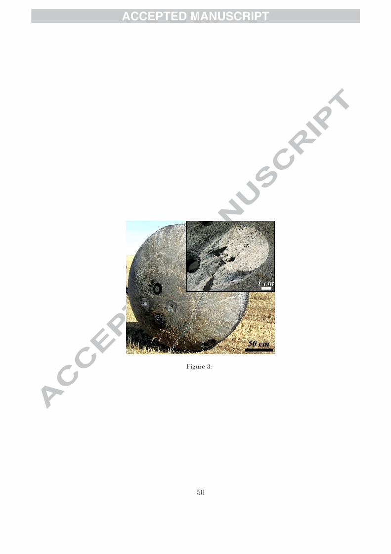
Figure 3: FOTON-M3 capsule after landing showing emplacement of the
rock sample around the stagnation point. Detail of the Kitty’s Gap Chert
sample, showing the presence of a white fusion crust over the whole of the
exposed surface and streaming of solidified molten rock and heat shield ma-
terial (insert).
Figure 4: (a) Sketch of transportation container and (b) photograph of
the sample in the container in the clean room at ESA-ESTEC.
Figure 5: Polished thin section 30 µm thick of the Kitty’s Gap Chert
sample after atmospheric re-entry. The fusion crust is at the top, denoted
by a thin (< 1 mm) bright white layer. Note the dark colour of the cement
in the lower part of the image.
43

ACCEPTED MANUSCRIPT
Figure 6: Analysis of the quartz in the fusion crust. (a) Raman spectra
of quartz (i) spectrum at 0.4 mm from the exposed surface (glassy state),
(ii) spectrum at 0.7 mm from the exposed surface (mixed state), and (iii)
spectrum at 1.2 mm from the exposed surface. (b) The optical image clearly
shows the presence of bubbles in the glassy fusion crust. (c) Raman map-
ping associated with the optical image represents the evolution of the width
of the 465 cm−1 peak of the quartz, the dark area being associated with fused
quartz.
Figure 7: (a) SEM image of cracks in and around a quartz particle after
slight etching (HF 5%, 30 min, distance from the exposed surface 7.6 mm).
(b) Hydromuscovite grains after loss of OH, surrounded by cracks in the
quartz matrix (distance from the exposed surface 4.6 mm).
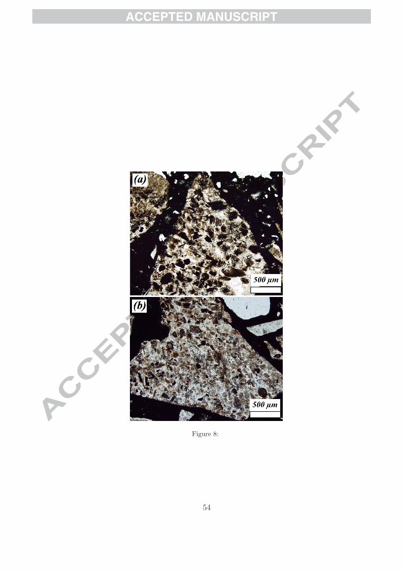
Figure 8: Optical view of the volcanic protoliths (a) in the middle of the
section (distance from the exposed surface 4 mm) with a dark appearance
and (b) at the back of the sample (distance from the exposed surface 8 mm).
Figure 9: Optical and Raman images of a melted hydromuscovite grain lo-
cated at 1.9 mm from the exposed surface. The H2O-filled bubbles are clearly
highlighted by Raman spectroscopy (colour coding: brown for quartz, blue
for melted hydromuscovite and green for water).
44

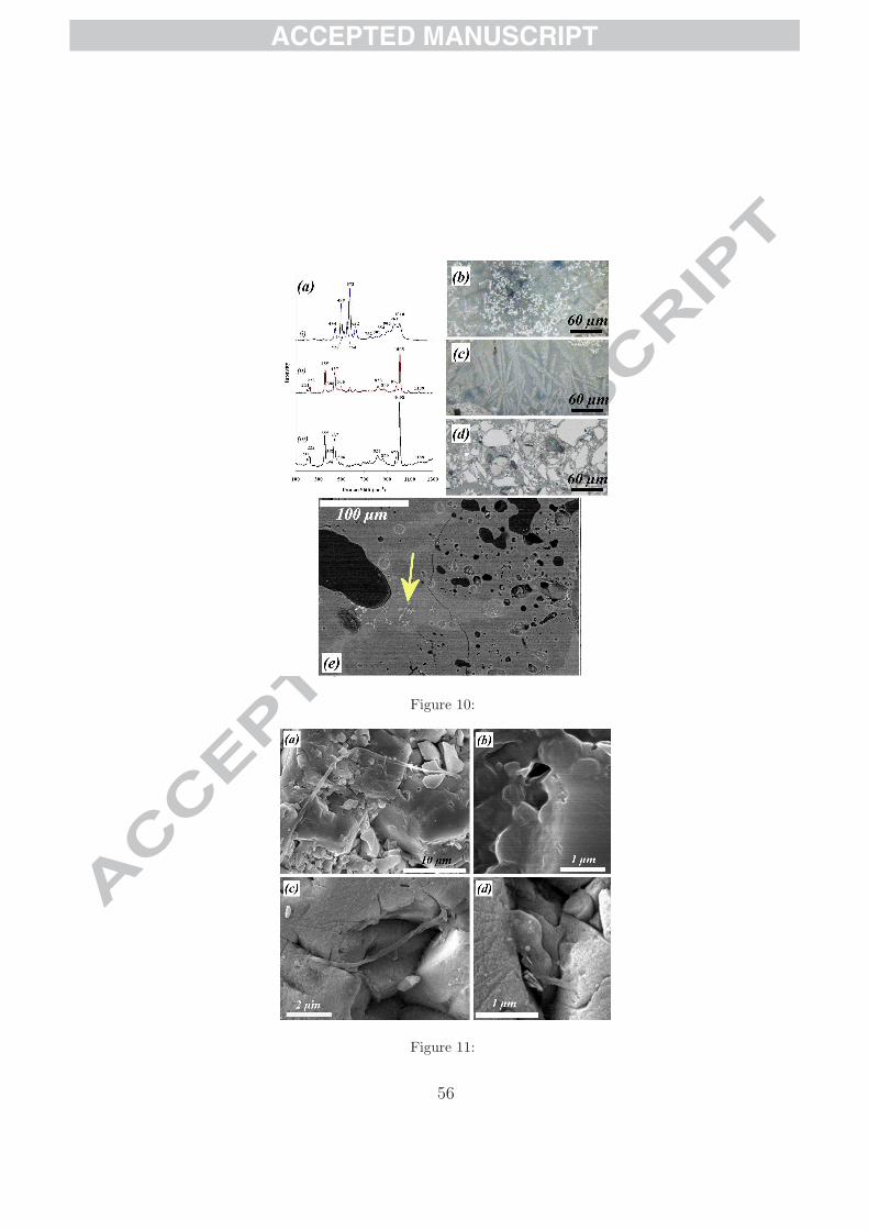
ACCEPTED MANUSCRIPT
Figure 10: (a) Raman spectra of the cement showing the transformation
of zircon into new phases: (i) spectrum of the small circular grains (b) ob-
served in the fusion crust (distance from exposed surface 1 mm), (ii) spectrum
of the dendritic structures (c) observed near the fusion crust crust (distance
from exposed surface 1.5 mm) and (iii) spectrum of the well preserved zircon
particles (d) at the back of the sample (distance from exposed surface 8 mm).
(e) SEM image showing the intermixing of the fused Mg-rich cement (lighter
coloured areas with dendritic crystals of zircon, arrow) and chert fragments
in the fusion crust (un-etched surface, distance from the exposed surface 0.8
mm). Note the bubbles of trapped air.
Figure 11: Comparison between (a) pre-flight and (b) post-flight micro-
fossils. (a) and (c) filaments (distance from exposed surface 7.3 mm) and (b)
and (d) coccoid structures (distance from exposed surface 7.2 mm).
Figure 12: (a) Optical and (b) Raman analysis of organic matter on the
surface of a hydromuscovite grain. The Raman image clearly shows the dis-
tribution of anatase and carbon around the grain (colour encoding: carbon
in yellow, anatase in pink, quartz in brown, muscovite in blue and rutile in
green).
Figure 13: (a) Optical transmitted light image of spots of organic matter
forming a dark aggregate (probably a fossilised colony of microorganisms, cf.
45
ACCEPTED MANUSCRIPT
Westall et al., 2006b) in the chert matrix. (b) Same area after HF etching
(24%, 45 min). (c) SEM image of the same area and (d) HRSEM image of
the aggregate (in black in (c)) showing a thick film of silicified EPS.(distance
from the exposed surface 5.5 mm).
Figure 14: (a) Optical and (b) Raman image of a dark carbonaceous
aggregate (distance from exposed surface 6.0 mm). The Raman image asso-
ciated with the optical image clearly shows the carbonaceous nature of the
spots (in yellow in the figure, corresponding to the 1350 cm−1 peak). The
presence of cracks in the quartz is enhanced by fluorescence in the image
(green in the figure). The intensity of the disorder peak of carbon at 1350
cm−1 is significantly higher than the intensity of the graphite peak at 1600
cm−1. The second order of carbon peaks located at 2700 and 2940 cm−1
are also shown. (colour encoding: quartz in brown, carbon in yellow and
fluorescence in green).
Figure 15: Raman spectra associated with large aggregates of spots of
carbonaceous matter (a) near the fusion crust (distance from the exposed
surface 2 mm) and (b) at the back of the sample (distance from the exposed
surface 6.2 mm). (c) Ratio of the graphite (G) and disorder Raman (D) peak
intensities versus distance from the exposed surface (x).
Figure 16: (a) SEM image showing tunnel structures (arrows) located
46
ACCEPTED MANUSCRIPT
around a hydromuscovite grain (distance from the fusion crust 9.8 mm). (b)
and (c) HRSEM images of the tunnel structures located in (a). A thick film
of silicified EPS has filled the tunnels (arrows). Filamentous structure ob-
served in the tunnel (c).is shown by AFM in (d) (80×80 µm2 in size, AFM
image processing using WSxM , Horcas et al., 2007).
Figure 17: (a) Optical view of Chroococcidiopsis bacteria. (b) HRSEM
image of the carbonized bacteria at the back of the sample after the experi-
ment.
Figure 18: Variation in temperature at the exposed surface of the flown
sample during entry into the Earth’s atmosphere as a function of heating
time. The ”stairs” curve used for the model have been reported.
Figure 19: Temperature distribution as a function of heating time and
depth in the sample, after smoothing of the curves. The temperatures of
relevant mineral alteration have been added to the figure.
Figure 20: Maximum temperature versus depth in a basalt sample esti-
mated from the analytical model. The maximal temperature for life has been
reported (113oC).
Table 1: Comparison of the compressive strengths of different rock types
47

ACCEPTED MANUSCRIPT
(after Brown (1981)).
48

ACCEPTED MANUSCRIPT
Figures
Figure 1:
Figure 2:
49
ACCEPTED MANUSCRIPT
Figure 3:
50
ACCEPTED MANUSCRIPT
Figure 4:
51
ACCEPTED MANUSCRIPT
Figure 5:
Figure 6:
52

ACCEPTED MANUSCRIPT
Figure 7:
53
ACCEPTED MANUSCRIPT
Figure 8:
54

ACCEPTED MANUSCRIPT
Figure 9:
55
ACCEPTED MANUSCRIPT
Figure 10:
Figure 11:
56

ACCEPTED MANUSCRIPT
Figure 12:
Figure 13:
57
ACCEPTED MANUSCRIPT
Figure 14:
Figure 15:
58
ACCEPTED MANUSCRIPT
Figure 16:
Figure 17:
Figure 18:
59

ACCEPTED MANUSCRIPT
Figure 19:
Figure 20:
Rock type Uniaxial compressiveStrength (MPa)
Fresh basalt, chert, diabase Gneiss, granite,quartzite
>250
Amphibolite, sandstone, Basalt, gabbro, gneiss,Granodiorite, peridotite, rhyolite, tuff
100-250
Limestone, marble, sandstone, schist 50-100
Phyllite, schist, siltstone 25-50
Chalk, claystone, potash, marl, siltstone, shale,rocksalt
2-25
Highly weathered or altered rock, shale 1-5
Table 1:
60


