
1
OECD GUIDANCE NOTES ON DERMAL ABSORPTION
DRAFT 22 OCTOBER 2010
2
FOREWORD
These Guidance notes on dermal absorption (Guidance Notes) are intended to provide practical
guidance to facilitate harmonised interpretation of experimental data from specific dermal absorption
studies, where they are available, and to provide advice on alternative ways to estimate dermal absorption
when there are no data or few specific data available. The Guidance Notes were prepared with the primary
focus on establishing appropriate dermal absorption values for occupational health and public health risk
assessment of pesticides and biocides. This guidance may also be relevant for other groups of chemicals,
such as veterinary medicines and industrial chemicals, thus enabling a consistent approach.
The Guidance Notes consider the type of data that may be available to risk assessors for estimating or
calculating the dermal absorption for the evaluation of public health or safety risks posed by a chemical.
The Guidance Notes also provide guidance on the interpretation of such data to facilitate a harmonised
approach.
These Guidance Notes outline core concepts and refer the reader to other useful sources when more
detailed or specific information is required. These Guidance Notes are intended to complement OECD Test
Guidelines and other publications by the OECD, especially OECD Test Guideline 427 (in vivo) and OECD
Test Guideline 428 (in vitro) (OECD 2004a and 2004b) and the OECD Guidance Document for the
Conduct of Skin Absorption Studies (OECD 2004c). These notes are also designed to complement the
WHO/IPCS Environmental Health Criteria 235: Dermal Absorption (WHO 2006) and guidance
documents developed by governments (e.g. EC 2004; EC 2006; and USEPA 1996 amongst others). All of
these documents encourage a harmonised approach to the conduct of dermal absorption studies. These
Guidance Notes do not comprehensively address the issue of test methodology and study performance,
recognising that there are numerous factors that can influence dermal penetration. The relevant test
guidelines and OECD GD 28 (2004c) should be consulted when designing dermal absorption studies.
The OECD and WHO/IPCS documents listed above should be read in conjunction with these
Guidance Notes. The WHO/IPCS (WHO 2006) document serves to introduce dermal absorption at a
broader level, and the OECD Test Guidelines advise on the conduct of the studies. In contrast, these
Guidance Notes are designed to help assess and interpret specific studies for the estimation of dermal
absorption values.
While dermal absorption values form an integral part of the risk assessment process, these Guidance
Notes do not address the entire risk assessment process. Although different regions and countries of the
world may have different approaches to the type of data required for the assessment of public health and
occupational safety of compounds, these Guidance Notes do not attempt to reconcile these differences of
approach.
The Guidance Notes were developed by the OECD Expert Group on Dermal Absorption, comprising
experts from Australia (lead country), Canada, Germany, the Netherlands, the United Kingdom, the United
States, the Business and Industry Advisory Committee to the OECD (BIAC), the International Programme
of Chemical Safety (IPCS), European Food Safety Authority (EFSA), and the OECD Secretariat.

3
TABLE OF CONTENTS
1. INTRODUCTION ....................................................................................................................... 5
2. SUMMARY OF RECOMMENDATIONS .............................................................................. 5 Part 1 .............................................................................................................................................. 5 Part 2 .............................................................................................................................................. 7
PART 1: INTERPRETATION OF DERMAL ABSORPTION STUDIES ..................................... 8
3. INTRODUCTION – TYPES OF DATA .................................................................................. 8 4. IN VITRO DATA ...................................................................................................................... 8
4.1 Introduction ............................................................................................................................. 8 4.2 Species selection ..................................................................................................................... 9
4.3 Skin samples to be used and details of the study design ...................................................... 10 4.4 Receptor fluid ....................................................................................................................... 11
5. IN VIVO DATA ...................................................................................................................... 13 6. COMBINATION OF ANIMAL AND HUMAN IN VITRO AND HUMAN IN VIVO DATA
..................................................................................................................................................... 15
6.1 Introduction: the ‘Triple Pack’ approach .............................................................................. 15 Example 1 ................................................................................................................................. 15
6.2 Use of the ‘Triple Pack’ approach in risk assessment ........................................................... 16
7. General considerations for the evaluation of dermal absorption studies ................................ 17
7.1 Chemical remaining in the skin ............................................................................................ 17 7.1.1 Definitions and existing guidance .................................................................................. 17
7.1.2 Tape stripping ................................................................................................................. 18 7.1.3 Completion of absorption in vivo ................................................................................... 18
7.2 Effect of formulation ............................................................................................................ 21 7.2.1 Test preparations ............................................................................................................ 21
7.2.2 Influence of formulation .................................................................................................. 21 7.2.3 Solid vs. liquid formulations .......................................................................................... 25
7.3 Metabolism in the skin .......................................................................................................... 25 7.4 Mass balance ......................................................................................................................... 26 7.5 Use of mean or centiles; treatment of outliers and use of rounding ..................................... 27
7.5.1 Introduction .................................................................................................................... 27
7.5.2 Variability is relatively low ............................................................................................ 27
7.5.3 Variability is high ........................................................................................................... 27 7.5.4 Outliers ........................................................................................................................... 28 7.5.5 Rounding ........................................................................................................................ 28
7.6 The ‘wash-in’ effect .............................................................................................................. 28
PART 2: ESTIMATION OF DERMAL ABSORPTION IN THE ABSENCE OF SPECIFIC
STUDIES ........................................................................................................................................ 29
4
PART 2: ESTIMATION OF DERMAL ABSORPTION IN THE ABSENCE OF SPECIFIC
STUDIES ........................................................................................................................................ 30
8. INTRODUCTION .................................................................................................................. 30 9. DEFAULT VALUES ............................................................................................................. 30 10. PREDICTION OF DERMAL ABSORPTION BY ALTERNATIVE APPROACHES ...... 31
10.1. Read-across ......................................................................................................................... 32 10.2. Quantitative structure-activity relationships (QSARs) ...................................................... 32 10.3. Use of flux or permeability coefficient KP ......................................................................... 34 10.4. Mathematical models .......................................................................................................... 34 10.5. Comparison of toxicity data from oral and dermal studies ................................................. 35
10.6. Other study types (including ADME and human in vivo data).......................................... 36 10.6.1. In vivo ADME studies ................................................................................................. 36
10.6.2. The ‘mass balance’ approach ...................................................................................... 36 10.6.3. Human in vivo dermal absorption studies .................................................................... 37 10.6.4. Human biomonitoring studies ...................................................................................... 37
REFERENCES ............................................................................................................................ 38
Study 1: In vivo dermal absorption of chem1 in the rat ............................................................... 46 Test material ................................................................................................................................ 46
Guidelines .................................................................................................................................... 46 Material and methods................................................................................................................... 46 Results .......................................................................................................................................... 46
Conclusions .................................................................................................................................. 48 Study 2: In vitro dermal absorption of chem1 in rat and human skin ......................................... 49
Test material ................................................................................................................................ 49 Guidelines .................................................................................................................................... 49
Material and methods................................................................................................................... 49 Results .......................................................................................................................................... 49
Conclusions:................................................................................................................................. 50 Discussion of dermal absorption of chem1.................................................................................. 51

5
1. INTRODUCTION
1. Chemicals in workplaces or other environments may come into contact with the skin and be
absorbed. Determining the extent of dermal absorption is a key step in the risk assessment of such
chemicals. Many factors can affect the numerical value that is used to represent the degree of dermal
absorption, such as exposure time, product formulation, dose, and the fate of the chemical in the skin. In
addition, there are also differences in the way that national agencies interpret the available data.
2. The assessment of dermal absorption studies was identified as a technical issue that could be a
constraint to international collaboration on the review of pesticide data. It was noted that guidance notes on
interpreting dermal absorption studies and consideration of default values for dermal absorption would
assist with technical harmonisation.
3. These guidance notes attempt to provide harmonised guidance to assist in the uniform
interpretation of dermal absorption studies and guidance on estimating dermal absorption values in the
absence of such studies. The purpose of this document is to provide:
An outline of the information that may be available for estimating dermal absorption.
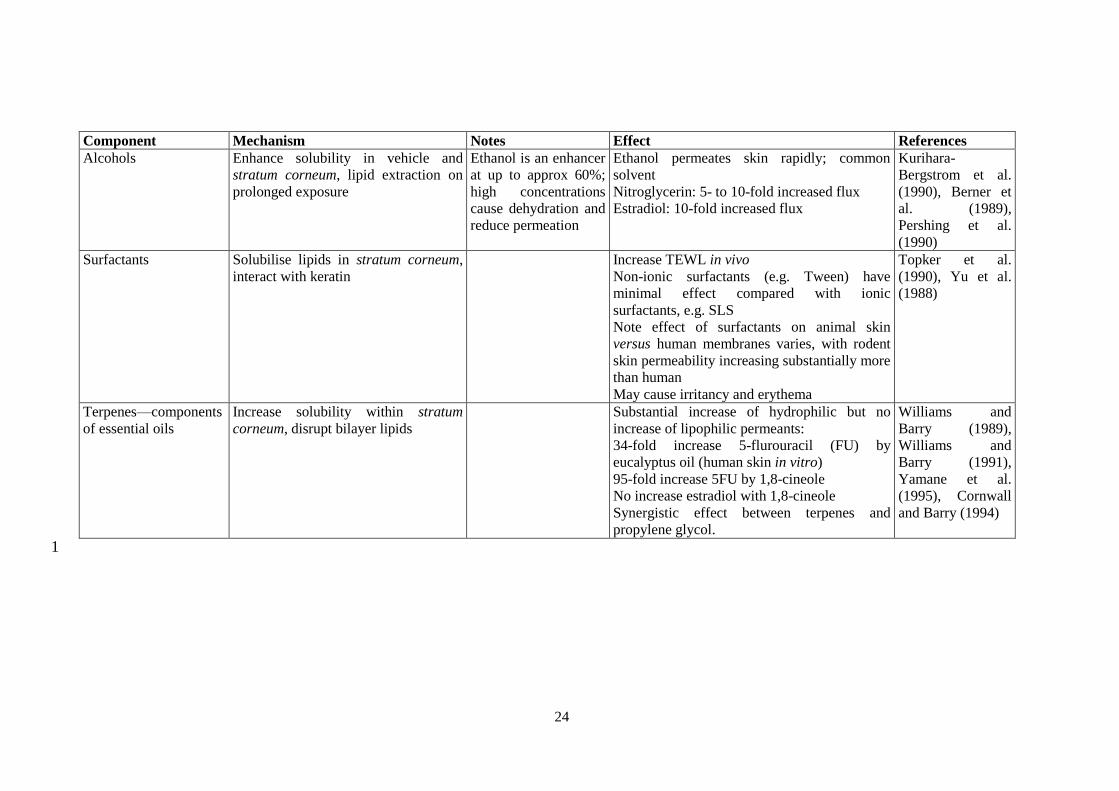
Practical guidance for using such information to estimate dermal absorption values.
4. Estimates of dermal absorption values are derived from experimental data in vivo or in vitro, or
both. Such data allow for direct or indirect estimation of dermal absorption of a test substance through
human skin. Part 1 of this document discusses issues that should be considered when evaluating such
experimental data. Part 1 also includes a discussion on combining in vivo and in vitro data in the ‘Triple
Pack’ approach. Part 2 of this document contains a general discussion on how to estimate the dermal
absorption of a chemical in the absence of experimental data.
2. SUMMARY OF RECOMMENDATIONS
Part 1
5. It is recognised that regulatory authorities around the world currently have differences in the
acceptability and use of certain types of data, and many regulatory authorities have guidance that should be
consulted where applicable. In general, all available data or relevant information are considered in a
weight-of-evidence approach to estimating a human dermal absorption value. The confidence in any
particular piece of information will determine the weight it is given in the overall risk assessment. The
guidance presented in this document will assist in evaluating the level of confidence that can be given to
any particular data.
6. In vitro studies (Section 4) should be conducted using OECD TG 428 (OECD 2004b) or a similar
protocol. In addition to providing guidance on evaluation of such studies, this section also includes
guidance on evaluating the acceptability of non-guideline studies:
Currently, regulatory authorities around the world have differences in the acceptability and use of
in vitro data. It is beyond the scope of these Guidance Notes to provide a harmonised regulatory
or scientific view on the use of in vitro data for regulatory risk assessment.
If it is to be used as ‘stand alone’ information, an isolated in vitro study on rat skin may be of
limited regulatory value because it is likely to give an overestimation of absorption, but it could

6
provide a rough estimate that could replace a worst case default value (of usually 100%) in risk
assessment.
The weight of evidence suggests that the predicted dermal absorption in humans will be
overestimated in most cases when the estimation is based on an in vitro study on human skin if
all of the test substance retained in the skin (following washing) is included.
7. In vivo studies (Section 5) should be conducted using OECD TG 427 (OECD 2004a) or a similar
protocol. Section 10.6 provides guidance on evaluating other study types, including ADME and human
in vivo studies:
As most substances have a higher permeability through rat (or rabbit) skin than through human
skin, an appropriately conducted in vivo study is unlikely to underestimate dermal absorption in
humans.
8. The term ‘Triple Pack’ refers to the combined use of three types of dermal absorption study:
1) in vivo animal; 2) in vitro animal; and 3) in vitro human (Section 6). The combined use of data from the
three studies and two testing systems offers the potential for greater accuracy in estimating human dermal
absorption because it corrects for the generally higher permeability of animal skin compared to human
skin. Application of the data to refine dermal absorption values can vary between regulatory authorities:
The ‘Triple Pack’ approach should be used to estimate a dermal absorption value only when the
three studies are conducted under the same experimental conditions.
In general, comparison of in vitro results using percentage absorption is preferred for finite dose
application, rather than flux.
9. When considering the fate of the chemical remaining in the skin at the end of a study, the existing
guidance should be consulted, in particular OECD GD 28 (OECD 2004c):
The current default approach taken by nearly all regulatory agencies is to determine the dermal
absorption value by adding the absorbed dose and the chemical remaining in the skin, following
washing. This is appropriate for both in vivo and in vitro studies, unless compelling evidence is
presented that demonstrates that some portion of the residue in the skin is unlikely to be
absorbed.
Section 7.1 should be read for guidance to assist in the consideration of whether to exclude some
portion of the residue in the skin.
10. The current test guidelines recommend that the test preparations should be the same or a realistic
surrogate to those which humans may be exposed to:
Data generated on the test substance in a preparation other than the commercial formulation
should be used only when the test preparation used in the study is very closely related to the
commercial formulation in terms of solvent, surfactant content, skin irritancy and concentration
of ingredients.
Co-formulants in the test preparation may have a significant impact on absorption, and the
outcome of a study in terms of flux or percentage absorption of the applied dose may be different
when another vehicle is used. Section 7.2 contains a table of solvents and co-formulants known
to affect dermal absorption.
7
Because of physicochemical considerations, it may be assumed that skin penetration of water-
based plant protection or biocide formulations and of solid materials (such as granules) will not
be higher than for organic solvent-based formulations of the same active compound at the same
concentration level, although there may be exceptions.
11. The anatomical location of exposure affects the dermal absorption:
Common exposure locations include abdominal or breast skin (human in vitro) or the forearm or
back (human in vivo), but other anatomical locations demonstrate greater (or lower) absorption.
The forearms and hands are potentially the areas most exposed to chemicals during occupational
use.
For some non-occupational uses of chemicals, such as topically applied insecticides or cosmetics,
which may involve application to other parts of the body, the anatomical location used in the
exposure study should be taken into account. A good discussion of differences between
anatomical locations can be found in EHC 235 (WHO 2006).
Part 2
12. In some cases, specific experimental data on dermal absorption are not available. Under such
circumstances, default values (Section 9) or alternative approaches to predict dermal absorption (Section
10) can be used:
In the absence of data, 100% dermal absorption has to be assumed to cover a ‘worst case’
scenario.
Many regulatory authorities will consider a reduction of the 100% default value to 10% if the
molecular weight is greater than 500 and logPow is either below –1 or above 4.
13. Other approaches are available to estimate dermal absorption in the absence of data. These other
approaches are outlined in Section 10 and have limited acceptability. They should be used only as a last
resort as they provide only crude estimates of dermal absorption. If approaches outlined in Section 10 are
used, the caveats described should be considered carefully, particularly if evaluating exposures to
compounds in formulations and mixtures:
‘Read-across’ is applicable only to chemicals that have been demonstrated to be very similar in
their chemical structure and physicochemical properties. Recently, the OECD has published
general guidance on read-across and the formation of chemical categories (OECD 2007a).
Modelling/QSAR is currently of limited applicability – the ‘learning set’ must contain a
reasonable number of closely related compounds. Even then, the formulated product may contain
several adjuvants and the interaction between components makes prediction in silico unreliable.
Studies used to evaluate ADME using the dermal route (e.g. using OECD TG 417; see
OECD 2010) may be used to provide an idea of the magnitude of dermal absorption and a
conservative rough estimate may be made.
Data from oral and dermal acute toxicity studies should not be used. Data from repeat dose oral
and dermal studies should be used only where there is close similarity between the two studies in
terms of design and effects seen.
8
PART 1: INTERPRETATION OF DERMAL ABSORPTION STUDIES
3. INTRODUCTION – TYPES OF DATA
14. Exposure to chemicals can occur, amongst others, through the oral, inhalation and dermal routes.
In occupational settings, it is the inhalation and dermal routes that are the major routes of exposure.
Occupational exposure to chemicals by inhalation has decreased to some extent, partly due to improved
technology to minimise the exposure. Consequently, the dermal route is considered to be the primary route
of exposure for occupational exposure to pesticides.
15. Ethical considerations have led to it becoming increasingly difficult to conduct dermal absorption
studies in humans. Therefore, risk assessors generally have to rely upon dermal absorption studies
conducted in animals or from studies using human or animal skin in vitro.
16. Most toxicity studies are conducted via the oral route, and a very limited number of studies are
conducted via the dermal and inhalation routes. For example, a pesticide for food use registration
conditionally requires (by US EPA: CFR 40 Part 158.500) only a 21-28 day dermal toxicity study, and
a 28-day inhalation toxicity study if exposure via the inhalation route is of concern for the risk assessment.
Often, the dermal toxicity studies are not suitable for risk assessment because the endpoints of concern
observed in oral studies are not evaluated in a dermal toxicity study and because of some of the limitations
of conducting the dermal toxicity study (for example, high dermal doses may lead to the ‘layering effect’).
17. As a result of these issues, it is necessary to use oral studies as a basis for estimating the risk of
exposure via dermal and inhalation routes. In order to conduct the route-to-route extrapolation, it is
important to know the dermal absorption of a chemical to estimate the internal dose. There are good
discussions on this subject available in the literature (for example, WHO 2006; EC 2004).
18. Dermal absorption studies are conducted using in vivo methods (US EPA 870.7600, OECD
TG 427) and in vitro methods (OECD TG 428). The results of the in vitro dermal absorption studies alone
are accepted for risk assessment purposes in the European Union and other countries; however, NAFTA
countries (the USA, Canada and Mexico) do not currently accept the results of in vitro studies alone for
risk assessment purposes (NAFTA Dermal Absorption Group Position Paper On Use of in vitro Dermal
Absorption Data in Risk Assessment, 2008). This is discussed further in Section 4.
19. In the absence of dermal absorption studies, risk assessors will need to estimate dermal
absorption using default assumptions and other considerations. The methods for estimating dermal
absorption in the absence of specific studies are discussed in Part 2 of the document.
4. IN VITRO DATA
4.1 Introduction
20. For the determination of dermal absorption values of chemicals for regulatory purposes, in vitro
studies can generally be used in one of the following two ways:1
1 Other scientific objectives such as investigations on partitioning of substances to the different skin layers or skin
metabolism are out of the scope of this document.

9
To compare the permeability of human and rat skin either in the same or in two separate studies
with a comparable design using finite doses. The resulting ratio can then be used to correct or
adjust the percentage of dermal absorption obtained in the rat in vivo (Section 5) provided the test
preparation was the same, and the applied concentrations were at least similar (i.e. the ‘Triple
Pack’ approach, see Section 6).
To use human skin in vitro as stand-alone data to predict the expected dermal absorption by
humans under field conditions without further conversion or correction.
21. It is generally recognised that regulatory authorities around the world currently have differences
in the acceptability and use of in vitro data. Depending on the views and decision practice of the
responsible national authorities, the use of the in vitro study on human skin may be considered as a basis
for establishing a dermal absorption value. During the European Union evaluation process for pesticides
under Directive 91/414/EEC, the dermal absorption values of many pesticides have been estimated this
way (e.g. EC 2003a; EC 2003b; EFSA 2006). However, in NAFTA countries (the USA, Canada and
Mexico), the results of the in vitro dermal absorption studies alone are not acceptable for risk assessment
purposes because it is considered that there is still too much uncertainty in results from differing protocols
to use in vitro human data as a stand-alone source of information. It is beyond the scope of these guidance
notes to provide a harmonised regulatory or scientific view on the use of in vitro data for regulatory risk
assessment.
4.2 Species selection
22. In most cases, in vitro studies on human skin or rat skin (or both) are available. Sometimes,
studies on skin obtained from other species such as monkey or pig or on artificial and cultured skin
(epidermis grown from keratinocytes) are submitted. Data show that rat, mouse and rabbit skin are
generally more permeable than human skin, but that the use of monkey or pig skin may not always result in
a conservative estimate (ECETOC 1993). Pig skin has been shown to be a good surrogate for human skin
and is commonly used in the cosmetic industry for in vitro studies. However it is noted that there are issues
of limited experience, technical problems and many uncertainties of the appropriateness of alternative
species for testing of pesticides and biocides. Specific expertise will be needed to justify the choice of such
a test system and for interpretation of the data. Accordingly, the use of such studies to provide an estimate
of dermal absorption values for the risk assessment of pesticides and biocides is not generally supported at
this time, unless sufficient justification can be provided.
23. An isolated in vitro study on rat skin without additional data may be of limited regulatory value
because it is likely to give an overestimate of absorption, but it could provide a rough estimate to replace a
worst-case default value (of mostly 100%) in risk assessment (see, for example, van Ravenzwaay and
Leibold (2004), or EHC 235 (WHO 2006)).
24. Published data give an inconsistent picture, but the weight of evidence suggests that the predicted
dermal absorption in humans will be overestimated in most cases when the estimation is based on an in
vitro study on human skin if all of the test substance retained in the skin is included. See Annex III and
EHC 235 (WHO 2006) for a review of the literature. It is generally acknowledged that a limitation of the
predictive value of in vitro data has been their high variability, which has been demonstrated in an inter-
laboratory comparison conducted in 2004 (van de Sandt et al. 2004); however, it can be expected that
increasing standardisation of experimental conditions after the adoption of OECD TG 428 (OECD 2004b)
will help to reduce this variability. It should be noted that, in other regulatory fields such as cosmetics,
dermal absorption estimates mainly rely on in vitro data.
10
25. Human skin for in vitro studies is either taken from autopsies (cadaver skin) or obtained during
cosmetic surgery. Permeability of human skin can be very different, depending on the site of body surface
from which the skin samples had been excised: for example, the forehead or scrotum are more permeable
than the back, the abdomen, the thighs or the forearms. The evaluator should, therefore, always consider
the source of the skin used in testing and the relevance to the exposure being assessed (see WHO 2006 for
a comparison of absorption from different anatomical locations).
26. During occupational exposure, less permeable body regions, such as the forearm are likely to be
exposed to a higher deposition of compounds and for a longer time interval. As these areas are more
relevant for real-world conditions, the data can be used in risk assessment. Neither the sex nor the racial
origin of the donors are considered to have a significant impact on dermal absorption (WHO 2006).
4.3 Skin samples to be used and details of the study design
27. For recently conducted studies, it can be expected, and it is generally required, that guideline
OECD TG 428 (OECD 2004) has been followed. However, because that guideline was adopted only in
2004, many studies that are to be evaluated will not be in full compliance with it. Regulators will have to
carefully check the acceptability of these studies on a case-by-case basis by comparing the study design
and the reporting quality with current requirements. Additionally, some countries have guidance for
specific groups of chemicals, for example pesticides and biocides (EC, 2004) or cosmetics (EC, 2006).
28. Crucial points might be the clear description of skin origin and preparation and the proof of skin
integrity prior to use by an appropriate method (see for example Davies et al. (2004)), temperature
(preferably around 32°C), the choice of a suitable receptor fluid (in which the test compound must be
adequately soluble, see Section 4.4), the description of the diffusion cells used, the actual area of skin
dosed, the number of cells/samples and donors, the duration of study/sampling period (preferably not more
than 24 hours for substances that penetrate the skin rapidly), and the determination of the amount retained
in skin after washing, irrespective of whether tape stripping was performed or not.
29. If the amount of chemical remaining in the treated skin has not been analysed, the study will
usually be considered as unacceptable. The dermal absorption should be given as a percentage of the
applied dose as collected during the whole sampling period and in the different compartments of the test
system (at least in receptor fluid, skin, and skin wash). More than the flux should be given in the study
report.
30. In vitro methods are designed to measure the penetration of chemicals into the skin and their
subsequent permeation across the skin into a fluid reservoir, as well as partition to the different skin layers
and possible deposition therein. Provided the excised skin sample is intact and its integrity has been proven
by appropriate methods, it can reasonably be assumed that its barrier function to what is generally a
diffusional process has been maintained in vitro. Then, in principle, the mechanism of skin penetration
may be regarded as the same as in vivo.
31. Accordingly, non-viable but intact skin can be used to investigate percutaneous absorption. In
addition, fresh, metabolically active (viable) skin can be used, but it should be recognised that many
enzymes present in the viable epidermis (e.g. P450 class) have little or no activity within a few hours
following resection (see Wilkinson and Williams 2008)). However, the latter case also allows limited
investigations on skin metabolism and its possible impact on the absorption process. According to OECD
TG 428 (OECD 2004b) and EHC 235 (WHO 2006), dermatomed (split-thickness, 200–400/500 µm) skin
can be used as well as (by heat or enzymatically) isolated epidermis (with stratum corneum) or, when
justified, full-thickness skin (consisting of stratum corneum, epidermis and dermis, up to 1000 µm thick).
Skin thickness may contribute considerably to variation in absorption, with thinner skin preparations
11
having increased flux and thicker preparations having a higher proportion of chemical retained in the skin
(Wilkinson et al. 2006). The impact of this variation is reduced if all or part of the chemical retained in the
skin is included in the dermal absorption value.
32. Both static (preferably with continuous stirring of the receptor fluid) and flow-through diffusion
cells can be used (for details, see EHC 235 (WHO 2006). The choice of occlusion or non-occlusion will
depend mainly on the properties of the test substance (for example, volatility) and sometimes also on the
exposure scenario. Non-occlusion is more likely to mimic pesticide operator exposure in the field.
33. Mostly, a finite dose experiment will be conducted since it better reflects occupational exposure.
For occupational exposure to chemicals, exposure time should be at least 6 to 10 hours before washing
with a relevant cleaning agent to remove the non-absorbed material: this time is consistent with the
duration of a normal working day. However, the study should not be terminated at this stage. Instead, a
sampling period of 24 hours is preferred as it allows the absorption process to be better characterised. That
is, the experiment should be terminated 14–18 hours after the skin has been washed.
34. Studies with a total exposure period of 24 hours are also acceptable if the skin surface is washed
at the termination of the study to remove the non-absorbed test material. This approach may be appropriate
for certain exposure scenarios, for example ‘leave-on’ cosmetic or topically applied insecticide products,
but it is likely to overestimate the exposure patterns for most agricultural or industrial uses of chemicals.
To improve the understanding of the absorption process and to allow a precise calculation of the flux,
frequent sampling of the receptor fluid should be undertaken as outlined in OECD (2004c): a total of 6–12
sampling points over 24 hours. Sampling after 8 or 10 hours is of particular importance since this value
might be used for refinement of the estimate.
35. A study duration of more than 24 hours is not recommended because skin tissue can be expected
to deteriorate. Of course, for some substances, in particular those that are lipophilic, it may take longer for
a chemical to migrate from a skin depot to the receptor fluid. From a regulatory point of view, however,
the resulting uncertainty can be readily overcome by including the amount found in the skin as potentially
absorbed (see Section 7.1 for further discussion on assessing chemicals remaining in the skin).
36. The dermal absorption value can be calculated as a percentage of the applied dose by measuring
the penetration of the test substance into the receptor fluid and the amount retained in the skin sample.
Partitioning can be described in greater detail if the different skin layers have been analysed separately. In
many studies, tape stripping is used to determine the percentage in the stratum corneum, although the
number of tape strips can vary (sometimes up to 10 or 15). There is currently some international
disagreement about whether or not part, or all, of the test substance retained in the stratum corneum should
be included in the calculation. This subject is discussed in Section 7.1.
37. To increase confidence in in vitro results, some countries have suggested the presentation of data
for reference compounds such a testosterone, caffeine, or benzoic acids that are obtained at the same
laboratory at a time that was the same or close to the dates of the study under review, but it should be noted
that OECD TG 428 does not require these reference compounds to be tested close to the study under
review.
4.4 Receptor fluid
38. The choice of receptor fluid is a very important factor while conducting in vitro dermal
absorption studies. OECD GD 28 (OECD 2004c) states that „Ideally, an estimate of the likely maximum
achieved concentration of test substance in receptor fluid should be made, based on previous in vivo or in
vitro study data. Physicochemical data or experimental results should be used to show that about 10 times

12
this concentration is achievable under the experimental conditions‟. For example, a saline solution may be
an appropriate receptor fluid for determining percutaneous absorption for hydrophilic compounds, but it is
unlikely to be appropriate for lipophilic compounds.
39. A major and frequently mentioned obstacle is the difficulty of estimating dermal absorption of
very lipophilic substances by in vitro methods. For example, Shah et al. (1989) reported the differences in
percutaneous absorption of several pesticides using the static and flow through systems. Both in vitro
methods significantly underestimated skin absorption of the highly lipophilic compounds chlordecone and
hexachlorobiphenyl. Lipophilic substances are poorly soluble in most receptor fluids, and partitioning will
be inhibited. In vivo, lipophilic compounds are readily taken up by blood once it enters the cutaneous
capillaries. The receptor fluid used in vitro should serve the same role as blood does in vivo. However,
unlike in vivo conditions, the receptor fluid volume may be more limited, particularly in static diffusion
cells. The effect of this can be minimised by use of frequent sampling (and subsequent replacement with
new receptor fluid, as should be done in studies of this type) or use of a flow-through system (USEPA
1992).
40. Studies on the penetration of the lipophilic chemical fluazifop-butyl through human epidermal
membranes showed that in vitro skin penetration results using an aqueous ethanol receptor fluid predicted
in vivo human results (Ramsey et al. 1994). However, in vitro receptor solutions consisting of tissue culture
medium and polyethylene glycol (PEG) underestimated human in vivo absorption.
41. In vitro and in vivo percutaneous absorption through rat skin has been measured for cypermethrin
(Scott and Ramsey 1987). Good agreement between absorption of cypermethrin through rat skin in vivo
and in vitro was observed when the receptor contained 50% aqueous ethanol, 6% Volpo 20, or 20% calf
serum.
42. Yang et al. (1989) compared the in vivo and in vitro percutaneous absorption of anthracene
through rat skin. Volpo-20 (6%) was added to the receptor fluid to increase the percutaneous absorption of
lipophilic compounds to mimic the in vivo absorption value.
43. This result may be due to the poor solubility of these chemicals into the receptor fluid. Bronaugh
and Stewart (1984) suggested that the lack of solubility of lipophilic compound penetrating into receptor
fluid for an in vitro method could contribute to discrepancies between in vivo and in vitro skin absorption
values.
44. Bronaugh and Stewart (1986) reported drastically low in vitro percutaneous absorption of DDT
(1.8%) and benzo(a)pyrene (BaP; 3.7%) when the receptor fluid was normal saline. However, the in vitro
percutaneous absorption was greatly enhanced for DDT (60.6%) and BaP (56%) when PEG-20 oleyl ether
was added in the receptor fluid. Additionally, the in vivo percutaneous absorption of DDT and BaP was
reported to be 69.5% for DDT and 48.3% BaP through rat skin, and the maximum absorption of cinnamyl
anthranilate was achieved when 6% PEG-20 oleyl ether was added to receptor fluid for static systems and
flow-through diffusion systems. Wester et al. (1985) reported a markedly different percutaneous
absorption value for triclocaban in human abdominal skin using a static and a flow-through system. The
relative insolubility of this compound in aqueous receptor fluid may be responsible for the discrepancy
between the results obtained in the static system (0.13-0.23%), flow-through system (6%), and in vivo
absorption value (7%).
45. The results summarised above clearly indicate that normal saline may be adequate as a receptor
fluid for hydrophilic compounds, but saline alone is likely to underestimate in vitro percutaneous
absorption of lipophilic compounds. Compounds such as anionic surfactants or other solvents must be
added to the receptor fluid in order to increase the uptake of lipophilic compounds. The addition of
13
surfactants to the receptor fluid may alter the permeability characteristics of the skin (Riley and
Kemppainen 1985), and skin integrity should be measured when such substances are added to the receptor
fluid.
46. For lipophilic compounds, the receptor fluid may contain solvent mixtures such as ethanol and
water (50% aqueous ethanol), < 6% polyoxyethelene (20) oleyl ether in water, or 5% bovine serum
albumin (Sartorelli et al. 2000; Bronaugh 2004). The use of 50% aqueous ethanol may enhance the
absorption. EHC 235 (WHO 2006) and OECD TG 428 (OECD 2004b) strongly recommend that, for
in vitro dermal absorption studies, the receptor fluid should not act as a rate-limiting step in the permeation
process due to the limited solubility of the test compound within the medium.
5. IN VIVO DATA
47. The main advantage of in vivo data is that they are generated from a physiologically and
metabolically intact system. As most substances have a higher permeability through rat (or rabbit) skin
than through human skin, this approach is unlikely to underestimate dermal absorption in humans. The
approach therefore provides an additional margin of safety. For further information and references see the
WHO/IPCS Environmental Health Criteria 235: Dermal Absorption (WHO 2006).
48. The rat is the most commonly used species for animal in vivo studies, because it is widely used in
other toxicity and toxicokinetic studies and the results are therefore directly comparable. Data from other
species (monkey and pig) may be used if skin absorption properties are more similar to those of humans
than of the rat. These two species are comparatively difficult and expensive to maintain as test species, and
there are ethical considerations for their uses.
49. There are several types of in vivo animal (rat) data that are useful for estimating the dermal
absorption value. This section describes guideline dermal absorption studies, and other types of in vivo
studies are discussed in Section 10.6.
50. Guideline studies for in vivo dermal absorption, including OECD TG 427 (OECD 2004a) and
EPA-870.7600 (USEPA 1996) produce the most comprehensive in vivo measurement of dermal absorption
because the quantities of chemical and/or its metabolites are determined throughout the animal and in the
excreta for an extended period. The guidelines require administration of the test substance in an appropriate
test preparation and in dilution(s) at realistic dose levels.
51. In vivo studies can be conducted with or without radiolabelled chemicals. Additional challenges
are present when unlabelled compounds are used and when extensive metabolism occurs without a clear
biomarker being available. Concerns about metabolism of radiolabelled chemicals are limited to the
position of the radiolabel on a potentially labile group. Further information and guidance on radiolabelling
and metabolism should be sought elsewhere, as these technical issues fall outside the scope of this
guidance.
52. The OECD TG 427 (OECD 2004a) proposes that the scheduled terminations of groups of
animals should occur at 24 hours and subsequent occasions, for example 48 and 72 hours. The EPA-
870.7600 (USEPA 1996) supplies an additional rationale for extending the number of groups to enable
data collection at longer time points:
„In some cases the difference between absorbed and retained material is sufficient to convert an
acceptable risk into an unacceptable risk if all the retained (in the skin) material is considered as
absorbed.

14
At 10 h one group from each dose is terminated ... In the remaining groups the skin is washed at 10 h,
the wash sample collected for analysis and the groups carried for one or more additional days, up to
14 days, in metabolism cages. A minimum of 14 days has been suggested by absorption data and a
maximum of 21 days has been suggested by cornified epidermal turnover time in the rat.‟
53. However, animal welfare issues mean that extending studies to the longer durations in the EPA-
870.7600 guidance is not a straightforward decision (USEPA 1996).
54. Studies conducted with extended post-application sampling can provide the following for the
duration of the study:
Blood plasma samples and residual carcass levels at multiple time points.
Eexcreta (urine, faeces, expired air) samples collected at 12- or 24-hour intervals, as this
sampling procedure enables elimination profiling.
Comparative values for the ‘reservoir effect’ at the application site (including profiling of the
residue through the stratum corneum) at different time points to enable an assessment of potential
absorption from any reservoir (the justification for the inclusion or exclusion of tape stripping is
discussed later in Section 7.1).
Determination of the rate of absorption over an extended period, including data to determine
whether the absorption has reached a plateau or may be insignificant.
55. Analysis of ALL the above factors allows the absorbed dose to be defined and permits a
justification for the exclusion of the application site. Further discussion on this can be found in Section 7.1.
56. The dermal absorption value (including or excluding the application site as appropriate) from the
final time point is generally the most appropriate regulatory value for a study conducted according to
OECD TG 427 (i.e. the value from the group sacrificed with the longest post-wash observation period).
The value from the final time point should be compared with those at other time points to ensure that the
selected value is consistent with the whole observation period. Note that if all animals are terminated at
cessation of exposure (i.e. if there is no post-application observation period included) the whole amount in
the application site skin after washing should be considered potentially absorbable (justification for the
inclusion or exclusion of tape strips is discussed later in Section 7.1).
57. If another time point is to be used, then it should be clearly justified. For example, this may be
appropriate if the final time point is clearly an outlier, or if the data have unusually high variability across
the time points (as seen in the example in Annex II).
58. Where the duration of exposure is longer than what is expected in the field (for example, a
24 hour exposure before wash-off for an agricultural pesticide), then it may not be appropriate to use the
value from the longest duration if this also represents an inappropriate exposure duration.

15
6. COMBINATION OF ANIMAL AND HUMAN IN VITRO AND HUMAN IN VIVO DATA
6.1 Introduction: the ‘Triple Pack’ approach
59. The term ‘Triple Pack’ refers to the combined use of three types of dermal absorption data from:
1) in vivo animal; 2) in vitro animal; and 3) in vitro human dermal absorption studies. The combined use of
data from the three studies and two testing systems offers the potential for greater accuracy in estimating
human dermal absorption because it corrects for animal skin generally having a higher permeability than
human skin. This produces a more reliable data set to inform the dermal risk assessment. Application of
the data to refine dermal absorption values can vary between regulatory authorities.
60. The dermal absorption estimate using data from the ‘Triple Pack’ is derived using the following
approach:
In vivo human absorption = (in vivo rat absorption) x (in vitro human absorption)
(in vitro rat absorption)
61. The following example is provided for illustration, and a further example is included in Annex II:
Example 1
62. The following hypothetical example demonstrates the approach using three studies that were
conducted using the same experimental conditions (e.g. the same test preparation and dose per square cm):
in vivo rat skin: 35%
in vitro human skin: 7%
in vitro rat skin: 49%
in vivo human dermal absorption is estimated to be 5% using ‘Triple Pack’ approach because there
is a 1:7 ratio in permeability between human and rat skin (35% x 7% / 49%)
63. Many regulatory authorities, such as the European Union, currently apply the ‘Triple Pack’
approach described above. The position of the NAFTA countries (USA, Canada) regarding triple packs is
outlined in the draft NAFTA Dermal Absorption Group Position Paper On Use of In Vitro Dermal
Absorption Data in Risk Assessment (2008). As discussion on the use of triple packs in these countries is
ongoing, further details regarding the NAFTA position will not be included in this guidance.
64. The ‘Triple Pack’ approach should be used to estimate a dermal absorption value only when the
three studies are conducted under the same experimental conditions, including using identical
concentrations of test substance applied per surface area, the same duration of exposure to skin, and the
same test preparation (for example, formulations such as emulsifiable concentrates or granules or in-use
spray dilutions).
65. The major disadvantage of the ‘Triple Pack’ approach is the ethical consideration of using large
numbers of animals and the cost of conducting these studies. The use of ‘Triple Pack’ is recommended
only when such data are already available or it is absolutely necessary to refine the dermal absorption value
due to concerns about high risk. In addition, some countries do not consider that in vitro studies are
sufficiently validated for use as part of a ‘Triple Pack’ approach for human health risk assessment
(see Section 4 for further discussion).
16
6.2 Use of the ‘Triple Pack’ approach in risk assessment
66. When valid (guideline-compliant and GLP or GLP-like) in vitro studies on human skin, in vitro
studies in animals and in vivo animal studies are available and conducted under the same experimental
conditions, then the ‘Triple Pack’ approach can be used to extrapolate the human dermal absorption values
for risk assessment.
67. The question of whether to include skin-bound residues is addressed in Section 7.1. For in vitro
studies, the OECD guideline (OECD 428) defines the ‘absorbable dose’ as ‘that present on or in the skin
following washing’. A similar approach is recommended for the in vivo studies.
68. For comparison purposes – to establish an interspecies ratio for permeability through human skin
versus rat skin – either the total absorption rate (in per cent) or the flux may be used. The flux describes the
penetration of the substance per area unit (square centimetres, cm2) and time (hours) and allows for semi-
quantitative determination of species differences. However, the main disadvantage of using the flux is that
the appearance of the test substance in the receptor fluid is the only relevant endpoint, and the amount
deposited in the different skin layers is not considered. This results in a possible underestimation of the
dermal absorption value when considering finite dose exposure. In general, the flux is not recommended
for use in the risk assessment of pesticides, and the percentage absorption is preferred (see Section 10.3).
However, if flux is used then the following should be considered:
69. The Permeability Coefficient (Kp) is a value, in units of centimetres per hour (cm/h) that
represents the rate at which a chemical penetrates the skin. This is calculated from the flux divided by the
applied concentration.
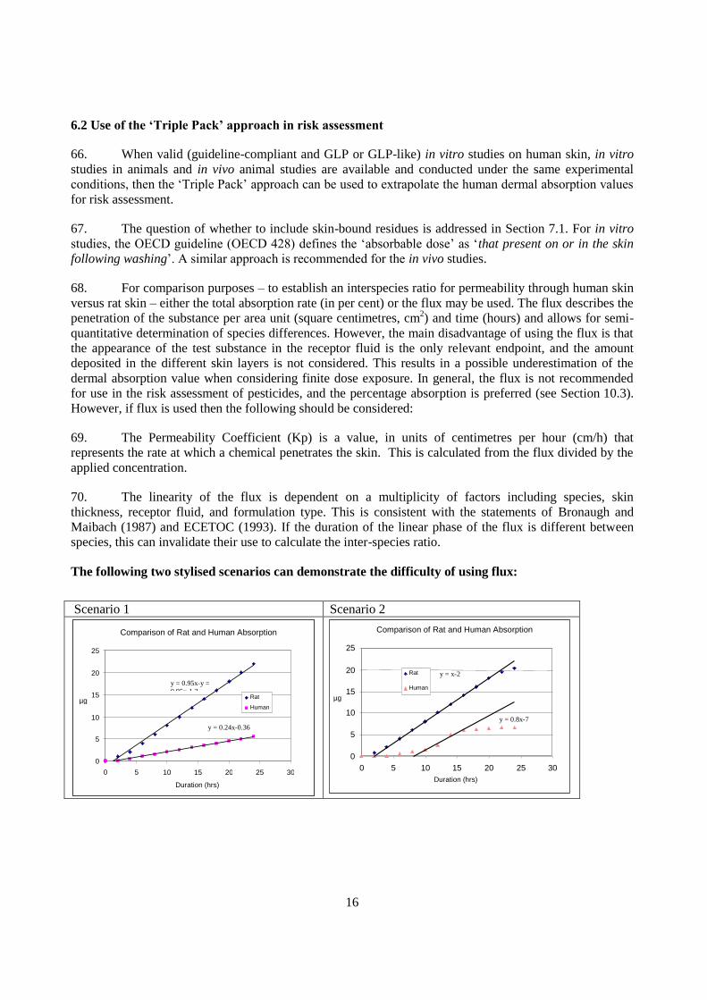
70. The linearity of the flux is dependent on a multiplicity of factors including species, skin
thickness, receptor fluid, and formulation type. This is consistent with the statements of Bronaugh and
Maibach (1987) and ECETOC (1993). If the duration of the linear phase of the flux is different between
species, this can invalidate their use to calculate the inter-species ratio.
The following two stylised scenarios can demonstrate the difficulty of using flux:
Scenario 1 Scenario 2
Comparison of Rat and Human Absorption
0
5
10
15
20
25
0 5 10 15 20 25 30 Duration (hrs)
µg
Rat Human
Comparison of Rat and Human Absorption
0
5
10
15
20
25
0 5 10 15 20 25 30 Duration (hrs)
µg Rat Human
y = 0.24x-0.36
y = 0.95x-y =
0.95x-1.2
y = x-2
y = 0.8x-7
17
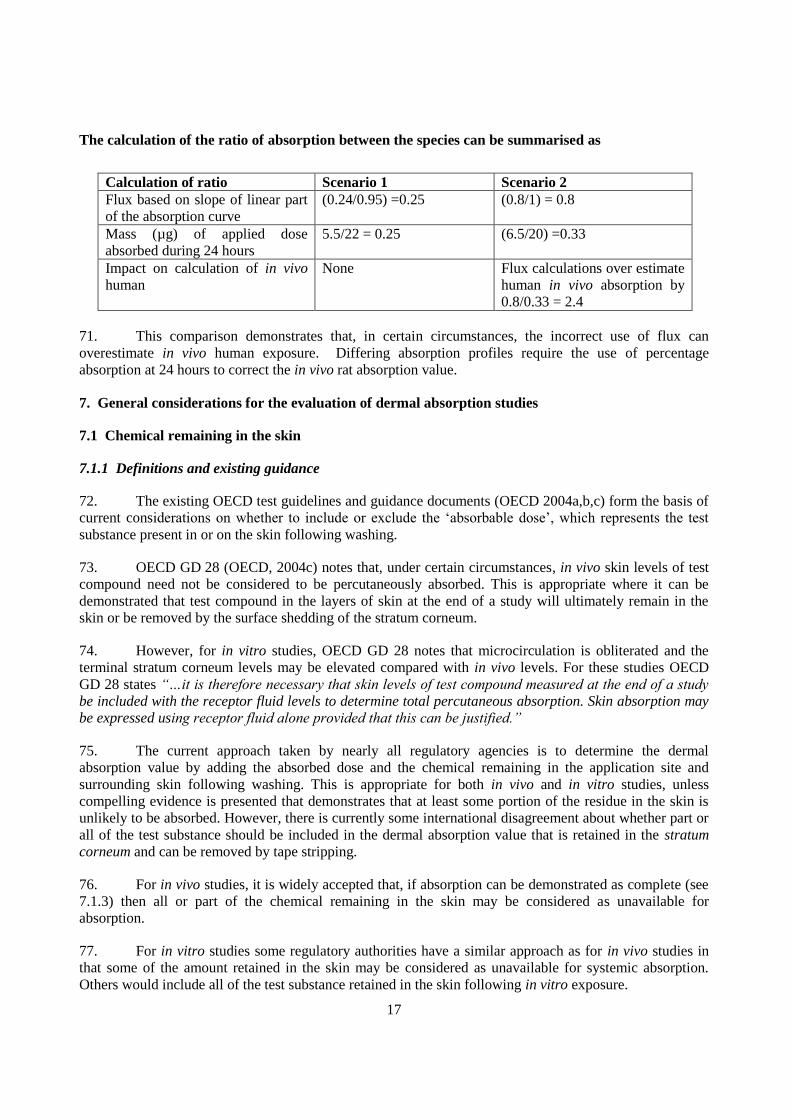
The calculation of the ratio of absorption between the species can be summarised as
Calculation of ratio Scenario 1 Scenario 2
Flux based on slope of linear part
of the absorption curve
(0.24/0.95) =0.25 (0.8/1) = 0.8
Mass (µg) of applied dose
absorbed during 24 hours
5.5/22 = 0.25 (6.5/20) =0.33
Impact on calculation of in vivo
human
None Flux calculations over estimate
human in vivo absorption by
0.8/0.33 = 2.4
71. This comparison demonstrates that, in certain circumstances, the incorrect use of flux can
overestimate in vivo human exposure. Differing absorption profiles require the use of percentage
absorption at 24 hours to correct the in vivo rat absorption value.
7. General considerations for the evaluation of dermal absorption studies
7.1 Chemical remaining in the skin
7.1.1 Definitions and existing guidance
72. The existing OECD test guidelines and guidance documents (OECD 2004a,b,c) form the basis of
current considerations on whether to include or exclude the ‘absorbable dose’, which represents the test
substance present in or on the skin following washing.
73. OECD GD 28 (OECD, 2004c) notes that, under certain circumstances, in vivo skin levels of test
compound need not be considered to be percutaneously absorbed. This is appropriate where it can be
demonstrated that test compound in the layers of skin at the end of a study will ultimately remain in the
skin or be removed by the surface shedding of the stratum corneum.
74. However, for in vitro studies, OECD GD 28 notes that microcirculation is obliterated and the
terminal stratum corneum levels may be elevated compared with in vivo levels. For these studies OECD
GD 28 states “…it is therefore necessary that skin levels of test compound measured at the end of a study
be included with the receptor fluid levels to determine total percutaneous absorption. Skin absorption may
be expressed using receptor fluid alone provided that this can be justified.”
75. The current approach taken by nearly all regulatory agencies is to determine the dermal
absorption value by adding the absorbed dose and the chemical remaining in the application site and
surrounding skin following washing. This is appropriate for both in vivo and in vitro studies, unless
compelling evidence is presented that demonstrates that at least some portion of the residue in the skin is
unlikely to be absorbed. However, there is currently some international disagreement about whether part or
all of the test substance should be included in the dermal absorption value that is retained in the stratum
corneum and can be removed by tape stripping.
76. For in vivo studies, it is widely accepted that, if absorption can be demonstrated as complete (see
7.1.3) then all or part of the chemical remaining in the skin may be considered as unavailable for
absorption.
77. For in vitro studies some regulatory authorities have a similar approach as for in vivo studies in
that some of the amount retained in the skin may be considered as unavailable for systemic absorption.
Others would include all of the test substance retained in the skin following in vitro exposure.

18
78. The following sections provide guidance to assist in the consideration of whether to exclude
some portion of the residue in the skin.
7.1.2 Tape stripping
79. OECD GD 28 states that skin fractionation may be conducted following exposure either in vitro
or in vivo, noting that tape stripping can be difficult in vitro with epidermal membranes, rodent skin, study
durations of more than 24 hours, or where the test preparation alters the stratum corneum.
80. Test substance retained in the top few layers of the stratum corneum (i.e. contained in the first
few tape strips) may be removed by desquamation and therefore may not be absorbed. This includes
substances retained in the top few layers of the stratum corneum as well as material that has not penetrated
into the stratum corneum but is protected from wash-off, for example in hair follicles or sweat ducts.
81. In the European Union and some other countries, it is general practice to exclude the amount that
was found in the first (upper) two tape strips at study completion both in vitro and in vivo.
82. Test substance in lower layers of the stratum corneum may penetrate into the dermis, or may be
removed by desquamation, and determination of the potential bioavailability of this test substance should
be made on a case-by-case basis.
83. Dermal absorption is primarily a diffusion-driven process, and therefore test substance in the
lower layers of the stratum corneum should be assumed to form a reservoir that may become systemically
available, unless it can be demonstrated in vivo that absorption is complete and this test substance will
remain in the stratum corneum until exfoliated (see 7.1.3).
84. In many studies conducted to date, separate analysis of the individual tape strips for radioactivity
has not been performed. Instead, all tape strips are pooled before measurement. In this case the whole
amount in the stratum corneum, as well as all the material retained in deeper layers, is generally considered
absorbable and should be included in the calculation of the dermal absorption value (unless it has been
demonstrated that absorption is complete). This highlights the importance of conducting a separate analysis
of each tape strip rather than pooling the strips. However, any such analysis should address potential
confounding factors such as those described in EHC 235 (WHO 2006).
7.1.3 Completion of absorption in vivo
85. Following an in vivo animal study, the ‘absorbable dose’ represents the amount of chemical
present on or in the skin following washing. The following examples are provided as guidance on whether
to include or exclude this absorbable dose in the calculation of the dermal absorption value:
1. In cases where an in vivo study is terminated just after cessation of exposure, there is no
chance to determine the fate of chemical remaining in the skin, and it should be assumed that
the dose remaining at the application site, including all material in the stratum corneum, is
available for absorption.
2. If during an in vivo animal study there is significant ongoing depletion of the dose from the
application site following washing and a corresponding increase in cumulative absorbed dose
over time, the dose remaining at the application site, including all material in the stratum
corneum (perhaps excluding the upper two tape strips), is considered to be available for
further skin absorption.

19
3. Where data show serial ‘non detects’ in excreta, then this indicates that chemical remaining in
the skin at the application site (including the stratum corneum) may be unavailable for further
absorption. This serial ‘non-detects’ approach is appropriate only in those cases where the
limit of detection is small in comparison to the amount excreted following wash-off – this
guidance exists to avoid providing benefits to studies with less sensitive limits of detection.
4. An in vivo dermal absorption study can be considered to have demonstrated completion of
absorption if 75% of the material absorbed by the end of the study (material in
excreta + exhaled gasses + the carcass excluding application site) is present in the excreta or
systemic compartment before the mid-point of the study. The reason for this approach is that
75% represents two half-times. If 75% of the absorption occurs within half of the study
duration, the total study duration should cover four half-times. Four half-times will cover
more than 93% of the potential absorption under normal (single) exponential conditions. In
this case, the bioavailability of any material remaining at the application site may be
considered to have a minimal impact on the overall conclusion for the percentage absorbed.
All material remaining in the skin at the application site (including the stratum corneum) may
be excluded from the amount absorbed.
Figure 1: Examples of representative absorption vs time profiles
Dermal absorption vs time curves
0%
10%
20%
30%
40%
50%
60%
70%
80%
90%
100%
0% 50% 100%
Time
Ab
sorp
tio
n
(as
a p
erce
nta
ge o
f th
e t
ota
l)
Absorption complete Absorption tapers off
Absorption not complete
20
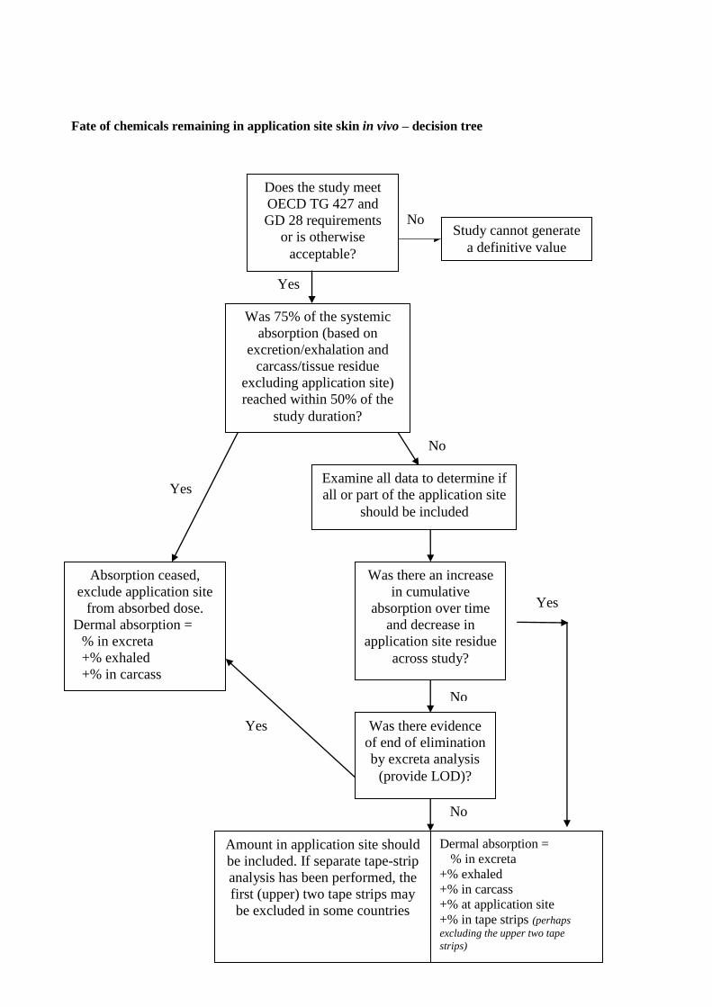
Fate of chemicals remaining in application site skin in vivo – decision tree
Yes
Yes
No
No
No
Yes
No
Yes
Does the study meet
OECD TG 427 and
GD 28 requirements
or is otherwise
acceptable?
Was 75% of the systemic
absorption (based on
excretion/exhalation and
carcass/tissue residue
excluding application site)
reached within 50% of the
study duration?
Study cannot generate
a definitive value
Absorption ceased,
exclude application site
from absorbed dose.
Dermal absorption =
% in excreta
+% exhaled
+% in carcass
Examine all data to determine if
all or part of the application site
should be included
Was there an increase
in cumulative
absorption over time
and decrease in
application site residue
across study?
Was there evidence
of end of elimination
by excreta analysis
(provide LOD)?
Amount in application site should
be included. If separate tape-strip
analysis has been performed, the
first (upper) two tape strips may
be excluded in some countries
Dermal absorption =
% in excreta
+% exhaled
+% in carcass
+% at application site
+% in tape strips (perhaps
excluding the upper two tape
strips)
21
7.2 Effect of formulation
7.2.1 Test preparations
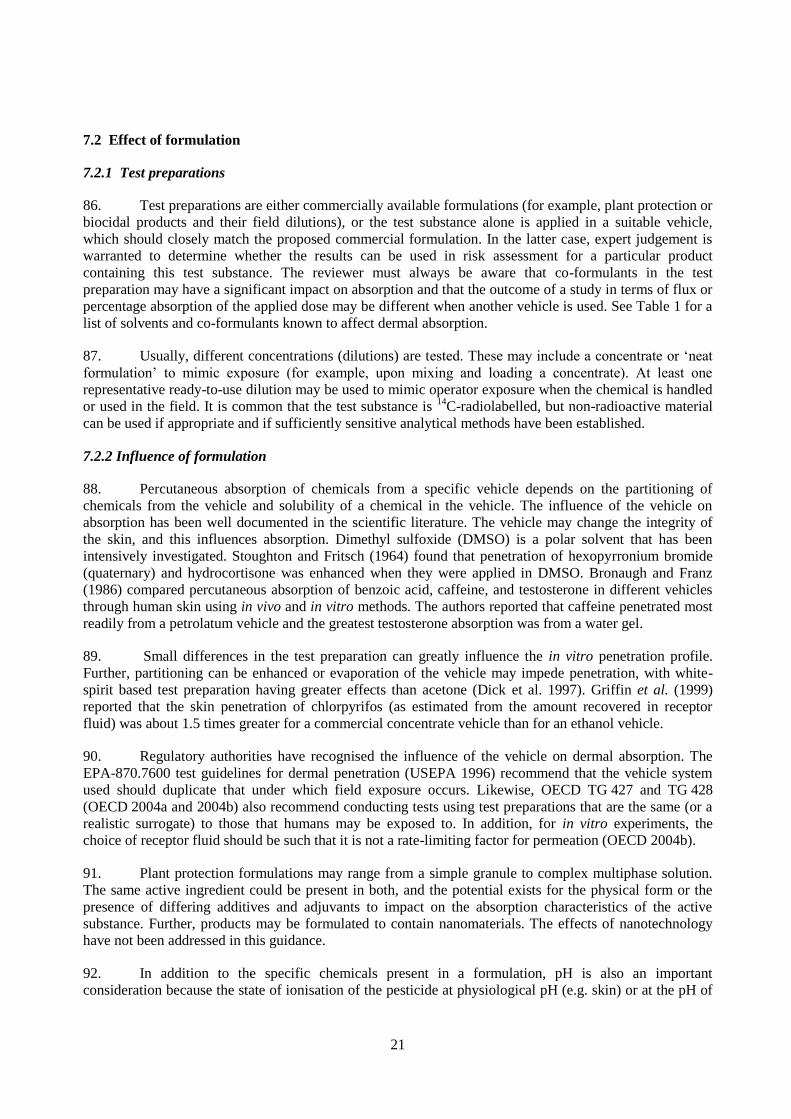
86. Test preparations are either commercially available formulations (for example, plant protection or
biocidal products and their field dilutions), or the test substance alone is applied in a suitable vehicle,
which should closely match the proposed commercial formulation. In the latter case, expert judgement is
warranted to determine whether the results can be used in risk assessment for a particular product
containing this test substance. The reviewer must always be aware that co-formulants in the test
preparation may have a significant impact on absorption and that the outcome of a study in terms of flux or
percentage absorption of the applied dose may be different when another vehicle is used. See Table 1 for a
list of solvents and co-formulants known to affect dermal absorption.
87. Usually, different concentrations (dilutions) are tested. These may include a concentrate or ‘neat
formulation’ to mimic exposure (for example, upon mixing and loading a concentrate). At least one
representative ready-to-use dilution may be used to mimic operator exposure when the chemical is handled
or used in the field. It is common that the test substance is 14
C-radiolabelled, but non-radioactive material
can be used if appropriate and if sufficiently sensitive analytical methods have been established.
7.2.2 Influence of formulation
88. Percutaneous absorption of chemicals from a specific vehicle depends on the partitioning of
chemicals from the vehicle and solubility of a chemical in the vehicle. The influence of the vehicle on
absorption has been well documented in the scientific literature. The vehicle may change the integrity of
the skin, and this influences absorption. Dimethyl sulfoxide (DMSO) is a polar solvent that has been
intensively investigated. Stoughton and Fritsch (1964) found that penetration of hexopyrronium bromide
(quaternary) and hydrocortisone was enhanced when they were applied in DMSO. Bronaugh and Franz
(1986) compared percutaneous absorption of benzoic acid, caffeine, and testosterone in different vehicles
through human skin using in vivo and in vitro methods. The authors reported that caffeine penetrated most
readily from a petrolatum vehicle and the greatest testosterone absorption was from a water gel.
89. Small differences in the test preparation can greatly influence the in vitro penetration profile.
Further, partitioning can be enhanced or evaporation of the vehicle may impede penetration, with white-
spirit based test preparation having greater effects than acetone (Dick et al. 1997). Griffin et al. (1999)
reported that the skin penetration of chlorpyrifos (as estimated from the amount recovered in receptor
fluid) was about 1.5 times greater for a commercial concentrate vehicle than for an ethanol vehicle.
90. Regulatory authorities have recognised the influence of the vehicle on dermal absorption. The
EPA-870.7600 test guidelines for dermal penetration (USEPA 1996) recommend that the vehicle system
used should duplicate that under which field exposure occurs. Likewise, OECD TG 427 and TG 428
(OECD 2004a and 2004b) also recommend conducting tests using test preparations that are the same (or a
realistic surrogate) to those that humans may be exposed to. In addition, for in vitro experiments, the
choice of receptor fluid should be such that it is not a rate-limiting factor for permeation (OECD 2004b).
91. Plant protection formulations may range from a simple granule to complex multiphase solution.
The same active ingredient could be present in both, and the potential exists for the physical form or the
presence of differing additives and adjuvants to impact on the absorption characteristics of the active
substance. Further, products may be formulated to contain nanomaterials. The effects of nanotechnology
have not been addressed in this guidance.
92. In addition to the specific chemicals present in a formulation, pH is also an important
consideration because the state of ionisation of the pesticide at physiological pH (e.g. skin) or at the pH of

22
the formulation will affect the overall net charge, which influences the ability to cross hydrophobic
membrane barriers such as skin.
93. The dermal absorption value of a test substance in a highly diluted product as measured in valid
experiments could be used to estimate skin penetration of a formulation that is of the same composition but
less diluted because, in many cases, the percentage dermal absorption from a less concentrated product is
higher and thus provides a conservative estimate for a more concentrated product. However, a recent study
by Buist et al. (2009) questioned this assumption, particularly for skin-irritating and volatile substances.
The study compared the results of published dermal absorption experiments for a total of 98 different
substances. In each study, more than one dilution was tested. The widely assumed principle of dermal
absorption value increasing with higher dilutions was confirmed for fewer than two thirds of the
138 dermal absorption experiments under review. In particular, less frequently seen was the expected
inverse relationship between dermal absorption and dermal loading for known skin-irritating and volatile
substances.
94. The following table lists some solvents that have been shown to increase the penetration of
certain chemicals. Care should be taken when a chemical is presented in a new formulation that contains
these solvents, and this may be a case where in vitro studies are particularly useful to bridge across
formulations. However, it should be noted that the effect of any particular solvent on any particular
chemical could not be easily predicted, with many differences not easily explained by a simple
classification into hydrophilic or hydrophobic chemicals.
23
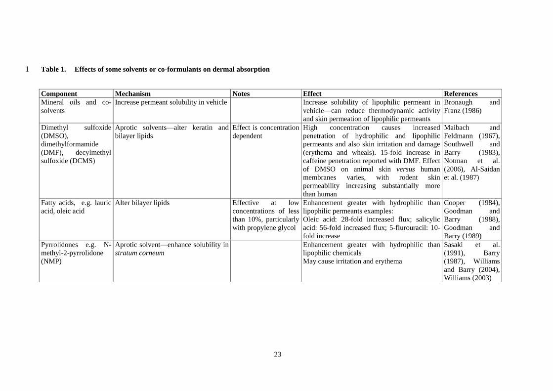
Table 1. Effects of some solvents or co-formulants on dermal absorption 1
Component Mechanism Notes Effect References
Mineral oils and co-
solvents
Increase permeant solubility in vehicle Increase solubility of lipophilic permeant in
vehicle—can reduce thermodynamic activity
and skin permeation of lipophilic permeants
Bronaugh and
Franz (1986)
Dimethyl sulfoxide
(DMSO),
dimethylformamide
(DMF), decylmethyl
sulfoxide (DCMS)
Aprotic solvents—alter keratin and
bilayer lipids
Effect is concentration
dependent
High concentration causes increased
penetration of hydrophilic and lipophilic
permeants and also skin irritation and damage
(erythema and wheals). 15-fold increase in
caffeine penetration reported with DMF. Effect
of DMSO on animal skin versus human
membranes varies, with rodent skin
permeability increasing substantially more
than human
Maibach and
Feldmann (1967),
Southwell and
Barry (1983),
Notman et al.
(2006), Al-Saidan
et al. (1987)
Fatty acids, e.g. lauric
acid, oleic acid
Alter bilayer lipids Effective at low
concentrations of less
than 10%, particularly
with propylene glycol
Enhancement greater with hydrophilic than
lipophilic permeants examples:
Oleic acid: 28-fold increased flux; salicylic
acid: 56-fold increased flux; 5-flurouracil: 10-
fold increase
Cooper (1984),
Goodman and
Barry (1988),
Goodman and
Barry (1989)
Pyrrolidones e.g. N-
methyl-2-pyrrolidone
(NMP)
Aprotic solvent—enhance solubility in
stratum corneum
Enhancement greater with hydrophilic than
lipophilic chemicals
May cause irritation and erythema
Sasaki et al.
(1991), Barry
(1987), Williams
and Barry (2004),
Williams (2003)
24
Component Mechanism Notes Effect References
Alcohols Enhance solubility in vehicle and
stratum corneum, lipid extraction on
prolonged exposure
Ethanol is an enhancer
at up to approx 60%;
high concentrations
cause dehydration and
reduce permeation
Ethanol permeates skin rapidly; common
solvent
Nitroglycerin: 5- to 10-fold increased flux
Estradiol: 10-fold increased flux
Kurihara-
Bergstrom et al.
(1990), Berner et
al. (1989),
Pershing et al.
(1990)
Surfactants Solubilise lipids in stratum corneum,
interact with keratin
Increase TEWL in vivo
Non-ionic surfactants (e.g. Tween) have
minimal effect compared with ionic
surfactants, e.g. SLS
Note effect of surfactants on animal skin
versus human membranes varies, with rodent
skin permeability increasing substantially more
than human
May cause irritancy and erythema
Topker et al.
(1990), Yu et al.
(1988)
Terpenes—components
of essential oils
Increase solubility within stratum
corneum, disrupt bilayer lipids
Substantial increase of hydrophilic but no
increase of lipophilic permeants:
34-fold increase 5-flurouracil (FU) by
eucalyptus oil (human skin in vitro)
95-fold increase 5FU by 1,8-cineole
No increase estradiol with 1,8-cineole
Synergistic effect between terpenes and
propylene glycol.
Williams and
Barry (1989),
Williams and
Barry (1991),
Yamane et al.
(1995), Cornwall
and Barry (1994)
1

25
7.2.3 Solid vs. liquid formulations
95. In occupational situations, workers may be exposed to chemicals through different formulations
such as emulsifiable concentrates, granules, wettable powders and adjuvants. Because of physicochemical
considerations, it may be assumed that skin penetration of water-based plant protection or biocide
formulations or of solid materials (such as granules) will not be higher than for organic solvent-based
formulations of the same active compound at the same concentration level, although there may be
exceptions. Provided there are no further co-formulants contained that might alter dermal uptake,
experimental data obtained with a solvent-based test preparation may be considered as a ‘worst case’.
Accordingly, these study results could be rounded to the closest figure (such as 10% or 25%) to give an
estimate for dermal absorption of the same or a comparable dilution of a water-based test preparation or of
granules or powder.
7.3 Metabolism in the skin
96. Skin plays an important role in the metabolism of endogenous chemicals such as carbohydrates,
lipids, proteins, and steroid hormones; and it plays an important role in the metabolism of exogenous
compounds. The highest metabolising capability of the skin is observed in the epidermis layer of the skin
and pilosebaceous glands. All of the major enzymes that are important for metabolism in the liver and
other tissues have been identified in the skin (Pannatier et al. 1978).
97. On a body-weight basis, Phase I metabolism (such as oxidation, hydrolysis, reduction) in the skin
is only a small fraction (2%) of that in the liver, but its importance should not be underestimated (Rice and
Cohen 1995). Skin metabolism can be extensive because of the large surface area and volume of the skin.
Mukhtar and Bickers (1981) reported that the activity of arylhydrocarbon hydroxylase (P450) activity in
the skin exceeds 20% of that in the whole body when neonate rats were dermally treated with
benzo(a)pyrene or Aroclor 1254.
98. The Phase II (such as conjugation, detoxification) metabolism capability in the skin has also been
demonstrated. Mukhtar and Bickers (1981) reported that the glutathione S-transferase activity in skin
cytosol was 15% of the corresponding hepatic activity in the neonatal rats dermally treated with
benzo(a)pyrene or Aroclor 1254. For more detailed discussions on metabolic activity of the skin, see
reviews by Kao and Carver (1990), Hotchkiss (1998), Hewitt et al. (2000), Bronaugh (2004a and 2004b)
and WHO (2006).
99. Metabolism processes can certainly alter the in vivo absorption of a chemical through the skin.
The influence of metabolism is much less significant for in vitro experiments due to lack of skin viability
reduced physiological functioning. Metabolism may not be an important consideration if the compound
remains in the stratum corneum. However, it becomes an important factor for lipophilic compounds that
cross the stratum corneum. Metabolism processes in the dermis can make the lipophilic compound more
hydrophilic and enhance the penetration of a chemical through the skin.
100. Where skin absorption data is used for risk assessment, metabolism is usually not a critical factor
in interpreting the data. This is because the total percentage penetration of a compound is usually
considered for the risk assessment. In addition, it is generally recommended that penetration studies be
conducted with radiolabelled compounds to increase the sensitivity of the method used for absorption
measurements. It is also recommended that the radio labelling position should be such that it is not easily
labile and follows the major portion of the compound.
101. In summary, the skin is a vital organ of the body containing major Phase I and Phase II
metabolising capabilities. Knowledge of the metabolism will enhance the interpretation of the dermal

26
absorption study results. If the intent of the study is to determine the extent of absorption for the risk
assessment then metabolism may not a critical factor.
7.4 Mass balance
102. OECD TG 427 and 428 (OECD 2004a and 2004b) require a mean mass balance recovery of the
test substance of between 90–110 % and the OECD GD28 (OECD 2004c) contains the same
recommendation, with a caveat that for volatile test substances and unlabelled test substances, a range of
80–120% is acceptable. However, with the in vivo study design, recoveries outside this range may be
acceptable but must be justified.
103. The criteria to justify absorption values outside the recovery acceptance range can be summarised
by the following examples:
1. Recovery values exceed the recommended range: If the recoveries exceed the accepted
maximum range, the data generated should not be normalised because that would result in
potentially underestimated absorption values. If these absorption values are not acceptable when
a risk assessment is conducted, then the study should be repeated to address any bias resulting
from excessive recoveries.
2. Recovery values below the recommended range: Low recoveries raise the concern that the value
for absorbed dose could be lower than that which would be achieved from a study where the
recoveries were within the guideline range. The reason for low recovery may be attributable to
the following factors:
incomplete application of dose
loss to the experimental equipment
incomplete extraction from matrices (or incomplete collection of exhaled CO2)
evaporation
unlabelled test preparations, metabolism or degradation.
104. If the low recovery is associated with losses of non-absorbed material, this will have minimal
impact of the value for absorption. If the low recovery is associated with losses from absorbed material,
correction for the recovery should be made. If the results from some individual replicates (animals or
in vitro test unit) show adequate recovery, then these can be compared with the low recovery replicates to
see if the losses arise from absorbed or non-absorbed material.
105. The potential impact of low recoveries on the amount absorbed needs to be evaluated. In cases
where the measured dermal absorption is low (e.g. less than 10%) then the low recovery may have a
greater proportional impact on the value used for risk assessment. Inclusion or exclusion of the ‘missing’
percentage should be considered on a case-by-case basis in the context of the study (i.e. what was
measured or collected and known limitations). For example, if the low recovery was due to incomplete
collection of exhaled CO2, then correction for recovery could be performed by assuming that all the
missing radioactivity could have been absorbed.

27
106. If the recovery rates are consistently below the recommended range across test animals, and the
fate of the unrecovered material is unclear, there are two potential approaches to normalise the data, both
of which could be applied on a case-by-case basis:
One approach would be to normalise the measured dermal absorption value. For example, if the
measured dermal absorption was 10% of the applied dose, and the recovery rate was 70%, then
normalisation of the measured absorption (10%) by the recovery ratio (100/70) would obtain a
new estimate of the dermal absorption (15%).
The other approach would be to include all the unrecovered material in the amount that is
potentially absorbed. Using the example above, the measured absorption (10%) would be added
to the unrecovered material (30%) to obtain an estimate of 40% absorption. For use in risk
assessment, this value might be flagged as a worst-case assumption.
7.5 Use of mean or centiles; treatment of outliers and use of rounding
7.5.1 Introduction
107. Results of in vivo and in vitro dermal absorption studies can produce results that exhibit a degree
of variability that is greater than that seen in many other types of studies used in human health risk
assessments. This variability does not necessarily indicate poor experimental technique – it can be
indicative of the physiological and biochemical inter-individual variability that exists in dermal absorption
processes. There are several factors that might contribute to this, such as differences between donors of
human skin samples, and slight damage to skin samples and application sites during preparation. When
there is a high degree of variability, the appropriateness of using the mean value can be questioned, as
could the overall ability to interpret the results in a consistent and meaningful manner. The guidance below
is aimed at providing a simple pragmatic approach that takes into account the data values normally
presented in dermal absorption studies conducted according to OECD TG 427 and 428 (OECD 2004a and
2004b).
7.5.2 Variability is relatively low
108. For values that are used to calculate the dermal absorption of an individual study where the
standard deviation is less than the mean value, the mean value should be used.
7.5.3 Variability is high
109. If there is significant variation between replicates (the standard deviation is equal to or greater
than the mean) consideration should be given to using a value other than the mean or possibly rejecting the
study entirely. Consideration should be given to outliers, and in particular the relative distribution of the
test substance through the skin. Where interindividual variation is high (i.e. the standard deviation is equal
to or greater than the mean) and group size is four or less (n ≤ 4), the dermal absorption should be quoted
as a range rather than an average, and consideration should be given to using the higher value in the range,
rather than the average when conducting the risk assessment. For larger group sizes, the addition of a
standard deviation to the mean value for absorption would give a value that covered the upper
87th percentile value of the results (Chebyshev’s theorem). Such an approach would be reasonably
conservative and could reduce the need to repeat studies (particularly in vivo ones).
110. Where results with large variability are applicable to values used to compare relative absorption
through rat and human skin in the ‘Triple Pack’ approach, a conservative method should be used.
For example, if the rat value had high variability, then the mean value should be used in determining the
28
ratio; if the variability was high for the human data, the standard deviation should be added to the mean
value.
7.5.4 Outliers
111. If any results are excluded as outliers (either in the preparation of the study report or by the
regulatory evaluator), the reasons should be clearly stated in the study report and summary text. In
addition, the full results from the samples considered to be outliers must be presented. It should be noted
that consideration of results treated as outliers should include spuriously low values as well as high ones.
7.5.5 Rounding
112. To avoid a false impression of accuracy on the results of a study: dermal absorption values of
more than 10% should be rounded to two significant figures, and values between 1% and 10% should be
rounded to one significant figure. There is currently some international disagreement on whether values of
less than 1% should be rounded up to 1%. The rounding should be applied only to the final calculated
value and not to intermediate values used in the calculation. For example, 8.7% becomes 9%; and 22.3
becomes 22%.
Example 3
113. The available dermal absorption studies of a hypothetical fungicide, all with low interindividual
variability, were assessed with the following results:
rat in vivo study 0.3% including application site residue
rat in vitro study 1.2%
human in vitro study about 0.2%
114. The consistency of the data from this example indicate that dermal absorption is likely to be
significantly less than 1% and rounding up to 1% is not justified. For this example, if the triple pack
approach is followed, a value of 0.05% is obtained (1:6 ratio of human: rat), which would be rounded up to
0.1%. If the rat in vivo study were used in isolation then the value would be 0.3%.
7.6 The ‘wash-in’ effect
115. The ‘wash-in’ effect refers to the enhanced absorption that may occur by washing skin for
cleansing purposes (Moody and Maibach 2006). This effect was reported for several pesticides in a series
of literature reports involving in vitro tests with guinea pig, rat, human, and human tissue culture skin. For
example, up to 32-fold enhanced penetration into the receptor solution of the insect repellent diethyl-m-
toluamide (DEET) was reported for excised human skin following soap washing (Moody and Maibach,
2006). Particularly in cases where an in vitro test is terminated by a skin washing procedure (no post-wash
sampling), consideration should be given to enhanced ‘wash-in’ absorption.
116. The mechanism(s) underlying the ‘wash-in’ effect is not fully understood, but it includes the
effects of the washing method itself, such as those involving the type of soap or cleanser used, the duration
and friction or pressure exerted on the skin surface, and possibly artefacts of in vitro methods, including
those related to the skin specimen (such as animal species, anatomic site, skin hydration and pH) (Moody
and Maibach, 2006).

29
117. As long as the skin depot is considered to be fully bioavailable, a ‘wash-in’ effect may not be as
important. However, as ‘wash-in’ may lead to a rapid release or ‘burst’ of chemical to blood, the
dermatotoxicokinetics should still be considered.
118. For in vivo studies conducted according to OECD TG 427 (OECD 2004a), the treated skin is
washed with a cleaning agent at the end of the exposure period, and excreta are collected (usually for a
number of days). Such protocols would include any chemical that has been made more bioavailable by
washing. However, for protocols terminated by skin soap washing (no sampling occurring after washing)
the potential of enhanced absorption needs to be considered. For these protocols, it is prudent to include all
chemical remaining in the skin as potentially absorbable. In all cases, irrespective of whether the study was
conducted by in vitro or in vivo methods, the skin washing method needs to be fully described. The
OECD Test Guidelines 417 and 418 (OECD 2004a,b) should also be followed to ensure the soap or other
cleansing agent used is relevant to the exposure scenario being modelled.
30
PART 2: ESTIMATION OF DERMAL ABSORPTION IN THE ABSENCE OF SPECIFIC
STUDIES
8. INTRODUCTION
119. If available, specific experimental data should be used to determine the dermal absorption value
for a chemical in a particular test preparation. However, in many cases, such information does not exist, is
not applicable (for example, because a certain formulation is not similar enough to the test preparation in
the available studies), or cannot be used because of data protection. Under such circumstances, either
default values must be used or alternative approaches to predict dermal absorption should be used. Some
methods have been developed for this purpose and were found to be more or less useful for chemicals of
interest. In contrast, their applicability to formulations (products with often more than one chemical of
interest, and various co-formulants such as solvents and surfactants) is generally limited or unclear.
9. DEFAULT VALUES
120. As a first step, a default value should be used in exposure calculations. Usually, to cover a ‘worst
case’ scenario, 100% dermal absorption has to be assumed. This conservative approach will result, in
almost all cases, in a clear overestimation of actual absorption that provides a high level of protection. In
practice, regulatory agencies will not ask for further data if, by calculating with the default value of 100%
for dermal absorption, the intended use is considered as acceptable.
121. The physicochemical properties of a substance have a major impact on its dermal penetration.
Thus, for example, it is widely assumed that for large molecules and those with either a very low or a very
high octanol water partition coefficient (logPow), the skin is much less permeable than it would be for other,
smaller molecules. Many authorities, particularly in Europe, consider this factor by reducing the 100%
default value to only 10% if the molecular weight is greater than 500 and logPow is either below –1 (that
will suggest a high affinity for the aqueous phase) or above 4 (which is associated with high lipophilicity).
This approach is explicitly mentioned in the European Union guidance document on dermal absorption
(EU 2004; based on proposals by De Heer et al. 1999) and can be considered current practice in most
European Union member states. However, the final scientific proof for neither this general assumption nor
the arbitrary default numeric value of 10% has been provided. This is especially the case for chemicals in
complex mixtures. Other countries may have different approaches and policies. In some countries the use
of information on physicochemical properties to reduce a default dermal absorption factor should be
corroborated with data from oral and dermal toxicity studies (Section 10.5).
122. Other physicochemical factors could also be important, but they have so far been used only
occasionally (or not at all) for regulatory purposes. Examples of such factors include the physical state of
the test preparation (for example, liquid, granules or powder), solubility in water and non-polar solvents,
melting point, ionogenic state/pKa or dipole moment, lipophilicity, hydrogen bonding donor/acceptor
potential, vapour pressure, surface tension, or corrosive properties due to extreme pH.
123. Further research is needed to establish clear relationships between physicochemical properties of
test substances and dermal absorption. Such studies might open up new ways towards a more scientifically
based choice of the appropriate default value in future.
124. Alikhan et al. (2009) tried to correlate dermal absorption in humans in vivo, based on urinary
excretion, with physicochemical parameters such as molecular weight, melting point, water solubility or
hydrogen bonding properties. The study examined a relatively small group of 12 pesticides from different
classes (with some of them being no longer in use). Only a nearly significant inverse correlation between

31
urinary excretion and molecular weight was found. In contrast, all the other physicochemical properties
failed to provide any explanations for the extreme differences in dermal absorption, as indicated by the
wide range (between 0.3% and 73.9% of the administered dose) that was found in urine. A similar picture
was obtained for a group of 21 ‘miscellaneous organic compounds’, and in that part of the study, molecular
weight was not a predictor for dermal absorption. In contrast, for a chemically more homogenous group of
15 steroids, a possible impact of molecular weight, melting point and hydrogen bonding properties (in
particular, the number of hydrogen acceptor groups on the molecule) could be shown. These results
underline the importance of choosing the appropriate chemical domain (see also the Section 10.2 below).
125. In addition to the use of the 100% or 10% default values, it has been sometimes argued that
dermal absorption cannot exceed the oral absorption rate. Accordingly, regulatory agencies might conclude
that dermal absorption of a certain substance for which no experimental data are available can be assumed
to have, for example, a maximum value of 60% if this percentage had been established for the oral
absorption rate in an ADME study. Further work is required to scientifically validate this assumption.
According to the European Union guidance document on dermal absorption, a direct comparison was
confined to only 12 pesticides and the data had not been published (EU 2004). However, based on practical
experience, it is very likely that it holds true for most substances despite the considerable differences in the
absorption mechanisms from the gut and through the skin, but there may be exceptions, particularly for
substances with very poor oral absorption. Furthermore, this practice would be applicable only for active
compounds but not for formulations. The reasons are that there are usually no oral ADME studies for
formulations, and the influence of co-formulants must not be ignored. For these reasons, estimates based
on oral absorption are of limited value, not fully reliable, and therefore generally not recommended.
126. Kroes et al. (2007) studied the applicability of the ‘threshold of toxicological concern’ (TTC)
concept to situations in which exposure is mainly by skin contact. For exposure calculations, they assumed
a dermal penetration of not more than 8%, but this conclusion was based on practical experience with only
15 cosmetic ingredients. Accordingly, the database was small and the applicability domain very different
to plant protection products in which, for example, solvents play a much bigger role, and for which much
higher dermal absorption values frequently have been shown. This lower value is therefore unsuitable as a
default value.
10. PREDICTION OF DERMAL ABSORPTION BY ALTERNATIVE APPROACHES
127. In some cases, default values are used in calculations, but the expected exposure is higher than
the systemic toxicity threshold value. For example, if 100% dermal absorption is assumed, then the
threshold value for operators, bystanders, residents, or workers may be exceeded for a certain product or
application. In such cases, estimates can be refined by exploring further sources of information to estimate
dermal absorption. Different approaches can be taken for this purpose, ranging from in silico predictions to
a comparison of toxicity data from oral and dermal studies. They may be also combined to increase overall
reliability.2 A sound scientific basis for choosing a certain alternative method of prediction must always be
provided. Companies are encouraged to submit these ‘theoretical considerations’, and regulatory agencies
should carefully check the validity of conclusions on a case-by-case basis because such efforts may save
resources and reduce the need for animal testing. However, it must be emphasised that, at least for the time
being, all these methods are mainly applicable to material in aqueous solution; their applicability to
formulations is limited (a more detailed discussion of formulation effects is included in Section 7.2). These
methods generally provide only a rough estimate of dermal absorption rather than precise values.
2 It is noted that an increase in reliability can be expected only if methods are combined that are based on principles
that are to some degree independent of each other.

32
Preferably, rounded values such as 10%, 25%, or 50% should be proposed to cover the high degree of
uncertainty that is inherent to all approaches. For the same reason, companies should not expect that a
proposal that was accepted by one regulatory authority will be necessarily considered sufficient by another.
128. In the following pages, the advantages and limitations of frequently used methods to estimate
dermal absorption by theoretical considerations are briefly described.
10.1. Read-across
129. According to an OECD definition (OECD 2007a), the term ‘read-across’ describes a technique of
filling data gaps. In the field of dermal absorption of chemicals, this approach has two main applications.
The first one is to predict skin penetration of a test substance on the basis of experimental data obtained
with a ‘similar’ compound, preferably from the same chemical group or class. The second application is to
conclude from existing data for a certain test preparation to dermal absorption of a different preparation
containing the same test substance (e.g. active ingredient). The first application is discussed below; the
second application is discussed in Section 7.2.
130. If valid experimental data revealed a dermal absorption value of, for example, 6% for substance
A, and if substance B, by expert judgement, is considered ‘similar’, a dermal absorption value of the same
magnitude for both compounds can be reasonably expected (one-to-one or ‘analogue’ approach). On this
basis, a regulatory agency might conclude that dermal absorption for substance B will not be higher than
10% and use this value for exposure calculations. This might happen if, for example, there was a reliable
‘Triple Pack’ for substance A that consists of in vivo and in vitro studies that were performed under GLP-
like conditions and that are in compliance with OECD TG 427 and 428 (OECD, 2004a and 2004b).
131. The availability of dermal absorption data for different compounds from the same chemical class
or group may enhance the confidence in this rough estimate (a many-to-one or ‘category’ approach). Thus,
as a first step, companies should search for as many appropriate chemical ‘analogues’ as possible, and they
should check whether those substances have been tested for dermal absorption. In many cases, it will be a
practical obstacle that the validity of (mostly brief) information taken from large databases cannot be
assessed. If there are no published data available, applicants will find it difficult to access dermal
absorption studies that are the confidential property of other companies.
132. However, from a scientific and regulatory point of view, the main weakness of this approach is
that ‘similarity’ is not clearly defined. It is obvious that two substances cannot be similar in absolute terms
but only in their relationship to a given property. Physicochemical properties (such as the size of the
molecule, logPow, melting point, ionogenic state) should be considered as well as chemical structures, and
these properties might be of even higher importance. Recently, the OECD has published general guidance
on read-across and the formation of chemical categories (OECD 2007a).
133. Additional uncertainty arises from the possible impact of different vehicles when data obtained
with ‘similar’ substances are taken into consideration. Kroes et al. (2007) emphasised that dermal
absorption of a test substance may differ substantially from formulation to formulation, depending on the
nature of the vehicle and the concentration of the test substance (a more detailed discussion of formulation
effects is included in Section 7.2).
10.2. Quantitative structure-activity relationships (QSARs)
134. Quantitative structure-activity relationships (QSARs), also known as quantitative structure-
permeability relationships (QSPeRs), have been frequently used to relate dermal absorption to various
physicochemical descriptors and structural properties of the molecule. The aim is to predict dermal
absorption of a chemical without a need for testing. QSARs provide statistically derived rules that have
33
been developed on the basis of a so-called ‘training set’ and are applicable to a certain chemical space
(‘domain’). Recently, the OECD has developed criteria for validation of QSARs (OECD 2007b). A
comprehensive overview on historical development and current use of QSARs and QSPeRs in the field of
dermal absorption is given in EHC 235 (WHO 2006). There are a number of principal technical problems
associated with modelling dermal absorption in silico, which have so far limited the applicability of
QSARs to estimate dermal absorption. One of the biggest challenges is that penetration is influenced not
only by molecular and physicochemical properties of the chemical itself but also by the properties of the
vehicle and the structure and properties of skin, along with their interactions (Alikhan et al. 2009).
Accordingly, Bouwman et al. (2008) reviewed 33 publicly available QSARs on skin absorption to assess
their applicability in regulation. They found only one of them3 suitable by giving reasonable predictions of
skin absorption for 62 test compounds for which valid in vitro data were available. The authors concluded
in 2008 that, ‘none of the publicly available QSARs is suitable for general use in quantitative risk
assessment’.
135. The only QSAR that is widely accepted in this field is the very simple one that takes into account
molecular weight and logPow to support reduction of the 100% default value in some cases (see the above
section on defaults). This approach should be further substantiated in the future.
136. The main difficulties that have prevented the applications of QSARs in the dermal absorption
field so far include the following:
The existing models have mostly been developed on small training sets of studies with substances
that do not (sufficiently) cover the pesticide or biocide ‘chemical space’. Thus, a few of the
‘classical’ in silico models (such as Potts and Guy 1992) are based on data of mostly hydrophilic
compounds, and their predictive value (r² = 0.67 when applied to the 93 compounds of the so-
called ‘Flynn data set’) may be lower if lipophilic compounds (such as most pesticides) are
assessed. Thus, there is concern about the applicability of these QSARs to the domain under
question.
Even if dermal absorption studies with pesticides or other biocides have been part of the training
set, the experimental conditions under which the results have been obtained (such as study
design) are often not known. Taking into account the high variability in the conduct of dermal
absorption studies (in particular before the OECD TG 427 and 428 came into force), it is
questionable whether the studies were in fact comparable to each other and acceptable from a
regulatory point of view.
Most QSARs do not account for the dependency of dermal absorption on the concentration and
dilution of the substance being assessed.
137. For prediction purposes, QSARs should relate certain physicochemical properties of a test
substance (for example, an active ingredient in a plant protection or biocide product) to a dermal
absorption value that is to be expected. They do not account for the influences of the vehicle or co-
formulants in the product.
138. Many QSARs in that field predict the dermal permeability co-efficient KP or the flux but not the
percentage of dermal absorption of a certain (finite) dose that was applied. However, this latter parameter
has to be used in exposure calculations (see also below).
3 According to Bouwman et al. (2008), this was the empirical model of Magnusson et al. (2004) that predicts flux in
terms of molecular weight as the only descriptor by means of a comparatively large training set of up to
278 compounds, although some multiple entries were found.
34
139. In spite of these problems, QSARs (perhaps in connection with read-across techniques) have the
potential to become very useful in the prediction of dermal absorption. Future development will depend on
the availability of large training sets from high-quality experimental dermal absorption data for as many
pesticides and biocides with comprehensive physicochemical parameters and reliable information on
vehicles, co-formulants, dilutions and study conditions as possible. On this basis, existing rules (such as
those evaluated by Bouwman et al. 2008) could be rigorously scrutinised. New rules might be developed.
10.3. Use of flux or permeability coefficient KP
140. Flux values are frequently reported, especially in the open literature, to describe dermal
absorption under infinite dose testing conditions. However, this parameter is of limited value in evaluating
risks arising from real-world exposure to finite amounts of dilute chemicals in a complex formulation.
141. The maximum flux at relevant exposure levels in milligrams per square centimetre per hour
(mg/cm2/h); calculated from the linear part of the absorption versus time curve) can be used for semi-
quantitative comparison of absorption of chemicals between species, for example, as part of a ‘Triple
Pack’, or to compare skin penetration of different compounds in the same species. As in vitro studies are
used to determine flux, the same considerations should apply to evaluating these studies as described in the
section on in vitro studies (Section 4).
142. The flux may also be applicable in situations where the exposure is similar to ‘infinite exposure’,
such as exposure to chemicals in swimming pools or from ‘leave-on’ topically applied insect repellents.
However, there is no reliable procedure for calculating the amount or percentage of absorbed material from
the flux for intermittent real-world exposure. Furthermore, an apparent disadvantage of using flux is that
the appearance of the test substance in the receptor fluid is the only relevant endpoint. The amount that is
retained in the skin is disregarded but should be taken into account at least partly for risk assessment as
potentially absorbed.
143. Many of the mathematical models that were designed to describe the process of dermal
absorption (see section below) will result in predicting flux. The available model calculations (for example,
Magnusson et al. 2004) will not provide an estimated value for the dermal absorption expressed as a
percentage of an applied dose.
144. In an in vitro dermal absorption experiment under infinite-dose steady-state conditions, KP is the
steady state flux, divided by the difference in concentrations of donor and acceptor chamber (however, if
the steady state is not reached within 24 hours, KP can be derived from the non-linear part of the
permeation kinetics).
145. The product of KP and measured (or estimated) solubility in the same vehicle (usually water)
provides an estimate of the maximum flux through the skin (Jones et al. 2004; Magnusson et al. 2004).
However, in real-life occupational exposure to chemicals, such a scenario is rarely encountered, and so this
parameter is not suitable for a risk assessment that is based on dermal absorption of finite doses (Korinth
et al. 2005). Again, even if KP were applicable in a certain regulatory situation (such as cosmetics or pool
chemicals), it will still be difficult to estimate a dermal absorption value from this parameter. Even so, the
steady permeability coefficient or maximum flux has been used together with the lag time to describe non-
steady-state or finite dose absorption (Roberts and Anissimov 2005).
10.4. Mathematical models
146. A remarkable number of very different mathematical models have been developed to describe the
process of percutaneous absorption and the partition of the absorbed material to the different skin layers

35
and compartments either in vitro or in vivo. Comprehensive overviews were given by Fitzpatrick et al.
(2004) and by Roberts and Anissimov (2005).
147. Mathematical models can be useful for better understanding of the skin penetration process and
its details, particularly when physiologically based pharmacokinetic models are used (van der Merwe et al.
2006). Nonetheless, their relevance for prediction of dermal absorption in terms of an amount or a
percentage of an applied dose appears rather limited.
148. For the time being, models are not considered to have been sufficiently validated for regulatory
purposes. Possible influences of the vehicle or co-formulants will remain an obstacle that is hard to
overcome. However, there are models that could be helpful to conclude from KP data obtained under
infinite dose conditions to estimates for dermal absorption after finite dose applications (WHO 2006).
Certain models may also contribute to address specific issues such as the bioavailability of skin-bound
residues in in vivo studies (Thongsinthusak et al. 1999).
10.5. Comparison of toxicity data from oral and dermal studies
149. Oral and dermal toxicity studies with repeated administration can be used to estimate dermal
absorption of a substance provided the following apply:
The species of animal used (mostly rat) and preferably the strain were the same in the studies,
and the duration of the studies are similar.
The range of parameters investigated is comparable and sufficiently extensive to detect target
organ toxicity. Usually, clinical observations, monitoring of body weight and food consumption,
haematology and clinical chemistry, gross pathology, organ weights and (any available)
histopathology should be included.
The number of animals per sex and group is sufficient for reliable statistical analysis.
At least in the oral study, there should be systemic effects that prove internal exposure, although
it is not mandatory that they be adverse (for example, a liver weight increase without
histopathological findings or clinical chemistry changes may be sufficient for this purpose).
However, meaningful comparison of the effects occurring after dermal administration is difficult
where effects are Cmax-driven . It is preferable that (similar) systemic effects be noted in the
dermal study, too. Clear no observable effect levels (NOELs) and lowest observable effect levels
(LOELs) should be established (rather than no observable adverse effect levels (NOAELs) or
lowest observable adverse effect levels (LOAELs)). In cases where no adverse effects are seen in
the dermal study, testing should be done up to the limit dose of 1000 mg/kg bw/day that will be
considered the NOEL. Further, in comparison of the observed oral and dermal effects, it is
considered appropriate to ignore local skin effects and only consider systemic observations.
All available information on potentially relevant differences in toxicokinetic properties of the
substance when administered by the two different routes, such as a significantly different
metabolism, a different distribution or excretion profile should be carefully considered and a
significant contribution of these factors can be ruled out.
150. When all these criteria are met, the NOELs and/or LOELs from the oral and dermal studies can
be compared. The resulting ratios would suggest the magnitude of dermal absorption and could allow a
rough estimate. For example: a NOEL of 20 mg/kg bw/day and a LOEL of 80 mg/kg bw/day in a 28-day

36
oral study in rats might be compared to the limit dose of 1000 mg/kg bw/day in a 28-day dermal study in
the same rat strain without any systemic effects occurring. The comparison of the oral LOEL to the dermal
NOEL represents the worst case assumption, providing a ratio of 1:12.5, whereas comparison of the oral
and dermal NOEL would give 1:50. It might be appropriate to make a conservative estimate of a dermal
absorption value of not more than 10 %.
151. Again, this approach will be usually be applicable only to the test substance in a simple test
preparation because studies of these types are usually not available for formulations.
152. A general criticism of comparing the results of oral and dermal toxicity studies has been that, at
dermal doses approaching the limit doses of approximately 1000 mg/kg bw/day, the depth of applied test
material could be such that much of it was not in contact with the skin and not available for absorption.
Such a situation would tend to compromise the reliability of the estimated systemic exposure, as opposed
to the applied dose, in the dermal toxicity study.
153. Another source of uncertainty is the possible impact of a first-pass effect following oral ingestion
or, in certain cases, also dermal application. To account for that, target organs and toxic effects, if
occurring, should be the same in oral and dermal studies.
154. Acute oral and dermal toxicity studies must not be used for comparison purposes because of the
very limited range of parameters investigated and because of their frequent conduct as limit tests.
Furthermore, in many cases, the different absorption kinetics would prevent meaningful comparison of the
effects.
155. The use of maternal toxicity data from developmental toxicity studies is also not recommended
because a direct comparison of effects in pregnant and non-pregnant animals should be avoided.
10.6. Other study types (including ADME and human in vivo data)
10.6.1. In vivo ADME studies
156. In rare cases, toxicokinetic studies (measuring absorption, distribution, metabolism, excretion and
mass balance) have used the dermal route. The methodology for these studies is detailed in OECD TG 417:
Toxicokinetics (OECD 2010). In principle, it is conceivable to compare results from such studies to those
obtained after oral or intravenous administration. However, for a reliable determination of dermal
absorption, it would be necessary to determine not only the plasma and blood level area under the curve,
urinary recovery and mass balance, but also the amount retained in treated skin and its different layers
following topical exposure. The recently updated OECD TG 417 recommends that an appropriate section
of treated skin should be analysed to determine residual substance. If more extensive data were available,
the study could be considered a more extensive in vivo dermal absorption study and interpreted as such
(see Section 5). If not, based on the internal dose, it would at best provide an idea of the magnitude of
dermal absorption and a conservative rough estimate could be made. Care should also be taken that the
exposure mimics ‘in-use’ exposure, including consideration, where relevant, of whether the test
preparations mimic exposure to a concentrated product and a more diluted ‘in-use’ preparation.
10.6.2. The ‘mass balance’ approach
157. A mass balance approach refers to an experiment where the dermal absorption is inferred from
the amount removed from the skin following the exposure period, together with urinary and faecal
excretion data. In some cases plasma levels may also be available. This approach is often used for human
studies and is sometimes seen in studies with laboratory animals.
37
158. This mass balance approach is problematic for chemicals with a relatively long half-life in the
body. There can be significant undetected absorption of chemical remaining in the body at the end of the
study, expired in air, or removed through normal skin cell turnover. Where possible, pharmacokinetics
following dermal absorption should be compared with pharmacokinetics following intravenous dosing.
Comparison of the plasma levels and excretion profiles for these studies can give a more accurate estimate
of percentage dermal absorption. Factors that should be considered include:
whether the sampling time is sufficiently long to allow nearly complete excretion – in cases
where the sampling time is too short, the total excretion may be modelled (see for example
Thongsinthusak et al. 1999)
whether the chemical is excreted mainly in urine, faeces or expired air – a significant portion of
the excreta may not be captured if only urine is sampled. The measured excretion should be
adjusted to reflect the actual excretion based on pharmacokinetic data from laboratory animals
whether the chemical is likely to accumulate in fatty tissues or undergo enterohepatic circulation
– caution must be used for these chemicals, as either excretion data or plasma levels may
significantly underestimate the absorption.
159. When radiolabelled test substances are not used, it is important to consider the applied dose and
the limit of detection (LOD). The maximum amount of chemical excreted should be relatively large
compared with the LOD. If this is not the case, then there may be significant undetected excretion,
particularly for chemicals with an extended excretion profile.
10.6.3. Human in vivo dermal absorption studies
160. A well-conducted in vivo dermal absorption study in human volunteers would be the gold
standard; however, such studies are unlikely to be available for most chemicals, and their use for
regulatory purposes may not be allowed in certain countries. There is no OECD test guideline to describe
how to conduct in vivo human dermal absorption studies. When evaluating these studies it may be
instructive to refer to examples of human studies in the literature (see EHC 235 (WHO 2006) for a list of
such studies).
161. The anatomical location of exposure is known to affect dermal absorption. The forearm or back is
commonly used as the exposure area in vivo in humans, but other anatomical locations demonstrate greater
(or lower) absorption. See EHC 235 (WHO 2006) for a review of the available literature. The forearms and
hands are potentially the areas most exposed to chemicals during occupational use. However non-
occupational uses of some chemicals, such as topically applied insecticides or cosmetics, may involve
application to other parts of the body. For example, the forehead has significantly higher absorption than
the forearm, and in cases where extended exposure to the forehead may occur, and where this is likely to
be a major source of exposure, the increased permeability of the forehead should be taken into account
when estimating the dermal absorption potential.
10.6.4. Human biomonitoring studies
162. Another approach that yields useful data is biomonitoring conducted on workers handling a
chemical in the field. While this approach may be useful for risk assessment (see for example EFSA 2010),
it is unlikely that the percentage dermal absorption can be extrapolated from such a study, as the exposure
– the amount of chemical in contact with the skin – is not accurately known.
38
REFERENCES
Alikhan A, Farahmand S, and Maibach HI (2009). Correlating percutaneous absorption with
physicochemical parameters in vivo in man: agricultural, steroid, and other organic compounds. J.
Appl. Toxicol., 2009 (published online: www.interscience.wiley.com, DOI10.1002/jat.1445).
Al-Saidan SM, Selkirk AB, Winfield AJ (1987). Effect of dimethylsulfoxide concentration on the
permeability of neonatal rat stratum corneum to alkanols. J. Invest. Dermatol., 89(4):426–429.
Barry BW (1987). Mode of action of penetration enhancers in human skin. J. Control. Rel., 6:85–87.
Berner B, Mazzenga GC, Otte JH, Steffens RJ, Juang RH, Ebert CD (1989). Ethanol: water mutually
enhanced transdermal therapeutic system II: skin permeation of ethanol and nitroglycerin. J. Pharm.
Sci., 78(5):402–407.
Bouwman T, Cronin MTD, Bessems JGM, van de Sandt JJM (2008). Improving the applicability of
(Q)SARs for percutaneous penetration in regulatory risk assessment. Human & Experimental
Toxicology, 27, 269–276.
Bronaugh R L, and Franz TJ (1986). Vehicle effects on percutaneous absorption. In vivo and in vitro
comparisons with human skin. Br. J. Dermatol. 115: 1–11.
Bronaugh RL (2004). Methods for in vitro percutaneous absorption. In: Zhai H and Maibach HI eds.
Dermatotoxicology, 6th ed. New York, CRC Press, pp 520–526.
Bronaugh RL (2004a) Methods for in vitro skin metabolism studies. In: Zhai H & Maibach HI eds.
Dermatotoxicology, 6th ed. New York, CRC Press, pp 622–630.
Bronaugh RL (2004b) Methods for in vitro percutaneous absorption. In: Zhai H & Maibach HI eds.
Dermatotoxicology, 6th ed. New York, CRC Press, pp 520–526.
Bronaugh RL and Stewart RF (1984). Methods for in vitro percutaneous absorption studies. III
Hydrophobic compounds. J. Pharm. Sci. 73: 1255–1257.
Bronaugh RL and Stewart RF (1986). Methods for in vitro percutaneous absorption studies. VI.
Preparation of the barrier layer. Am. Pharm. 75(5):487–491.
Bronaugh RL, Stewart RB, Congdon ER, Giles AL (1982). Methods for in vitro percutaneous absorption
studies. I. Comparison with in vivo results. Toxicol. Appl. Pharmacol., 62, 474–480.
Buist HE, Schaafsma G, van de Sandt JJM (2009). Relative absorption and dermal loading of chemical
substances: Consequences for risk assessment. Regul. Toxicol. Pharmacol., 4, 221–228.
Cnubben NHP, Elliott GR, Hakkert BC, Meuling WA, van de Sandt JJM (2002). Comparative in vitro-in
vivo percutaneous penetration of the fungicide ortho-phenylphenol. Regul. Toxicol. Pharmacol., 35,
198–208.
Cooper ER (1984). Increased skin permeability for lipophilic molecules. J. Pharm. Sci., 73(8):1153–1156.
Cornwell PA, Barry BW (1994). Sesquiterpene components of volatile oils as skin penetration enhancers
for the hydrophilic permeant 5-fluorouracil. J. Pharm. Pharmacol., 46(4):261–269.

39
Davies, DJ, Ward RJ, and Heylings JR (2004). Multi-species assessment of electrical resistance as a skin
integrity marker for in vitro percutaneous absorption studies. Toxicol. in Vitro, 18, 351–358.
De Heer C, Wilschut A, Stevenson H, and Hackkert BC (1999). Guidance document on the estimation of
dermal absorption according to a tiered approach: an update. TNO Report V981237, Zeist, The
Netherlands.
Dick IP, Blain PG, Williams FM (1997). The percutaneous absorption and skin distribution of lindane in
man. II. In vitro studies. Hum. Exp. Toxicol., 16(11): 652–657.
EC (2003a). Review report for the active substance flufenacet. European Commisssion. Health and
Consumer Protection Directorate-General. 7469/VI/98-Final, 3 July 2003.
EC (2003b). Review report for the active substance propineb. European Commisssion. Health and
Consumer Protection Directorate-General. SANCO/7574/VI/97-Final, 26 February 2003.
EC (2004) Guidance Document on Dermal Absorption. European Commission, Health & Consumer
Protection Directorate-General, Sanco/222/2000 rev. 7, 19 March 2004.
EC (2006) Opinion on Basic Criteria for the In Vitro Assessment of Dermal Absorption of Cosmetic
Ingredients. Scientific Committee On Consumer Products (SCCP/0970/06). Updated March 2006
ECETOC (1993) Monograph Report No 20: Percutaneous absorption. European Centre for Ecotoxicology
and Toxicology of Chemicals. August 1993.
EFSA (2006). Conclusion regarding the peer review of the pesticide risk assessment on the active
substance fenitrothion (finalised 13 January 2006). EFSA Scientific Report, 59, 1–80.
EFSA (2010) Proposal for a Revision of the Guidance Document on Dermal Absorption. Question number:
EFSA-Q-2009–00554. Accepted for publication on 28 April 2010.
Fitzpatrick D, Corish J, Hayes B (2004). Modelling skin permeability in risk assessment – The future.
Chemosphere, 55, 1309–1314.
Franz TJ (1975). Percutaneous absorption. On the relevance of in vitro data. The Journal of Investigative
Dermatology, 64, 190–195.
Goodman M, Barry BW (1988). Action of penetration enhancers on human skin as assessed by the
permeation of model drugs 5-fluorouracil and estradiol. I. Infinite dose technique. J. Invest.
Dermatol., 91(4):323–327.
Goodman M, Barry BW (1989). Lipi-protein-partitioning (LPP) theory of skin penetration enhancer: finite
dose technique. Int. J. Pharm. 57:29–40.
Griffin P, Mason H, Heywood K, Cocker J (1999). Oral and dermal absorption of chlorpyrifos: a human
volunteer study. Occup. Environ. Med. 56:10–3.
Hewitt PG, Perkins J, Hotchkiss SA (2000). Metabolism of fluroxypyr, fluroxypyr methyl ester, and the
herbicide fluroxypyr methylheptyl ester. II: In rat skin homogenates. Drug Metab Dispos, 28(7):
755–759.

40
Hotchkiss SAM (1998). Dermal metabolism. In: Roberts MS & Walter KA eds. Dermal absorption and
toxicity assessment. New York, Marcel Dekker, pp 43–101 (Drug and the Pharmaceutical Sciences
Vol. 91).
Jones AD et al. (2004). CEFIC workshop on methods to determine dermal penetration for human risk
assessment. European Chemical Industry Council, December, pp 1–86 (Research Report TM/04/07).
http://www.iom-world.org/pubs/IOM_TM0407.pdf.
Kao J, Carver MP (1990). Cutaneous metabolism of xenobiotics. Drug Metab. Rev. 22(4):363–410.
Korinth G, Schaller KH, Drexler H (2005). Is the permeability coefficient KP a reliable tool in
percutaneous absorption studies? Arch. Toxicol., 79, 155–159.
Kroes R, Renwick AG, Feron C, Galli CL, Gibney M, Greim H, Guy RH, Lhuguenot JC, van de Sandt JJM
(2007). Application of the threshold of toxicological concern (TTC) to the safety evaluation of
cosmetic ingredients. Food Chem. Toxicol., 45, 2533–2562.
Kurihara-Bergstrom T, Knutson K, DeNoble LJ, Goates CY (1990). Percutaneous absorption enhancement
of an ionic molecule by ethanol-water systems in human skin. Pharm. Res., 7(7):762–766.
Magnusson BM, Anissimov YG, Cross SE, Roberts MS (2004). Molecular size as the main determinant of
solute maximum flux across skin. J Invest. Dermatol., 122, 993–999.
Maibach HI, Feldmann RJ (1967). The effect of DMSO on percutaneous penetration of hydrocortisone and
testosterone in man. Ann. N.Y. Acad. Sci. 141(1):423–427.
Moody RP and Maibach HI (2006). Skin decontamination: importance of the wash-in effect. Food Chem.
Toxicol. 44(11): 1783–1788.
Mukhtar H, and Bickers DR (1981). Drug metabolism in skin. Comparative activity of the mixed-function
oxidases, epoxide hydratase, and glutathione S-transferase in liver and skin of the neonatal rat. Drug
Metabolism and diso. 9(4):311–314.
Notman R, Noro M, O'Malley B, Anwar J (2006). Molecular basis for dimethylsulfoxide (DMSO) action
on lipid membranes. J Am Chem Soc 128(43):13982–13983.
OECD (2004a). Test Guideline 427: Skin Absorption: in vivo Method. Paris, Organisation for Economic
Co-operation and Development.
OECD (2004b). Test Guideline 428: Skin Absorption: in vitro Method. Paris, Organisation for Economic
Co-operation and Development.
OECD (2004c). Guidance Document for the Conduct of Skin Absorption Studies. OECD Series on Testing
and Assessment Number 28. Paris, Organisation for Economic Co-operation and Development.
OECD (2007a). Guidance on grouping of chemicals. Series on Testing and Assessment, No. 80.
ENV/JM/MONO(2007)28; OECD Homepage.
OECD (2007b). Guidance document on the validation of (Quantitative) Structure-Activity Relationships
[(Q)SAR]models. ENV/JM/MONO(2007)2; OECD Homepage.
41
OECD (2010). Test Guideline 417: Toxicokinetics. Paris, Organisation for Economic Co-operation and
Development. Adopted 22 July 2010.
Pannatier A, Jenner P, Testa B, Etter JC (1978). The skin as a drug-metabolizing organ. Drug Metab. Rev.,
8(2): 319–343.
Pershing LK, Lambert LD, Knutson K (1990). Mechanism of ethanol-enhanced estradiol permeation
across human skin in vivo. Pharm. Res., 7(2):170–175.
Potts RO and Guy RH (1992). Predicting skin permeability. Pharm. Res., 9, 663–669.
Ramsey J, Woollen BH, Auton TR and Scott RC (1994). The predictive accuracy of in vitro measurements
for the dermal absorption of a lipophilic penetrant (fluazifop-butyl) through rat and human skin.
Fundam. Appl. Toxicol. 23: 230–236.
Rice RH and Cohen DE (1995). Toxic Responses of the Skin. In Casarett and Doull’s Toxicology the
Basic Science of Poisons, 5th edition, edited by Klaassen CD, McGraw-Hill, 1995.
Riley RT and Kemppainen BW (1985). Receptor fluid penetrant interactions and the in vitro cutaneous
penetration of chemicals. In: Bronaugh R.L.; Maibach, H.I., eds. Percutaneous absorption. New
York, NY: Marcel Dekker; pp. 387–392.
Roberts MS and Anissimov Y (2005). Mathematical models in percutaneous absorption. In: Bronaugh RL
and Maibach HI (eds.): Percutaneous Absorption, Fourth Edition (2005), Marcel Dekker (Ney Work,
Basel), ISBN 0–8247–1966–2, pp.1–44.
Sartorelli P, Andersen HR, Angerer J, Corish J, Drexler H, Göen T, Griffin P, Hotchkiss SAM, Larese F,
Montomoli L, Perkins J, Schmelz M, van de Sandt J, Williams F (2000). Percutaneous penetration
studies for risk assessment. Environ. Toxicol. Pharmacol., 8(2): 133–152.
Sasaki H, Kojima M, Mori Y, Nakamura J, Shibasaki J (1991). Enhancing effect of pyrrolidone derivatives
on transdermal penetration of 5-fluorouracil, triamcinolone acetonide, indomethacin, and
flurbiprofen. J. Pharm. Sci., 80(6):533–538.
Scott RC, Ramsey JD (1987). Comparison of the in vivo and in vitro percutaneous absorption of a
lipophilic molecule (cypermethrin, a pyrethroid insecticide) J. Invest. Dermatol. 89: 142–146.
Shah PV, Fisher HL, Sumler MR, and Hall LL (1989). Dermal absorption and pharmacokinetics of
pesticides in rats. In Biological Monitoring of Pesticides Exposure: Measurement, Estimation and
Risk reduction, eds R. Wang, CA Franklin, RC Honeycutt and JC Reinert, pp 169–187, ACS Symp.
Ser 283, Washington, DC. American Chemical Society.
Southwell D, Barry BW (1983). Penetration enhancers for human skin: mode of action of 2-pyrrolidone
and dimethylformamide on partition and diffusion of model compounds water, n-alcohols, and
caffeine. J. Invest. Dermatol. 80(6):507–514.
Stoughton R B and Fritsch W (1964). Influence of dimethyl sulfoxide (DMSO) on human percutaneous
absorption. Arch. Dermatol 90: 512–517.
Thongsinthusak T, Ross JH, Saiz SG,Krieger RI (1999). Estimation of dermal absorption using the
exponential saturation model. Regul. Toxicol. Pharmacol., 29, 37–43.

42
Tupker RA, Pinnagoda J, Nater JP (1990). The transient and cumulative effect of sodium lauryl sulphate
on the epidermal barrier assessed by transepidermal water loss: inter-individual variation. Acta.
Derm. Venereol., 70(1):1–5.
USEPA (1992). EPA/600/8–91/011B January 1992 Interim Report Dermal Exposure Assessment:
Principles and Applications Exposure Assessment Group Office of Health and Environmental
Assessment U.S. Environmental Protection Agency, Washington.
USEPA (1996). Health Effects Test Guidelines OPPTS 870.7600 Dermal Penetration. United States
Environmental Protection Agency.
USEPA (2004) In vitro dermal absorption rate testing of certain chemicals of interest to the Occupational
Safety and Health Administration; Final rule. Fed Regist, 69(80):22402–22441.
van de Sandt JJM, Meuling WJA, Elliot GR, Cnubben NHP, Hakkert BC (2000). Comparative in vitro-in
vivo percutaneous absorption of the pesticide propoxur. Toxicol. Sciences, 58, 15–22.
van de Sandt JJM, van Burgsteden JA, Cage S, Carmichael PL, Dick I, Kenyon S, Korinth G, Larese F,
Limasset JC, Maas WJM, Montomoli L, Nielsen JB, Payan J-P, Robinson E, Sartorelli P, Schaller
KH, Wilkinson SC, and Williams FM (2004). In vitro predictions of skin absorption of caffeine,
testosterone, and benzoic acid: a multi-centre comparison study. Regul. Toxicol. Pharmacol., 39,
271–281.
van der Merwe D, Brooks JD, Gehring R, Baynes RE, Monteiro-Riviere NA, Riviere JE (2006). A
physiologically based pharmacokinetic model of organophosphate dermal absorption. Toxicol.
Sciences, 89, 188–204.
van Ravenzwaay B and Leibold E (2004). A comparison between in vitro rat and human and in vivo rat
skin absorption studies. Human Exper. Toxicol., 23, 421–430.
Wester RC, Maibach HI, Surinchak J, Bucks DAW (1985). Predictability of in vitro diffusion systems.
Effect of skin types and ages on percutaneous absorption of triclocarban. In: Bronaugh RL and
Maibach HI, eds. Percutaneous absorption. New York, NY. Marcel Dekker; pp. 223–226.
WHO (2006). Dermal Absorption. Environmental Health Criteria (EHC) 235. ISBN 978 92 4 1572 35 4.
Wilkinson SC and Williams FM (2008) Cutaneous Metabolism. In: Roberts MS & Walter KA eds. Dermal
absorption and toxicity assessment. New York, Marcel Dekker, pp 89–115 (Drugs and the
Pharmaceutical Sciences Vol. 91).
Wilkinson SC, Maas WJM, Nielsen JB, Greaves LC, van de Sandt JJM, Williams FM (2006). Interactions
of skin thickness and physicochemical properties of test compounds in percutaneous penetration
studies. International Archives in Occupational and Environmental Health 79, 405–413.
Williams AC (2003). Transdermal and Topical Drug Delivery. London: Pharmaceutical Press.
Williams AC, Barry BW (1989). Urea analogues in propylene glycol as penetration enhancers in human
skin. Int. J. Pharm., 36:43–50.
Williams AC, Barry BW (1991). Terpenes and the lipid-protein-partitioning theory of skin penetration
enhancement. Pharm. Res., 8(1):17–24.
43
Williams AC, Barry BW (2004). Penetration enhancers. Adv. Drug Delivery Rev., 56(5):603–618.
Williams F M (2006). In vitro studies – how a good are they at replacing in vivo studies for measurement
of skin absorption? Environ. Toxicol. and Pharmacol., 21, 199–203.
Yamane MA, Williams AC, Barry BW (1995). Terpene penetration enhancers in propylene glycol/water
co-solvent systems: effectiveness and mechanism of action. J. Pharm. Pharmacol., 47(12A):978–
989.
Yang JJ, Roy T A and Mackerer T (1986). Percutaneous Absorption of Benzo[a]Pyrene in the Rat:
Comparison of in vivo and in vitro Results. Toxicology and Industrial Health, Vol. 2, No. 1, 79–84.
Yu D, Sanders LM, Davidson GW, 3rd, Marvin MJ, Ling T (1988). Percutaneous absorption of nicardipine
and ketorolac in rhesus monkeys. Pharm. Res., 5(7):457–462.

44
ANNEX I
DEFINITIONS
The following definitions were taken from OECD TG 427 and 428 and OECD GD 28 (OECD
2004,a,b,c).
Absorbed dose: (in vivo) comprises that present in urine, cage wash, faeces, expired air (if measured),
blood, tissues (if collected) and the remaining carcass, following removal of application site skin.
Absorbed dose: (in vitro) mass of test substance reaching the receptor fluid or systemic circulation within a
specified period of time.
Absorbable dose: represents that present on or in the skin following washing.
Unabsorbed dose: represents that washed from the skin surface after exposure and any present on the
nonocclusive cover, including any dose shown to volatilise from the skin during exposure.
Penetration enhancer: Adjuvant, which facilitates penetration of the test substance through skin.
Dermal absorption: the process by which a substance is transported across the skin and taken up into the
living tissue of the body.
Applied dose: mass of test preparation containing a specified mass of test substance applied per cm2of skin.
Exposure period: time from application of test preparation to removal at skin washing.
Dermal delivery: sum of the applied dose found in the treated skin and the absorbed dose at the end of the
experiment.
Infinite dose: Amount of test preparation applied to the skin where a maximum absorption rate of the test
substance (per unit area of skin) is achieved and maintained.
Finite dose: amount of test preparation applied to the skin where a maximum absorption rate of the test
substance may be achieved for a certain time interval but is not maintained.
‘In-use’ preparation: the preparation of test substance that relates directly to potential human exposure (e.g.
cosmetic or agrochemical formulations and dilutions thereof, a mixture of industrial chemicals in a solvent,
etc.).
Flux: mass of test substance passing through a unit area of skin per unit of time under steady-state
conditions in micrograms per square centimetre per hour (μg/cm2/h).

45
Dislodgeable dose: mass of test substance that is removable from the application site.
Exposure period: time from application of test preparation to removal at skin washing.
Permeability coefficient (KP): a value, in units of cm/h, which represents the rate at which a chemical
penetrates the skin. This is calculated from the flux divided by the applied concentration.
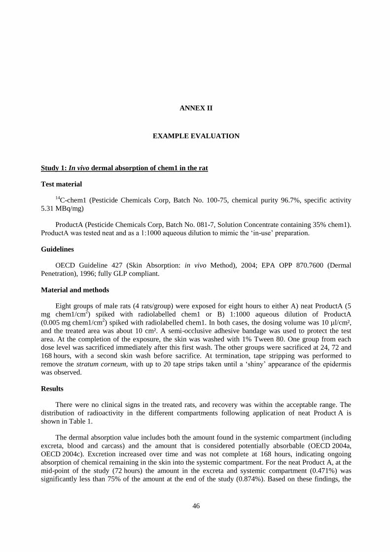
46
ANNEX II
EXAMPLE EVALUATION
Study 1: In vivo dermal absorption of chem1 in the rat
Test material
14C-chem1 (Pesticide Chemicals Corp, Batch No. 100-75, chemical purity 96.7%, specific activity
5.31 MBq/mg)
ProductA (Pesticide Chemicals Corp, Batch No. 081-7, Solution Concentrate containing 35% chem1).
ProductA was tested neat and as a 1:1000 aqueous dilution to mimic the ‘in-use’ preparation.
Guidelines
OECD Guideline 427 (Skin Absorption: in vivo Method), 2004; EPA OPP 870.7600 (Dermal
Penetration), 1996; fully GLP compliant.
Material and methods
Eight groups of male rats (4 rats/group) were exposed for eight hours to either A) neat ProductA (5
mg chem1/cm2) spiked with radiolabelled chem1 or B) 1:1000 aqueous dilution of ProductA
(0.005 mg chem1/cm2) spiked with radiolabelled chem1. In both cases, the dosing volume was 10 µl/cm²,
and the treated area was about 10 cm². A semi-occlusive adhesive bandage was used to protect the test
area. At the completion of the exposure, the skin was washed with 1% Tween 80. One group from each
dose level was sacrificed immediately after this first wash. The other groups were sacrificed at 24, 72 and
168 hours, with a second skin wash before sacrifice. At termination, tape stripping was performed to
remove the stratum corneum, with up to 20 tape strips taken until a ‘shiny’ appearance of the epidermis
was observed.
Results
There were no clinical signs in the treated rats, and recovery was within the acceptable range. The
distribution of radioactivity in the different compartments following application of neat Product A is
shown in Table 1.
The dermal absorption value includes both the amount found in the systemic compartment (including
excreta, blood and carcass) and the amount that is considered potentially absorbable (OECD 2004a,
OECD 2004c). Excretion increased over time and was not complete at 168 hours, indicating ongoing
absorption of chemical remaining in the skin into the systemic compartment. For the neat Product A, at the
mid-point of the study (72 hours) the amount in the excreta and systemic compartment (0.471%) was
significantly less than 75% of the amount at the end of the study (0.874%). Based on these findings, the
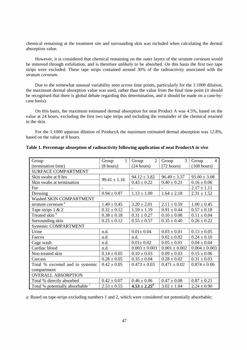
47
chemical remaining at the treatment site and surrounding skin was included when calculating the dermal
absorption value.
However, it is considered that chemical remaining on the outer layers of the stratum corneum would
be removed through exfoliation, and is therefore unlikely to be absorbed. On this basis the first two tape
strips were excluded. These tape strips contained around 30% of the radioactivity associated with the
stratum corneum.
Due to the somewhat unusual variability seen across time points, particularly for the 1:1000 dilution,
the maximum dermal absorption value was used, rather than the value from the final time point (it should
be recognised that there is global debate regarding this determination, and it should be made on a case-by-
case basis).
On this basis, the maximum estimated dermal absorption for neat Product A was 4.5%, based on the
value at 24 hours, excluding the first two tape strips and including the remainder of the chemical retained
in the skin.
For the 1:1000 aqueous dilution of ProductA the maximum estimated dermal absorption was 12.8%,
based on the value at 8 hours.
Table 1. Percentage absorption of radioactivity following application of neat ProductA in vivo
Group
(termination time)
Group 1
(8 hours)
Group 2
(24 hours)
Group 3
(72 hours)
Group 4
(168 hours)
SURFACE COMPARTMENT
Skin swabs at 8 hrs 99.41 ± 1.16
94.12 ± 3.82 96.49 ± 3.37 93.00 ± 3.08
Skin swabs at termination 0.43 ± 0.22 0.40 ± 0.21 0.16 ± 0.06
Fur - - - 2.17 ± 1.11
Dressing 0.94 ± 0.87 1.13 ± 1.00 1.64 ± 2.18 2.31 ± 1.52
Washed SKIN COMPARTMENT
stratum corneum a 1.49 ± 0.45 3.20 ± 2.01 2.11 ± 0.59 1.00 ± 0.45
Tape strips 1 & 2 0.32 ± 0.12 1.59 ± 1.19 0.91 ± 0.44 0.57 ± 0.18
Treated skin b 0.38 ± 0.18 0.31 ± 0.27 0.10 ± 0.08 0.11 ± 0.04
Surrounding skin 0.25 ± 0.12 0.55 ± 0.57 0.35 ± 0.40 0.26 ± 0.22
Systemic COMPARTMENT
Urine n.d. 0.01± 0.04 0.03 ± 0.01 0.13 ± 0.05
Faeces n.d. n.d. 0.02 ± 0.02 0.24 ± 0.10
Cage wash n.d. 0.01± 0.02 0.05 ± 0.01 0.04 ± 0.04
Cardiac blood n.d. 0.003 ± 0.003 0.001 ± 0.002 0.004 ± 0.003
Non-treated skin 0.14 ± 0.05 0.10 ± 0.03 0.09 ± 0.03 0.15 ± 0.06
Carcass 0.28 ± 0.05 0.35 ± 0.04 0.28 ± 0.02 0.31 ± 0.03
Total % excreted and in systemic
compartment
0.42 ± 0.05 0.473 ± 0.03 0.471 ± 0.02 0.874 ± 0.06
OVERALL ABSORPTION
Total % directly absorbed 0.42 ± 0.07 0.46 ± 0.06 0.47 ± 0.08 0.87 ± 0.21
Total % potentially absorbable c 2.53 ± 0.55 4.53 ± 2.25
d 3.02 ± 1.04 2.24 ± 0.90
a: Based on tape-strips excluding numbers 1 and 2, which were considered not potentially absorbable;
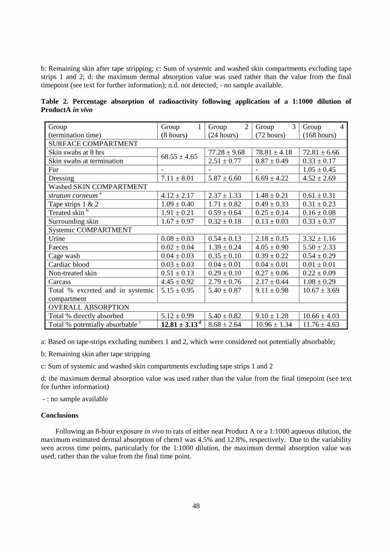
48
b: Remaining skin after tape stripping; c: Sum of systemic and washed skin compartments excluding tape
strips 1 and 2; d: the maximum dermal absorption value was used rather than the value from the final
timepoint (see text for further information); n.d. not detected; - no sample available.
Table 2. Percentage absorption of radioactivity following application of a 1:1000 dilution of
ProductA in vivo
Group
(termination time)
Group 1
(8 hours)
Group 2
(24 hours)
Group 3
(72 hours)
Group 4
(168 hours)
SURFACE COMPARTMENT
Skin swabs at 8 hrs 68.55 ± 4.65
77.28 ± 9.68 78.81 ± 4.18 72.81 ± 6.66
Skin swabs at termination 2.51 ± 0.77 0.87 ± 0.49 0.33 ± 0.17
Fur - - - 1.05 ± 0.45
Dressing 7.11 ± 8.01 5.87 ± 6.60 6.69 ± 4.22 4.52 ± 2.69
Washed SKIN COMPARTMENT
stratum corneum a 4.12 ± 2.17 2.37 ± 1.33 1.48 ± 0.21 0.61 ± 0.31
Tape strips 1 & 2 1.09 ± 0.40 1.71 ± 0.82 0.49 ± 0.33 0.31 ± 0.23
Treated skin b 1.91 ± 0.21 0.59 ± 0.64 0.25 ± 0.14 0.16 ± 0.08
Surrounding skin 1.67 ± 0.97 0.32 ± 0.18 0.13 ± 0.03 0.33 ± 0.37
Systemic COMPARTMENT
Urine 0.08 ± 0.03 0.54 ± 0.13 2.18 ± 0.15 3.32 ± 1.16
Faeces 0.02 ± 0.04 1.39 ± 0.24 4.05 ± 0.90 5.50 ± 2.33
Cage wash 0.04 ± 0.03 0.35 ± 0.10 0.39 ± 0.22 0.54 ± 0.29
Cardiac blood 0.03 ± 0.03 0.04 ± 0.01 0.04 ± 0.01 0.01 ± 0.01
Non-treated skin 0.51 ± 0.13 0.29 ± 0.10 0.27 ± 0.06 0.22 ± 0.09
Carcass 4.45 ± 0.92 2.79 ± 0.76 2.17 ± 0.44 1.08 ± 0.29
Total % excreted and in systemic
compartment
5.15 ± 0.95 5.40 ± 0.87 9.11 ± 0.98 10.67 ± 3.69
OVERALL ABSORPTION
Total % directly absorbed 5.12 ± 0.99 5.40 ± 0.82 9.10 ± 1.28 10.66 ± 4.03
Total % potentially absorbable c 12.81 ± 3.13
d 8.68 ± 2.64 10.96 ± 1.34 11.76 ± 4.63
a: Based on tape-strips excluding numbers 1 and 2, which were considered not potentially absorbable;
b: Remaining skin after tape stripping
c: Sum of systemic and washed skin compartments excluding tape strips 1 and 2
d: the maximum dermal absorption value was used rather than the value from the final timepoint (see text
for further information)
- : no sample available
Conclusions
Following an 8-hour exposure in vivo to rats of either neat Product A or a 1:1000 aqueous dilution, the
maximum estimated dermal absorption of chem1 was 4.5% and 12.8%, respectively. Due to the variability
seen across time points, particularly for the 1:1000 dilution, the maximum dermal absorption value was
used, rather than the value from the final time point.

49
Study 2: In vitro dermal absorption of chem1 in rat and human skin
Test material
14C-chem1 (Pesticide Chemicals Corp, Batch No. 100-75, chemical purity 96.7%, specific activity
5.31 MBq/mg)
ProductA (Pesticide Chemicals Corp, Batch No. 081-7, Solution Concentrate containing 35% chem1).
ProductA was tested neat and as a 1:1000 aqueous dilution to mimic the ‘in-use’ preparation.
Guidelines
OECD Guideline 428 (Skin Absorption: in vitro Method), 2004; fully GLP compliant.
Material and methods
Samples of shaved rat skin from the dorsal region and dermatomed human abdominal skin were
exposed for eight hours to either neat Product A or a 1:1000 aqueous dilution of Product A as specified in
Study 1. The integrity of the skin samples was assayed using trans-epidermal water loss.
Following addition of the test substance, samples of the receptor fluid were taken at various
timepoints. At the completion of the 8-hour exposure, the skin was washed with 1% Tween 80. Another
skin wash was conducted at 24 hours, after which tape stripping was performed and radioactivity
measured.
Results
Recovery was within the expected range, and the test substance was adequately soluble in the receptor
fluid (0.2 mg/mL). There was generally low absorption of the concentrate in vitro in both human and rat
skin.
Chemical remaining on the outer layers of the stratum corneum may be removed through exfoliation.
On this basis the first two tape strips may be excluded. For human skin, these first two tape strips contained
around 85% of the radioactivity associated with the stratum corneum, while for rat skin these strips
contained around 40%. Due to this significant difference between rat and human skin the in vitro dermal
absorption was calculated both with and without the first two tape strips.
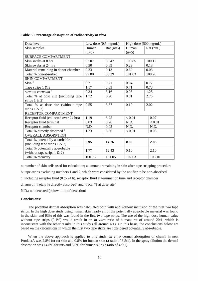
50
Table 3. Percentage absorption of radioactivity in vitro
Dose level Low dose (0.5 mg/mL) High dose (500 mg/mL)
Skin samples Human
(n=5)
Rat (n=5) Human
(n=5)
Rat (n=6)
SURFACE COMPARTMENT
Skin swabs at 8 hrs 97.07 85.47 100.85 100.12
Skin swabs at 24 hrs 0.50 0.69 0.29 0.13
Material remaining in donor chamber 0.23 0.13 0.69 0.03
Total % non-absorbed 97.80 86.29 101.83 100.28
SKIN COMPARTMENT
Skin a 0.21 0.71 0.04 0.77
Tape strips 1 & 2 1.17 2.33 0.71 0.73
stratum corneum b 0.34 3.16 0.05 1.25
Total % at dose site (including tape
strips 1 & 2)
1.72 6.20 0.81 2.75
Total % at dose site (without tape
strips 1 & 2)
0.55 3.87 0.10 2.02
RECEPTOR COMPARTMENT
Receptor fluid (collected over 24 hrs) 1.19 8.25 < 0.01 0.07
Receptor fluid terminal 0.03 0.26 N.D. < 0.01
Receptor chamber N.D. 0.05 N.D. N.D.
Total % directly absorbed c 1.23 8.56 < 0.01 0.08
OVERALL ABSORPTION
Total % potentially absorbable d
(including tape strips 1 & 2) 2.95 14.76 0.82 2.83
Total % potentially absorbable
(without tape strips 1 & 2) 1.77 12.43 0.10 2.10
Total % recovery 100.73 101.05 102.63 103.10
n: number of skin cells used for calculation; a: amount remaining in skin after tape stripping procedure
b: tape-strips excluding numbers 1 and 2, which were considered by the notifier to be non-absorbed
c: including receptor fluid (0 to 24 h), receptor fluid at termination time and receptor chamber
d: sum of ‘Totals % directly absorbed’ and ‘Total % at dose site’
N.D.: not detected (below limit of detection)
Conclusions:
The potential dermal absorption was calculated both with and without inclusion of the first two tape
strips. In the high dose study using human skin nearly all of the potentially absorbable material was found
in the skin, and 93% of this was found in the first two tape strips. The use of the high dose human value
without tape strips (0.1%) would result in an in vitro ratio of human: rat of around 20:1, which is
inconsistent with the other results in this study (all around 4:1). On this basis, the conclusions below are
based on the calculations in which the first two tape strips are considered potentially absorbable.
When the above approach is applied to this study, in vitro dermal absorption of chem1 in neat
ProductA was 2.8% for rat skin and 0.8% for human skin (a ratio of 3.5:1). In the spray dilution the dermal
absorption was 14.8% for rats and 3.0% for human skin (a ratio of 4.9:1).
51
Discussion of dermal absorption of chem1
The in vivo study on rats demonstrated dermal absorption values of 4.5% for chem1 in neat ProductA
and of 12.8% for a 1:1000 ‘in use’ spray dilution.
The comparative in vitro study on human and rat skin demonstrated that, as with most substances, rat
skin was more permeable than human skin and dermal absorption of the dilution was higher than that of
the concentrate. The ratios of rat: human skin were 3.5:1 for chem1 in neat ProductA and 4.9:1 for the ‘in
use’ spray dilution.
On this basis the dermal absorption value for use in risk assessment is 1.3% (4.5%/3.5) for chem1 in
ProductA and 2.6% (12.8%/4.9) for the ‘in use’ spray dilution. Values of 2% (for the concentrate) and
3% (for the in-use dilution) are therefore recommended for use in risk assessment.
52
ANNEX III
PREDICTIVE VALUE OF IN VITRO STUDIES ON HUMAN SKIN
Franz (1975) compared the penetration of 12 organic compounds (no pesticides included) through
samples of human abdominal skin under finite dose conditions with previously published results for dermal
absorption of these chemicals through the forearm skin of human volunteers. The in vivo absorption was
first based on urinary excretion over five days. However, results obtained after intravenous application
were subsequently used to calculate total absorption. The flux was taken as the relevant parameter to
describe the process of absorption, and generally a good correlation between in vitro and in vivo results
was found. The only exception was thiourea, for which the in vivo experiment had to be repeated using the
abdomen, rather than the forearm, to be in agreement with the in vitro prediction. With regard to total
absorption as a percentage of the administered dose, in vitro testing revealed a higher or nearly equal (in
two cases only) absorption than the in vivo experiment for nine substances. This suggests the prediction
would be sufficiently conservative. However, with salicylic acid, caffeine, and dinitrochlorbenzene,
approximately two to five times higher dermal absorption was obtained in the human volunteer study. The
ranking of the substances for total absorption, based on median values, also revealed remarkable
differences between the two sets of studies, although substances of low absorption in vivo (lower than
10%) were all clearly confirmed as such in vitro.
In rats, Bronaugh et al. (1982) examined the dermal absorption of benzoic acid, acetylsalicylic acid,
urea, and caffeine by summing up the urinary excretion and an estimate for the amount retained in the body
that was calculated from the achieved blood concentrations. The absorption values in terms of percentages
of the applied doses were in excellent agreement with in vitro data (rat, full thickness skin) for all
substances except caffeine. For urea, acetylsalicylic and benzoic acid, the absorption rates were in the same
magnitude as determined by Franz (1975) in humans or on human skin. It must be taken into account that,
in both studies, skin analysis was not performed.
Van de Sandt et al. (2000) postulated that it is difficult if not impossible to directly compare in vivo
data with in vitro data because of the very different experimental conditions. Therefore, they tried to
perform studies under standardised conditions with respect to dose, vehicle and exposure duration to
overcome this lack of comparability. Their experiments with the pesticide propoxur demonstrated that the
outcome of the in vitro study on human skin using viable full-thickness membranes (1.7 or 9.7% dermal
absorption after 4 or 24 hours, respectively, including the amount that was retained in skin) correlated well
with the human in vivo situation (mean values of 0.5 and 3.7%, based on urinary excretion). The actual in
vivo absorption might be higher because the amount in skin remains unknown. Nonetheless, the level of
dermal absorption is likely to be slightly overestimated if only the in vitro study is available.
For the more lipophilic fungicide ortho-phenylphenol, the same group found a 33% penetration
through human viable skin at 48 hours after commencement of a four-hour exposure period, whereas about
15% of the total dermal dose had been excreted in the urine of human volunteers within the same time
interval. Thus, good predictivity and sufficient conservatism in the estimation of dermal absorption by an
in vitro experiment on human skin was again demonstrated (Cnubben et al. 2002), even though there might
be doubts about the integrity of skin in vitro when the study duration exceeds 24 hours. This could explain

53
overestimation of dermal absorption in vitro. Penetration through an epidermal membrane was much
higher in the study (nearly 100%), and the authors suspected some deposition in the dermis as previously
hypothesised for lipophilic substances.
Van Ravenzwaay and Leibold (2004) compared the dermal absorption of 14 pesticides (including
eight fungicides, five herbicides and one insecticide, all developed and manufactured by a single company)
at two or three exposure levels in vivo in rats and in vitro on rat and human skin. Skin-bound residues were
included. It could be shown that, for all substances, the in vitro results on rat skin usually overestimated the
dermal absorption as measured in vivo. Only very few data were in the same magnitude; whereas for the
majority of compounds and dilutions, the in vitro absorption was generally two to nearly 80 times higher.
Furthermore, rat skin proved to be more permeable than human skin in all cases, although the individual
ratios, as well as those for the comparison of in vivo and in vitro in rats, differed very much among the
various substances.
Williams (2006) reviewed a number of studies for comparability of in vivo and in vitro dermal
absorption of chemicals from different regulatory fields and found the in vitro data in general sufficiently
predictive. However, some reservations were expressed about the use of such studies in estimating the
dermal absorption rate of very lipophilic substances due to the strong reservoir effects to be expected in
vitro that perhaps may not be similar enough to the in vivo situation.
A more comprehensive overview of the literature is given in EHC 235 (WHO 2006), which drew the
conclusion that there is a generally good correlation between in vivo and in vitro dermal absorption data,
and that properly conducted in vitro measurements can be used to predict in vivo absorption. The best
results in terms of a similar absorption rate were achieved when viable full-thickness human skin
membranes were used for the in vitro experiments and when the absorbed percentages of the applied total
dose were compared instead of flux.


