© 2000 by the American Society for Dermatologic Surgery, Inc. • Published by Blackwell Science, Inc.ISSN: 1076-0512/00/$15.00/0 • Dermatol Surg 2000;26:835–843
Noninvasive Rejuvenation of Photodamaged Skin Using Serial, Full-Face Intense Pulsed Light Treatments
Patrick H. Bitter, Jr., MD
Los Gatos, California
background.
Photodamaged skin is characterized not only byrhytides, but also by epidermal and dermal atrophy, rough skintexture, irregular pigmentation, telangiectasias, laxity, and en-larged pores. There is growing interest in the development ofnoninvasive methods to treat photodamaged skin. Skin photo-rejuvenation is the visible improvement of photodamaged skinusing a laser or other light source. A noncoherent, broadband,pulsed light source is effective in the treatment of vascular andpigmented lesions of the skin. This study evaluates the role ofintense pulsed light in the rejuvenation of photo aged skin.
objective.
The purpose of this study was to evaluate andquantify the degree of visible improvement in photodamagedskin following a series of full-face, intense pulsed light treat-ments.
methods.
Forty-nine subjects with varying degrees of photo-
damage were treated with a series of four or more full-facetreatments at 3-week intervals using a nonablative, nonlaser in-tense pulsed visible light source. Fluences varied from 30 to 50
J/cm
2
. Subject evaluation and skin biopsies were used to assesstreatment results.
results.
All aspects of photodamage including wrinkling, skincoarseness, irregular pigmentation, pore size, and telangiecta-sias showed visible improvement in more than 90% of subjectswith minimal downtime and no scarring. Eighty-eight percentof subjects were satisfied with the overall results of their treat-ments.
conclusion.
Treatment of photodamaged facial skin using aseries of full-face treatments with intense pulsed light is a newand effective noninvasive method of skin rejuvenation withminimal risk and no patient downtime.
PHOTODAMAGE IS the result of chronic ultraviolet(UV) light exposure inducing characteristic epidermaland dermal changes. The visible signs of photodamageare characterized by thinning of the epidermis and der-mis, coarse skin texture, wrinkling, pigmentation alter-ations, telangiectasias, and in some cases actinic kera-toses and epidermal malignancies. The clinical pictureof photodamaged skin is more than just rhytides. Con-sequently treatments that focus only on improvement inrhytides will, by nature, produce results limited to im-proving only one visible component of photodamagedskin. Conversely a treatment that is able to improveeach of the different components of photodamaged skinwill result in a more dramatic visible improvement.
Numerous treatments have been developed to im-prove the appearance and health of photoaged skin.These include various topical agents such as glycolicacid,
1
retinoids,
2,3
ascorbic acid,
4
a variety of chemicalpeeling agents,
5
dermabrasion,
6
epidermabrasion andlaser skin resurfacing.
7–9
To date the most effective
methods for improving photodamaged skin have beeninvasive. The major disadvantage of invasive treat-ments is the requisite recovery period following proce-dures such as laser skin resurfacing. In addition, scar-ring has been reported with each of the invasiveprocedures including laser resurfacing.
10,11
There has been great interest in the development ofnoninvasive and nonablative methods to effectivelyimprove the appearance of photodamaged skin with-out patient downtime. Recently a 1320 nm laser hasshown some promise in the nonablative treatment ofwrinkles.
12,13
In addition, the pulsed dye laser has beenused to treat selected facial rhytides.
14,15
Electrosur-gery has also been used to improve rhytides in a nona-blative method.
16
A noncoherent, nonlaser, filtered flash-lamp emitting a broadband visible light has been shownto be highly effective in the treatment of a variety of vas-cular and pigmented lesions of the skin.
17
Most recently,Goldberg
18
reported the results of intense pulsed light inthe treatment of superficial rhytides.
This study describes a new application of intensepulsed light technology and evaluates the visible im-provements seen in photodamaged skin following a se-ries of full-face treatments. The role of intense pulsedlight in skin rejuvenation and the advantages and po-tential benefits of this procedure in the amelioration ofphotodamage are discussed.
No outside funding was provided for this study or for preparation ofthis manuscript. The author has received honoraria from ESC Medical,Inc. for lectures.Address correspondence and reprint requests to: Patrick Bitter Jr., MD,16300 Camino Del Sol, Los Gatos, CA 95032.
836
bitter: skin rejuvenation using intense pulsed light
Dermatol Surg 26:9:September 2000
Methods
Subjects
Forty-nine successive patients (43 female and 6 male), ages30–64 years, with varying degrees of photodamage andwrinkling who completed between four and six full-facetreatments participated in the study. All patients were ofFitzpatrick skin types I–III. All patients had visible evidenceof photoaging (wrinkling, irregular pigmentation, telang-iectasias). All patients to be treated underwent physicianhistory and examination to exclude recent oral retinoid druguse, sensitivity to infrared radiation or visible light, use ofphotosensitizing medications, or the presence of any suspi-cious lesions. Patients were placed on a simple skin care reg-imen with a gentle cleanser, moisturizer, and sunblock. Pa-tients using topical tretinoin or topical or oral medicationsfor rosacea continued these treatments as needed. No othertreatments were permitted. All patients gave informed con-sent for treatments and photographs.
Treatment Protocol
A noncoherent, filtered broadband pulsed flashlamp (Vascu-light, ESC/Sharplan, Norwood, MA) emitting in the range of500–1200 nm was used for all treatments. Each patient un-derwent at least four and up to six full-face treatments. Theaverage number of treatments for the 49 patients was 4.94.
Treatments were administered at 3-week intervals. Theentire face including the lower eyelids, but excluding the up-per eyelids, was treated at each session. Eyelids were pro-tected by small external plastic shields. Treatment fluencesvaried between 30 and 50 J/cm
2
. Energy was delivered indouble or triple pulse trains of 2.4–4.7 msec with pulse de-lays of 10–60 msec. Cut-off filters of 550 or 570 nm wereused for all treatments. A chilled colorless ultrasonic gel wasapplied directly to the light guide of the cutoff filter prior toapplication to the skin. Specific treatment parameters werechanged at each treatment for each patient depending on theresults of the previous treatment. At each subsequent treatmentthe fluence was generally increased by 1–2 J/cm
2
. The entireface was treated at each session except in some of the male sub-jects (50%) who elected to avoid treatment of the beard areabecause of potential hair loss. Treatment parameters were cho-sen to minimize or prevent purpura.
In working with the double-pulse parameters, it was ob-served that lengthening the second pulse duration (to 4.0msec or more) and/or the interpulse interval (to 20 msec ormore) and maintaining a fluence of 36 J/cm
2
or less almostentirely eliminated the appearance of purpura. In addition,it was observed that carefully maintaining a skin/light guidedistance of 1 mm or more was also very important in pre-venting purpura or blister formation. The object of eachtreatment was to prevent any patient downtime while maxi-mizing the improvement in the vascular and pigmented le-sions and overall skin appearance. Subjects were asked toevaluate the degree of change visible in their skin for each ofthe following criteria: fine wrinkling, skin smoothness, skin
laxity, irregular pigmentation, redness, flushing, presence oftelangiectasias, and pore size. Subjects were also asked torate their overall improvement as well as the degree of dis-comfort, any adverse outcomes, and any “downtime” as de-fined as any time missed from usual activities due to treat-ments. Pretreatment with a 4% lidocaine cream (ELA-Max,Ferndale Labs, Ferndale, MI) for topical anesthesia wasused 30 minutes prior to treatment. Subject evaluationswere obtained 3–4 weeks after the final treatment. Skin bi-opsies were obtained from the forehead of one patient priorto treatment and 4 weeks after the final treatment. Subjectphotographs were obtained pretreatment after two treat-ments and 3–4 weeks after the final treatment.
Results
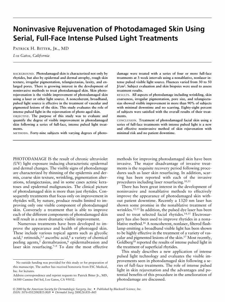
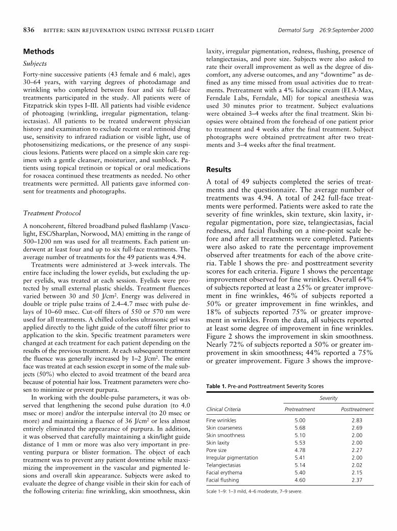
A total of 49 subjects completed the series of treat-ments and the questionnaire. The average number oftreatments was 4.94. A total of 242 full-face treat-ments were performed. Patients were asked to rate theseverity of fine wrinkles, skin texture, skin laxity, ir-regular pigmentation, pore size, telangiectasias, facialredness, and facial flushing on a nine-point scale be-fore and after all treatments were completed. Patientswere also asked to rate the percentage improvementobserved after treatments for each of the above crite-ria. Table 1 shows the pre- and posttreatment severityscores for each criteria. Figure 1 shows the percentageimprovement observed for fine wrinkles. Overall 64%of subjects reported at least a 25% or greater improve-ment in fine wrinkles, 46% of subjects reported a50% or greater improvement in fine wrinkles, and18% of subjects reported 75% or greater improve-ment in wrinkles. From the data, all subjects reportedat least some degree of improvement in fine wrinkles.Figure 2 shows the improvement in skin smoothness.Nearly 72% of subjects reported a 50% or greater im-provement in skin smoothness; 44% reported a 75%or greater improvement. Figure 3 shows the improve-
Table 1.
Pre-and Posttreatment Severity Scores
Severity
Clinical Criteria Pretreatment Posttreatment
Fine wrinkles 5.00 2.83Skin coarseness 5.68 2.69Skin smoothness 5.10 2.00Skin laxity 5.53 2.00Pore size 4.78 2.27Irregular pigmentation 5.41 2.00Telangiectasias 5.14 2.02Facial erythema 5.40 2.15Facial flushing 4.60 2.37
Scale 1–9: 1–3 mild, 4–6 moderate, 7–9 severe.
Dermatol Surg 26:9:September 2000
bitter: skin rejuvenation using intense pulsed light
837
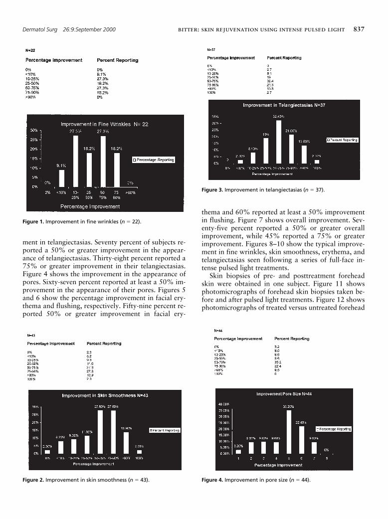
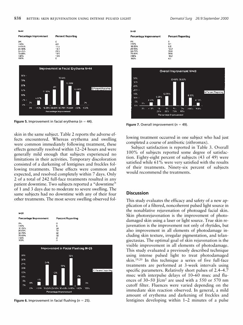
ment in telangiectasias. Seventy percent of subjects re-ported a 50% or greater improvement in the appear-ance of telangiectasias. Thirty-eight percent reported a75% or greater improvement in their telangiectasias.Figure 4 shows the improvement in the appearance ofpores. Sixty-seven percent reported at least a 50% im-provement in the appearance of their pores. Figures 5and 6 show the percentage improvement in facial ery-thema and flushing, respectively. Fifty-nine percent re-ported 50% or greater improvement in facial ery-
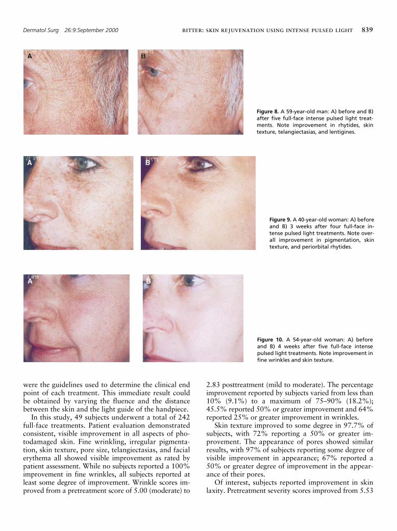
thema and 60% reported at least a 50% improvementin flushing. Figure 7 shows overall improvement. Sev-enty-five percent reported a 50% or greater overallimprovement, while 45% reported a 75% or greaterimprovement. Figures 8–10 show the typical improve-ment in fine wrinkles, skin smoothness, erythema, andtelangiectasias seen following a series of full-face in-tense pulsed light treatments.
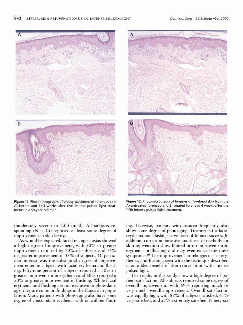
Skin biopsies of pre- and posttreatment foreheadskin were obtained in one subject. Figure 11 showsphotomicrographs of forehead skin biopsies taken be-fore and after pulsed light treatments. Figure 12 showsphotomicrographs of treated versus untreated forehead
Figure 1. Improvement in fine wrinkles (n 5 22).
Figure 2. Improvement in skin smoothness (n 5 43).
Figure 3. Improvement in telangiectasias (n 5 37).
Figure 4. Improvement in pore size (n 5 44).
838
bitter: skin rejuvenation using intense pulsed light
Dermatol Surg 26:9:September 2000
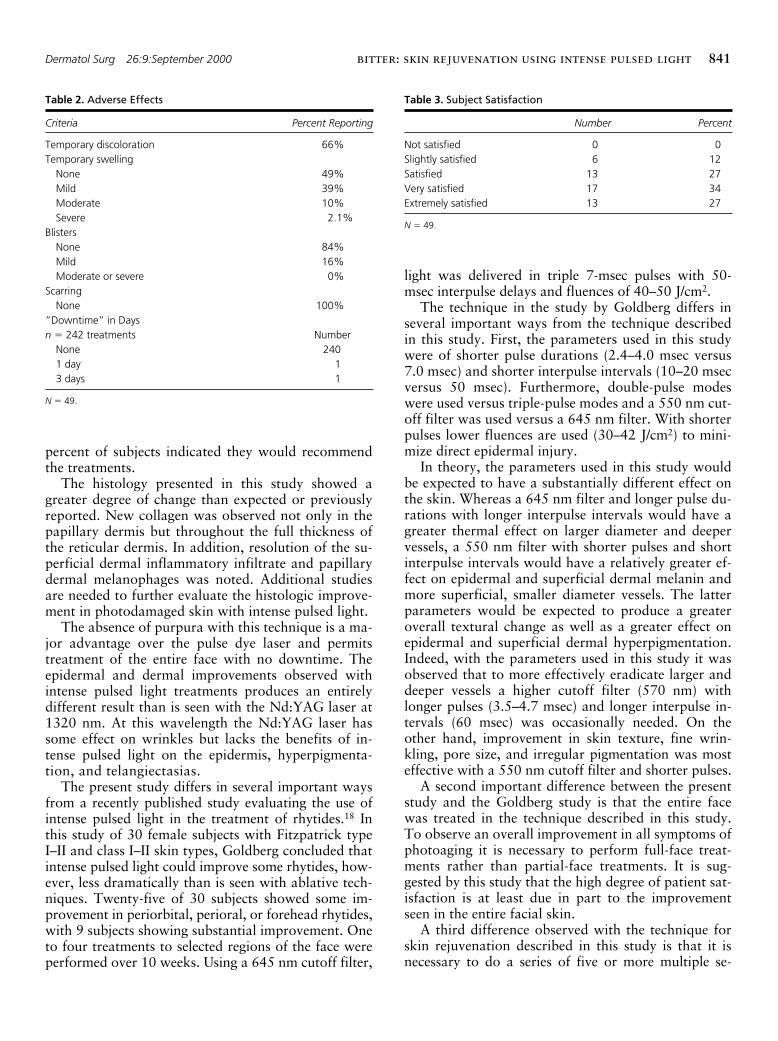
skin in the same subject. Table 2 reports the adverse ef-fects encountered. Whereas erythema and swellingwere common immediately following treatment, theseeffects generally resolved within 12–24 hours and weregenerally mild enough that subjects experienced nolimitations in their activities. Temporary discolorationconsisted of a darkening of lentigines and freckles fol-lowing treatments. These effects were common andexpected, and resolved completely within 7 days. Only2 of a total of 242 full-face treatments resulted in anypatient downtime. Two subjects reported a “downtime”of 1 and 3 days due to moderate to severe swelling. Thesame subjects had no downtime with any of their fourother treatments. The most severe swelling observed fol-
lowing treatment occurred in one subject who had justcompleted a course of antibiotic (zithromax).
Subject satisfaction is reported in Table 3. Overall100% of subjects reported some degree of satisfac-tion. Eighty-eight percent of subjects (43 of 49) weresatisfied while 61% were very satisfied with the resultsof their treatments. Ninety-six percent of subjectswould recommend the treatments.
Discussion
This study evaluates the efficacy and safety of a new ap-plication of a filtered, noncoherent pulsed light source inthe nonablative rejuvenation of photoaged facial skin.Skin photorejuvenation is the improvement of photo-damaged skin using a laser or light source. True skin re-juvenation is the improvement not only of rhytides, butalso improvement in all elements of photodamage in-cluding skin texture, irregular pigmentation, and telan-giectasias. The optimal goal of skin rejuvenation is thevisible improvement in all elements of photodamage.This study evaluated a previously described techniqueusing intense pulsed light to treat photodamagedskin.
19,20
In this technique a series of five full-facetreatments are performed at 3-week intervals usingspecific parameters. Relatively short pulses of 2.4–4.7msec with interpulse delays of 10–60 msec and flu-ences of 30–50 J/cm
2
are used with a 550 or 570 nmcutoff filter. Fluences were varied depending on theimmediate skin reaction observed. In general, a mildamount of erythema and darkening of freckles andlentigines developing within 1–2 minutes of a pulse
Figure 5. Improvement in facial erythema (n 5 44).
Figure 6. Improvement in facial flushing (n 5 25).
Figure 7. Overall improvement (n 5 49).
Dermatol Surg 26:9:September 2000
bitter: skin rejuvenation using intense pulsed light
839
were the guidelines used to determine the clinical endpoint of each treatment. This immediate result couldbe obtained by varying the fluence and the distancebetween the skin and the light guide of the handpiece.
In this study, 49 subjects underwent a total of 242full-face treatments. Patient evaluation demonstratedconsistent, visible improvement in all aspects of pho-todamaged skin. Fine wrinkling, irregular pigmenta-tion, skin texture, pore size, telangiectasias, and facialerythema all showed visible improvement as rated bypatient assessment. While no subjects reported a 100%improvement in fine wrinkles, all subjects reported atleast some degree of improvement. Wrinkle scores im-proved from a pretreatment score of 5.00 (moderate) to
2.83 posttreatment (mild to moderate). The percentageimprovement reported by subjects varied from less than10% (9.1%) to a maximum of 75–90% (18.2%);45.5% reported 50% or greater improvement and 64%reported 25% or greater improvement in wrinkles.
Skin texture improved to some degree in 97.7% ofsubjects, with 72% reporting a 50% or greater im-provement. The appearance of pores showed similarresults, with 97% of subjects reporting some degree ofvisible improvement in appearance; 67% reported a50% or greater degree of improvement in the appear-ance of their pores.
Of interest, subjects reported improvement in skinlaxity. Pretreatment severity scores improved from 5.53
Figure 8. A 59-year-old man: A) before and B)after five full-face intense pulsed light treat-ments. Note improvement in rhytides, skintexture, telangiectasias, and lentigines.
Figure 9. A 40-year-old woman: A) beforeand B) 3 weeks after four full-face in-tense pulsed light treatments. Note over-all improvement in pigmentation, skintexture, and periorbital rhytides.
Figure 10. A 54-year-old woman: A) beforeand B) 4 weeks after five full-face intensepulsed light treatments. Note improvement infine wrinkles and skin texture.
840
bitter: skin rejuvenation using intense pulsed light
Dermatol Surg 26:9:September 2000
(moderately severe) to 2.00 (mild). All subjects re-sponding (
N
5
15) reported at least some degree ofimprovement in skin laxity.
As would be expected, facial telangiectasias showeda high degree of improvement, with 50% or greaterimprovement reported by 70% of subjects and 75%or greater improvement in 38% of subjects. Of partic-ular interest was the substantial degree of improve-ment noted in subjects with facial erythema and flush-ing. Fifty-nine percent of subjects reported a 50% orgreater improvement in erythema and 60% reported a50% or greater improvement in flushing. While facialerythema and flushing are not exclusive to photodam-age, they are common findings in the Caucasian popu-lation. Many patients with photoaging also have somedegree of concomitant erythema with or without flush-
ing. Likewise, patients with rosacea frequently alsoshow some degree of photoaging. Treatments for facialerythema and flushing have been of limited success. Inaddition, current noninvasive and invasive methods forskin rejuvenation show limited or no improvement inerythema or flushing and may even exacerbate thesesymptoms.
10
The improvement in telangiectasias, ery-thema, and flushing seen with the technique describedis an added benefit of skin rejuvenation with intensepulsed light.
The results in this study show a high degree of pa-tient satisfaction. All subjects reported some degree ofoverall improvement, with 69% reporting much orvery much overall improvement. Overall satisfactionwas equally high, with 88% of subjects satisfied, 61%very satisfied, and 27% extremely satisfied. Ninety-six
Figure 11. Photomicrographs of biopsy specimens of forehead skinA) before and B) 4 weeks after five intense pulsed light treat-ments in a 59-year-old man.
Figure 12. Photomicrograph of biopsies of forehead skin from theA) untreated forehead and B) treated forehead 4 weeks after thefifth intense pulsed light treatment.
Dermatol Surg 26:9:September 2000
bitter: skin rejuvenation using intense pulsed light
841
percent of subjects indicated they would recommendthe treatments.
The histology presented in this study showed agreater degree of change than expected or previouslyreported. New collagen was observed not only in thepapillary dermis but throughout the full thickness ofthe reticular dermis. In addition, resolution of the su-perficial dermal inflammatory infiltrate and papillarydermal melanophages was noted. Additional studiesare needed to further evaluate the histologic improve-ment in photodamaged skin with intense pulsed light.
The absence of purpura with this technique is a ma-jor advantage over the pulse dye laser and permitstreatment of the entire face with no downtime. Theepidermal and dermal improvements observed withintense pulsed light treatments produces an entirelydifferent result than is seen with the Nd:YAG laser at1320 nm. At this wavelength the Nd:YAG laser hassome effect on wrinkles but lacks the benefits of in-tense pulsed light on the epidermis, hyperpigmenta-tion, and telangiectasias.
The present study differs in several important waysfrom a recently published study evaluating the use ofintense pulsed light in the treatment of rhytides.
18
Inthis study of 30 female subjects with Fitzpatrick typeI–II and class I–II skin types, Goldberg concluded thatintense pulsed light could improve some rhytides, how-ever, less dramatically than is seen with ablative tech-niques. Twenty-five of 30 subjects showed some im-provement in periorbital, perioral, or forehead rhytides,with 9 subjects showing substantial improvement. Oneto four treatments to selected regions of the face wereperformed over 10 weeks. Using a 645 nm cutoff filter,
light was delivered in triple 7-msec pulses with 50-msec interpulse delays and fluences of 40–50 J/cm
2
.The technique in the study by Goldberg differs in
several important ways from the technique describedin this study. First, the parameters used in this studywere of shorter pulse durations (2.4–4.0 msec versus7.0 msec) and shorter interpulse intervals (10–20 msecversus 50 msec). Furthermore, double-pulse modeswere used versus triple-pulse modes and a 550 nm cut-off filter was used versus a 645 nm filter. With shorterpulses lower fluences are used (30–42 J/cm
2
) to mini-mize direct epidermal injury.
In theory, the parameters used in this study wouldbe expected to have a substantially different effect onthe skin. Whereas a 645 nm filter and longer pulse du-rations with longer interpulse intervals would have agreater thermal effect on larger diameter and deepervessels, a 550 nm filter with shorter pulses and shortinterpulse intervals would have a relatively greater ef-fect on epidermal and superficial dermal melanin andmore superficial, smaller diameter vessels. The latterparameters would be expected to produce a greateroverall textural change as well as a greater effect onepidermal and superficial dermal hyperpigmentation.Indeed, with the parameters used in this study it wasobserved that to more effectively eradicate larger anddeeper vessels a higher cutoff filter (570 nm) withlonger pulses (3.5–4.7 msec) and longer interpulse in-tervals (60 msec) was occasionally needed. On theother hand, improvement in skin texture, fine wrin-kling, pore size, and irregular pigmentation was mosteffective with a 550 nm cutoff filter and shorter pulses.
A second important difference between the presentstudy and the Goldberg study is that the entire facewas treated in the technique described in this study.To observe an overall improvement in all symptoms ofphotoaging it is necessary to perform full-face treat-ments rather than partial-face treatments. It is sug-gested by this study that the high degree of patient sat-isfaction is at least due in part to the improvementseen in the entire facial skin.
A third difference observed with the technique forskin rejuvenation described in this study is that it isnecessary to do a series of five or more multiple se-
Table 2.
Adverse Effects
Criteria Percent Reporting
Temporary discoloration 66%Temporary swelling
None 49%Mild 39%Moderate 10%Severe 2.1%
BlistersNone 84%Mild 16%Moderate or severe 0%
ScarringNone 100%
”Downtime” in Days
n
5
242 treatments NumberNone 2401 day 13 days 1
N
5
49.
Table 3.
Subject Satisfaction
Number Percent
Not satisfied 0 0Slightly satisfied 6 12Satisfied 13 27Very satisfied 17 34Extremely satisfied 13 27
N
5
49.

842
bitter: skin rejuvenation using intense pulsed light
Dermatol Surg 26:9:September 2000
quential treatments. This allows for a gradual, progres-sive visible improvement while enabling treatments tobe gentle enough to avoid adverse effects or patientdowntime. This contrasts with the Goldberg studywhere some subjects had only one treatment. Finally,the Goldberg study focused on intense pulsed lighttreatment of wrinkles. In contrast, this study investi-gated a new and unique application of intense pulsedlight in the visible improvement of photoaged facialskin, including wrinkles.
Though it is not known whether additional treat-ments beyond the five described in this study wouldproduce substantially greater results, observations todate suggest that at least some patients continue toshow progressive improvement with additional treat-ments.
In conclusion, the technique described in this studyshows efficacy in visibly improving all aspects of pho-toaging, including fine wrinkles, irregular pigmenta-tion, skin texture, pore size, and telangiectasias. A se-ries of full-face intense pulsed light treatments are safeand readily tolerated by patients with minimal adverseeffects, minimal downtime, and minimal risk of scar-ring. Treatments are relatively easy for patients to un-dergo. The average treatment duration is 20 minutes,and the discomfort of treatments is made quite tolera-ble with the pretreatment use of Ela-Max cream (theauthor’s unpublished data).
The ultimate goal of any cosmetic procedure is asatisfied patient. In the treatment of photoaged skin,patients are seeking some degree of visible improve-ment in all aspects of aging. While patient expecta-tions may vary, to some degree all patients desire avisible improvement in wrinkling, skin texture, irregu-lar pigmentation, and telangiectasias. In addition, pa-tients are increasingly seeking noninvasive or minimallyinvasive methods to improve the effects of aging. Atreatment that can accomplish consistent visible im-provement in the skin with no downtime is highly desir-able. This study shows that the technique for skin reju-venation using serial, full-face treatments with a filtered,noncoherent pulsed light source achieves a high degreeof patient satisfaction noninvasively, with no downtimeand minimal adverse effects. Further investigation toevaluate long-term results, increased benefits of addi-tional treatments, degree of histologic improvement,
and additional benefits from combined technologies ispresently under way.
References
1. Ditre, LM, Griffin TD, Murphy GF, et al. The effects of alpha hy-droxy acids (AHAS) on photoaged skin: a pilot clinical, histologicaland ultrastructural study. J Am Acad Dermatol 1996;34:187–95.
2. Kligman AM, Grove GL, Hirose R, et al. Topical retinoic acid forphotoaged skin. J Am Acad Dermatol 1986;15:836–59.
3. Weiss JS, Ellis CN, Headington JT, et al. Topical retinoic acid inthe treatment of aging skin. J Am Acad Dermatol 1988;19:169–75.
4. Darr D, Dunston S, Faust H, et al. Effectiveness of antioxidants (vi-tamin C and E) with and without sunscreens as topical photo-pro-tectants. Acta Derm Venereol 1996;176:264–8.
5. Glogau RG, Matarasso SL. Chemical peels: trichloroacetic acid andphenol. Dermatol Clin 1995;13:263–76.
6. Winton GR, Salasche SJ. Dermabrasion of the scalp as a treatmentfor actinic damage. J Am Acad Dermatol 1986;14:661–8.
7. Lowe NJ, Lask G, Griffin ME, et al. Skin resurfacing with the Ul-traPulse carbon dioxide laser: observations on 100 patients. Der-matol Surg 1995;21:1025–9.
8. Fitzpatrick RE, Goldman MP, Sotur NM, Type WD. Pulsed carbondioxide laser resurfacing of photoaged skin. Arch Dermatol 1996;132:395–402.
9. McDaniel DH, Ash K, Lord J, et al. The erbium:YAG laser: a re-view and preliminary report on resurfacing of the face, neck andhands. Aesthetic Surg J 1997;17:157.
10. Goldman MP, Fitzpatrick RE, Smith SS. Resurfacing complicationsand their management. In: Coleman WP, Lawrence N, ed. Laser re-surfacing. Baltimore: William & Williams, 1997.
11. Fitzpatrick RE, Geronemus RG, Grevelink JM, et al. The incidenceof adverse healing reactions occurring with UltraPulse CO
2
resurfac-ing during a multicenter study. Lasers Surg Med Suppl 1996;8:34.
12. Goldberg DJ. Non-ablative subsurface remodeling: clinical and his-tologic evaluation of a 1320-nm Nd:YAG laser. J Cutan Laser Ther1999;I:153–7.
13. Menaker GM, Wrone DA, Williams RM, Moy RL. Treatment offacial rhytides with a nonablative laser: a clinical and histologicstudy. Dermatol Surg 1999;25:440–44.
14. Kilmer SL, Chotzen VA. Pulse dye laser treatment of rhytides. La-sers Surg Med 1997;19(suppl 9):194.
15. Zelickson BD, Kilmer SL, Bernstein E, et al. Pulsed dye laser for sundamaged skin. Lasers Surg Med 1999;25:229–36.
16. Zachary CB. Electro surgical skin resurfacing. Presented at Contro-versies and Conversations in Cutaneous Laser Surgery, Napa, CA,August 1999.
17. Goldman MP. Treatment of benign vascular lesions with the Pho-toDerm VL high intensity pulsed light source. Adv Dermatol 1998;13:503–21.
18. Goldberg DJ. Nonablative improvement of superficial rhytides withintense pulsed light. Lasers Surg Med 2000;26(suppl 2):196–200.
19. Bitter PH Jr. Successful treatment of rosacea, flushing, erythemaand photoaging with intense pulsed light [abstract]. Presented atthe XXth Congress of the International Society for DermatologicSurgery, September 1999.
20. Bitter PH Jr, Goldman MP. Non-ablative skin rejuvenation usingintense pulsed light. Lasers Surg Med Suppl 2000;12:16.
Dermatol Surg 26:9:September 2000
bitter: skin rejuvenation using intense pulsed light
843
Commentary
The concept of rejuvenation of photo-aged skin is one of thehottest topics among dermatologic surgeons and the generalpublic. Those of us who treat patients in cosmetic dermatologyknow of the daily multiple requests to improve texture, color,pore size, broken blood vessels, and wrinkling. As dermatolo-gists we have endeavored to improve and perfect numeroustechniques such as chemical peeling, ablative and non-ablativeresurfacing, dermabrasion, and various visible light and near IRlaser treatments. More recently we have pursued newer modali-ties such as micro-abrasion to accomplish skin textural im-provement with no patient “downtime.”
What perfect sense to use a light source (IPL, intense pulsedlight) which combined many wavelengths (yellow, red, andnear IR wavelengths) to accomplish improvement of many as-pects of photo-aging. Dr. Patrick Bitter, Jr’s retrospective studyof 49 patients treated with standardized parameters and tech-nique with one light source breaks new ground in the conceptof IPL rejuvenation of photodamaged skin. While dermatologicsurgeons like Dr. Mitchel Goldman and Dr. Margaret Weissand many others have helped improve IPL parameters since1994 and have observed great utility for use in reduction of tel-angiectasias and poikiloderma,
1
observations of accompanyingimproved skin texture and reduced wrinkling were anecdotal.Dr. Bitter has put this all together with a coordinated series of 5treatments with careful evaluation of photographs, patient self-assessment, and some histology. Although photographs may bedifficult to interpret for fine wrinkling and textural changes, hisdistribution of improvement scores appears to be valid. Telan-giectasias were improved in the vast majority by 50–90 %which is similar to what we have observed. Reductions in facialflushing hint that IPL may also help alleviate symptoms of rosa-cea. Improvement of mottled pigmentation was not quantitatedbut is illustrated dramatically in the accompanying figures.Most significantly is the concept of multiple treatments to al-
low for gradual improvement with treatment parameters belowthe threshold for serious adverse effects.
Until others confirm these findings, however, there will bemuch controversy over the quantity and quality of results. Butstudies such as Zelickson and Kist
2
showing increased collagenI, III, collagenase, elastin, hyaluronate receptor, and pro-col-lagen in histologically examined skin after treatment withpulsed yellow dye laser and IPL give additional evidence in sup-port of the results reported in this article. Studies are presentlyunderway with even more sophisticated techniques, ie, skin sur-face microscopy, to document textural and pore size changeswith IPL.
What remains for us now is to elucidate how to best utilizeall the non-invasive modes of collagen remodeling and texturalimprovement—IPL, pulsed dye laser, 1320 nm, light chemicalpeels, and micro-abrasion to develop the optimal regimen forphotodamaged patient. What is clear is that IPL can treat manyof the signs and symptoms of photo-aging, what is equally clearis that the dermatologic surgery community will be the groupthat discerns the best indications for and sequence of the vari-ous minimally invasive modalities available in the comingyears.
Robert A. Weiss, MD
Baltimore, Maryland
Reference
1. Weiss RA, Goldman MP, Weiss MA. Treatment of poikiloderma ofcivatte with an intense pulsed light source. Dermatol Surg 2000;26:823–8.
2. Zelickson B, Kist D.Effect of pulsed dye laser and intense pulsedlight source on the dermal extracellular matrix remodeling. LasersSurg Med 2000;12 (suppl): 17.


