
Noninvasive Cardiac Imaging in Noninvasive Cardiac Imaging in Myocardial InfarctionMyocardial Infarction
Noninvasive Cardiac Imaging in Noninvasive Cardiac Imaging in Myocardial InfarctionMyocardial InfarctionMyocardial InfarctionMyocardial InfarctionMyocardial InfarctionMyocardial Infarction
Sangchol LeeSangchol LeeSangchol LeeSangchol Lee
Sungkyunkwan UniversitySungkyunkwan UniversitySamsung Medical CenterSamsung Medical CenterSungkyunkwan UniversitySungkyunkwan UniversitySamsung Medical CenterSamsung Medical CenterSamsung Medical CenterSamsung Medical CenterSamsung Medical CenterSamsung Medical Center

Current Guidelines for DiagnosisCurrent Guidelines for DiagnosisCurrent Guidelines for DiagnosisCurrent Guidelines for DiagnosisCurrent Guidelines for Diagnosis Current Guidelines for Diagnosis of AMIof AMI
Current Guidelines for Diagnosis Current Guidelines for Diagnosis of AMIof AMI
•• Chest painChest painST h EKGST h EKG
•• Chest painChest painST h EKGST h EKG•• ST change on EKGST change on EKG
•• Cardiac EnzymesCardiac Enzymes•• ST change on EKGST change on EKG•• Cardiac EnzymesCardiac EnzymesCardiac Enzymes Cardiac Enzymes Cardiac Enzymes Cardiac Enzymes

Do We Need Imaging for AMIDo We Need Imaging for AMIDo We Need Imaging for AMIDo We Need Imaging for AMIDo We Need Imaging for AMI Do We Need Imaging for AMI Management?Management?
Do We Need Imaging for AMI Do We Need Imaging for AMI Management?Management?
•• Confusing results on EKGConfusing results on EKGTi d d f t iTi d d f t i
•• Confusing results on EKGConfusing results on EKGTi d d f t iTi d d f t i•• Time needed for enzymes to riseTime needed for enzymes to rise
•• Chest painChest pain -- typical? atypical?typical? atypical?•• Time needed for enzymes to riseTime needed for enzymes to rise•• Chest painChest pain -- typical? atypical?typical? atypical?Chest pain Chest pain typical? atypical?typical? atypical?Chest pain Chest pain typical? atypical?typical? atypical?
•• Measurement of infarct sizeMeasurement of infarct size•• Measurement of infarct sizeMeasurement of infarct size•• PostPost--PCI complicationsPCI complications•• A t f id l i blA t f id l i bl•• PostPost--PCI complicationsPCI complications•• A t f id l i blA t f id l i bl•• Assessment of residual viable Assessment of residual viable
myocardiummyocardium•• Assessment of residual viable Assessment of residual viable
myocardiummyocardium

Current Noninvasive ImagingCurrent Noninvasive ImagingCurrent Noninvasive ImagingCurrent Noninvasive ImagingCurrent Noninvasive Imaging Current Noninvasive Imaging Modalities for AMI DiagnosisModalities for AMI DiagnosisCurrent Noninvasive Imaging Current Noninvasive Imaging Modalities for AMI DiagnosisModalities for AMI Diagnosis
•• EchocardiographyEchocardiographyR di lid f iR di lid f i
•• EchocardiographyEchocardiographyR di lid f iR di lid f i•• Radionuclide perfusion scanRadionuclide perfusion scan
•• Cardiac CTCardiac CT•• Radionuclide perfusion scanRadionuclide perfusion scan•• Cardiac CTCardiac CTCardiac CTCardiac CT•• Cardiac MRICardiac MRI
Cardiac CTCardiac CT•• Cardiac MRICardiac MRI
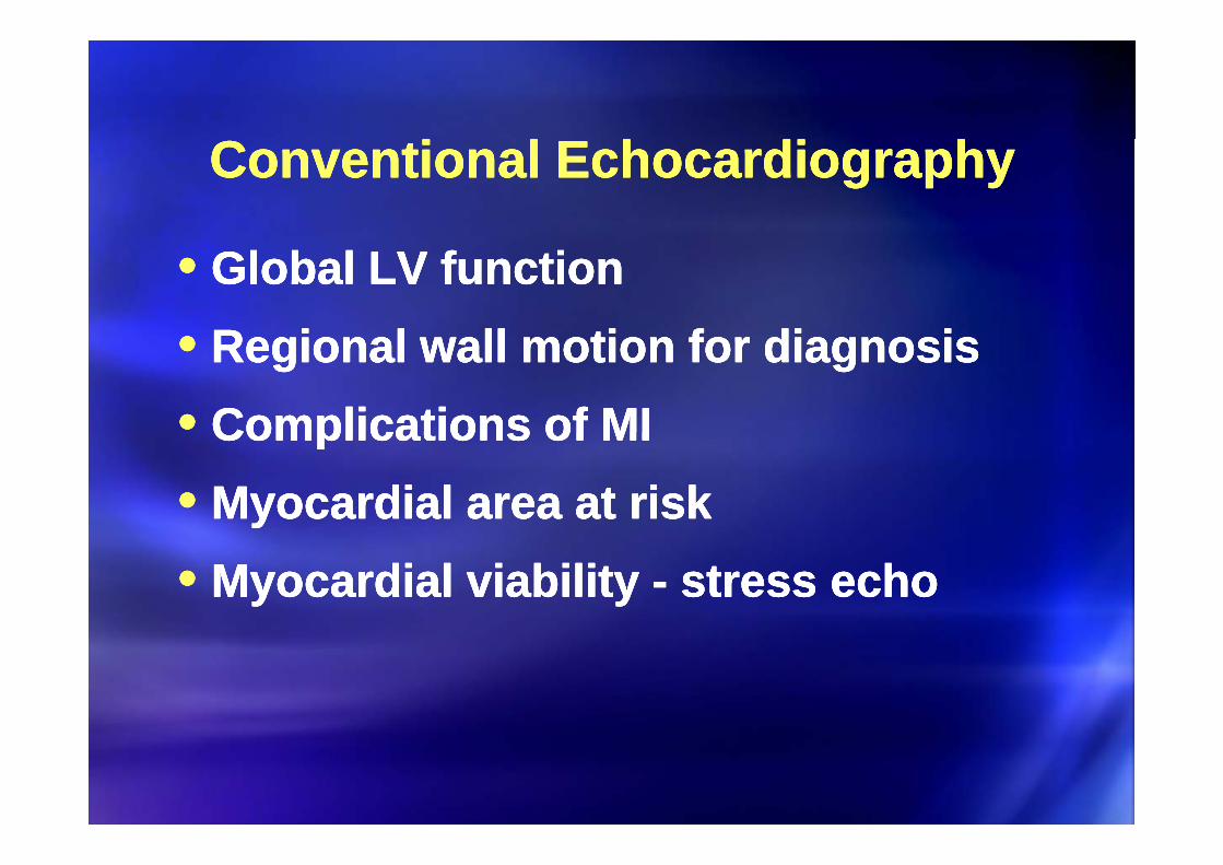
Conventional EchocardiographyConventional EchocardiographyConventional EchocardiographyConventional Echocardiography
•• Global LV functionGlobal LV functionR i l ll ti f di iR i l ll ti f di i
•• Global LV functionGlobal LV functionR i l ll ti f di iR i l ll ti f di i•• Regional wall motion for diagnosisRegional wall motion for diagnosis
•• Complications of MIComplications of MI•• Regional wall motion for diagnosisRegional wall motion for diagnosis•• Complications of MIComplications of MIComplications of MIComplications of MI•• Myocardial area at riskMyocardial area at risk
Complications of MIComplications of MI•• Myocardial area at riskMyocardial area at risk•• Myocardial viability Myocardial viability -- stress echostress echo•• Myocardial viability Myocardial viability -- stress echostress echo
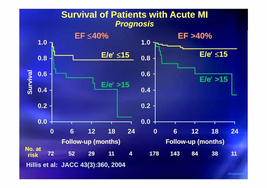
Survival of Patients with Acute MIPrognosis
Survival of Patients with Acute MIPrognosis
1.01.0EF 40%EF 40% EF >40%EF >40%
E/ 1E/ 1
0 6
0.8
0 6
0.8
alal
E/e 15E/e 15 E/e 15E/e 15
0.4
0.6
0.4
0.6
Surv
iva
Surv
iva
E/e >15E/e >15E/e >15E/e >15
0.20.2
SS
0.00 6 12 18 24
0.00 6 12 18 24
Follow-up (months)Follow-up (months)
7272 5252 2929 1111 44No. atrisk
No. atrisk 178178 143143 8484 3838 1111
Follow-up (months)Follow-up (months)
Hillis et al: JACC 43(3):360, 2004Hillis et al: JACC 43(3):360, 2004CP1141593-3

Prognostic Value of E/EPrognostic Value of E/E'' After Acute MIAfter Acute MIPrognostic Value of E/EPrognostic Value of E/E'' After Acute MIAfter Acute MI
1.0 1.0E/e' 15E/e' 15 E/ ' 15E/ ' 15
In DTBT <90In DTBT <90 In DTBT 90In DTBT 90h)h)
0.9
h)h)
0.9
E/e' 15E/e' 15
E/e' >15E/e' >15
E/e' 15E/e' 15
al (d
eath
al (d
eath
0.8
al (d
eath
al (d
eath
0.8
E/e 15E/e 15P<0.0001P<0.0001
Surv
iva
Surv
iva
0.7
Surv
iva
Surv
iva
0.7 E/e' >15E/e' >15P=0.06P=0.06
0.6 0.6
0.50 200 400 600 800
0.50 200 400 600 800
CP1302151-7
Follow-up (days)Follow-up (days) Follow-up (days)Follow-up (days)
Park SJ, Ting H, Oh JK Unpublished

Unconventional EchocardiographyUnconventional EchocardiographyUnconventional EchocardiographyUnconventional Echocardiography
•• Myocardial strain imagingMyocardial strain imagingC t t h di hC t t h di h
•• Myocardial strain imagingMyocardial strain imagingC t t h di hC t t h di h•• Contrast echocardiographyContrast echocardiography•• Contrast echocardiographyContrast echocardiography

Strain ImagingStrain ImagingStrain ImagingStrain Imaging• Strain: Deformation of an object, relative to its
original length• Strain: Deformation of an object, relative to its
original lengthoriginal lengthoriginal length
L - L0L - L0
L0L0
LL==
L0L0==
L0L0 L0L0
If 10 cm original length is shortened to 7 5 cmshortened to 7.5 cm,
strain is (-) 25 %.
CP1237840-8
Normal strain is > 20 %.

2D Speckle Tracking ImageNormal
Radial strain (SAX) Transversal strain (A4)
Aplio (SSA-770A, Toshiba Japan)K.Ogawa, T.Hozumi et al. AJC 2006

Longitudinal strain: normal case DisplayLongitudinal strain: normal case DisplayLongitudinal strain: normal case DisplayLongitudinal strain: normal case Display
4C 2C Lengthening
Shortening
APLAX
Shortening
Bull’s Eye

Bull’s Eye Mapping of StrainBull’s Eye Mapping of StrainBull’s Eye Mapping of StrainBull’s Eye Mapping of StrainBull s Eye Mapping of StrainBull s Eye Mapping of StrainBull s Eye Mapping of StrainBull s Eye Mapping of StrainLengthening+20% g g
Shortening-20% g
Anteroseptal MI (LAD) Inferior MI (RCA)Inferolateral MI (LCX)

C E h di hC E h di hC E h di hC E h di hContrast EchocardiographyContrast EchocardiographyContrast EchocardiographyContrast Echocardiography
Perfusion Defect in the apical segments

Contrast Perfusion Echo for ViabilityContrast Perfusion Echo for Viability2 patients with Anterior STEMI and PCI2 patients with Anterior STEMI and PCI
Contrast Perfusion Echo for ViabilityContrast Perfusion Echo for Viability2 patients with Anterior STEMI and PCI2 patients with Anterior STEMI and PCI2 patients with Anterior STEMI and PCI2 patients with Anterior STEMI and PCI2 patients with Anterior STEMI and PCI2 patients with Anterior STEMI and PCIBaseline Follow up

Is there apical thrombus?Is there apical thrombus?Is there apical thrombus?Is there apical thrombus?Is there apical thrombus?Is there apical thrombus?Is there apical thrombus?Is there apical thrombus?

CT for Myocardial ImagingCT for Myocardial ImagingCT for Myocardial ImagingCT for Myocardial Imaging
•• Coronary CT angiographyCoronary CT angiographyA t i l h di l i iA t i l h di l i i
•• Coronary CT angiographyCoronary CT angiographyA t i l h di l i iA t i l h di l i i•• Arterial phase myocardial imagingArterial phase myocardial imaging
•• Myocardial motion interpretationMyocardial motion interpretation•• Arterial phase myocardial imagingArterial phase myocardial imaging•• Myocardial motion interpretationMyocardial motion interpretationMyocardial motion interpretationMyocardial motion interpretation•• Viability imaging with delayed Viability imaging with delayed
Myocardial motion interpretationMyocardial motion interpretation•• Viability imaging with delayed Viability imaging with delayed
enhancementenhancementenhancementenhancement

Role of CCT in AMIRole of CCT in AMIRole of CCT in AMIRole of CCT in AMI
•• Evaluation of acute chest painEvaluation of acute chest painM di l i bilitM di l i bilit
•• Evaluation of acute chest painEvaluation of acute chest painM di l i bilitM di l i bilit•• Myocardial viabilityMyocardial viability•• Myocardial viabilityMyocardial viability


Quantification of Infarct SizeQuantification of Infarct SizeQuantification of Infarct SizeQuantification of Infarct Size

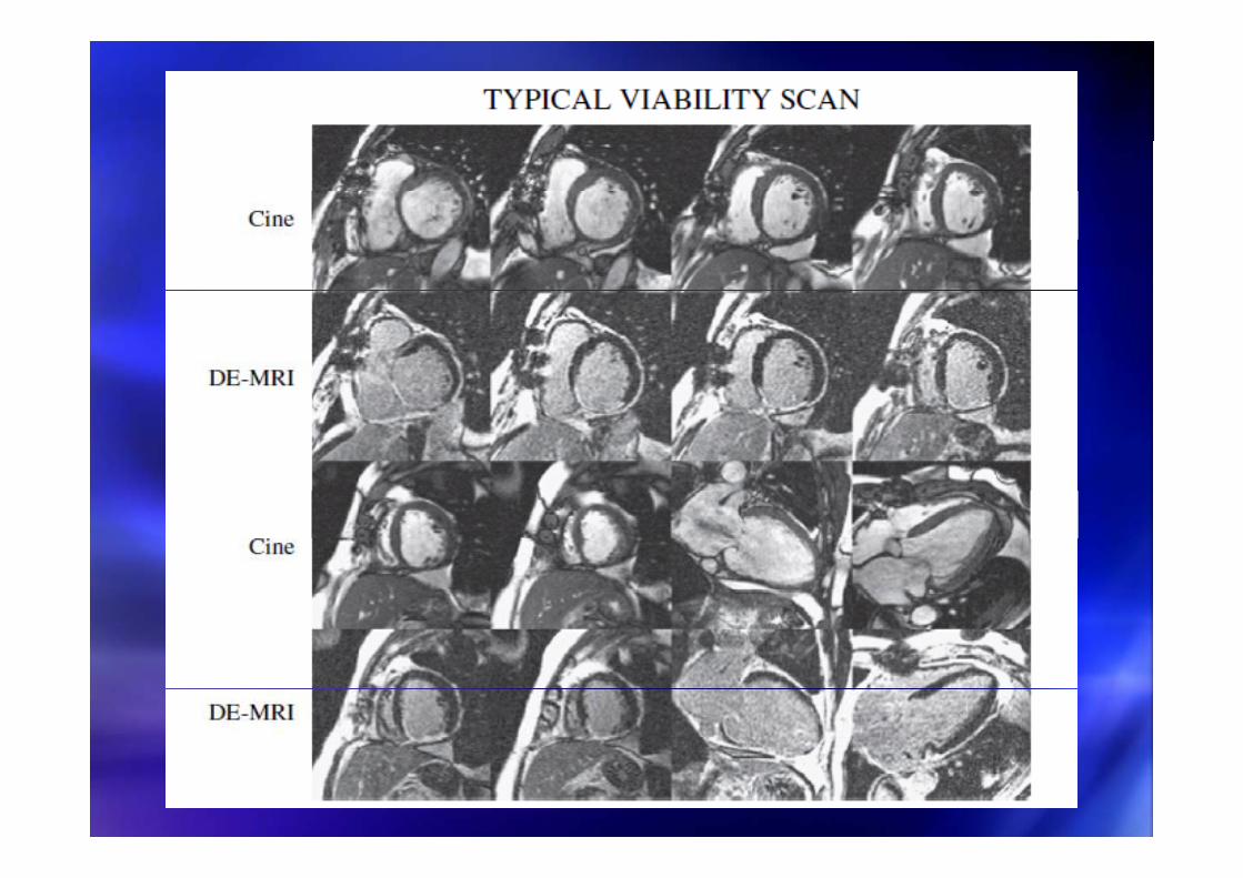
Myocardial ViabilityMyocardial ViabilityMyocardial ViabilityMyocardial Viability

DualDual--Phase CTPhase CTDualDual--Phase CTPhase CT

DualDual--Phase CTPhase CTDualDual--Phase CTPhase CT

CMR: Delayed EnhancementCMR: Delayed EnhancementCMR: Delayed EnhancementCMR: Delayed Enhancementyyyy

Two Patients with Inferior STEMITwo Patients with Inferior STEMISoon after PCISoon after PCI
Two Patients with Inferior STEMITwo Patients with Inferior STEMISoon after PCISoon after PCISoon after PCISoon after PCISoon after PCISoon after PCI
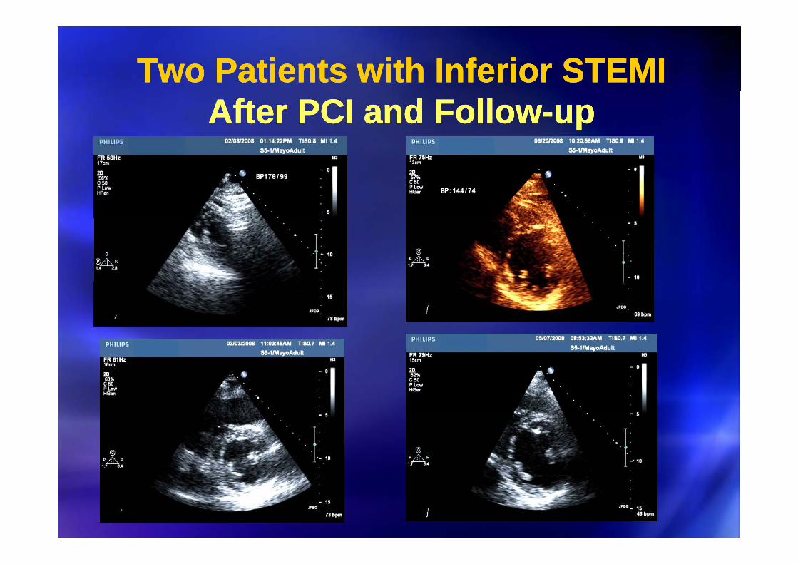
Two Patients with Inferior STEMITwo Patients with Inferior STEMITwo Patients with Inferior STEMITwo Patients with Inferior STEMIAfter PCI and FollowAfter PCI and Follow--upupAfter PCI and FollowAfter PCI and Follow--upup

Two Patients with Inferior STEMITwo Patients with Inferior STEMITwo Patients with Inferior STEMITwo Patients with Inferior STEMIFollowFollow--up Echo and Baseline MRIup Echo and Baseline MRIFollowFollow--up Echo and Baseline MRIup Echo and Baseline MRI
Small DE by Gd
Large DE by Gd

Myocardial Function and DHEMyocardial Function and DHEMyocardial Function and DHEMyocardial Function and DHE
•• Transmural extent of infarction and contractilityTransmural extent of infarction and contractility•• Transmural extent of infarction and contractilityTransmural extent of infarction and contractility
Marholdt, JACC 2003Marholdt, JACC 2003

All DysfunctionalS t
All DysfunctionalS t
Segments withSevere Hypokinesia,
Aki i D ki i
Segments withSevere Hypokinesia,
Aki i D ki iSegments with
Aki i D ki iSegments with
Aki i D ki i
100%
)%
) (128(128
(12of 12)
(12of 12)
SegmentsSegments Akinesia, or DyskinesiaAkinesia, or Dyskinesia Akinesia or DyskinesiaAkinesia or Dyskinesia
80
ility
(%ili
ty (% (256
of 329)(256
of 329)
(128of 148)
(128of 148)
(56 of(56 of
(23of 28)
(23of 28)
60
ontr
act
ontr
act (109
of 183)(109
of 183)
(45 of110)
(45 of110)
86)86)
(29 of68)
(29 of68)
(9of 20)
(9of 20)
20
40
oved
co
oved
co 110)110) 68)68) ))
0
20
Impr
oIm
pro (13 of
124)(13 of124)
(1 of58)
(1 of58)
(10 of103)
(10 of103)
(0 of57)
(0 of57)
(4of 54)
(4of 54) (0 of
46)(0 of46)0 0 1-25
26-50
51-75
76-100
0 1-25
26-50
51-75
76-100
0 1-25
26-50
51-75
76-100
CP1302210-5
Transmural extent of hyperenhancement (%)Transmural extent of hyperenhancement (%)
R. Kim et al NEJM 2000

Prognosis Associated with DHEPrognosis Associated with DHEPrognosis Associated with DHEPrognosis Associated with DHE
Chest pain without known OMIChest pain without known OMI PostPost--STEMISTEMI
Kwong, Circ 2006Kwong, Circ 2006 Wu, Heart 2008Wu, Heart 2008

Additional Information:Additional Information:Additional Information:Additional Information:Additional Information:Additional Information:Microvascular ObstructionMicrovascular Obstruction
Additional Information:Additional Information:Microvascular ObstructionMicrovascular Obstruction
Sakuma, JMRI 2007Sakuma, JMRI 2007

Functional Recovery after AMI andFunctional Recovery after AMI andFunctional Recovery after AMI andFunctional Recovery after AMI andFunctional Recovery after AMI and Functional Recovery after AMI and MOMO
Functional Recovery after AMI and Functional Recovery after AMI and MOMO
Nijveldt, JACC 2008Nijveldt, JACC 2008

MO and PrognosisMO and PrognosisMO and PrognosisMO and Prognosis
Wu, Circulation 1998Wu, Circulation 1998

AMI with EdemaAMI with EdemaAMI with EdemaAMI with Edema
Sakuma, JMRI 2007Sakuma, JMRI 2007

T2WI for Edema in MIT2WI for Edema in MIT2WI for Edema in MIT2WI for Edema in MI
Myoview ScanT2WI-triple IR

Acute MI with SwellingAcute MI with SwellingAcute MI with SwellingAcute MI with Swelling
T2WI
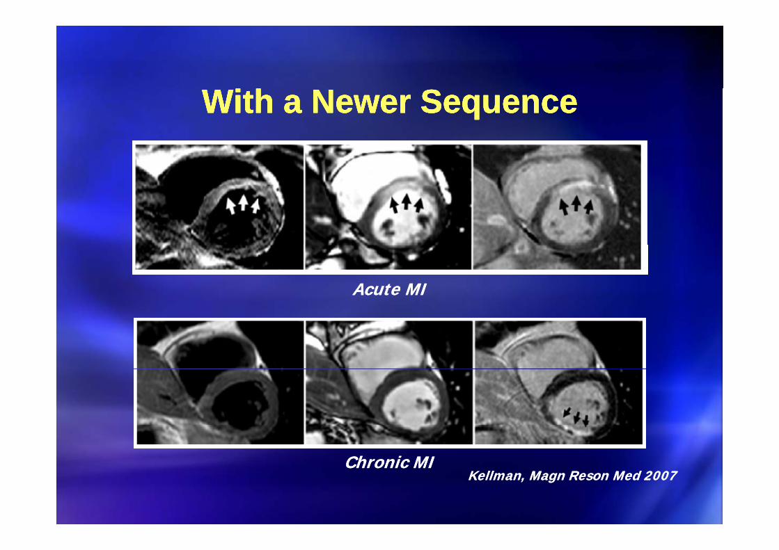
With a Newer SequenceWith a Newer SequenceWith a Newer SequenceWith a Newer Sequence
Acute MIAcute MI
Chronic MIChronic MIChronic MIChronic MIKellman, Magn Reson Med 2007Kellman, Magn Reson Med 2007

PeriPeri--Infarct Zone EnhancementInfarct Zone EnhancementPeriPeri--Infarct Zone EnhancementInfarct Zone EnhancementPeriPeri Infarct Zone Enhancement Infarct Zone Enhancement (The “Grey” Zone)(The “Grey” Zone)
PeriPeri Infarct Zone Enhancement Infarct Zone Enhancement (The “Grey” Zone)(The “Grey” Zone)
2-3 SD
>3 SD3 SD
Yan AT et al. Circulation 2006;114;32-39.

STEMI ?STEMI ?STEMI ?STEMI ?Chest and left arm pain for 1 hr. Chest and left arm pain for 1 hr.
Increased TroponinIncreased TroponinChest and left arm pain for 1 hr. Chest and left arm pain for 1 hr.
Increased TroponinIncreased Troponinpppp

65 year old woman with chest pain65 year old woman with chest pain65 year old woman with chest pain65 year old woman with chest pain

Cine Delayed EnhancementCine Delayed EnhancementCine Delayed EnhancementCine Delayed Enhancement
Courtesy of Siemens Med. Systems

Cardiac MRI New Technology
Diffusion Tensor Diffusion Tensor ImagingImaging
Diffusion Tensor Diffusion Tensor ImagingImagingg gg gg gg g
Myocardial Strain Myocardial Strain ImagingImaging

Evaluation of Chest PainEvaluation of Chest Pain
ImagingImagingImagingImagingPrognosisViability
PrognosisViability
FunctionInfarct sizeFunction
Infarct sizeCADViabilityViability Infarct sizeInfarct sizeCAD
Unstable Hemodynamics d C li ti
Unstable Hemodynamics d C li ti
CP1210291-8
and Complicationsand Complications


