DNA Repair and Recombination in Plants 3Susan Schropfer, Alexander Knoll, Oliver Trapp, and Holger Puchta
Contents
Introduction . . . . . . . . . . . . . . . . . . . . . . . . . . . . . . . . . . . . . . . . . . . . . . . . . . . . . . . . . . . . . . . . . . . . . . . . . . . . . . . . . . . . . . . 52
DNA-Damaging Factors and DNA Lesions . . . . . . . . . . . . . . . . . . . . . . . . . . . . . . . . . . . . . . . . . . . . . . . . . . . . . 53
Enzymatic Reversal of DNA Damage . . . . . . . . . . . . . . . . . . . . . . . . . . . . . . . . . . . . . . . . . . . . . . . . . . . . . . . . . . . 60
Base Excision Repair (BER) . . . . . . . . . . . . . . . . . . . . . . . . . . . . . . . . . . . . . . . . . . . . . . . . . . . . . . . . . . . . . . . . . . . . . 63
Nucleotide Excision Repair (NER) . . . . . . . . . . . . . . . . . . . . . . . . . . . . . . . . . . . . . . . . . . . . . . . . . . . . . . . . . . . . . . 70
Mismatch Repair (MMR) . . . . . . . . . . . . . . . . . . . . . . . . . . . . . . . . . . . . . . . . . . . . . . . . . . . . . . . . . . . . . . . . . . . . . . . . 75
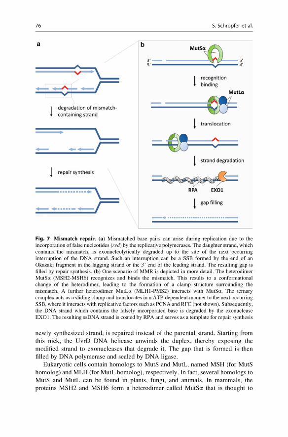
Repair of Double-Strand Breaks . . . . . . . . . . . . . . . . . . . . . . . . . . . . . . . . . . . . . . . . . . . . . . . . . . . . . . . . . . . . . . . . . 78
Tolerance and Repair Processes at Damaged Replication Forks . . . . . . . . . . . . . . . . . . . . . . . . . . . . . . . 84
Cellular Changes and Signaling After DNA Damage . . . . . . . . . . . . . . . . . . . . . . . . . . . . . . . . . . . . . . . . . . . 89
Future Directions . . . . . . . . . . . . . . . . . . . . . . . . . . . . . . . . . . . . . . . . . . . . . . . . . . . . . . . . . . . . . . . . . . . . . . . . . . . . . . . . . 91
References . . . . . . . . . . . . . . . . . . . . . . . . . . . . . . . . . . . . . . . . . . . . . . . . . . . . . . . . . . . . . . . . . . . . . . . . . . . . . . . . . . . . . . . . 92
Abstract
• DNA damage can be caused by a large number of internal and external, biotic
and abiotic sources and can affect cell viability and can lead to mutations.
• Depending on the type of damage, different evolutionarily conserved repair
pathways are used.
• Some specific lesions caused by UV radiation and DNA alkylation can be
repaired by direct enzymatic reversal.
• The base excision repair pathway is used for the removal of a variety of
damaged bases.
S. Schropfer • A. Knoll • O. Trapp • H. Puchta (*)
Botanical Institute II, Karlsruhe Institute of Technology, Karlsruhe, Germany
e-mail: [email protected]; [email protected]; [email protected];
# Springer Science+Business Media New York 2014
S.H. Howell (ed.), Molecular Biology, The Plant Sciences 2,DOI 10.1007/978-1-4614-7570-5_2
51
• When larger modifications of nucleotides are present, the nucleotide excision
repair pathway is active.
• The mismatch repair pathway can reverse the incorporation of
noncomplementary nucleotides by replicative polymerases.
• DNA double-strand breaks can be repaired by the pathways of
nonhomologous end-joining, single-strand annealing, and homologous
recombination which lead to different outcomes.
• Different DNA damage tolerance and repair pathways can deal with DNA
lesions at damaged replication forks.
• Repair of DNA has to be tightly regulated with respect to other cellular
processes.
• DSB repair pathways form the basis for recently developed techniques for
directed modification of genomes for research and agronomy.
Introduction
DNA is a biomolecule which represents the basis for all living organisms by
encoding the information for all processes in life. The maintenance of genome
stability by counteracting changes in DNA is a great challenge, which has to be
achieved in every single cell.
Different kinds of mutations with diverse consequences for the cell can arise due
to a multitude of factors. Point mutations are changes of a single nucleotide, for
example, a substitution of one base by another base. An exchange of a pyrimidine
by a pyrimidine base (e.g., T/C) or a purine by a purine base (e.g., A/G) is defined as
a transition. In contrast, a substitution of a pyrimidine with a purine base (e.g., T/G
or A) or a purine with a pyrimidine base (e.g., A/C or T) is called a transversion. A
point mutation in an open reading frame (ORF) of a gene can lead to a substitution
of an amino acid in the resulting protein, which might change the properties of the
respective protein. Nonsense point mutations, e.g., by introducing a stop codon in
the middle of an ORF, can result in a complete loss of function of the encoded
protein. Other changes in DNA caused by insertions or deletions of one or more
nucleotides might also drastically affect protein biosynthesis, e.g., by causing a
frame shift. In contrast to mutations which concern a single DNA sequence, also
large rearrangements of chromosomes such as inversions and translocations can
arise due to aberrant recombination processes. Furthermore, huge insertions and
deletions can occur as well. Insertions of many long DNA sequences into a genomic
locus such as coding or regulating DNA sequences can be due to an insertion of a
mobile DNA element such as a transposon. Certain bacterial species can insert huge
pieces of foreign DNA into a plant genome, such as Agrobacterium tumefaciens,which transfers a so-called transfer DNA (T-DNA) into plant cells. T-DNA is
randomly integrated into the plant genome and codes for metabolic enzymes
which reprogram the metabolism of the host plant and provide nutrients specifically
for the bacterium.
52 S. Schropfer et al.
Mutations can arise due to a variety of DNA damage. DNA is constantly affected
by DNA-damaging factors which can cause modifications in the chemical compo-
sition and structure of DNA. In contrast to the majority of other biomolecules,
lesions in DNA may lead to the formation of stable mutations which will be
inherited by all daughter cells and may affect their metabolism permanently.
Therefore, the manifold lesions that can arise in DNA can be repaired by a complex
and conserved system of DNA repair mechanisms to avoid the expression of such
mutations in the genome. In plants, the repair of damaged DNA has a special
relevance because plants do not have a predetermined germ line. Mutations in
DNA can originate in somatic tissue which later gives rise to germ cells and can
thus be passed on to the next generation.
Endogenous factors such as genotoxic by-products of the cellular metabolism or
errors in DNA replication and recombination can lead to spontaneous DNA dam-
age. Furthermore, exogenous factors coming from the abiotic and biotic environ-
ment can also affect DNA. Some examples of the various exogenous factors are
energy-rich radiation such as ionizing or solar UV radiation, naturally occurring
chemical genotoxins produced by some bacteria or fungi, as well as synthetic
chemical agents. In the following sections the formation of different types of
DNA damage by various DNA-damaging factors is discussed, followed by a
description of the current state of knowledge on the diverse pathways that repair
these lesions.
DNA-Damaging Factors and DNA Lesions
Replication-associated DNA damage: During the duplication of chromosomes in
S phase of the cell cycle, DNA is replicated by DNA polymerases. An incorporation
of a noncomplementary nucleotide opposite to the parental template DNA strand
can be detected and counteracted by the proofreading activity of the replicative
DNA polymerases. In such case, the misincorporated nucleotide is removed from
the newly synthesized DNA strand mediated by an intrinsic exonuclease domain.
Despite such proofreading activity of the replicative DNA polymerases, a small
fraction of misincorporated nucleotides persist in DNA, leading to a mutation rate
of about 10�8 per replication round. Because of nonmatching and different base
pairing properties of the opposing nucleotides, a so-called DNA mismatch is
formed which also affects the structure and stability of DNA. If a mismatch is not
detected until the next round of DNA replication is initiated, the incorrectly
incorporated nucleotide is then a component of the parental template stand, which
causes a stable establishment of the mutation by the incorporation of the comple-
mentary nucleotide.
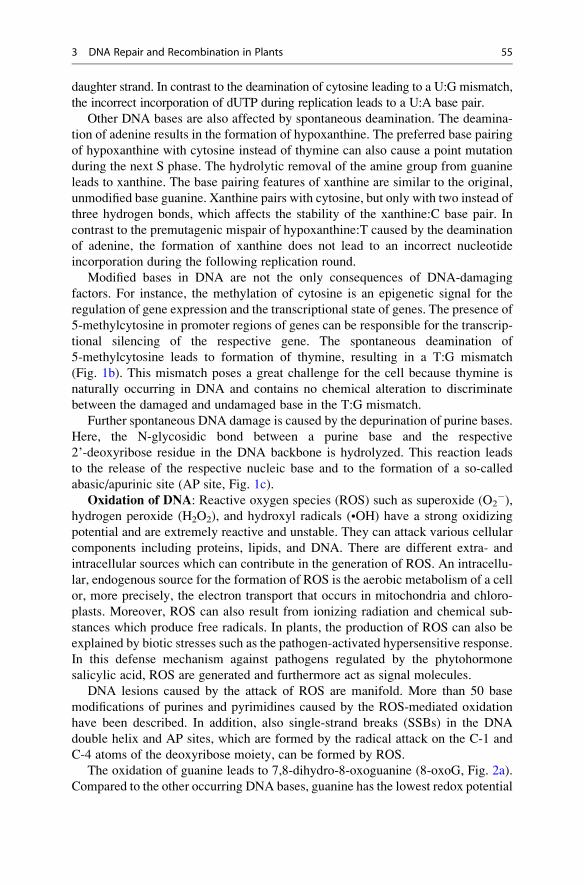
Spontaneous hydrolytic DNA damage: DNA is exposed to spontaneous mod-
ifications by hydrolysis of chemical bonds, such as deamination and depurination,
leading to a change of the chemical composition of the DNA molecule. A deam-
ination of DNA causes the removal of an amine group from DNA bases, whereas a
depurination leads to a complete loss of a purine base (Fig. 1).
3 DNA Repair and Recombination in Plants 53
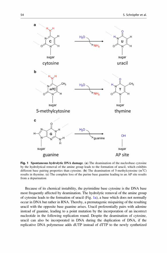
Because of its chemical instability, the pyrimidine base cytosine is the DNA base
most frequently affected by deamination. The hydrolytic removal of the amine group
of cytosine leads to the formation of uracil (Fig. 1a), a base which does not normally
occur in DNA but rather in RNA. Thereby, a premutagenic mispairing of the resulting
uracil with the opposite base guanine arises. Uracil preferentially pairs with adenine
instead of guanine, leading to a point mutation by the incorporation of an incorrect
nucleotide in the following replication round. Despite the deamination of cytosine,
uracil can also be incorporated in DNA during the duplication of DNA, if the
replicative DNA polymerase adds dUTP instead of dTTP to the newly synthetized
Fig. 1 Spontaneous hydrolytic DNA damage. (a) The deamination of the nucleobase cytosine
by the hydrolytical removal of the amine group leads to the formation of uracil, which exhibits
different base pairing properties than cytosine. (b) The deamination of 5-methylcytosine (m5C)
results in thymine. (c) The complete loss of the purine base guanine leading to an AP site results
from a depurination
54 S. Schropfer et al.
daughter strand. In contrast to the deamination of cytosine leading to a U:G mismatch,
the incorrect incorporation of dUTP during replication leads to a U:A base pair.
Other DNA bases are also affected by spontaneous deamination. The deamina-
tion of adenine results in the formation of hypoxanthine. The preferred base pairing
of hypoxanthine with cytosine instead of thymine can also cause a point mutation
during the next S phase. The hydrolytic removal of the amine group from guanine
leads to xanthine. The base pairing features of xanthine are similar to the original,
unmodified base guanine. Xanthine pairs with cytosine, but only with two instead of
three hydrogen bonds, which affects the stability of the xanthine:C base pair. In
contrast to the premutagenic mispair of hypoxanthine:T caused by the deamination
of adenine, the formation of xanthine does not lead to an incorrect nucleotide
incorporation during the following replication round.
Modified bases in DNA are not the only consequences of DNA-damaging
factors. For instance, the methylation of cytosine is an epigenetic signal for the
regulation of gene expression and the transcriptional state of genes. The presence of
5-methylcytosine in promoter regions of genes can be responsible for the transcrip-
tional silencing of the respective gene. The spontaneous deamination of
5-methylcytosine leads to formation of thymine, resulting in a T:G mismatch
(Fig. 1b). This mismatch poses a great challenge for the cell because thymine is
naturally occurring in DNA and contains no chemical alteration to discriminate
between the damaged and undamaged base in the T:G mismatch.
Further spontaneous DNA damage is caused by the depurination of purine bases.
Here, the N-glycosidic bond between a purine base and the respective
2’-deoxyribose residue in the DNA backbone is hydrolyzed. This reaction leads
to the release of the respective nucleic base and to the formation of a so-called
abasic/apurinic site (AP site, Fig. 1c).
Oxidation of DNA: Reactive oxygen species (ROS) such as superoxide (O2�),
hydrogen peroxide (H2O2), and hydroxyl radicals (•OH) have a strong oxidizing
potential and are extremely reactive and unstable. They can attack various cellular
components including proteins, lipids, and DNA. There are different extra- and
intracellular sources which can contribute in the generation of ROS. An intracellu-
lar, endogenous source for the formation of ROS is the aerobic metabolism of a cell
or, more precisely, the electron transport that occurs in mitochondria and chloro-
plasts. Moreover, ROS can also result from ionizing radiation and chemical sub-
stances which produce free radicals. In plants, the production of ROS can also be
explained by biotic stresses such as the pathogen-activated hypersensitive response.
In this defense mechanism against pathogens regulated by the phytohormone
salicylic acid, ROS are generated and furthermore act as signal molecules.
DNA lesions caused by the attack of ROS are manifold. More than 50 base
modifications of purines and pyrimidines caused by the ROS-mediated oxidation
have been described. In addition, also single-strand breaks (SSBs) in the DNA
double helix and AP sites, which are formed by the radical attack on the C-1 and
C-4 atoms of the deoxyribose moiety, can be formed by ROS.
The oxidation of guanine leads to 7,8-dihydro-8-oxoguanine (8-oxoG, Fig. 2a).
Compared to the other occurring DNA bases, guanine has the lowest redox potential
3 DNA Repair and Recombination in Plants 55
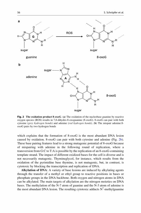
which explains that the formation of 8-oxoG is the most abundant DNA lesion
caused by oxidation. 8-oxoG can pair with both cytosine and adenine (Fig. 2b).
These base pairing features lead to a strong mutagenic potential of 8-oxoG because
of mispairing with adenine in the following round of replication, where a
transversion from G:C to T:A is possible by the replication of an 8-oxoG-containing
template strand. The impact of different oxidized bases for the cell is diverse and is
not necessarily mutagenic. Thymineglycol, for instance, which results from the
oxidation of the pyrimidine base thymine, is not mutagenic, but, in contrast, is
cytotoxic by blocking the transcription and replication of DNA.
Alkylation of DNA: A variety of base lesions are induced by alkylating agents
through the transfer of a methyl or ethyl group to reactive positions in bases or
phosphate groups in the DNA backbone. Both oxygen and nitrogen atoms in DNA
can be alkylated. The main targets of alkylation are the nitrogen moieties on DNA
bases. The methylation of the N-7 atom of guanine and the N-3 atom of adenine is
the most abundant DNA lesion. The resulting cytotoxic adducts N7-methylguanine
Fig. 2 The oxidation product 8-oxoG. (a) The oxidation of the nucleobase guanine by reactive
oxygen species (ROS) results in 7,8-dihydro-8-oxoguanine (8-oxoG). 8-oxoG can pair with both
cytosine (grey hydrogen bonds) and adenine (red hydrogen bonds). (b) The mispair adenine:8-
oxoG pairs by two hydrogen bonds
56 S. Schropfer et al.
(N7-MeGua) and N3-methyladenine (N3-MeAde), which affect proper transcription
and replication, account for more than 80 % of all methylation events. In contrast,
the methylation of the oxygen bound to C-6 of guanine leading to O6-
methylguanine (O6-MeGua) also has mutagenic potential because of changed
base pairing features. O6-MeGua predominately pairs with thymine. Accordingly,
the persistence of O6-MeGua in the parental DNA strand leads to an incorporation
of thymine instead of cytosine during DNA replication.
Alkylating agents such as nitrogen mustards, nitrosoureas and alkyl sulfonates,
triazines, and ethylenimines are separated into two subgroups dependent on their
reaction mechanism. SN1-type agents can alkylate oxygen as well as nitrogen atoms,
whereas SN2-type chemicals are only able to alkylate nitrogen atoms in nucleic acids.
The most important environmental alkylating agent is methyl methanesulfonate
(MMS), which methylates ring nitrogen residues in DNA bases, resulting in partic-
ular in N7-MeGua and N3-MeAde. Like another mutagenic agent ethyl
methanesulfonate (EMS), used for the generation of plant mutants in research and
agriculture, MMS is an SN2-type agent and belongs to the group of alkyl sulfonates.
DNA damage induced by energy-rich radiation: Different types of energy-
rich radiation types like ionizing radiation and UV radiation can lead to the
formation of damaged DNA.
X-rays and γ-radiation have ionizing features and are defined as electromagnetic
waves like light, but they transmit much more energy than visible light. On the one
hand, ionizing radiation can have a direct effect on cellular compounds. In such a
case, SSBs and double-strand beaks (DSBs) in DNA can be directly generated.
Especially DSBs represent a great danger for the integrity of the genome. For
instance, the presence of a single DSB during replication may lead to the complete
loss of chromosome fragments and all genetic information encoded therein. Addi-
tionally, the repair of DSBs by different recombination pathways (discussed in
more detail in section Mismatch Repair (MMR)) is associated with a high risk of
mutations like insertions, deletions, and chromosomal rearrangements. On the other
hand, ionizing radiation can also have an indirect effect on DNA by the production
of ROS through the interaction with water molecules. DNA lesions caused by
oxidation through ROS were described above.
Visible sunlight, on one hand, is essential for plant life to power photosynthesis.
But on the other hand, the UV fraction of sunlight represents a near constant source
of DNA damage. Solar UV radiation is categorized as UV-C (180–290 nm), UV-B
(290–320 nm), and UV-A (320–400 nm). The energy content of radiation is
inversely proportional to wavelength. For that reason, UV-C is the most and
UV-A the least energetic UV radiation. Animals and plants are most affected by
UV-B, the main fraction of genotoxic sunlight, penetrating and damaging their
genomes. In contrast to animals, plants are not able to reduce the exposure to solar
UV radiation by changing their location. The strategy of plants to minimize
UV-induced DNA damage is to accumulate secondary metabolites (e.g.,
UV-absorbing flavonoids) in the epidermal layers which capture UV radiation
and attenuate the UV dose. Despite such shielding, a portion of UV radiation
attacking DNA reaches epidermal levels and tissues beyond.
3 DNA Repair and Recombination in Plants 57
The DNA-damaging effect of UV radiation is explained by the absorption
spectrum of the DNA molecule. DNA has a maximum of absorption at 260 nm.
The absorption of energy from UV radiation by DNA leads to the generation of
so-called bulky DNA lesions, which are formed through the dimerization of
neighboring pyrimidine bases. These bulky DNA lesions such as cyclobutane
pyrimidine dimers (CPDs) and pyrimidine (6–4) pyrimidone photoproducts (6–4
PPs) are not able to base pair anymore and inhibit replication and transcription by
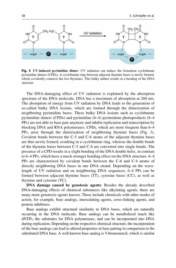
blocking DNA and RNA polymerases. CPDs, which are more frequent than 6–4
PPs, arise through the dimerization of neighboring thymine bases (Fig. 3).
Covalent bonds between the C-5 and C-6 atoms of the adjacent thymine bases
are thus newly formed, resulting in a cyclobutane ring, whereas the double bonds
of the thymine bases between C-5 and C-6 are converted into single bonds. The
presence of a CPD results in a slight bending of the DNA double helix, in contrast
to 6–4 PPs, which have a much stronger bending effect on the DNA structure. 6–4
PPs are characterized by covalent bonds between the C-6 and C-4 atoms of
directly neighboring DNA bases in one DNA strand. Depending on the wave-
length of UV radiation and on neighboring DNA sequences, 6–4 PPs can be
formed between adjacent thymine bases (TT), cytosine bases (CC), as well as
thymine and cytosine (TC).
DNA damage caused by genotoxic agents: Besides the already described
DNA-damaging effects of chemical substances like alkylating agents, there are
many more genotoxic agents known. These include chemicals with other modes of
action, for example, base analogs, intercalating agents, cross-linking agents, and
protein inhibitors.
Base analogs exhibit structural similarity to DNA bases, which are naturally
occurring in the DNA molecule. Base analogs can be metabolized much like
dNTPs, the substrates for DNA polymerases, and can be incorporated into DNA
during replication. Depending on the respective chemical structure, the incorporation
of the base analogs can lead to altered properties in base pairing in comparison to the
substituted DNA base. A well-known base analog is 5-bromouracil, which is similar
Fig. 3 UV-induced pyrimidine dimer. UV radiation can induce the formation cyclobutane
pyrimidine dimers (CPDs). A cyclobutane ring between adjacent thymine bases is newly formed,
which covalently connects the two thymines. This bulky adduct results in a bending of the DNA
structure
58 S. Schropfer et al.
to thymine. The frequently occurring enol tautomer of 5-bromouracil can pair with
guanine instead of adenine, which explains the mutagenic effect of 5-bromouracil.
DNA intercalating agents such as proflavine, ethidium bromide, or acridine
orange are flat molecules containing a polycyclic ring system. They can intercalate
into DNA by inserting between stacked base pairs, which may introduce changes in
DNA winding, and subsequently lead to insertions or deletions during replication.
The mutagenic potential is explained by failures in DNA synthesis caused by the
introduced changes in DNA winding. For instance, DNA polymerase might incor-
porate an additional nucleotide if an intercalating agent is integrated in the template
strand, resulting in an insertion event, or the polymerase can also jump over a
nucleotide causing a deletion.
DNA cross-linkers are able to form covalent adducts with DNA and can generate
cross-links (CLs) in one DNA strand (intrastrand CL) or between both complemen-
tary DNA strands (interstrand CL). The synthetic molecule cisplatin, for example,
preferentially produces intrastrand CLs. In contrast, mitomycin C (MMC), which is
a secondary metabolite of the bacterium Streptomyces caespitosus, leads predom-
inantly to the formation of interstrand CLs. After the uptake of the cross-linker into
the cell, the molecule is bioactivated, resulting in an instable and reactive interme-
diate. Following a first reaction step, in which the cross-linker forms a monoadduct
on DNA, the second reactive center of the cross-linker can form a further covalent
bond with DNA.
Genotoxic agents can also act as inhibitors of enzymes which are involved in the
DNA metabolism. The alkaloid camptothecin (CPT), which is present in all organs
of the Happy tree Camptotheca acuminata, is an inhibitor of the topoisomerase
I. Topoisomerase I catalyzes the relaxation of supercoiled DNA arising during
replication and transcription by the formation of a reversible SSBs in DNA, the
strand passage, and the subsequent resealing of the break. CPT binds the
DNA-topoisomerase I complex and inhibits the resealing of the introduced SSB,
which then persists in DNA. During replication, topoisomerase I-bound SSBs can
also be converted into DSBs when the replication fork meets the SSB. Caffeine,
another genotoxin, comes from the secondary metabolism of different plants, most
notably coffee and tea plants. Caffeine can lead to the accumulation of various
types of DNA damage by inhibiting the important kinases ATM and ATR, which
have a role in cell cycle progression after DNA damage (see section Tolerance and
Repair Processes at Damaged Replication Forks).
The accumulation of DNA damage leads to genotoxic stress and is a risk for
genome stability. The chemical modifications of DNA damage often result in
structural changes in DNA, which can be detected by the DNA repair machinery.
The following sections of this chapter deal with different specialized DNA
repair mechanisms, which are indispensable to maintain genome stability.
For more information about DNA damage and repair pathways in plants,
see also other general reviews on this topic (Vonarx et al. 1998;
Kunz et al. 2005; Roldan-Arjona and Ariza 2009; Tuteja et al. 2009; Spampinato
and Gomez-Casati 2012).
3 DNA Repair and Recombination in Plants 59
Enzymatic Reversal of DNA Damage
The formation of various DNA lesions, such as UV photoproducts or modified
bases, can be enzymatically reversed instead of being repaired through the excision
pathways (see also reviews Weber 2005; Goosen and Moolenaar 2008; Dalhus
et al. 2009; He et al. 2011). In this section the reversion of these lesions is discussed.
Direct repair mechanisms are quite simple, as they only need a single enzyme,
compared to the complex multi-protein excision repair pathways.
Most of the research in this field was done on bacterial enzymes, but further
work demonstrated that there is a very high level of conservation of these proteins
between the different kingdoms. Some of the information presented here was
gained through research in bacteria, but the basic findings are also applicable to
the plant proteins.
The direct reversal reactions of UV photoproducts, termed photoreactivation, are
extremely important for plants, because they cannot avoid UV radiation like
animals due to their sessile lifestyle. First, the photoreactivation will be described,
followed by a short excursion to cryptochromes, which are related to photolyases
but offer only limited DNA repair capability. Instead, most of them have acquired a
role as light receptor and signaling component. Afterward, the direct reversal of
modified bases, which works in parallel to excision repair pathways, is explained in
the following sections.
Photoreactivation by photolyases: UV radiation is very toxic for the genome of
a cell, as the wavelength of the energetic UV light (180–400 nm) overlaps with the
absorption spectrum of DNA, which has a maximum at around 260 nm. The most
energy-rich UV-C radiation (180–290 nm) is effectively filtered out by the ozone
layer around the earth and therefore plays almost no role as a genotoxic factor.
UV-A radiation (320–400 nm) is not energetic enough to harm DNA directly, but
can be mutagenic through the production of harmful intermediates, like reactive
oxygen species. The principal component of UV radiation that damages DNA is
UV-B (290–320 nm), which mainly produces pyrimidine dimers. 70–80 % of all
UV photoproducts are CPDs and 20–30 % are 6–4 PPs. The type of damage is in
this case dependent on the DNA sequence and structure, but both exhibit genotoxic
effects by blocking the polymerases during transcription and replication and are
important factors for the development of skin cancer.
Plants usually accumulate shielding compounds like flavonoids that absorb UV
radiation in order to minimize the potential of getting harmed, but of course this
does not render them immune to UV radiation. In order to deal with the described
DNA lesions induced by UV radiation, plants can utilize the nucleotide excision
repair pathway to cut out the damaged DNA strand or use a direct reversion
pathway, photoreactivation.
The name “photoreactivation” comes from the fact that it is a direct reversal of
harmful photoproducts (“reactivation”) and its need for light (“photo”) energy to
function. It is facilitated by specialized enzymes called “photolyases,” which are
basic and widespread DNA repair proteins that are conserved in most of the species
living today. They act as monomers and can be classified by their specific substrates
60 S. Schropfer et al.
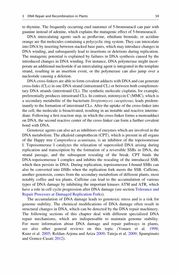
into CPD photolyases and 6–4 PP photolyases. Both kinds of photolyases recognize
and bind to a pyrimidine dimer, which is then extruded out of the DNA into the
active site of the photolyase. Subsequently, light-activated photolyases transfer an
electron to a pyrimidine dimer, which induces the dissolution of covalent bonds
between the neighboring pyrimidine bases (see Fig. 4).
CPD and 6–4 PP photolyases each contain a flavin adenine dinucleotide (FAD)
as a catalytic cofactor, which is needed for splitting pyrimidine dimers. In CPD
photolyases either pterin methenyltetrahydrofolate (MTHF) or deazaflavin
8-hydroxy-5-deazariboflavin (8-HDF) is non-covalently bound as a second cofac-
tor. These second cofactors are required to harvest light and make photoreactivation
more efficient. Concerning 6–4 PP photolyases, data shows that they might only use
MTHF as a second cofactor.
CPD photolyases are very well studied and can be further divided into two
classes by their amino acid similarity. Class I photolyases are generally found in
microbial organisms, while class II photolyases are mainly found in more complex
organisms, for example, in Arabidopsis thaliana and the fruit fly Drosophilamelanogaster. The human genome contains no active photolyase genes, and
humans rely only on the excision pathways in order to repair UV-induced DNA
damage, which could lead to mutations and skin cancer, if not repaired.
Photolyases repair DNA damage caused by the energy-rich, but for human eyes
invisible, UV light. Photolyases use less energetic wavelengths in the visible spectrum
in order to divide pyrimidine dimers. This is achieved through an electron transfer from
a two-electron reduced FADH- to the pyrimidine dimer. The FAD cofactor in the
photolyases can be directly excited by light in order to facilitate photoreactivation, but
this is a very inefficient process, asFADH- showsonlyweak absorption ofwavelengths
under 400 nm and almost no absorption of longer wavelengths. In order to increase
efficiency and absorbance spectra, photolyases harbor secondary chromophores that
serve as a light antenna. Methenyltetrahydrofolate (MTHF)-containing photolyases
exhibit their maximum catalytic activity when light between 377 and 410 nm is
present, and the deazariboflavin photolyases work best when irradiated by light of
wavelengths between 430 and 460 nm. The secondary antenna chromophores increase
the absorption and catalytic activity of photolyases significantly. The energy absorbed
by the second chromophore is transferred via resonance energy transfer onto the FAD
in order to generate the fully reduced and catalytically active FAHD- and excite it.
Fig. 4 Photoreactivation by photolyases. The photolyase recognizes and binds the thymine
dimer. Then, the photolyase is activated by visible light and catalyzes the dissolution of covalent
bonds between the neighboring thymine bases by an electron transfer onto the thymine dimer
3 DNA Repair and Recombination in Plants 61
Crystal structure analyses of CPD photolyases have shown that FAD is
non-covalently bound in a C-terminal groove in a unique U-shaped conformation
and that it is needed for the interaction of the photolyase with the damaged DNA.
The area around this groove is important for the contact between the protein and
the DNA. The pyrimidine dimer gets “flipped out” of the DNA double strand in a
way that it resides in the groove in close proximity to the FADH-. The excited
FADH- then transfers an electron to the CPD, thereby generating a semiquinone
FADH• radical and a CPD anion radical, which then undergoes monomerization
and transfers the electron back to the FADH• radical. By this reaction,
the CPD lesion is effectively repaired and the photolyase dissociates from the
DNA double strand.
Despite CPD and 6–4 PP photolyases being very similar at the protein sequence
level, much less information is available about 6–4 PP photolyases compared to
CPD photolyases. But from today’s point of view, although 6–4 PP photolyases are
not able to repair CPDs, the molecular mechanisms with which they repair 6–4 PPs
seem to be identical to the ones described for CPD photolyases.
Cryptochromes: Closely related to CPD photolyases are another group of pro-
teins, called cryptochromes. Cryptochromes were first identified in Arabidopsisthaliana and are less widespread than the photolyases (see review Chaves
et al. 2011). They are found in many plants and animals, but are rare in other
eukaryotes and prokaryotes. The proteins are basically photolyases that have lost
their ability to repair UV-induced lesions in the genome. Instead, they have
acquired a role as important blue light receptors and are involved in many pro-
cesses, like development, defense response, stress response, induction of flowering,
and the circadian clock.
Apart from the cryptochromes involved in blue light reception and signaling,
there is another class of cryptochromes, cryDASH, where DASH stands for Dro-sophila, Arabidopsis, Synechocystis, Homo. However, it needs to be mentioned that
despite their name, they are not present in Drosophila and humans. cryDASH
cryptochromes are still able to facilitate repair by light-induced photoreactivation
of pyrimidine dimers, but only on damaged single-stranded DNA (ssDNA) sub-
strates, whereas photolyases can efficiently repair ssDNA and double-stranded
DNA (dsDNA). Research showed that this is based on the fact that cryDASH
cannot flip the damaged base out of a DNA double strand. Another difference is
that cryDASH proteins only use MTHF, but no deazaflavin, as second cofactor,
although they are structurally similar to the photolyases. Whether cryDASH pro-
teins also exhibit signaling activity like other cryptochromes, is, however, not yet
known.
As all of the abovementioned photolyase-related protein classes are present in
plants, a view of the evolution of these proteins is very interesting: All classes seem
to have originated from one photolyase predecessor and are independent from each
other. Even cryDASH and cryptochromes represent independent classes and have
not developed from each other, although the functional characteristics of cryDASH
proteins would place them in between photolyases and cryptochromes.
62 S. Schropfer et al.
Enzymatic repair of base modifications: Not only UV-induced DNA damage
but also potentially mutagenic base modifications can be repaired directly through
specialized enzymes and are not necessarily restricted to repair through the excision
repair pathways. Little is known about these proteins in plants to date, and most of the
studies on the basic mechanisms of these direct reversal proteins have been conducted
in bacterial or mammalian cells, leading to merely a basic understanding in plants.
Through endogenous or exogenous substances, a lot of bases are modified every
day in each cell. These modified bases are potentially cytotoxic as they may block
replication and transcription or be mutagenic by having different base pairing
characteristics. One such example is the alkylated guanine O6-MeGua, which pre-
dominantly pairs with thymine instead of cytosine and therefore can lead to muta-
tions, if it is not repaired. The methylguanine-methyltransferase is a specialized repair
enzyme that can directly reverse this damage by removing the methyl group from the
guanine and transferring it to one of its own cysteines. The covalent binding of the
methyl group is irreversible, and therefore the protein can only repair one lesion
before it needs to be degraded. By definition, the methylguanine-methyltransferase is
not even a proper enzyme, as it is not able to catalyze the reaction more often.
Another example is the direct removal of 1-methyladenine and 3-methylcytosine,
which can be reverted by the oxidoreductase ALKBH2 in Arabidopsis thaliana, ahomolog to AlkB from E. coli, where initial studies were conducted.
Direct repair mechanisms are quite conserved throughout evolution and pose
important ways to secure genomic stability. However, as mentioned above, such
lesions can not only be repaired through enzymatic reversal but also through the
excision pathways.
Base Excision Repair (BER)
Instead of a direct repair of the damaged nucleotides, DNA lesions can also be
removed from the DNA by different excision repair systems such as base excision
repair (BER) and nucleotide excision repair (NER), which are discussed in this and
the following sections.
BER is a well-studied repair mechanism in mammals, and it is highly conserved
in prokaryotes and eukaryotes (see also reviews Fortini et al. 2003; Baute and
Depicker 2008; Dalhus et al. 2009; Roldan-Arjona and Ariza 2009; Wallace
et al. 2012). BER is a cellular process with a variety of different enzymatic
functions that occur in sequential steps, including the damage-specific recognition
and then removal of the base lesion followed by cleavage of the sugar-phosphate
backbone and excision of the abasic site (AP site). Subsequently, the resulting
single-stranded gap is filled and resealed, using the opposite, undamaged DNA
strand as template for DNA synthesis. By this repair mechanism, different types of
DNA lesions can be repaired, such as modified bases originating from deamination,
oxidation, and alkylation. Also AP sites resulting from the spontaneous hydrolysis
of a base are repaired by BER.
3 DNA Repair and Recombination in Plants 63
Principal process of BER: The initiating step of BER is carried by a DNA
glycosylase which recognizes the damaged base and hydrolyzes the N-glycosidic
bond between the damaged base and the respective deoxyribose residue. By the
action of the glycosylase, an AP site is generated (Fig. 5). For the further processing
of the AP site by the endonucleolytic cleavage of the DNA backbone, there are two
different possibilities (Fig. 5).
(1) Besides the recognition and the generation of AP sites, so-called bifunctional
DNA glycosylases can further process the AP site by their intrinsic 3’ AP lyase
activity. Here, a 5’ phosphate and a 3’ blocking lesion such as a 3’ α,β-unsaturatedaldehyde (PUA) after a β-elimination is generated. (2) Another way to process AP
sites is mediated by an AP endonuclease (such as the human APE1) after base
lesion removal by a monofunctional DNA glycosylase. The activity of the AP
endonuclease results in other products flanking the gap, compared to the action of
bifunctional glycosylases. At the 3’ terminus, a free hydroxyl (–OH) group is
generated, which can be directly used by the DNA polymerase without further
processing. At the opposite 5’ terminus of the single-strand gap, a deoxyribose-
phosphate (5’ dRP) is left by the AP endonuclease.
To fill the gap via polymerase-dependent DNA synthesis and a subsequent
ligation reaction, a 3’ OH group and a 5’ phosphate flanking the gap are required.
For that reason the 3’α,β-unsaturated aldehydes and also the 5’ dRPs have to be
converted into conventional 3’ OH and 5’ phosphate ends to allow gap filling
(Fig. 5). (1) 3’ blocking lesions generated by bifunctional DNA glycosylases are
removed by the 3’ diesterase activity of the AP endonuclease, whereas (2) 5’ dRPs
resulting from the action of an AP endonuclease are removed by the 5’ dRPase
activity of polymerase β.For gap filling and rejoining, there also exist two different sub-pathways:
(A) short-patch BER and (B) long-patch BER, which differ in the size of the repair
gap and also in the enzymes involved in the pathway (Fig. 5). (A) Short-patch BER,
which is also called single nucleotide BER, is characterized by the incorporation of
the correct single nucleotide and the processing of the 5’ end by polymerase β(or bacterial polymerase I), followed by the ligation of the remaining nick by a
complex consisting of DNA ligase III and XRCC1 (X-ray cross-complementing
1 or bacterial ligase I). (B) During long-patch BER, polymerase β also incorporatesthe initial missing nucleotide, but instead of a direct resealing of the nick, further
DNA synthesis occurs by the replicative polymerases δ or ε while the DNA strand
downstream of the initial damage site is displaced. Because of this differing
mechanism in long-patch BER, additional protein factors are required which have
also a well-known function in DNA replication. For example, the clamp protein
PCNA (proliferating cell nuclear antigen) is needed for the loading of the replica-
tive polymerases. Furthermore, the overhanging displaced DNA single strand, also
called a DNA flap, is removed by FEN1 (flap endonuclease 1), which endonucleo-
lytically cleaves at the base of the DNA flap structure. The resulting nick is sealed
by ligase I.
DNA glycosylases: Organisms possess a set of different DNA glycosylases,
which all exhibit several specificities for damaged DNA bases. In humans, there
64 S. Schropfer et al.
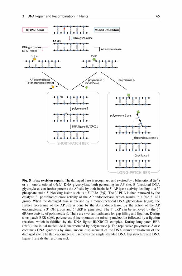
Fig. 5 Base excision repair. The damaged base is recognized and excised by a bifunctional (left)or a monofunctional (right) DNA glycosylase, both generating an AP site. Bifunctional DNA
glycosylases can further process the AP site by their intrinsic 3’ AP lyase activity, leading to a 5’
phosphate and a 3’ blocking lesion such as a 3’ PUA (left). The 3’ PUA is then removed by the
catalytic 3’ phosphodiesterase activity of the AP endonuclease, which results in a free 3’ OH
group. When the damaged base is excised by a monofunctional DNA glycosylase (right), thefurther processing of the AP site is done by the AP endonuclease. By the action of the AP
endonuclease, a 3’ OH group and 5’ dRP is generated. The 5’ dRP can be removed by the 5’
dRPase activity of polymerase β. There are two sub-pathways for gap filling and ligation. During
short-patch BER (left), polymerase β incorporates the missing nucleotide followed by a ligation
reaction, which is fulfilled by the DNA ligase III/XRCC1 complex. During long-patch BER
(right), the initial nucleotide is incorporated by polymerase β. The replicative polymerase δ or εcontinues DNA synthesis by simultaneous displacement of the DNA strand downstream of the
damaged site. The flap endonuclease 1 removes the single stranded DNA flap structure and DNA
ligase I reseals the resulting nick
3 DNA Repair and Recombination in Plants 65
have been eight DNA glycosylases described at present. In most cases, there is a
broad substrate spectrum of the DNA glycosylases that explain the overlapping
specificities of different glycosylases in some cases. The recognition of the DNA
damage by DNA glycosylases can be described as a pinch-push-plug-pull mecha-
nism. The DNA glycosylase slides along the minor grove of the DNA helix
scanning for specific modified bases, thereby bending the DNA double helix. A
kind of reading head is inserted into the minor groove of DNA (pinch), which then
pushes on the damaged base. This leads to a base flipping of the damaged base from
the interior of the DNA double helix into the active site pocket of the glycosylase,
where hydrogen bonding groups interact with the extrahelical base (plug and pull).
The active site pocket of the glycosylase determines their specificity, because the
extruded base has to match within. The substrate specificity depends on the shape,
the hydrogen binding, and also the electrostatic potential of the base.
There are two classes of DNA glycosylases. Monofunctional DNA glycosylases
only exhibit a glycosylase activity using an activated water molecule as a nucleo-
phile for the attack to release the damaged base. Thereby, no covalent intermediates
between the DNA glycosylase and the nucleotide are formed, and there is no
interruption of the DNA backbone generated by the DNA glycosylase. For the
breakage of the DNA backbone, a different enzyme, an AP endonuclease, is
required. In contrast, bifunctional glycosylases combine base excision by the
glycosylase activity with a DNA nicking activity leading to an SSB. The base
excision mechanism of a bifunctional glycosylase includes the formation of a
covalent Schiff base intermediate between a conserved lysine located in the active
site pocket of the glycosylase and the aldehyde group of the sugar moiety of the
nucleotide. A subsequent β-elimination reaction leads to the release of the
damaged base.
There are different hypotheses about the scanning mechanism for damaged DNA
bases. First, it was speculated that every base is flipped out of the DNA and
presented into the active site pocket of the DNA glycosylase. This would imply a
massive consumption of energy. A second hypothesis is founded on the breathing of
DNA and the spontaneous extrusion of damaged bases. The DNA glycosylase may
stabilize the open conformation during DNA breathing and recognize damaged
bases. The third hypothesis considers the destabilizing effect of modified bases on
the base pairing and the stability of the DNA double helix. The DNA glycosylase,
which inserts an intercalating reading head into the DNA double helix, could
examine very quickly the structure and the energetics of the base pairs. A damaged
base would be discovered by the further destabilization of the target base pair. To
support this hypothesis, fast movement of the DNA glycosylase OGG1 along
normal DNA duplexes could be visualized.
OGG1 (8-Oxoguanine DNA glycosylase 1) is a bifunctional DNA glycosylase
and is able to recognize and remove 8-oxoG paired with cytosine. 8-oxoG results
from the oxidation of guanine and is the most abundant DNA lesion caused by
oxidation. In bacteria, there is a different DNA glycosylase, MutM, which can also
initiate the BER of oxidized purines including 8-oxoG. Interestingly, plants possess
homologs with redundant functions for both enzymes, OGG1 and MutM. It was
66 S. Schropfer et al.
speculated that both glycosylases might be located in different organelles of the
plant cell, for instance, in the nucleus and the chloroplast. Alternatively, both
proteins might have evolved different specificities in plants during evolution.
The repair of uracil in DNA can be initiated by the monofunctional uracil DNA
glycosylase (UDG). Uracil can be generated in DNA by the spontaneous deamina-
tion of cytosine, representing the most frequent product of deamination, or be
wrongly incorporated during replication. UDGs are well conserved throughout
evolution and present in bacteria, yeast, plants, and animals. In humans, the UDG
named UNG2, located in the nucleus, is cell cycle-regulated with highest levels in
the G1-to-S transition. Because of this expression pattern, it is likely that the major
role of UNG2 consists in counteracting U:A base pair formation due to the
misincorporation of uracil during replication. The specialized uracil glycosylase
activity could also be identified in many plants such as carrot (Daucus carota),onion (Allium cepa), pea (Pisum sativum), and wheat (Triticum aestivum). In the
model organisms Arabidopsis thaliana and rice, a homolog to UDG could be
identified in silico, but is yet not characterized.
Interestingly, studies revealed that plants also possess two plant-specific DNA
glycosylases for which no homolog outside of the plant kingdom is known yet to
exist. These genes are called ROS1 (repressor of silencing 1) and DME (DEME-
TER). ROS1 and DME code for bifunctional DNA glycosylases which surprisingly
are not involved in the repair of damaged bases. Rather, they play a role in the
regulation of gene expression by mediating the demethylation of 5-methylcytosine
in an indirect manner (see review Zhu 2009). 5-methylcytosine, representing a
signal for transcriptional gene silencing, is recognized and removed through the
action of these glycosylases initiating BER. In this way, 5-methylcytosine is
replaced by the unmethylated base cytosine. DME is specifically required to
regulate the expression of the imprintedMEDEA gene, which is involved in proper
female gametophyte and seed development. The MEDEA gene is generally meth-
ylated and inactivated in vegetative tissue. By the demethylating activity of the
glycosylase DME, the maternal target alleleMEDEA is specifically expressed in the
central cell of the female gametophyte.
AP endonuclease: The AP endonuclease APE1 is involved in short- as well as in
long-patch BER. The endonuclease activity of APE1 is characterized by nicking the
phosphodiester backbone 5’ to the AP site, which results in a 3’ hydroxyl group and
a 5’ dRP flanking the nucleotide gap. APE1 also possesses further enzymatic
activities such as a 3’ phosphodiesterase or 3’ phosphatase activity that can remove
3’ terminal blocking groups formed by the 3’ AP β-lyase activity of bifunctional
glycosylases. Additionally, also a 3’-5’ exonuclease function for 3’ termini of
internal nicks in DNA has been described.
Besides catalytic functions, APE1 also plays a role in a variety of interactions
with several factors involved in BER. APE1 can stimulate the activity of DNA
glycosylases. By direct protein-protein interaction with polymerase β, APE1
facilitates the binding of polymerase β to the AP site and stimulates the removal
of 5’ dRPs. APE1 is also involved in the direct recruitment of long-patch BER
factors by the interaction with PCNA. Later steps in long-patch BER such as
3 DNA Repair and Recombination in Plants 67
the trimming of the DNA flap as well as ligation are also stimulated by the
interaction of APE1 with FEN1 and ligase I, respectively.
Repair synthesis and ligation in short-patch BER: Different polymerases are
alternatively involved in the DNA repair synthesis of BER. But in short-patch BER,
only the activity of polymerase β is needed. Polymerase β acts as a DNA polymer-
ase incorporating the missing nucleotide and also as a 5’ dRP lyase. Because
polymerase β does not possess a proofreading activity, the polymerase
β-mediated DNA synthesis is error prone and shows low fidelity (error rate of
10�4). The scaffold protein XRCC1 binds to polymerase β. XRCC1 possesses no
enzymatic activities but interacts with most of the factors involved in short-patch
BER, which emphasizes its function in the coordination of BER. XRCC1 can
stimulate the enzymatic activity of APE1. The direct protein-protein interaction
with both APE1 and polymerase β may be important for the positioning of poly-
merase β to perform its lyase activity. Furthermore, XRCC1 provides physical
linkage between the polymerase and DNA ligase IIIα, by the formation of a stable
complex. DNA ligase IIIα can bind to nicked DNA via its DNA-binding domain
and reseal the nick.
It is not clear whether short-patch BER occurs in plants because some of the
proteins involved in this sub-pathway are missing in plants. No homologs of
polymerase β have been identified in any plant genome. For that reason, it has
been proposed that polymerase λ can assume the function of polymerase β in plants.As it has been shown in rice, polymerase λ exhibits a 5’ dRP lyase activity like
polymerase β. Furthermore, plants lack DNA ligase IIIα. However, plant DNAligases I and/or IV may function as DNA ligase IIIα. In accordance with the lack ofpolymerase β and DNA ligase IIIα in plants, the plant homolog of XRCC1 does not
contain the interaction domains which are responsible for the protein-protein
interaction with polymerase β and ligase IIIα in humans. But XRCC1 contains
the PARP1 interaction domain, which represents an additional factor involved in
long-patch BER. Altogether, it seems likely that only the long-patch sub-pathway is
present in plants.
Repair synthesis, flap removal, and ligation in long-patch BER: DNA repair
synthesis in long-patch BER is mediated by different polymerases. Polymerase βincorporates the initial nucleotide, followed by a switch of polymerases during
repair synthesis. The further synthesis of the repaired strand and the displacement
of the DNA single-strand downstream of the initial damage site are mediated by
polymerases δ or ε, which are involved in long-patch BER and DNA replication,
but not in short-patch BER. FEN1 is responsible for the removal of the resulting
DNA flap. Like the replicative polymerases, FEN1 fulfills functions in long-patch
BER and also in DNA replication, processing the 5’ ends of Okazaki fragments
during lagging-strand synthesis. The highly conserved structure-specific endonu-
clease cleaves at branched DNA structures containing an overhanging single-
stranded 5’ flap. To accomplish this, FEN1 tracks along the ssDNA flap from the
5’ end to the site of cleavage. Modifications of the 5’ end like the dRP residue, left
by the action of the AP endonuclease, are simultaneously removed with the
68 S. Schropfer et al.
trimming of the DNA flap. Thus, a dRPase activity mediated by DNA polymerases
is not absolutely necessary in long-patch BER. Apart from the catalytic activity of
FEN1, it is also known that FEN1 can stimulate strand displacement and DNA
synthesis by polymerase β. The religation of the remaining nick is done by DNA
ligase I, which is also involved in different DNA repair pathways and replication.
Besides the described factors, several additional proteins like PCNA, RFC,
PARP1, RPA, and WRN are also involved in long-patch BER. RFC (replication
factor C) binds to the 3’ terminus, which serves as DNA synthesis primer. There,
RFC helps to load PCNA onto DNA. PCNA forms a ring-shaped clamp, tracking
along DNA and serves as a docking platform for the replicative DNA polymerases.
Therefore, PCNA is required for the loading of the replicative polymerases onto
DNA and also enhances DNA polymerase activity. Furthermore, PCNA can
enhance the binding stability of FEN1 and modulate the activity of the endonucle-
ase by protein-protein interaction. The stable association of DNA ligase I to nicked
DNA duplexes also requires PCNA. RPA (replication protein A) binds to ssDNA
and is needed for DNA synthesis by the replicative DNA polymerases. The poly
(ADP-ribose)polymerase PARP1 binds to the SSB, which leads to the activation of
poly-ADP-ribosylation of specific nuclear proteins. By its activity, PARP1 is
important for the protection of the SSB and thus for preserving the substrate for
BER. WRN (Werner syndrome helicase) is a RecQ helicase possessing an addi-
tional 3’-5’ exonuclease activity. It was shown that WRN can stimulate the strand
displacement by polymerase β dependent on its helicase activity.
SSB repair and pathway choice: As described above, BER is a highly coordi-
nated DNA repair pathway in which DNA repair intermediates are transferred from
one repair protein to the next. This transfer mechanism may avoid the occurrence of
unfinished DNA repair intermediates which may have a cytotoxic effect on the cell.
SSBs, for instance, can arise directly due to DNA-damaging factors or indirectly as
intermediates of BER. The repair of SSBs by the SSB repair pathway (SSBR) is
similar to the process of BER. In the SSBR pathway, PARP1 is involved in the
recognition and the binding of the SSB, followed by a recruitment of XRCC1,
which also acts in SSBR as a molecular scaffold protein. Dependent on the types of
modifications, the ssDNA ends flanking the gap are processed by specific AP
endonucleases for DNA synthesis and ligation. The processed ends act as substrate
for short- or long-patch BER.
Different factors can influence the choice between the two BER sub-pathways,
short- and long-patch BER. One factor is the type of DNA termini flanking the
single-stranded gap. The occurrence of unconventional ends such as 5’ dRP triggers
long-patch BER, whereas the presence of conventional 5’ ends leads predominantly
to short-patch BER. A second parameter determining the pathway choice is the
local concentration of BER components and protein-protein interactions at the
repair site, which differ between short- and long-patch BER. Furthermore, the
phase of the cell cycle plays a role in selection of the respective pathway. It could
be shown that long-patch BER is more frequent in cells passing through S phase
than in non-replicating cells.
3 DNA Repair and Recombination in Plants 69
Nucleotide Excision Repair (NER)
A multitude of diverse types of DNA damage is repaired by the nucleotide excision
repair (NER) pathway including numerous of bulky adducts on DNA such as
UV-induced DNA lesions like CPDs and 6–4 PPs (see also reviews de Laat
et al. 1999; Costa et al. 2003; Kunz et al. 2005; Roldan-Arjona and Ariza 2009;
Spampinato and Gomez-Casati 2012). NER proteins are also able to recognize
structural changes in the DNA, e.g., those leading to a distortion in the double helix.
In contrast to BER, not only a single base but also a 24–32-nucleotide-long
oligomer containing the DNA lesion is excised, resulting in a single-stranded gap
in DNA. Afterward, the undamaged DNA strand is used as a template for repair
synthesis to fill the gap. The NER pathway is class-divided into two sub-pathways:
global genome NER (GG-NER) and transcription-coupled NER (TC-NER).
GG-NER recognizes DNA damage which is randomly distributed in the genome,
whereas TC-NER repairs DNA lesions which are present in transcribed DNA
strands and thus blocking transcription.
The NER proteins of eukaryotes are conserved during evolution. In humans,
defects in NER can lead to the hereditary disease xeroderma pigmentosum (XP),
which is characterized by an extremely high sensitivity to sunlight, in particular to
UV radiation, causing various lesions in skin tissue and a predisposition for skin
cancer. Mutations in seven genes involved in DNA repair are associated with XP,
and for that reason they are named XPA–XPG. It was shown that XPA, XPC, XPD,
XPF, and XPG are involved in NER. Like XP, the distinct recessive disorders called
Cockayne syndrome (CS) and trichothiodystrophy (TTD) are also associated with
defects in NER and share the common clinical feature of photosensitive skin.
Recognition and recruitment by XPC in GG-NER: The DNA damage sensor
and recruitment protein XPC (xeroderma pigmentosum group C) is the first factor
which is specifically involved in the GG-NER sub-pathway. XPC is able to detect
DNA lesions and directly binds to damaged DNA (Fig. 6), with a high affinity for
both ssDNA and dsDNA and a preference for UV-damaged DNA. Deformations in
the DNA double helix are also recognized by XPC. By its binding to DNA, XPC
introduces changes in the DNA conformation around the lesion which produces a
local distortion in the DNA double helix structure. Furthermore, XPC is capable of
recruiting further factors in the NER repair machinery to the DNA lesion such as the
transcription factor TFIIH (see below). A second protein HR23B, which forms a
complex with XPC, is involved in these processes by stimulating the XPC activity.
The affinity of the XPC/HR23B complex for different DNA lesions as well as its
localization in terms of accessibility of the respective lesion affects the DNA repair
rate of GG-NER. An additional factor, CEN2 (CENTRIN2), is required for the
stabilization of the complex and the stimulation of NER.
Recognition of DNA damage in transcription-coupled NER: The detection of
DNA lesions and the activation of further repair processes in TC-NER are inde-
pendent of XPC, which is the crucial DNA-damaging sensing and recruitment
factor in GG-NER. Despite an active sensing of DNA lesions in GG-NER, the
detection of DNA lesions in TC-NER is rather coincidental, depending on the
70 S. Schropfer et al.
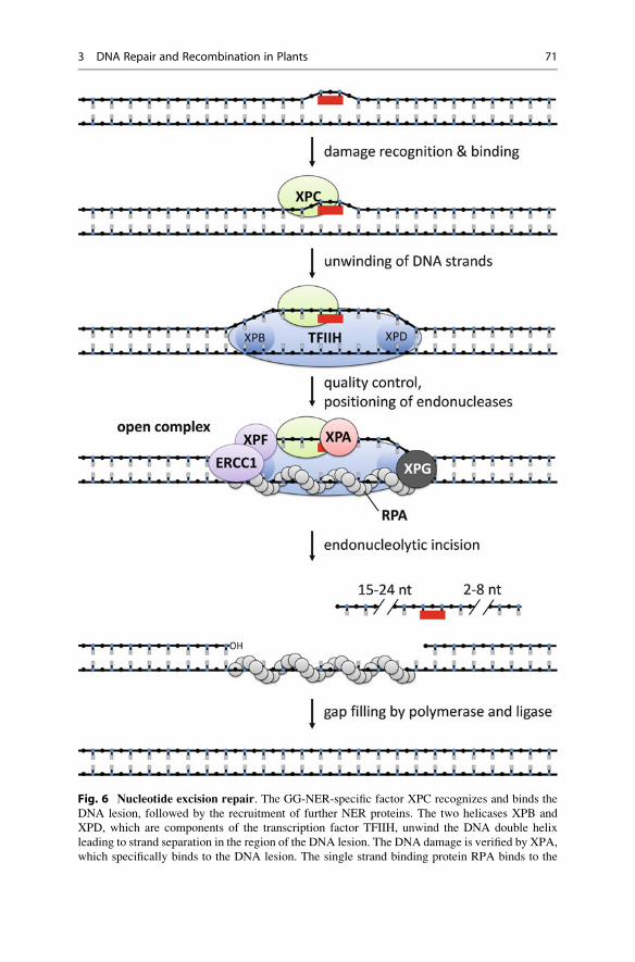
Fig. 6 Nucleotide excision repair. The GG-NER-specific factor XPC recognizes and binds the
DNA lesion, followed by the recruitment of further NER proteins. The two helicases XPB and
XPD, which are components of the transcription factor TFIIH, unwind the DNA double helix
leading to strand separation in the region of the DNA lesion. The DNA damage is verified by XPA,
which specifically binds to the DNA lesion. The single strand binding protein RPA binds to the
3 DNA Repair and Recombination in Plants 71
transcription process in actively expressed genes. DNA lesions in the transcribed
DNA strand lead to a block of RNA polymerase II while it is moving along DNA.
The transcription factor TFIIH, which is involved in both transcription and NER, is
associated with the captured RNA polymerase sitting on the active, transcribed
gene. From this point of view, RNA polymerases encountering a DNA lesion
represent a further class of proteins acting as efficient damage sensors. Until now,
it is not clear whether the RNA polymerase II is displaced or dissociated from DNA,
in order to allow the accessibility for further NER factors. TC-NER requires
additional repair factors which are specifically involved in this sub-pathway, such
as the assembly factors CSA and CSB (for Cockayne Syndrome protein A and B,
respectively). CSB, for instance, possesses nucleosome remodeling activity and
therefore is able to alter the conformation of DNA and is possibly involved in the
recruitment of TFIIH.
Open complex formation by TFIIH with XPB and XPD: The transcription
factor TFIIH is a multifunctional complex which is involved in the initiation of
transcription of DNA by RNA polymerase II, as well as in both sub-pathways of
NER. The TFIIH protein complex is organized in a ringlike structure and consists of
10 subunits including the two helicases XPB (also known as ERCC3) and XPD
(also known as ERCC2). The directionality of the helicase function differs between
XPB and XPD. XPB can unwind dsDNA in a 3’-5’ direction, whereas XPD has the
opposite direction of activity (it unwinds dsDNA in a 5’-3’ direction). By having
the two helicases as integral components, TFIIH mediates the separation of the
DNA strands at the damaged site, initiating the formation of a so-called open
complex (Fig. 6). The fully open complex spans about 20–30 base pairs
(bp) around the DNA lesion and contains ss- to dsDNA transition sites, which are
required for the cleavage by the structure-specific endonucleases XPG and the
ERCC1-XPF complex (described below). The formation of the fully open complex
occurs in a two-step reaction. The initial opening spans about <10 bp and requires
XPC and TFHIIH. The subsequent extension of the open complex to about 30 bp is
dependent on further NER factors such as XPA, RPA, and XPG.
Quality control and positioning of the endonucleases: After the separation of
the DNA strands surrounding the DNA lesion, the DNA-binding protein XPA
validates the DNA damage in the open complex formation. XPA binds the damaged
DNA (Fig. 6), preferentially at NER-specific types of DNA damage including
CPDs and 6–4 PPs.
In principle, the endonucleases ERCC1-XPF and XPG, which catalyze the
incision in the damaged DNA strand, are able to cut both DNA strands at the border
of the open DNA intermediate. But in NER, the incisions by the endonucleases are
�
Fig. 6 (continued) undamaged DNA strand and is involved in the positioning of the endonucleases
ERCC1-XPF and XPG. After the formation of the fully open complex, XPG cleaves at the 3’ site
2–8 nucleotides distant from the lesion and ERCC1-XPF catalyzes the 5’ incision 15–24 nucle-
otides away from the DNA lesion. The gap filling is done by replicative polymerases and DNA
ligase I
72 S. Schropfer et al.
restricted to the damaged DNA strand only. This fact is crucial for the success of
DNA repair by NER and points to the important role of nuclease positioning by the
NER machinery via protein-protein interactions.
Aside from binding of the damaged DNA, XPA also interacts with several
factors in the NER pathway such as TFIIH, the endonuclease ERCC1-XPF1, as
well as RPA. By mediating these protein-protein interactions, XPA has a role in the
correct assembly and positioning of the DNA repair machinery around the DNA
lesion. An additional protein, RPA, is also involved in the positioning of the
endonucleases by direct protein-protein interactions. Primarily, RPA binds to the
undamaged ssDNA strand by its ssDNA-binding activity to stabilize the open
complex (Fig. 6). RPA has a defined DNA-binding orientation which is relevant
for the coordination of the nucleases. The 3’-oriented side of RPA interacts with
ERCC1-XPF, whereas the 5’-oriented side binds XPG (Fig. 6). Furthermore, RPA
stimulates the endonucleolytic cleavage of the damaged DNA strand and inhibits
incisions in the undamaged DNA strand. Altogether, the assembly of the fully open
complex is dependent on a variety of protein-protein interactions. For example, the
positioning of ERCC1-XPF requires the interaction with RPA and XPA, which
facilitates and stabilizes the positioning of the endonuclease.
Incision by the endonucleases ERCC1-XPF and XPG: Two different endo-
nucleases, ERCC1-XPF and XPG, are recruited to the pre-incision complex as
described above. The activities of both endonucleases lead to an asymmetrical
cleavage with respect to the DNA lesion site (Fig. 6). First, XPG cleaves at the 3’
site 2–8 nucleotides distant from the lesion. Following the 3’ incision, a second
incision at the opposite 5’ site introduced by ERCC1-XPF is 15–24 nucleotides
away from the DNA lesion. The exact incision positions are dependent on the type
of DNA damage and the sequence context. For this reason, the size of the replaced
DNA oligonucleotide varies from 24 to 32 nucleotides.
XPG is a structure-specific endonuclease responsible for the 3’ incision at the
border of the open DNA intermediate. There, XPG acts with a defined cleavage
polarity which is characterized by the incision in one strand of the DNA duplex at
the ss to dsDNA transition site. Furthermore, XPG also has another important role
during NER, as it is required for the fully open complex formation described above.
The two proteins ERCC1 (excision repair cross-complementing 1) and XPF
form a stable complex which acts as a structure-specific endonuclease. Besides
NER, the complex is also required in other DNA repair pathways such as
interstrand cross-link repair and homologous recombination. During NER,
ERCC1-XPF catalyzes the incision at the 5’ site of the open complex resulting in
a free hydroxyl group at the 3’ end. In contrast to XPG, ERCC1-XPF does not
appear to have a structural function in the open complex formation.
Resealing of the gap: The 3’ end flanking the single-stranded gap, which results
from the incision by ERCC1-XPF, can be directly used as a DNA primer for DNA
synthesis to fill the gap (Fig. 6). Repair synthesis in NER requires several factors
which are also involved in DNA replication, such as RPA, RFC, PCNA, and
polymerases δ and ε (for further details see section Base Excision Repair). The
role of RPA in NER is manifold. Besides the previously described function in
3 DNA Repair and Recombination in Plants 73
positioning of the endonucleases, RPA is also involved in repair synthesis. The
DNA binding of RPA leads to the protection of the undamaged DNA strand against
nucleases, which then serves as a template for repair synthesis. Furthermore, RPA
facilitates DNA synthesis because its presence stimulates the activity of the repli-
cative DNA polymerases δ and ε. The ligation reaction of the remaining nick
between the newly synthesized DNA strand and the original DNA strand is carried
out by DNA ligase I.
Kinetics of NER: The recruitment and the activity of NER proteins is dependent
on different parameters such as the location and the type of the DNA lesion.
Therefore it was suggested that there are two different NER responses. The
immediate NER response is characterized by the removal of DNA lesions which
have a great effect on the DNA structure or which are detected through the
transcription of genes. The remaining DNA lesions are repaired at a much slower
rate in a secondary NER response. For instance, 6–4 PPs, leading to large distor-
tions of the DNA double helix, are repaired five times faster than CPDs, which have
only a slight bending effect on the DNA structure.
Evolution of NER: The basic NER mechanisms including recognition of DNA
lesions, DNA incision, fragment excision, and repair synthesis have been function-
ally conserved during evolution. The basics of NER in eukaryotes and prokaryotes
are similar, but more complex in eukaryotes because of the involvement of more
than 25 factors, compared to only four in prokaryotes. These so-called Uvr proteins
are required to detect and remove the DNA damage. The UvrA/UvrB complex
scans the genome to find distortions of DNA. After the detection and binding of the
DNA lesion by the complex, UvrA dissociates and UvrB catalyzes the local melting
of the DNA double strand at the site of the DNA damage. UvrC associates with
UvrB and cleaves the damaged DNA strand eight nucleotides upstream (5’) and 4–5
nucleotides downstream (3’) from the DNA lesion. By this endonucleolytic cleav-
age, a 12–13-nucleotide-long oligomer containing the DNA lesion is created. By
the unwinding activity of the UvrD helicase, the damaged ssDNA strand is excised
from the DNA duplex. However, despite an obvious conservation of the NER
mechanism itself, the enzymes involved in this process differ between prokaryotes
and eukaryotes. Because of the lack of sequence homology, when comparing the
NER proteins of the both groups, it is likely that the analogous functions in the NER
mechanisms evolved independently in prokaryotes and eukaryotes.
In eukaryotes, not only the NER mechanism but also the involved proteins are
well conserved, suggesting a conserved repair pathway in these organisms. Most of
the genes involved in NER in yeast and mammals can also be found in plant
genomes, such as XPC, HR23B, CEN2, XPB, XPD, RPA, XPG, ERCC1, XPF,and XPE. Like human patients with defects in the NER pathway, plants containing
mutations in NER genes display UV hypersensitivity. The same holds for different
Arabidopsis mutants with defects in the plant homologs of XPD (Atuvh6), XPG(Atuvh3), XPF (Atrad1), ERCC1 (Atuvr7), and CEN2 (Atcen2).
In the case of the helicase gene XPB, two homologs AtXPB1 and AtXPB2 have
been identified in the genome of Arabidopsis thaliana. Both duplicated homologs
contain a DNA-binding domain and seven helicase motifs, which are present in
74 S. Schropfer et al.
human XPB, as well. Further, the other helicase in the TFIIH complex, XPD, is also
present in Arabidopsis and shows conservation of the helicase domain. It is likely
that the function of AtXPD is conserved during evolution, because it was shown
that the plant XPD homolog can interact with TFIIH components in yeast. Another
example of a high degree of conservation is the endonuclease complex ERCC1-
XPF. Both components of the complex could be identified by sequence similarity in
plants, and it was shown that XPF from Arabidopsis (also called AtRAD1) is able tointeract with AtERCC1.
Despite conservation of most of the NER proteins in plants, there are some
differences between the phylogenetic groups present. No homolog of XPA, which
is involved in the quality control by binding the DNA damage and assembly of the
NER machinery in mammals and yeast, could be identified in plants until now. It is
possible that functions which are fulfilled by XPA in mammals are not essential for
the NER mechanism in plants, or another unknown plant protein has functions
similar to mammalian XPA.
Mismatch Repair (MMR)
Mismatched base pairs in dsDNA lead to distortions of the double helix that may
hinder or block replication and transcription. If unrepaired, such mismatches can
become fixed mutations after another round of replication. Mismatched bases may
occur through chemical reactions, e.g., by spontaneous deamination or by chemical
genotoxins. A further source of mismatches is replicative DNA polymerases. Even
though they possess a proofreading activity to correct errors, they nonetheless place
the wrong base opposite to their template strand in about one in 100,000,000 (10�8)
bases. To counteract this source of mutations, a specific repair pathway called
mismatch repair (MMR) has evolved (see also reviews Jiricny 2006; Spampinato
and Gomez-Casati 2012). It can detect and correct 99 % of mismatched bases
introduced by replicative DNA polymerases, reducing the error rate to about 10�10.
Although details vary in the MMR pathway between prokaryotes and eukary-
otes, the basic steps are the same (see Fig. 7): At first, the mismatched bases have to
be found by proteins that recognize the distortion of the double helix. Then, a
stretch of the newly synthesized strand in which the wrong nucleotide has been
introduced is removed. Finally, using the correct sequence information of the
parental strand, the single-stranded gap is closed by a polymerase and ligase.
In E. coli, where MMR was initially studied, recognition of a mismatch is done
by a homodimer of the protein MutS which forms a ring structure that encloses
dsDNA. After it has found a mismatch, the MutS homodimer interacts with a
homodimer of MutL and moves away from the mismatch along DNA until it
encounters the endonuclease MutH bound to dsDNA at GATC sequences. In
E. coli, the GATC sites are normally methylated on the adenine, but following
replication, the newly synthesized daughter strand is not methylated for a short
time. MutS-MutL can now activate the MutH endonuclease activity, which only
cuts the unmethylated strand. This ensures that that source of the mismatch, the
3 DNA Repair and Recombination in Plants 75
newly synthesized strand, is repaired instead of the parental strand. Starting from
this nick, the UvrD DNA helicase unwinds the duplex, thereby exposing the
modified strand to exonucleases that degrade it. The gap that is formed is then
filled by DNA polymerase and sealed by DNA ligase.
Eukaryotic cells contain homologs to MutS and MutL, named MSH (for MutS
homolog) and MLH (for MutL homolog), respectively. In fact, several homologs to
MutS and MutL can be found in plants, fungi, and animals. In mammals, the
proteins MSH2 and MSH6 form a heterodimer called MutSα that is thought to
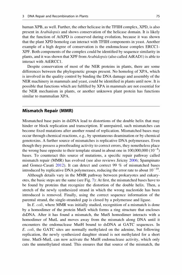
Fig. 7 Mismatch repair. (a) Mismatched base pairs can arise during replication due to the
incorporation of false nucleotides (red) by the replicative polymerases. The daughter strand, which
contains the mismatch, is exonucleolytically degraded up to the site of the next occurring
interruption of the DNA strand. Such an interruption can be a SSB formed by the end of an
Okazaki fragment in the lagging strand or the 3’ end of the leading strand. The resulting gap is
filled by repair synthesis. (b) One scenario of MMR is depicted in more detail. The heterodimer
MutSα (MSH2-MSH6) recognizes and binds the mismatch. This results to a conformational
change of the heterodimer, leading to the formation of a clamp structure surrounding the
mismatch. A further heterodimer MutLα (MLH1-PMS2) interacts with MutSα. The ternary
complex acts as a sliding clamp and translocates in n ATP-dependent manner to the next occurring
SSB, where it interacts with replicative factors such as PCNA and RFC (not shown). Subsequently,
the DNA strand which contains the falsely incorporated base is degraded by the exonuclease
EXO1. The resulting ssDNA strand is coated by RPA and serves as a template for repair synthesis
76 S. Schropfer et al.
act similarly to the MutS homodimer in E. coli. Furthermore, MSH2 and MSH3
form the heterodimer MutSβ that is involved in a more specialized repair of large
DNA loops formed by insertions or deletions. A heterodimer of the MutL homologs
MLH1 and PMS2, named MutLα, can interact with MutSα to form a ternary
complex similar to the MutS-MutL complex in bacteria.
Comparable to MutS-MutL in bacteria, the eukaryotic MutSα-MutLα com-
plex moves along dsDNA after mismatch recognition through MutSα. The
critical step in the endonucleolytic cleavage by MutH in the daughter strand,
however, is specific to gram-negative bacteria, since MutH is not conserved
outside this group. So how can the degradation of the daughter strand be initiated
in eukaryotes, and how is it ensured that the parental strand is not modified? In
contrast to bacteria, it is thought that eukaryotic cells use preexisting gaps and
nicks present during replication specifically in the newly synthesized daughter
strand: the free 3’ end of the leading strand or both ends of the Okazaki
fragments in the lagging strand (Fig. 7). In accordance with this idea, it was
shown that MSH6 from MutSα can interact with proteins usually found in the
replication complex at the elongating 3’ end of DNA: the sliding clamp protein
PCNA and its clamp loader RFC. Such a free DNA end may now serve to initiate
strand degradation in MMR. Depending on the orientation of the gap to the
mismatch, a number of exonuclease activities with either 3’-5’ or 5’-3’ polarity
are known to be involved in this degradation. While the parental strand is
stabilized by the ssDNA-binding protein RPA, DNA polymerase δ closes the
gap, and DNA ligase I seals the nick.
In plants, the MMR pathway seems to work similarly as described for mammals
above. Counterparts of all mammalian MMR proteins are also found in plants, and
mutants of MSH2 show an expected increase in point mutations and genome
instability. Loss of the MMR system in plants specifically affects morphology,
fertility, and seed development in a generation-dependent manner. In addition to the
role of MMR proteins in the repair of mismatches during replication, there is also a
further role in meiosis. In A. thaliana, the plant-specific MSH7 interacts with MSH2
to form another heterodimer, MutSγ, which is required for the detection of mis-
matches during meiotic recombination. Furthermore, the MSH4-MSH5
heterodimer and the MLH1-MLH3 heterodimer are also involved in meiotic
recombination rather than classical MMR (also see section Repair of Double-
Strand Breaks).
Since mismatched bases may also arise in the pairing of similar, but not
identical, sequences during homologous recombination (see section Repair of
Double-Strand Breaks), several proteins from the MMR pathway have been
found to affect the outcomes of this, so-called homologous, recombination. Organ-
isms containing polyploid genomes with homologous chromosomes from hybrid-
ization events, e.g., hexaploid wheat, are especially prone to pair homologous
sequences during HR. Here, it is thought that MMR proteins recognize mismatches
in heteroduplex DNA and abort the deleterious recombination reaction. Loss of
MMR protein therefore leads to an increase in homologous recombination in
addition to the elevated level in point mutations.
3 DNA Repair and Recombination in Plants 77
Repair of Double-Strand Breaks
DSBs, the most severe type of DNA lesion, require timely repair. A single
unrepaired DSB may lead to cell death due to failure to correctly distribute
chromosomes to the daughter cells. Thus it is not surprising that a number of
different pathways, which at least partially can compete with each other, have
evolved to cope with DSBs (see also reviews Schuermann et al. 2005; Waterworth
et al. 2011). These pathways differ in the frequency they are used to repair lesions
as well as in the fidelity of repair.
Repair of DSBs by nonhomologous end-joining: The predominantly used and
mechanistically most simple pathway in somatic cells is called nonhomologous
end-joining (NHEJ), which ligates the broken ends at DSBs (Fig. 8a). In the case of
a clean break, such a ligation will result in the wild-type sequence without any
mutations. In most cases, however, a break will not be blunt ended or it will be
chemically modified, making a direct ligation without mutations impossible. DSB
repair by NHEJ therefore is a quick, but highly mutagenic pathway.
Following the formation of a DSB, the heterodimer KU70/KU80 binds to the
ends of the break. The KU heterodimer forms a circular structure with positively
charged amino acids on the inside and a diameter just wide enough for dsDNA,
which enables it to slide onto the broken ends, but not elsewhere along the
chromosomes. Recruitment of further repair factors, such as the trimeric MRN
complex (for MRE11, RAD50, NBS1), enables processing of the two ends and
brings them into close proximity to each other by interaction of the MRN com-
plexes. Finally, the ends are ligated to close the break by a heterodimer of the DNA
ligase IV (LIG4) and XRCC4.
Fig. 8 Repair of a DSB by NHEJ,MMEJ and SSA. (a) In the NHEJ pathway, the DSB ends are
processed and directly ligated. (b) In contrast to NHEJ, during MMEJ short single-stranded
overhangs are formed at the ends of the break. A small complementary region of about 5–25 bp
(green, micro homologies) are used for hybridization; through which the two ends are reconnected.
(c) Followed by an extensive resection in the SSA pathway, the generated single-stranded over-
hangs can hybridize using direct sequence repeats (orange), which are flanking the DSB site. All
three DSB repair pathways are associated with genomic changes. Non-homologous overhangs are
removed by nucleases (triangle)
78 S. Schropfer et al.
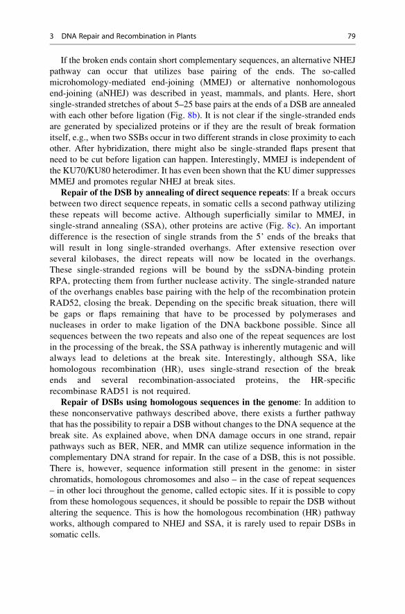
If the broken ends contain short complementary sequences, an alternative NHEJ
pathway can occur that utilizes base pairing of the ends. The so-called
microhomology-mediated end-joining (MMEJ) or alternative nonhomologous
end-joining (aNHEJ) was described in yeast, mammals, and plants. Here, short
single-stranded stretches of about 5–25 base pairs at the ends of a DSB are annealed
with each other before ligation (Fig. 8b). It is not clear if the single-stranded ends
are generated by specialized proteins or if they are the result of break formation
itself, e.g., when two SSBs occur in two different strands in close proximity to each
other. After hybridization, there might also be single-stranded flaps present that
need to be cut before ligation can happen. Interestingly, MMEJ is independent of
the KU70/KU80 heterodimer. It has even been shown that the KU dimer suppresses
MMEJ and promotes regular NHEJ at break sites.
Repair of the DSB by annealing of direct sequence repeats: If a break occurs
between two direct sequence repeats, in somatic cells a second pathway utilizing
these repeats will become active. Although superficially similar to MMEJ, in
single-strand annealing (SSA), other proteins are active (Fig. 8c). An important
difference is the resection of single strands from the 5’ ends of the breaks that
will result in long single-stranded overhangs. After extensive resection over
several kilobases, the direct repeats will now be located in the overhangs.
These single-stranded regions will be bound by the ssDNA-binding protein
RPA, protecting them from further nuclease activity. The single-stranded nature
of the overhangs enables base pairing with the help of the recombination protein
RAD52, closing the break. Depending on the specific break situation, there will
be gaps or flaps remaining that have to be processed by polymerases and
nucleases in order to make ligation of the DNA backbone possible. Since all
sequences between the two repeats and also one of the repeat sequences are lost
in the processing of the break, the SSA pathway is inherently mutagenic and will
always lead to deletions at the break site. Interestingly, although SSA, like
homologous recombination (HR), uses single-strand resection of the break
ends and several recombination-associated proteins, the HR-specific
recombinase RAD51 is not required.
Repair of DSBs using homologous sequences in the genome: In addition to
these nonconservative pathways described above, there exists a further pathway
that has the possibility to repair a DSB without changes to the DNA sequence at the
break site. As explained above, when DNA damage occurs in one strand, repair
pathways such as BER, NER, and MMR can utilize sequence information in the
complementary DNA strand for repair. In the case of a DSB, this is not possible.
There is, however, sequence information still present in the genome: in sister
chromatids, homologous chromosomes and also – in the case of repeat sequences
– in other loci throughout the genome, called ectopic sites. If it is possible to copy
from these homologous sequences, it should be possible to repair the DSB without
altering the sequence. This is how the homologous recombination (HR) pathway
works, although compared to NHEJ and SSA, it is rarely used to repair DSBs in
somatic cells.
3 DNA Repair and Recombination in Plants 79
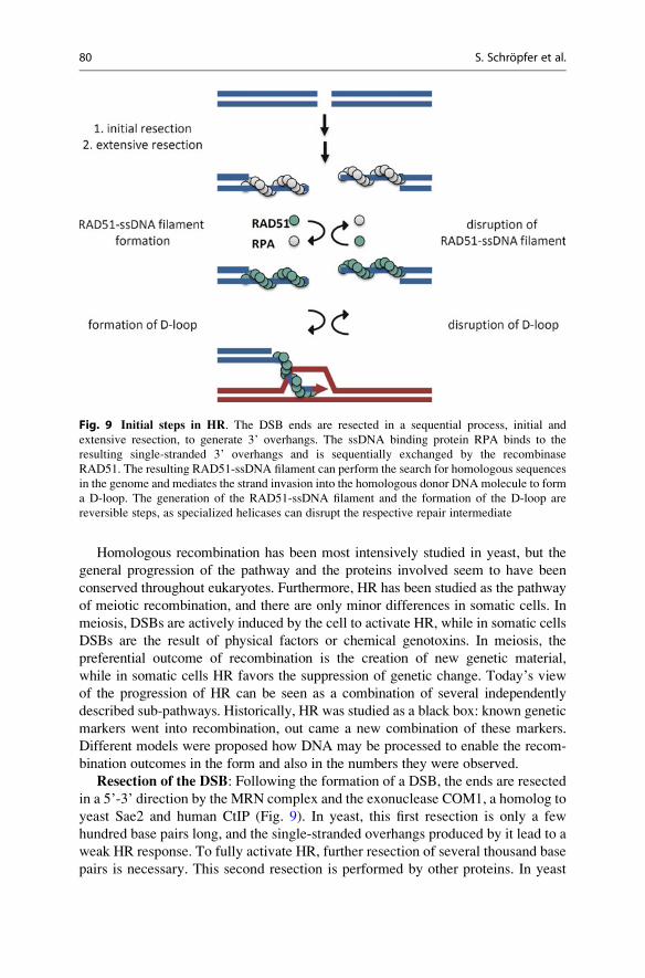
Homologous recombination has been most intensively studied in yeast, but the
general progression of the pathway and the proteins involved seem to have been
conserved throughout eukaryotes. Furthermore, HR has been studied as the pathway
of meiotic recombination, and there are only minor differences in somatic cells. In
meiosis, DSBs are actively induced by the cell to activate HR, while in somatic cells
DSBs are the result of physical factors or chemical genotoxins. In meiosis, the
preferential outcome of recombination is the creation of new genetic material,
while in somatic cells HR favors the suppression of genetic change. Today’s view
of the progression of HR can be seen as a combination of several independently
described sub-pathways. Historically, HR was studied as a black box: known genetic
markers went into recombination, out came a new combination of these markers.
Different models were proposed how DNA may be processed to enable the recom-
bination outcomes in the form and also in the numbers they were observed.
Resection of the DSB: Following the formation of a DSB, the ends are resected
in a 5’-3’ direction by the MRN complex and the exonuclease COM1, a homolog to
yeast Sae2 and human CtIP (Fig. 9). In yeast, this first resection is only a few
hundred base pairs long, and the single-stranded overhangs produced by it lead to a
weak HR response. To fully activate HR, further resection of several thousand base
pairs is necessary. This second resection is performed by other proteins. In yeast
Fig. 9 Initial steps in HR. The DSB ends are resected in a sequential process, initial and
extensive resection, to generate 3’ overhangs. The ssDNA binding protein RPA binds to the
resulting single-stranded 3’ overhangs and is sequentially exchanged by the recombinase
RAD51. The resulting RAD51-ssDNA filament can perform the search for homologous sequences
in the genome and mediates the strand invasion into the homologous donor DNAmolecule to form
a D-loop. The generation of the RAD51-ssDNA filament and the formation of the D-loop are
reversible steps, as specialized helicases can disrupt the respective repair intermediate
80 S. Schropfer et al.
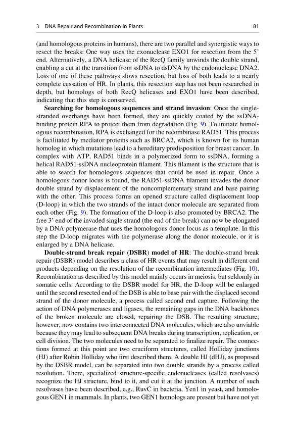
(and homologous proteins in humans), there are two parallel and synergistic ways to
resect the breaks: One way uses the exonuclease EXO1 for resection from the 5’
end. Alternatively, a DNA helicase of the RecQ family unwinds the double strand,
enabling a cut at the transition from ssDNA to dsDNA by the endonuclease DNA2.
Loss of one of these pathways slows resection, but loss of both leads to a nearly
complete cessation of HR. In plants, this resection step has not been researched in
depth, but homologs of both RecQ helicases and EXO1 have been described,
indicating that this step is conserved.
Searching for homologous sequences and strand invasion: Once the single-
stranded overhangs have been formed, they are quickly coated by the ssDNA-
binding protein RPA to protect them from degradation (Fig. 9). To initiate homol-
ogous recombination, RPA is exchanged for the recombinase RAD51. This process
is facilitated by mediator proteins such as BRCA2, which is known for its human
homolog in which mutations lead to a hereditary predisposition for breast cancer. In
complex with ATP, RAD51 binds in a polymerized form to ssDNA, forming a
helical RAD51-ssDNA nucleoprotein filament. This filament is the structure that is
able to search for homologous sequences that could be used in repair. Once a
homologous donor locus is found, the RAD51-ssDNA filament invades the donor
double strand by displacement of the noncomplementary strand and base pairing
with the other. This process forms an opened structure called displacement loop
(D-loop) in which the two strands of the intact donor molecule are separated from
each other (Fig. 9). The formation of the D-loop is also promoted by BRCA2. The
free 3’ end of the invaded single strand (the end of the break) can now be elongated
by a DNA polymerase that uses the homologous donor locus as a template. In this
step the D-loop migrates with the polymerase along the donor molecule, or it is
enlarged by a DNA helicase.
Double-strand break repair (DSBR) model of HR: The double-strand break
repair (DSBR) model describes a class of HR events that may result in different end
products depending on the resolution of the recombination intermediates (Fig. 10).
Recombination as described by this model mainly occurs in meiosis, but seldomly in
somatic cells. According to the DSBR model for HR, the D-loop will be enlarged
until the second resected end of the DSB is able to base pair with the displaced second
strand of the donor molecule, a process called second end capture. Following the
action of DNA polymerases and ligases, the remaining gaps in the DNA backbones
of the broken molecule are closed, repairing the DSB. The resulting structure,
however, now contains two interconnected DNA molecules, which are also unviable
because theymay lead to subsequent DNA breaks during transcription, replication, or
cell division. The two molecules need to be separated to finalize repair. The connec-
tions formed at this point are two cruciform structures, called Holliday junctions
(HJ) after Robin Holliday who first described them. A double HJ (dHJ), as proposed
by the DSBR model, can be separated into two double strands by a process called
resolution. There, specialized structure-specific endonucleases (called resolvases)
recognize the HJ structure, bind to it, and cut it at the junction. A number of such
resolvases have been described, e.g., RuvC in bacteria, Yen1 in yeast, and homolo-
gous GEN1 in mammals. In plants, two GEN1 homologs are present but have not yet
3 DNA Repair and Recombination in Plants 81
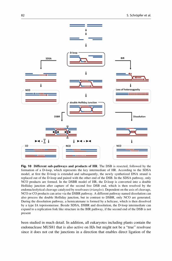
been studied in much detail. In addition, all eukaryotes including plants contain the
endonuclease MUS81 that is also active on HJs but might not be a “true” resolvase
since it does not cut the junctions in a direction that enables direct ligation of the
Fig. 10 Different sub-pathways and products of HR. The DSB is resected, followed by the
formation of a D-loop, which represents the key intermediate of HR. According to the SDSA
model, at first the D-loop is extended and subsequently, the newly synthetized DNA strand is
replaced out of the D-loop and paired with the other end of the DSB. In the SDSA pathway, only
NCO products are formed. In the DSBR model of HR, the D-loop is converted into a double
Holliday junction after capture of the second free DSB end, which is then resolved by the
endonucleolytical cleavage catalyzed by resolvases (triangles). Dependent on the axis of cleavage,NCO or CO products can arise via the DSBR pathway. A different pathway named dissolution can
also process the double Holliday junction, but in contrast to DSBR, only NCO are generated.
During the dissolution pathway, a hemicatenane is formed by a helicase, which is then dissolved
by a type IA topoisomerase. Beside SDSA, DSBR and dissolution, the D-loop intermediate can
expand to a replication fork like structure in the BIR pathway, if the second end of the DSB is not
present
82 S. Schropfer et al.
products. After the resolvase activity, a DNA ligase seals the breaks and finalizes
DSB repair. Depending on the orientation of the cut at the two HJs, the product of the
HR reaction may be either a crossover (CO) or a noncrossover (NCO). In a NCO,
which has also been called gene conversion, a nonreciprocal exchange of genetic
information takes place. Information is copied from the donor to the damaged DNA
double strand at the break site. When a CO happens, on the other hand, flanking
sequences of the break site are exchanged between donor and damaged double strand.
In meiosis COs are the basis for the mixing of parental genomes.
Synthesis-dependent strand-annealing (SDSA) model of HR: Since the
resolvases acting according to the DSBR model cannot differentiate between the
two possible ways to cut a HJ, one would expect a distribution of 50 % COs and
50 % NCOs. However, experiments have shown that there are far more NCOs
produced. An alternative to the DSBR model is the synthesis-dependent strand-
annealing (SDSA) model (Fig. 10), in which HR proceeds from a DSB as described
above. In this model, single DNA strands are resected from the broken ends, and
they are coated first by RPA and then RAD51, followed by homology search and
strand invasion to form a D-loop. From this step onward, the SDSA model differs
from the DSBR model. SDSA-like recombination occurs during meiosis as well as
in somatic cells. After elongation of the invaded strand using the donor molecule as
a template, the SDSA model proposes – instead of second-end capture – the
rejection of the invaded strand from the D-loop, resulting in a disruption of the
D-loop. This allows for the annealing of the single-stranded break ends, followed
by repair synthesis to fill the gaps and religation to seal the backbone. Such a
process cannot lead to a CO product, but only to NCO outcomes.
Regulation of pathway choice: Since both the DSBR and SDSA pathways of
HR require the formation of a D-loop, the pathway choice is made at this crucial step.
Which pathway will be used depends on the ability of the second, free end of the DSB
to anneal to the displaced donor strand at the D-loop. Therefore, either elongation or
stabilization of the D-loop will increase the chance of second-end capture and,
therefore, the chance of the DSBR pathway, while early disruption of the D-loop
due to displacement of the invaded single strand will promote SDSA. Two classes of
proteins have been shown to be involved in this pathway decision process in meiosis:
DNA helicases and ring- or clamp-forming proteins. In the case of DNA helicases
(see reviewKnoll and Puchta 2011), the specific activity of the respective protein will
determine which of the two pathways will be promoted. Simply by the act of strand
separation, a helicase can enlarge the D-loop, forming a larger region of unpaired
donor DNA and helping the capture of the second strand. Alternatively, a similar
activity can separate donor strand and invaded strand, thereby promoting SDSA. A
number of DNA helicases have been described that might act in such processes. The
yeast Srs2 helicase as well as the animal Rtel1 helicase has been shown to disrupt
model D-loops in vitro and to promote SDSA outcomes in vivo. Homologs of both
proteins are conserved in plants. On the other hand, in meiotic recombination the
DNA helicase ROCK-N-ROLLERS (RCK)/MER3 has been shown to promote CO
formation via the DSBR pathway, most probably through D-loop elongation. Also in
meiotic recombination, where CO formation is absolutely required, a heterodimer of
3 DNA Repair and Recombination in Plants 83
the bacterial MutS homologous proteins MSH4 and MSH5 is thought to form a ring
structure that encloses the D-loop to suppress its disruption. Loss of either of the two
proteins leads to a strong reduction in the number of crossovers observed, since more
D-loops will enter the SDSA pathway.
Dissolution of double Holliday junctions: In addition to the classical DSBR
repair pathway, in which dHJs are processed by resolvases, it has been shown that at
least one further distinct pathway exists to process dHJs. Although it was theoretically
proposedabout30yearsago, itwasdemonstrated invitroand invivoonlyduringthe last
decade. Like SDSA, this pathway also can only form noncrossover products, but will
proceed throughDSBrepair in the samemanneras theDSBRpathwayup to thepointof
dHJ formation, facilitating a rather late decision for NCOs compared to the earlier
decision at the D-loop in the SDSA pathway. This pathway does not require endonu-
cleolytic resolvases and is therefore called “dissolution” to differentiate it from the
resolution step inDSBR (Fig. 10).But howcan the twodsDNAmolecules be separated
from each other without the activity of endonucleases?Dissolution requires in its most
basic form aDNAhelicase and a topoisomerase, although some structural proteins are
necessary as well to facilitate the interaction of these proteins. Starting from a dHJ
structure, a DNA helicase from the widely conserved RecQ family – in ArabidopsisRECQ4A specifically – can migrate the two HJs toward each other through its branch
migration activity. When the two HJs meet, a new structure is formed, a so-called
hemicatenane, in which one strand of each dsDNA molecule is topologically
interlocked with the other. This hemicatenane represents the substrate for type IA
topoisomerases, such as yeast Top3 or plant and animal TOP3α, which can process
this structure to unlink the two DNA molecules. The helicase and topoisomerase-
dependent dissolution reaction results exclusively in the formation of NCO products.
Inaddition to these twoenzymes, the structuralproteinsRMI1andRMI2areneeded for
the interaction of the helicase and the topoisomerase, and loss ofRMI proteins impacts
the dissolution reaction to a similar extent as the loss of the enzyme partners in the
complex. In yeast, in animals, and also in plants, it has been shown that this so-called
RTR(forRecQhelicase, topoisomerase3/3α,RMI) complex suppresses the formation
ofCOproducts afterDSBformation in somatic cells.Additionally, twomembers of the
plant RTR complex are essential for the completion of meiotic recombination: muta-
tions in TOP3A or RMI1 lead to severe chromosome fragmentation during meiotic
recombination and arrest at the end of meiosis I. Therefore, it seems that at least some
meiotic recombination intermediates must be processed through the dissolution path-
way and that topoisomerase TOP3α is the only plant protein capable of acting on
hemicatenane structures.
Tolerance and Repair Processes at Damaged Replication Forks
DNA damage often occurs in nonproliferating cells. However, if DNA is damaged
in proliferating cells, there are a number of checkpoints that will arrest the cell cycle
until the damaged DNA has been repaired. In some cases it is still possible that cells
enter S phase making DNA repair processes at the replication fork necessary.
84 S. Schropfer et al.
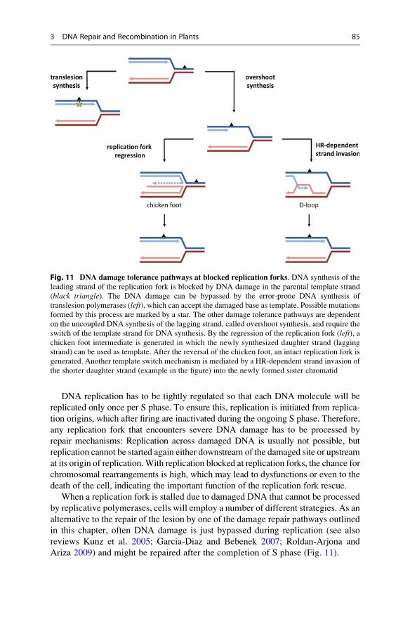
DNA replication has to be tightly regulated so that each DNA molecule will be
replicated only once per S phase. To ensure this, replication is initiated from replica-
tion origins, which after firing are inactivated during the ongoing S phase. Therefore,
any replication fork that encounters severe DNA damage has to be processed by
repair mechanisms: Replication across damaged DNA is usually not possible, but
replication cannot be started again either downstream of the damaged site or upstream
at its origin of replication.With replication blocked at replication forks, the chance for
chromosomal rearrangements is high, which may lead to dysfunctions or even to the
death of the cell, indicating the important function of the replication fork rescue.
When a replication fork is stalled due to damaged DNA that cannot be processed
by replicative polymerases, cells will employ a number of different strategies. As an
alternative to the repair of the lesion by one of the damage repair pathways outlined
in this chapter, often DNA damage is just bypassed during replication (see also
reviews Kunz et al. 2005; Garcia-Diaz and Bebenek 2007; Roldan-Arjona and
Ariza 2009) and might be repaired after the completion of S phase (Fig. 11).
Fig. 11 DNA damage tolerance pathways at blocked replication forks. DNA synthesis of the
leading strand of the replication fork is blocked by DNA damage in the parental template strand
(black triangle). The DNA damage can be bypassed by the error-prone DNA synthesis of
translesion polymerases (left), which can accept the damaged base as template. Possible mutations
formed by this process are marked by a star. The other damage tolerance pathways are dependent
on the uncoupled DNA synthesis of the lagging strand, called overshoot synthesis, and require the
switch of the template strand for DNA synthesis. By the regression of the replication fork (left), achicken foot intermediate is generated in which the newly synthesized daughter strand (lagging
strand) can be used as template. After the reversal of the chicken foot, an intact replication fork is
generated. Another template switch mechanism is mediated by a HR-dependent strand invasion of
the shorter daughter strand (example in the figure) into the newly formed sister chromatid
3 DNA Repair and Recombination in Plants 85
DNA damage bypass by translesion polymerases: To bypass damaged nucle-
otides, the replicative DNA polymerase cannot be used since it is not able to fit both
the damaged parental strand and a newly synthesized daughter strand into its active
center. To facilitate damage bypass, alternative proteins are therefore required.
There are two different strategies to bypass damaged DNA, with the first strategy
being error prone and the second error-free. The error-prone strategy, called
translesion synthesis, uses alternative DNA polymerases instead of the replicative
DNA polymerase (Fig. 11). Such translesion polymerases possess modified active
centers that are able to accommodate damaged DNA to place a new nucleotide in
the daughter strand. Since such polymerases most of the time are not able to
correctly recognize chemically modified nucleotides, they cannot add the correct
nucleotide to the growing DNA strand, leading to the incorporation of wrong
nucleotides and possibly point mutations later on. Several translesion polymerases
from a number of different DNA polymerase families have been described to date,
but the best understood are the polymerases of the Y family. DNA polymerase η of
the Y family of polymerases is conserved in fungi, animals, and plants and has the
ability to correctly bypass T-T CPDs, enabling error-free translesion synthesis
(TLS) of this lesion. Another Y-family polymerase, REV1, has a propensity to
insert a cytosine opposite to a number of DNA lesions. Here, it is thought that this is
a “best guess” strategy for the repair of abasic sites following the frequent sponta-
neous depurination of guanine. REV1 also interacts with another TLS polymerase,
ζ of the B family, that is used to elongate the strand following REV1 activity.
Polymerase ζ by itself is also able to bypass CPD lesions, but only with about 10 %
fidelity. This illustrates a general rule of translesion synthesis: Whether the bypass
of a lesion by a TLS polymerase will be error prone or error-free greatly depends on
the type of lesion and the kind of polymerases available or involved.
DNA damage tolerance by postreplication repair: Besides the strategy of
translesion synthesis that facilitates damage bypass by direct polymerase synthesis,
there is also a second strategy that switches daughter strand synthesis to a new
template. In the so-called postreplication repair (PRR) pathway, a feature of a
stalled replication fork is used by the cell (Fig. 11).
Since damage to nucleotides usually occurs only on one strand, replication can
proceed on the second, undamaged parental strand for some time, leading to
replication fork uncoupling and finally stalling. So-called overshoot synthesis
results in a newly synthesized daughter strand that contains exactly the sequence
information missing in the damaged parental strand. To use this information, the
stalled replication fork has to be rewound by a DNA helicase – in Arabidopsisprobably the homolog of yeast Rad5, RAD5A, and the plant RecQ helicase
RECQ4A – so that both daughter strands can anneal to each other instead of their
respective parental strands, forming a DNA structure also known as a “chicken
foot.” This process is also called replication fork regression. Here, the shorter
daughter strand, whose synthesis was blocked by the DNA damage, can be elon-
gated by a DNA polymerase using the longer daughter strand as template. Once
DNA synthesis has proceeded past the damaged site, the chicken foot structure can
be rewound, again by a DNA helicase, to reform the replication fork. Now,
86 S. Schropfer et al.
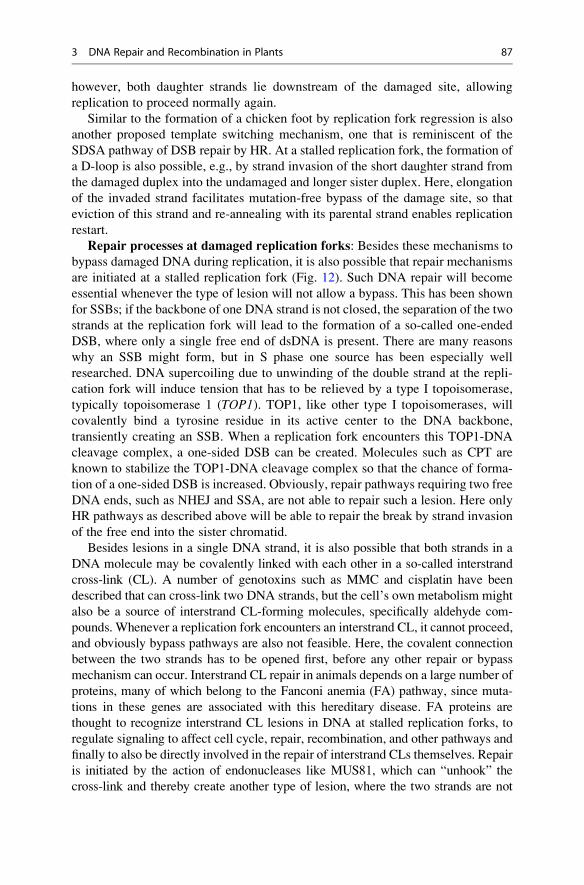
however, both daughter strands lie downstream of the damaged site, allowing
replication to proceed normally again.
Similar to the formation of a chicken foot by replication fork regression is also
another proposed template switching mechanism, one that is reminiscent of the
SDSA pathway of DSB repair by HR. At a stalled replication fork, the formation of
a D-loop is also possible, e.g., by strand invasion of the short daughter strand from
the damaged duplex into the undamaged and longer sister duplex. Here, elongation
of the invaded strand facilitates mutation-free bypass of the damage site, so that
eviction of this strand and re-annealing with its parental strand enables replication
restart.
Repair processes at damaged replication forks: Besides these mechanisms to
bypass damaged DNA during replication, it is also possible that repair mechanisms
are initiated at a stalled replication fork (Fig. 12). Such DNA repair will become
essential whenever the type of lesion will not allow a bypass. This has been shown
for SSBs; if the backbone of one DNA strand is not closed, the separation of the two
strands at the replication fork will lead to the formation of a so-called one-ended
DSB, where only a single free end of dsDNA is present. There are many reasons
why an SSB might form, but in S phase one source has been especially well
researched. DNA supercoiling due to unwinding of the double strand at the repli-
cation fork will induce tension that has to be relieved by a type I topoisomerase,
typically topoisomerase 1 (TOP1). TOP1, like other type I topoisomerases, will
covalently bind a tyrosine residue in its active center to the DNA backbone,
transiently creating an SSB. When a replication fork encounters this TOP1-DNA
cleavage complex, a one-sided DSB can be created. Molecules such as CPT are
known to stabilize the TOP1-DNA cleavage complex so that the chance of forma-
tion of a one-sided DSB is increased. Obviously, repair pathways requiring two free
DNA ends, such as NHEJ and SSA, are not able to repair such a lesion. Here only
HR pathways as described above will be able to repair the break by strand invasion
of the free end into the sister chromatid.
Besides lesions in a single DNA strand, it is also possible that both strands in a
DNA molecule may be covalently linked with each other in a so-called interstrand
cross-link (CL). A number of genotoxins such as MMC and cisplatin have been
described that can cross-link two DNA strands, but the cell’s own metabolism might
also be a source of interstrand CL-forming molecules, specifically aldehyde com-
pounds. Whenever a replication fork encounters an interstrand CL, it cannot proceed,
and obviously bypass pathways are also not feasible. Here, the covalent connection
between the two strands has to be opened first, before any other repair or bypass
mechanism can occur. Interstrand CL repair in animals depends on a large number of
proteins, many of which belong to the Fanconi anemia (FA) pathway, since muta-
tions in these genes are associated with this hereditary disease. FA proteins are
thought to recognize interstrand CL lesions in DNA at stalled replication forks, to
regulate signaling to affect cell cycle, repair, recombination, and other pathways and
finally to also be directly involved in the repair of interstrand CLs themselves. Repair
is initiated by the action of endonucleases like MUS81, which can “unhook” the
cross-link and thereby create another type of lesion, where the two strands are not
3 DNA Repair and Recombination in Plants 87
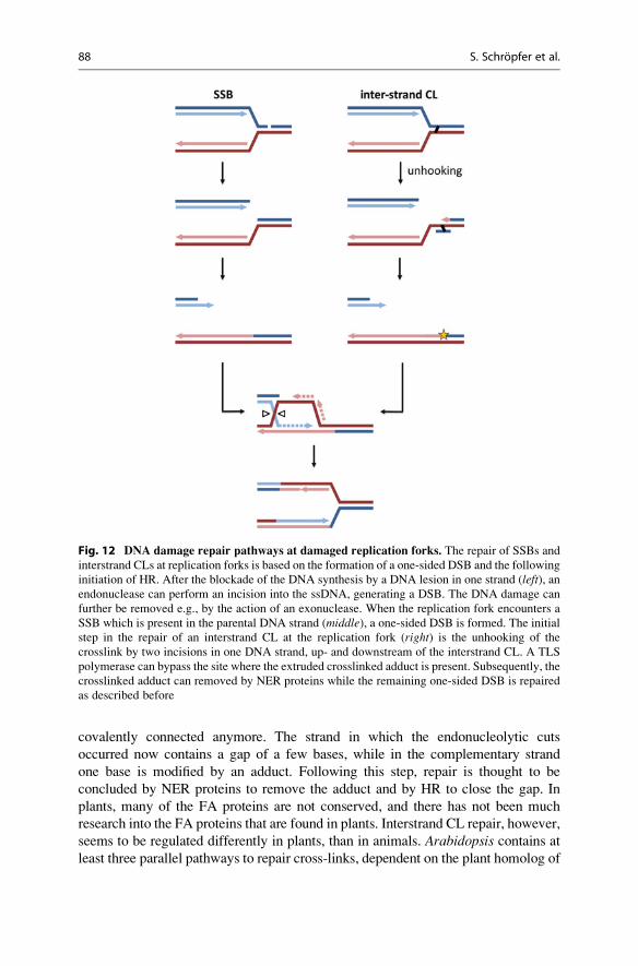
covalently connected anymore. The strand in which the endonucleolytic cuts
occurred now contains a gap of a few bases, while in the complementary strand
one base is modified by an adduct. Following this step, repair is thought to be
concluded by NER proteins to remove the adduct and by HR to close the gap. In
plants, many of the FA proteins are not conserved, and there has not been much
research into the FA proteins that are found in plants. Interstrand CL repair, however,
seems to be regulated differently in plants, than in animals. Arabidopsis contains atleast three parallel pathways to repair cross-links, dependent on the plant homolog of
Fig. 12 DNA damage repair pathways at damaged replication forks. The repair of SSBs and
interstrand CLs at replication forks is based on the formation of a one-sided DSB and the following
initiation of HR. After the blockade of the DNA synthesis by a DNA lesion in one strand (left), anendonuclease can perform an incision into the ssDNA, generating a DSB. The DNA damage can
further be removed e.g., by the action of an exonuclease. When the replication fork encounters a
SSB which is present in the parental DNA strand (middle), a one-sided DSB is formed. The initial
step in the repair of an interstrand CL at the replication fork (right) is the unhooking of the
crosslink by two incisions in one DNA strand, up- and downstream of the interstrand CL. A TLS
polymerase can bypass the site where the extruded crosslinked adduct is present. Subsequently, the
crosslinked adduct can removed by NER proteins while the remaining one-sided DSB is repaired
as described before
88 S. Schropfer et al.
MUS81, the helicase RAD5A that is also involved in PRR (see above), and the RecQ
family helicase RECQ4A. So while it seems possible that the unhooking of the
interstrand CL by MUS81 is also important for the repair of this type of lesion, there
seem to be alternative pathways in plants that do not depend on it.
Break-induced replication (BIR): Just as a damaged replication forks can
initiate repair mechanisms, situations have been described where HR reactions
initiate replication-like DNA synthesis to the end of the chromosome. When there
is only a single end of dsDNA present, for example, following a cut of one fork at a
stalled replication fork, initiation of an HR reaction will be possible, creating a
typical D-loop. However, since there is no second end to reconnect and since
replication in such a situation cannot be reinitiated after repair, the D-loop migrates
with the elongating broken end toward the end of the chromosome (Fig. 10). Such a
reaction, termed break-induced replication (BIR), will result in the loss of all
genetic information from the broken chromosome from the breakpoint to the
telomere. BIR reactions have been repeatedly described in yeast, but have not yet
been found in plants.
Cellular Changes and Signaling After DNA Damage
The previous subchapters described a complex network of DNA repair pathways
needed for the efficient repair of different kinds of DNA lesions. These repair
pathways need to be tightly regulated in order to efficiently repair the different
kinds of DNA damage and ensure cell survival. Unfortunately, the mechanisms and
pathways for regulation and signaling after DNA damage are still poorly under-
stood in plants. Many of the factors involved in DNA repair are conserved between
the different kingdoms; therefore, it is tempting to speculate that the regulation of
the different repair pathways might show a high level of conservation, too. One
very important factor of orchestrating the different DNA damage repair pathways is
a change in the expression of genes involved in the DNA damage response (see also
review Mannuss et al. 2012). This is quite well studied in plants and will be the
main focus of this section.
Cells do not waste resources in order to have all the repair proteins present in
abundance when DNA damage occurs. It is rather a dynamic process to synthesize
the enzymes that are needed to repair the particular damage. Many of the mentioned
repair proteins, like RAD51 or BRCA1, which is thought to be an important mediator
of DNA damage repair via HR, are present in a low number in plant cells, but if DSBs
are induced in the genome, the expression of the corresponding genes is elevated
several hundredfold. The Arabidopsis homolog of the breast cancer gene BRCA1 is
the gene that is most upregulated in response to DSBs in plants, which is consistent
with its important function in DNA repair via HR. The large subunits of the single-
strand binding protein RPA, which covers the single-stranded DNA recombination
intermediates and the open complex intermediate during NER, are also among the
genes that are most highly expressed in response to DNA damage. Not only are the
proteins involved in the repair of DSBs through homologous recombination
3 DNA Repair and Recombination in Plants 89
upregulated, but also the proteins that work in different DSB repair pathways, such as
NBS1, which is part of theMRN complex, are upregulated, too. KU70 and KU80 that
are involved in NHEJ are also expressed at higher levels following induction of
DSBs. Not only genes involved in the repair of DSBs are upregulated but also genes
involved in the BER pathway, such as XRCC1 and PARP1, although there might be
an overlap of functions of these proteins with the NHEJ pathway. Interestingly, most
of the NER proteins are not upregulated upon DNA damage, not even through UV
radiation, where it is an important repair pathway.
Apart from sophisticated repair genes, other genes – for example, the replicative
DNA polymerases Polδ and Polε, which are needed for proper replication and
furthermore also have a function in NER – are also upregulated after γ-irradiation.Which factors are involved in the signaling between detection of DNA damage
and the change of gene expression is an important question. Interestingly, two
factors, the kinases ATM and ATR, mediate the signaling cascade following DNA
damage through phosphorylation of downstream factors. ATM is mainly activated
by DSBs, and ATR is primarily activated by single-stranded regions following
stalled replication forks. It has to be mentioned though that there seems to be some
level of redundancy between the two kinases in plants. ATM is responsible for most
of the observed changes in gene expression after DSBs, whereas dependency on
ATR is observed in only a few cases. It was demonstrated for Arabidopsis that
especially upregulation of genes needed for the repair of DNA damage is dependent
on ATM. The importance of ATM and ATR for proper signaling after DNA
damage and the subsequent repair in plant cells is further strengthened by the fact
that atm/atr double mutants show growth defects and sterility.
In the next step the signal is passed on from ATM and ATR to a transcription
factor called SOG1 (suppressor of gamma response 1), which is activated by the
kinases through phosphorylation. Subsequently, SOG1 is responsible for the
changes in regulation of almost all genes in response to DNA damage. Since it
operates downstream from both ATM and ATR, it has been proposed to be a master
regulator of gene expression after DNA damage. In mammals, the most prominent
tumor suppressor is a transcription factor called p53 that, among other functions,
regulates gene expression after DNA damage. Although plants possess no direct
homolog to p53, SOG1 might have a similar function after DNA damage.
An additional factor involved in the regulation of gene expression after DNA
damage is called TEBICHI. It was postulated to regulate the expression of genes
through chromatin remodeling, which is an important means for gene regulation.
How the signal is transmitted to TEBICHI is not known, yet. Furthermore, some
E2F transcription factors are strongly downregulated after DNA damage. E2Fs are
a family of transcription factors that are highly conserved in plants and mammals.
In normal plant cells, the transcription factor E2Fe suppresses the expression of a
number of DNA repair factors, for example, the aforementioned BRCA1, PARP1,and SOG1 and the photolyase gene PHR1. Through downregulation of E2Fe, the
expression of the DNA repair genes can be increased. Therefore the E2F transcrip-
tion factors seem to represent another level of regulation of gene expression after
DNA damage.
90 S. Schropfer et al.
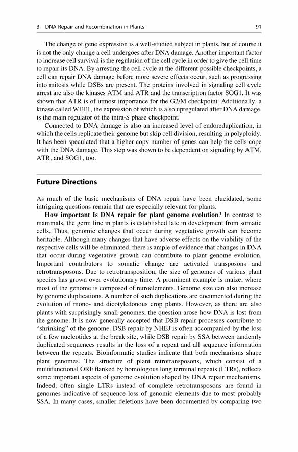
The change of gene expression is a well-studied subject in plants, but of course it
is not the only change a cell undergoes after DNA damage. Another important factor
to increase cell survival is the regulation of the cell cycle in order to give the cell time
to repair its DNA. By arresting the cell cycle at the different possible checkpoints, a
cell can repair DNA damage before more severe effects occur, such as progressing
into mitosis while DSBs are present. The proteins involved in signaling cell cycle
arrest are also the kinases ATM and ATR and the transcription factor SOG1. It was
shown that ATR is of utmost importance for the G2/M checkpoint. Additionally, a
kinase called WEE1, the expression of which is also upregulated after DNA damage,
is the main regulator of the intra-S phase checkpoint.
Connected to DNA damage is also an increased level of endoreduplication, in
which the cells replicate their genome but skip cell division, resulting in polyploidy.
It has been speculated that a higher copy number of genes can help the cells cope
with the DNA damage. This step was shown to be dependent on signaling by ATM,
ATR, and SOG1, too.
Future Directions
As much of the basic mechanisms of DNA repair have been elucidated, some
intriguing questions remain that are especially relevant for plants.
How important Is DNA repair for plant genome evolution? In contrast to
mammals, the germ line in plants is established late in development from somatic
cells. Thus, genomic changes that occur during vegetative growth can become
heritable. Although many changes that have adverse effects on the viability of the
respective cells will be eliminated, there is ample of evidence that changes in DNA
that occur during vegetative growth can contribute to plant genome evolution.
Important contributors to somatic change are activated transposons and
retrotransposons. Due to retrotransposition, the size of genomes of various plant
species has grown over evolutionary time. A prominent example is maize, where
most of the genome is composed of retroelements. Genome size can also increase
by genome duplications. A number of such duplications are documented during the
evolution of mono- and dicotyledonous crop plants. However, as there are also
plants with surprisingly small genomes, the question arose how DNA is lost from
the genome. It is now generally accepted that DSB repair processes contribute to
“shrinking” of the genome. DSB repair by NHEJ is often accompanied by the loss
of a few nucleotides at the break site, while DSB repair by SSA between tandemly
duplicated sequences results in the loss of a repeat and all sequence information
between the repeats. Bioinformatic studies indicate that both mechanisms shape
plant genomes. The structure of plant retrotransposons, which consist of a
multifunctional ORF flanked by homologous long terminal repeats (LTRs), reflects
some important aspects of genome evolution shaped by DNA repair mechanisms.
Indeed, often single LTRs instead of complete retrotransposons are found in
genomes indicative of sequence loss of genomic elements due to most probably
SSA. In many cases, smaller deletions have been documented by comparing two
3 DNA Repair and Recombination in Plants 91
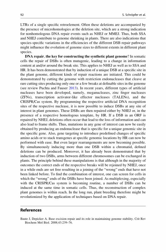
LTRs of a single specific retroelement. Often these deletions are accompanied by
the presence of microhomologies at the deletion site, which are a strong indication
for nonhomologous DNA repair events such as NHEJ or MMEJ. Thus, both SSA
and NHEJ contribute to genome shrinking in plants. There are also indications that
species-specific variations in the efficiencies of the different DSB repair pathways
might influence the evolution of genome sizes to different extents in different plant
species.
DNA repair, the key for constructing the synthetic plant genome? In somatic
cells the repair of DSBs is often mutagenic, leading to a change in information
content at and/or around the break site. This applies to NHEJ as well as to SSA and
HR. It has been demonstrated that by induction of a unique DSB at a specific site in
the plant genome, different kinds of repair reactions are initiated. This could be
demonstrated by cutting the genome with restriction endonucleases that cleave at
rare cutting sites producing only one or a few breaks at definable sites in the genome
(see review Puchta and Fauser 2013). In recent years, different types of artificial
nucleases have been developed, namely, meganucleases, zinc finger nucleases
(ZFNs), transcription activator-like effector nucleases (TALENs), and the
CRISPR/Cas system. By programming the respective artificial DNA recognition
sites of the respective nuclease, it is now possible to induce DSBs at any site of
interest in plant genomes. These DSBs are then repaired either by NHEJ or, in the
presence of a respective homologous template, by HR. If a DSB in an ORF is
repaired by NHEJ, deletions often occur that lead to the loss of information and can
also lead to frame shifts. Thus, mutations in any gene of interest can now be easily
obtained by producing an endonuclease that is specific for a unique genomic site in
the specific gene. Also, gene targeting to introduce predefined changes of specific
amino acids or to stack transgenes at specific genomic locations by HR can now be
performed with ease. But even larger rearrangements are now becoming possible.
By simultaneously inducing more than one DSB within a chromatid, defined
deletions can be produced. Moreover, it has already been demonstrated that by
induction of two DSBs, arms between different chromosomes can be exchanged in
plants. The principle behind these manipulations is that although in the majority of
outcomes the correct ends of the respective breaks will be rejoined by NHEJ, once
in a while ends are set free resulting in a joining of the “wrong” ends that have not
been linked before. To find the combination of interest, one can screen for cells in
which the “wrong” ends of the DSBs have been joined. As multiplexing, especially
with the CRISPR/Cas system is becoming routine, a number of DSBs can be
induced at the same time in somatic cells. Thus, the reconstruction of complex
plant genomes is within reach. In the long run, plant breeding therefore might be
revolutionized by the application of techniques based on DNA repair.
References
Baute J, Depicker A. Base excision repair and its role in maintaining genome stability. Crit Rev
Biochem Mol Biol. 2008;43:239–76.
92 S. Schropfer et al.
Chaves I, Pokorny R, Byrdin M, Hoang N, Ritz T, et al. The cryptochromes: blue light photore-
ceptors in plants and animals. Annu Rev Plant Biol. 2011;62:335–64.
Costa RM, Chigancas V, Galhardo Rda S, Carvalho H, Menck CF. The eukaryotic nucleotide
excision repair pathway. Biochimie. 2003;85:1083–99.
Dalhus B, Laerdahl JK, Backe PH, Bjøras M. DNA base repair–recognition and initiation of
catalysis. FEMS Microbiol Rev. 2009;33:1044–78.
de Laat WL, Jaspers NG, Hoeijmakers JH. Molecular mechanism of nucleotide excision repair.
Genes Dev. 1999;13:768–85.
Fortini P, Pascucci B, Parlanti E, D’Errico M, Simonelli V, et al. The base excision repair:
mechanisms and its relevance for cancer susceptibility. Biochimie. 2003;85:1053–71.
Garcia-Diaz M, Bebenek K. Multiple functions of DNA polymerases. CRC Crit Rev Plant Sci.
2007;26:105–22.
Goosen N, Moolenaar GF. Repair of UV damage in bacteria. DNA Repair (Amst). 2008;7:353–79.
He XJ, Chen T, Zhu JK. Regulation and function of DNA methylation in plants and animals. Cell
Res. 2011;21:442–65.
Jiricny J. The multifaceted mismatch-repair system. Nat Rev Mol Cell Biol. 2006;7:335–46.
Knoll A, Puchta H. The role of DNA helicases and their interaction partners in genome stability
and meiotic recombination in plants. J Exp Bot. 2011;62:1565–79.
Kunz BA, Anderson HJ, Osmond MJ, Vonarx EJ. Components of nucleotide excision repair and
DNA damage tolerance in Arabidopsis thaliana. Environ Mol Mutagen. 2005;45:115–27.
Mannuss A, Trapp O, Puchta H. Gene regulation in response to DNA damage. Biochim Biophys
Acta. 2012;1819:154–65.
Puchta H, Fauser F. Synthetic nucleases for genome engineering in plants: prospects for a bright
future. Plant J. 2013. doi:10.1111/tpj.12338.
Roldan-Arjona T, Ariza RR. Repair and tolerance of oxidative DNA damage in plants. Mutat Res.
2009;681:169–79.
Schuermann D, Molinier J, Fritsch O, Hohn B. The dual nature of homologous recombination in
plants. Trends Genet. 2005;21:172–81.
Spampinato CP, Gomez-Casati DF. Research on plants for the understanding of diseases of
nuclear and mitochondrial origin. J Biomed Biotechnol 2012: 836196
Tuteja N, Ahmad P, Panda BB, Tuteja R. Genotoxic stress in plants: shedding light on DNA
damage, repair and DNA repair helicases. Mutat Res. 2009;681:134–49.
Vonarx EJ, Mitchell HL, Karthikeyan R, Chatterjee I, Kunz BA. DNA repair in higher plants.
Mutat Res. 1998;400:187–200.
Wallace SS, Murphy DL, Sweasy JB. Base excision repair and cancer. Cancer Lett.
2012;327:73–89.
Waterworth WM, Drury GE, Bray CM, West CE. Repairing breaks in the plant genome: the
importance of keeping it together. New Phytol. 2011;192:805–22.
Weber S. Light-driven enzymatic catalysis of DNA repair: a review of recent biophysical studies
on photolyase. Biochim Biophys Acta. 2005;1707:1–23.
Zhu JK. Active DNA demethylation mediated by DNA glycosylases. Annu Rev Genet.
2009;43:143–66.
Further Readings
Aguilera A, Rothstein R. Molecular genetics of recombination. Berlin/New York: Springer; 2007.
Friedberg EC, Elledge SJ, Lehmann AR. DNA repair, mutagenesis, and other responses to DNA
damage: a subject collection from Cold Spring Harbor perspectives in biology. Cold Spring
Harbor: Cold Spring Harbor Laboratory; 2013.
Friedberg EC, Walker GC, Siede W. DNA repair and mutagenesis. Washington, DC: ASM Press;
2006.
3 DNA Repair and Recombination in Plants 93


