
Mohammed Alshareef
Vibhor Krishna, M.D. Mark Kindy, PhD.
Tarek Shazly, PhD.
Excerpt from the Proceedings of the 2012 COMSOL Conference in Boston
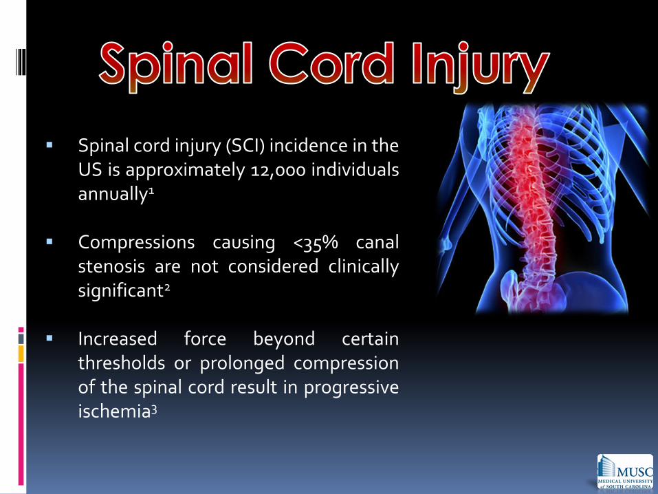
Spinal cord injury (SCI) incidence in the US is approximately 12,000 individuals annually1
Compressions causing <35% canal stenosis are not considered clinically significant2
Increased force beyond certain thresholds or prolonged compression of the spinal cord result in progressive ischemia3
Most current research focuses on clinical assessment of spinal cord injury
The state of spinal blood flow at subclinical forces has not been well understood
Characterize the relative extent to which various modes of compressive mechanical loading compromise blood flow in the anterior spinal arterial supply.

3-D finite element model of the cervical spinal cord was developed using Comsol Multiphysics 4.0a
Fluid-structure interaction physics module
Quantifying changes in outlet flow as a result of compression
Applied Loads based on the most common spinal injuries: Anterior, Posterior, Axial
Changes in Mechanical properties: Spinal cord elastic modulus, anterior spinal artery elastic modulus

Model includes a 1 cm segment of the cervical spinal cord, surrounding dura mater, the anterior spinal artery, and 5 arterial branches
Measurements based on bovine and porcine experiments
May be extrapolated to human studies

All materials in the model were characterized as linear elastic materials
Blood was modeled as a Newtonian fluid with a density of 1060 kg/m3 and a dynamic viscosity of 5e-3 Pa.s.
Blood flow was induced with an average inlet velocity of 0.3 m/s
Adaptive free-tetrahedral meshing
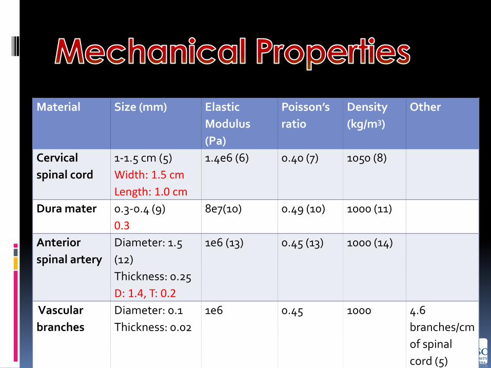
Material Size (mm) Elastic
Modulus
(Pa)
Poisson’s
ratio
Density
(kg/m3)
Other
Cervical
spinal cord
1-1.5 cm (5)
Width: 1.5 cm
Length: 1.0 cm
1.4e6 (6) 0.40 (7) 1050 (8)
Dura mater 0.3-0.4 (9)
0.3
8e7(10) 0.49 (10) 1000 (11)
Anterior
spinal artery
Diameter: 1.5
(12)
Thickness: 0.25
D: 1.4, T: 0.2
1e6 (13) 0.45 (13) 1000 (14)
Vascular
branches
Diameter: 0.1
Thickness: 0.02
1e6 0.45 1000 4.6
branches/cm
of spinal
cord (5)


Cannot induce acute mechanical damage
Spinal cord vascular auto-regulation is not simulated
Linear Elastic Material used to model materials
Lack of a cerebrospinal fluid layer
Newtonian fluid & steady state flow for blood flow
Collateral circulation & posterior spinal arteries were not included
Anterior loading results in reduced flow and increased deformation in the ASA.
may induce maladaptive vascular remodeling
may disrupt auto-regulation mechanism
Posterior loading reduces perfusion substantially within the spinal cord
limits blood flow in the arterial branches
minimally affects the ASA
may lead to ischemia of the supplied tissues
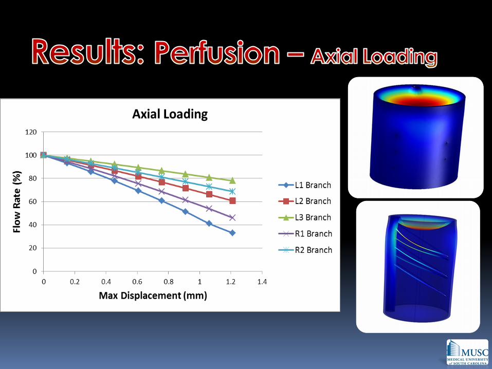
Axial loading affects arterial branches predominantly in proximity of the loading site.
Decreased blood flow caused by spinal compression may contribute to progressive ischemia of the spinal cord.

Passive and active mechanical testing of anterior spinal artery
Ex-vivo testing of compressive loading on spinal cord
Update model using constitutive equations for vascular tissue for quantitative analysis

Dr. Vibhor Krishna, M.D.
Dr. Mark Kindy, Ph.D.
Dr. Tarek Shazly, Ph.D.
Funding
Dr. Sunil Patel, M.D. of the Department of Neurosurgery at MUSC
The Summer Health Professionals Research Program, Medical University of South Carolina

References 1. National Spinal Cord Injury Statistical Center (NSCISC). Spinal Cord Injury Facts and Figures at a Glance. 2010;
https://www.nscisc.uab.edu/. Accessed July 10th, 2012.
2. Shields CB, Zhang YP, Shields LB, Han Y, Burke DA, Mayer NW. The therapeutic window for spinal cord decompression in a rat spinal cord injury model. Journal of neurosurgery. Spine. Oct 2005;3(4):302-307.
3. Ducker TB, Kindt GW, Kempe LG: Pathological findings in acute experimental cord trauma. J Neurosurg 35:700-708, 1971.
4. Tator CH, Fehlings MG. Review of the secondary injury theory of acute spinal cord trauma with emphasis on vascular mechanisms. Journal of neurosurgery. Jul 1991;75(1):15-26.
5. Anatomy of the Spinal Cord . (n.d.). Neuroscience Online. Retrieved July 24, 2012, from http://neuroscience.uth.tmc.edu/s2/chapter03.html
6. Mazuchowski, E. L., & Thibault, L. E. (2003). Biomechanical Properties of the Human Spinal Cord and Pia Mater. Key Biscayne: Summer Bioengineering Conference.
7. Ichihara, K., Taguchi, T., Shimada, Y., Sakuramoto, I., Kawano, S., & Kawai, S. (2001). Gray matter of the bovine cervical spinal cord is mechanically more rigid and fragile than the white matter. Journal of Neurotrauma, 18(3), 361-367.
8. Nelson SR, Mantz M-L, Maxwell JA (1971) Use of specific gravity in the measurement of cerebral edema. J Appl Physio130: 268 - 271
9. Reina, M. A., A. Lopez-Garcia, et al. (1996). "[Structural analysis of the thickness of human dura mater with scanning electron microscopy]." Rev Esp Anestesiol Reanim 43(4): 135-137.
10. Persson, C., Evans, S., Marsh, R., Summers, J., & Hall, R. (2010). Poisson's ratio and strain rate dependency of the constitutive behavior of spinal dura mater.. Ann Biomed Eng., 38(3), 975-83.
11. Persson, C., Summers, J., & Hall, R. M. (2011). The Effect of Cerebrospinal Fluid Thickness on Traumatic Spinal Cord Deformation. Journal of Applied Biomechanics, 27, 330-335.
12. Zhao, S., Logan, L., Schraedley, P., & Rubin, G. (2009). Assessment of the anterior spinal artery and the artery of Adamkiewicz using multi-detector CT angiography. Chinese Medical
13. Torii, R., Oshima, M., Kobayashi, T., Takagi, K., & Tezduyar, T. (2005). Influence of wall elasticity in patient-specific hemodynamic simulations.Computers & Fluids, 36(1), 160-168.
14. Tezduyar, T., Sathe, S., Keedy, R., & Stein, K. (2006). Space–time finite element techniques for computation of fluid–structure interactions. Computer Methods in Applied Mechanics and Engineering, 195(17-18), 2002-2027.


