
Mohammad Faizal Ahmad Fauzi, Ph.D. Associate Professor
Faculty of Engineering
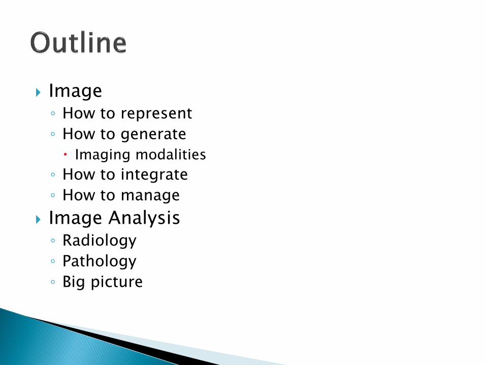
Image ◦ How to represent
◦ How to generate
Imaging modalities
◦ How to integrate
◦ How to manage
Image Analysis ◦ Radiology
◦ Pathology
◦ Big picture

Imaging Informatics ◦ Subfield of Biomedical Informatics
Deals with ◦ Image generation
◦ Image manipulation
◦ Image management
◦ Image integration
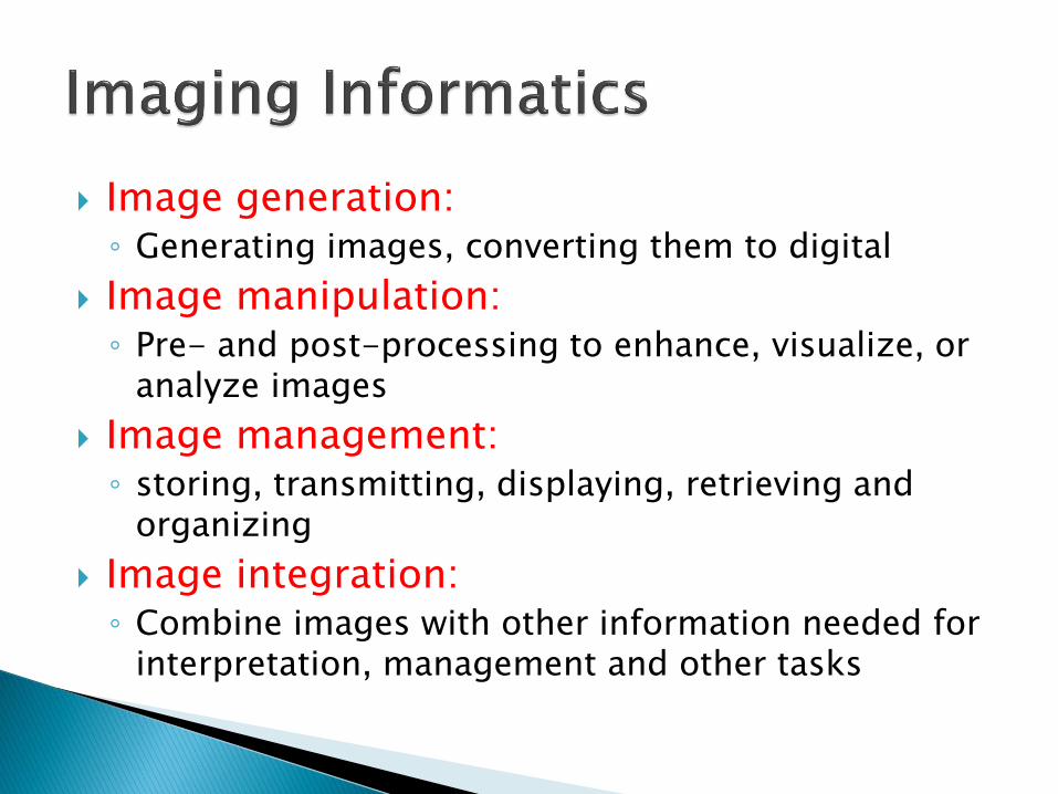
Image generation: ◦ Generating images, converting them to digital
Image manipulation: ◦ Pre- and post-processing to enhance, visualize, or
analyze images
Image management: ◦ storing, transmitting, displaying, retrieving and
organizing
Image integration: ◦ Combine images with other information needed for
interpretation, management and other tasks

Images ◦ 2D
◦ 3D
◦ 4D
Diagnostic Imaging Modalities ◦ Anatomical: X-ray, fluoroscopy, CT, MRI, US
◦ Functional: PET, SPECT, fMRI
Display and Organization Systems

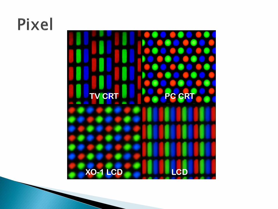
Two dimensional array of numbers


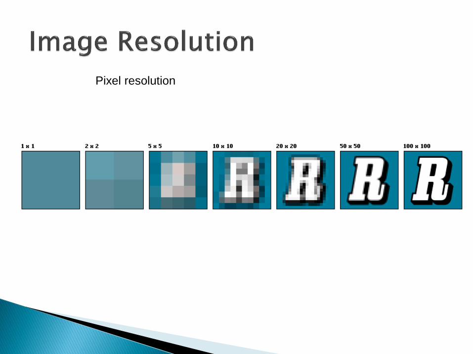
Pixel resolution


Spatial resolution: How well the modality can
distinguish points that are close to each other

Distance
Pixel connectivity
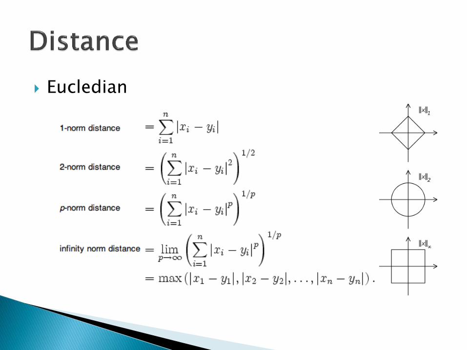
Eucledian


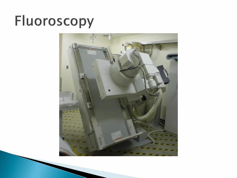
• Anatomical • Projection radiography (X-ray)
• Fluorography
• Computed Tomography
• Magnetic Resonance Imaging
• Ultrasound
• Functional • Nuclear Medicine and Positron Emission
Tomography

Source: X-ray
Detector
X-ray attenuation: Density of tissues


Source: Continuous low-power X-ray beam
Detector: X-ray image intensifier
Continuous acquisition of a sequence of X-ray images over time

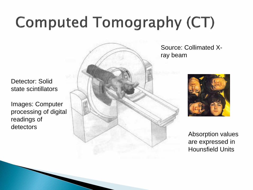
Source: Collimated X-
ray beam
Detector: Solid
state scintillators
Images: Computer
processing of digital
readings of
detectors Absorption values
are expressed in
Hounsfield Units

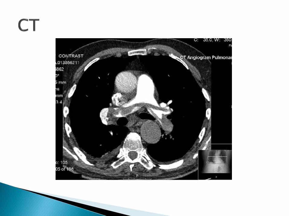


Hounsfield Units • 1000: Bone
• -1000: Air
• 0: Water


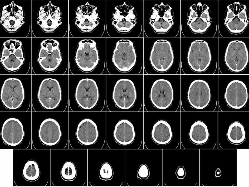
Source: High Intensity magnetic field and radio frequency pulses (on/off)
Detector: Phased array receiver
RF excitations of the protons results in absorption and subsequent release of energy -> magnetic characteristics of the tissue
Pictures of organs, bone, soft tissue
Computer generated images

Non-
iodizing Excellent
soft-tissue
contrast
detail
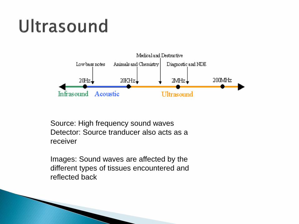
Source: High frequency sound waves
Detector: Source tranducer also acts as a
receiver
Images: Sound waves are affected by the
different types of tissues encountered and
reflected back


Source: X-ray or γ-ray emitting radio-isotopes are injected, inhaled or
ingested
Detector: Gamma camera – measures the radioactive decay of the
active agent
Image: Functional information


Core function: storage, distribution and display of medical images
Further strengthened by a hospital’s other IT infrastructure
Hospital Information System (HIS)
Electronic Medical Records System (EMR)
Radiology Information System (RIS)

Uses: ◦ Hard copy replacement
◦ Remote access – teleradiology
◦ Integration with other electronic systems
◦ Radiology workflow management


Digital Imaging and Communication in Medicine
Standard format for PACS files and messages ◦ A standard for handling, storing,
printing, and transmitting information in medical imaging
◦ File format definition and network communication protocol
DICOM files can be exchanged
between two entities that are capable
of receiving image and patient data in DICOM format.

eFilm
OsiriX – open source
ImageJ


