
Tumor and Stem Cell Biology
miR-34a and miR-34b/c Suppress IntestinalTumorigenesisLongchang Jiang1 and Heiko Hermeking1,2,3
Abstract
The p53-inducible miR-34a and miR-34b/c genes are fre-quently silenced in colorectal cancer. To address the in vivorelevance of miR-34a/b/c function for suppression of intestinaltumor formation, we generated ApcMin/þ mice with deletions ofthe miR-34a and/or miR-34b/c genes separately or in combina-tion. Combined deletion of miR-34a/b/c increased the numberof intestinal stem cells as well as Paneth and Goblet cells,resulting in enlarged intestinal crypts. miR-34a/b/c-deficientApcMin/þ mice displayed an increased tumor burden and gradeand decreased survival. miR-34a/b/c-deficient adenomasshowed elevated proliferation and decreased apoptosis anddisplayed pronounced bacterial infiltration, which may be dueto an observed decrease in infiltrating immune cells and down-regulation of barrier proteins. mRNA induction in miR-34a/b/c-
deficient tumors was enriched for miR-34a/b/c seed-matchingsites and for mRNAs encoding proteins related to epithelial–mesenchymal transition, stemness, and Wnt signaling. Accord-ingly, cells explanted from miR-34a/b/c-deficient adenomasformed tumor organoids at an increased rate. Several upregu-lated miR-34 targets displayed elevated expression in primaryhuman colorectal cancers that was associated with lymph-nodemetastases (INHBB, AXL, FGFR1, and PDFGRB) and upregula-tion of INHBB and AXL in primary colorectal cancer wasassociated with poor patient survival. In conclusion, our resultsshow thatmiR-34a/b/c suppress tumor formation caused by lossof Apc and control intestinal stem cell and secretory cellhomeostasis by downregulation of multiple target mRNAs.Cancer Res; 77(10); 2746–58. �2017 AACR.
IntroductionColorectal cancer is a leading cause of cancer death. In the
United States alone, 134,490 new cases and 49,190 deaths fromcolorectal cancer were expected in 2016 (1). Colorectal cancer is amultistep process driven by mutational activation of severaloncogenes and genetic/epigenetic inactivation of tumor suppres-sors (2). The majority of colorectal cancers originate from benignadenomas, which are caused by inactivating mutations in theadenomatous polyposis coli (APC) gene. APC mutation leads to theconstitutive activation of the Wnt/b-catenin pathway, whichresults in the increased expression of b-catenin, a critical regulatorof intestinal epithelial cell homeostasis (3). During colorectalcancer progression, additional mutations in key oncogenes andtumor suppressor genes, such as p53, are acquired. ApcMin/þ miceinherit a mutant Apc allele that results in a truncation of the APCprotein at amino acid 850 (4) and spontaneously lose the wild-type Apc allele, resulting in multiple benign adenomas mainlythroughout the small intestine (5). Thus, ApcMin/þ mice allow to
study the influence of putative tumor suppressor genes on theinitiation of intestinal tumorigenesis in vivo (6).
The tumor suppressor gene p53 is inactivated in the majority ofcolorectal cancers and encodes a transcription factor, which isposttranscriptionally induced by DNA damage and a number ofadditional cellular stresses (7). Besides regulating the expressionof mRNAs, p53 also controls the expression of noncoding RNAs,such as lncRNAs andmicroRNAs (miRNA; refs. 8, 9).Many tumorsuppressive functions of p53 are thought to be mediated byp53-induced miRNAs (8, 10). Among the p53-induced micro-RNA-encoding genes, miR-34a and miR-34b/c display the mostconsistent and pronounced induction, which may explain whytheywere identified as one of thefirst p53-regulatedmiRNAs (11).
Via downregulation of key-regulators, miR-34a/b/c suppressnumerous cancer-associated processes, such as cell prolifera-tion and survival (12), epithelial–mesenchymal transition(EMT; ref. 13), and stemness (14). Moreover, miR-34a/b/cpresumably function as cell-fate determinants in colonic cancerstem cells (CSC) by suppressing Notch activity (15). Further-more, miR-34a/b/c regulate the homeostasis of normal stemcells (16) and suppress the formation of induced pluripotentstem cells (IPSCs) (17).
Consistent with a tumor suppressive role in tumorigenesis,downregulation of miR-34a/b/c expression has been observed ina variety of human cancers (8). miR-34a/b/c expression is alsoepigenetically silenced by CpG methylation in colorectal cancercell lines (18)and inprimary colorectal cancers (19). Thesefindingssuggest an important role of miR-34a/b/c as downstream effectorsof p53 and potential tumor suppressors in colorectal cancer. In linewith its tumor suppressive potential miR-34a mimetics are cur-rently tested in the clinics for treatment of advanced cancer (20).
Here, we report the generation of ApcMin/þ mice carryingtargeted deletions of the miR-34a and miR-34b/c genes and thecharacterization of the resulting phenotypes at the organismal
1Experimental and Molecular Pathology, Institute of Pathology, Ludwig-Max-imilians-University, M€unchen, Germany. 2German Cancer Consortium (DKTK),Partner site Munich, Munich, Germany. 3German Cancer Research Center(DKFZ), Heidelberg, Germany.
Note: Supplementary data for this article are available at Cancer ResearchOnline (http://cancerres.aacrjournals.org/).
Corresponding Author: Heiko Hermeking, Ludwig-Maximilians-UniversityM€unchen, Thalkirchner Strasse 36, Munchen D-80337, Germany. Phone: 11-49-2180-73685; Fax: 11-49-89-2180-73697; E-mail:[email protected]
doi: 10.1158/0008-5472.CAN-16-2183
�2017 American Association for Cancer Research.
CancerResearch
Cancer Res; 77(10) May 15, 20172746
on May 1, 2019. © 2017 American Association for Cancer Research. cancerres.aacrjournals.org Downloaded from
Published OnlineFirst March 31, 2017; DOI: 10.1158/0008-5472.CAN-16-2183

and molecular level. Our findings provide genetic proof of atumor suppressive function of the miR-34a and miR-34b/c genesduring intestinal tumorigenesis.
Materials and MethodsAnimals
The generation of miR-34a�/� mice with a C57BL6/SV129background has beendescribed previously (21).miR-34b/cfl/flmicewere kindly providedbyDr. AlexanderNikitin (Cornell University,Ithaca, NY; ref. 16). In brief, gene-specific deletions were generatedby using homologous recombination with a vector containingmiR-34a or miR-34b/c sequences flanked by loxP sites and anintronic Neomycin resistance (Neo) cassette flanked frt sites indi-vidually (21). The Neo cassette was removed by crossing with flp-mice and germline miR-34a or miR-34b/c knock-out mice weregenerated by crossing with CMV-Cre mice. miR-34a�/�; b/c�/�
compound mice were generated by crossing miR-34a�/� andmiR-34b/c�/�. The resulting genotypes were obtained in theexpected Mendelian ratios and the offspring displayed no overtphenotype. miR-34a�/�, miR-34b/c�/�, miR-34a�/�; miR-34b/c�/�
and miR-34aþ/þ; miR-34bcþ/þ mice were crossed with ApcMin/þ
mice to obtain mice with the following genotypes: miR-34a�/�;ApcMin/þ, miR-34b/c�/�; ApcMin/þ, miR-34a/b/c�/�; ApcMin/þ, andmiR-34a/b/cþ/þApcMin/þ. Mice were housed in individually venti-lated cages (IVC). Animal studies were approved by the Govern-ment of Upper Bavaria, Germany (AZ 55.2-1-54-2532-4-2014).
Tissue preparation and tumor countMice were sacrificed at 18 weeks of age. Whole intestines were
isolated, separated into four equal parts,flushedwithPBS, openedlongitudinally, photographed, and fixed as Swiss rolls in 4%buffered formaldehyde. Tumor numbers and size were evaluatedusing ImageJ software.
Histology and IHCThree-micron paraffin sections were used for hematoxylin and
eosin staining and periodic acid–Schiff (PAS) staining accordingto standard protocols. Tumors from ApcMin/þmice were classifiedas adenomas with either low- or high-grade dysplasia, based onnuclear-cytoplasmic ratio, nucleus location, prominence ofnucleoli, gland architecture, amount of interglandular stroma,and the presence ofmucus secretion. Immunohistochemistry wasperformed using antibodies and reagents listed in SupplementaryTable S1. For each immunohistochemical detection � 40 tumorsper genotype were examined. Positive staining was evaluated byImage-Pro plus and ImageJ software.
In situ hybridization and fluororescence in situ hybridizationFor detection of intestinal stem cells an Olfm4-specific, DIG-
labeled RNA probe was generated using a murine Olfm4 vector(kindly providedbyDr.HansClevers,Hubrecht Institute,Utrecht,The Netherlands) in combination with aDIGNorthern Starter Kit(Roche Diagnostics). in situ hybridization (ISH) was performedas described (22). To visualize intestinal bacteria by fluorescencein situ hybridization, the universal eubacteria probe (EUB338)and negative control probe (NON338) were employed. The 50-FITC-labeled EUB338 DNA (50-GCTGCCTCCCGTAGGAGT-30)and 50-Cy3-labeled NON338 DNA (50-CGACGGAGGGCAT-CCTCA-30) probes were synthesized by Metabion (Planegg) andhybridized to 3-mm paraffin sections.
Transcriptomic analysisTotal RNA from tumors was isolated using the RNeasy Plus
Mini Kit (Qiagen) with an on-column DNase digestion (threeRNA samples per genotype; each tumorRNA sample represented apool of 3 tumors isolated from the samemouse). RandomprimedcDNA libraries were generated and sequenced using theHiSeq2500 (Illumina) platform by GATC (Konstanz). Each sam-ple was covered by at least 35million single reads of 50 bp length.Data were normalized in R with the RUVSeq (23) module anddifferential expression analysis was performed using Chipster(24). The overlap of DEseq2 and edgeR results were consideredto represent the most significantly, differentially expressed genes.GeneOntology (GO) andKEGGpathway analysis was performedwithDAVID Bioinformatics Resources (25). Gene Set EnrichmentAnalysis (GSEA) was performed using the GSEA (26) software.miR-34a/b/c targets were predicted with TargetScan 6.2 (27).Expression data were deposited in the Gene Expression Omnibuswebsite (accession no. GSE84138).
Cell lines and culture, generation of cell pools with conditionalpri-miR-34a expression, and tumor organoid culture
H1299 lung cancer cells used for 30-UTR reporter assays werefrom own stocks and their negativity for p53 was validated.SW620 colorectal cancer cells used for ectopic pri-miR-34a expres-sion were from own stocks and authenticated by STR analysis in2014 (Eurofins Medigenomix Forensik GmbH, Ebersberg, Ger-many). H1299 and SW620 cancer cell lines were kept in Dulbec-co's modified Eagle's medium (DMEM) and 10% fetal calf serum(Invitrogen) at 5% CO2. SW620 cells were transfected withepisomal pRTR-pri-miR-34a expression plasmids as describedbefore (13). After 24 hours, cell pools were selected by additionof puromycin (2 mg/mL) for 10 days. GFP expression was eval-uated by fluorescence microscopy 48 hours after addition of100 ng/mL doxycycline (DOX) to the cell pools. Intestinal ade-noma cells from ApcMin/þmice were isolated and counted using ahemocytometer. Single cells (15,000) were embedded in 50 mLMatrigel per well in 24-well plates. The tumor organoid culturemedium was formulated as described before (28).
30-UTR reporter assaysThe 30-UTR of Wasf1 was PCR amplified from genomic DNA
isolated from mouse tissue, inserted into pGL3-control-MCSvector downstream of a firefly luciferase ORF, and verified bysequencing. H1299 cells were seeded in a 12-well plate with 3 �104 cells/well and transfected with 100 ng of pGL3-Wasf1 plas-mid, 20 ng of Renilla reporter plasmid for normalization, and 25nmol/L miR-34a or miR-34c pre-miRNAs or a negative controloligonucleotide (Ambion). Forty-eight hours later, luciferaseassays were performed with the Dual-Luciferase Reporter 1000Assay System (Promega). Fluorescence intensities were measuredwith an Orion II Microplate Luminometer (Titertek-Berthold).Primers used for cloning and sequencing are listed in Supple-mentary Table S2.
Quantitative real-time PCR and Western blot analysisTotal RNA was isolated from tumor samples or cultured cell
lines using the RNeasy Plus Mini Kit (Qiagen). cDNA was gen-erated by Verso cDNA kit (Thermo Scientific) and quantitativereal-time PCR (qPCR) was performed by using Fast SYBR GreenMaster Mix (Applied Biosystems) and a LightCycler 480 II (RocheDiagnostics). Relative gene expression was determined using the
Genetic Analysis of miR-34a/b/c in Intestinal Tumorigenesis
www.aacrjournals.org Cancer Res; 77(10) May 15, 2017 2747
on May 1, 2019. © 2017 American Association for Cancer Research. cancerres.aacrjournals.org Downloaded from
Published OnlineFirst March 31, 2017; DOI: 10.1158/0008-5472.CAN-16-2183
2�DDCt method (29). The individual mRNA levels were normal-ized to b-actin. Primers used for qPCR are listed in SupplementaryTable S3.
For protein lysates tumor samples or cultured cells were lysedin RIPA buffer containing cOmplete Mini protease inhibitorcocktail tablets (Roche Diagnostics). Lysates were sonicatedand centrifuged. Whole-lysate proteins (30–60 mg) were loadedper lane. Gel electrophoresis and transfer to PVDF membranes(Millipore) were carried out using standard protocols (Bio-RadLaboratories). Primary antibodies (Supplementary Table S1)were used in combination with HRP-coupled secondary anti-bodies. ECL (Millipore) signals were recorded with a 440CFimaging system (Kodak).
Analysis of online expression data setsThe Cancer Genome Atlas (TCGA; ref. 30) gene expression
data and follow-up information of colon adenocarcinomas(COAD) were downloaded from NCI's Genomic Data Com-mons website (GDC; https://gdc.cancer.gov/). NormalizedRSEM counts were used to determine the expression of relevantmRNAs. Clinical outcome data were divided into high, inter-mediate, and low expression groups according to the expressionvalue of individual genes.
Statistical analysisA two-tailed Student t test was used to compare continuous
variables. Categorical variables were compared using the c2 meth-od. Kaplan-Meier calculations were used to display the overallsurvival time and the results were compared with a log-rank test.The Sidak method was used to adjust P values when multiplecomparisons were performed. P values less than 0.05 were consid-ered significantand indicatedbyasterisks (�,P<0.05; ��,P<0.01;or���, P < 0.0001). Univariate, age/gender/tumor grade-adjustedhazard ratios, and 95% confidence intervals (CI) were estimatedwith a Cox's proportional hazard model. Prism 6 (GraphPadsoftware) or SPSS (IBM) programs were used for calculations.
ResultsmiR-34a/b/c deletion modulates the architecture of the smallintestine in mice
We generated mice carrying deletions of the miR-34a or miR-34b/c loci and combinations of these. Deletion of miR-34a, miR-34b/c, or miR-34a/b/c in the germline did not significantly influ-ence the lifespan of mice (Fig. 1A). However, we observed agender-independent increase in the width and depth of crypts ofthe small intestine in miR-34a/b/c-deficient mice, which presum-ably caused the minor increase in the total length of the smallintestine (Fig. 1B). Notably, the numbers of Paneth cells per cryptandGoblet cells per villus were significantly increased inmiR-34a/b/c-deficient mice (Fig. 1C and D). In addition, the frequency ofstem cells at the crypt base was significantly increased inmiR-34a/b/c-deficient mice as determined by detection of the stem cellmarker Olfm4 using in situ hybridization (Fig. 1E). These pheno-types were independent of the gender (Fig. 1B–E).
In order to determine, whether inactivation of miR-34 genesaffects intestinal tumor formation,wegeneratedApcMin/þmicewithdeletions of miR-34a, miR-34b/c, or miR-34a/b/c. Also in ApcMin/þ
mice deficiency for miR-34a/b/c resulted in an increase of thenumber of Paneth, Goblet, and Olmf4-positive cells per crypt inuntransformed tissue of the small intestine (Supplementary Fig.S1A–D). Similarly, thewidth anddepthof the cryptswere increased
significantly inmiR-34a/b/c-deficient ApcMin/þ mice, but the minorincrease in the length of the small intestine was not statisticallysignificant (Supplementary Fig. S1E). Again, these architecturalchanges were observed in both, male and female mice. Therefore,deletion of one APC allele did not influence the morphologicalchanges in the intestine caused by deletion of miR-34a/b/c.
miR-34a/b/c loss enhances tumorigenesis in ApcMin/þ miceNotably, miR-34a/b/c�/�; ApcMin/þ mice showed signs of mor-
bidity sooner and exhibited a shorter life-span than wild-typeApcMin/þ mice (Fig. 2A). In contrast, ApcMin/þ mice deficient foreither themiR-34aor themiR-34b/calleledidnot showastatisticallysignificant change in life-span (Fig. 2A).Whenmice were sacrificedat the age of 18 weeks and the entire small intestinal tract wasexamined, miR-34a/b/c�/�; ApcMin/þ and, to a lesser extent, miR-34a�/�; ApcMin/þ knockout mice showed a significantly increasednumberof tumorswhencomparedwithApcMin/þmice (Fig. 2BandC). Furthermore, the total tumor area in miR-34a/b/c�/�; ApcMin/þ
micewas�4 times larger than inwild-typeApcMin/þmice (Fig. 2D).The frequency of large tumors (>6 mm2) was significantly higherwhen miR-34a/b/c, but not when only miR-34a or miR-34b/c geneshad been deleted in ApcMin/þ mice (Fig. 2E). We also noted asignificantly increased number of adenomas with high-grade dys-plasia in miR-34a/b/c-deficient ApcMin/þ mice, whereas the inacti-vation of eithermiR-34a ormiR-34b/c alone did not have this effect(Fig. 2F). Taken together, our findings show that the inactivation ofmiR-34a/b/c promotes intestinal tumor formation by increasingtumor initiation and enhancing the growth of tumors in ApcMin/þ
mice.As the combined inactivationofmiR-34aandmiR-34b/cgeneswasnecessary for these effects, thesemiRNAsmayhaveoverlappingfunctions and compensate each other. In addition, these results arein accordance with the combined epigenetic inactivation ofmiR-34a and miR-34b/c by DNA methylation, which has beendetected in more than 75% of analyzed colorectal cancer samples(31). In the subsequent analyses, we therefore focused on studyingmice with simultaneous deletion of miR-34a and miR-34b/c.
miR-34a/b/c loss affects proliferation, apoptosis, andinfiltration by bacteria and immune cells of adenomas
Notably, tumors inmiR-34a/b/c knockoutApcMin/þmice showeda significant increase in proliferation and a significantly reducedrate of apoptosis (Fig. 3A). The frequency of stromal cells withinthe tumors was not affected bymiR-34a/b/c deletion as determinedby detection of vimentin. Because it was recently shown thatbacterial infiltration promotes tumorigenesis in ApcMin/þ mice(32), we asked whether miR-34a/b/c deletion affects the presenceof bacteria in intestinal tumors. Bydetection of bacterial 16S rRNA,we found that adenomas from miR-34a/b/c�/�; ApcMin/þ micedisplay extensive bacterial infiltration regardless of tumor size(Fig. 3A) or tumor grade (Supplementary Fig. S2A). However, theadjacent normal intestinal epithelium of all mice analyzedherewas devoid of bacteria. Therefore,miR-34a/b/c-deficientmicedo not display a general intestinal barrier defect. Unexpectedly, thefrequencyof immune cells, such asmacrophages, T- andB-cell,wasdecreased in miR-34a/b/c-deficient adenomas (Fig. 3B; Supple-mentary Fig. S2B). Therefore, the increased bacterial infiltrationof adenomas may be due to a defect in the tumor-associatedimmune defense in miR-34a/b/c-deficient mice.
Expression profiling of miR-34a/b/c-deficient adenomasTo further illuminate the mechanisms by which miR-34a/b/c
loss promotes intestinal tumorigenesis in ApcMin/þ mice, we
Jiang and Hermeking
Cancer Res; 77(10) May 15, 2017 Cancer Research2748
on May 1, 2019. © 2017 American Association for Cancer Research. cancerres.aacrjournals.org Downloaded from
Published OnlineFirst March 31, 2017; DOI: 10.1158/0008-5472.CAN-16-2183

A
B
C
D
E
Surv
ival
(%)
miR-34a –/–; miR-34bc+/+ (n = 66)
miR-34a/b/c–/–(n = 54)miR-34a+/+; miR-34bc –/–(n = 38)
miR-34a/b/c+/+ (n = 44)
Days
100
80
60
40
20
01,0006002000 1,200800400
PAS
miR-34a/b/c –/–
miR-34a/b/c+/+
Pane
th c
ells
/cry
pt
4
6
8
10
12
miR-34a –/–+/+–/–+/+miR-34bc +/+ –/––/–+/+
***
MaleFemale
PAS
miR-34a/b/c –/–miR-34a/b/c+/+
Gob
let c
ells
/vill
us
0
5
10
15
20
miR-34a –/–+/+–/–+/+miR-34bc +/+ –/––/–+/+
***
MaleFemale
Olfm4
miR-34a/b/c –/–miR-34a/b/c+/+
miR-34a –/–+/+–/–+/+miR-34bc +/+ –/––/–+/+
3
5
6
7
8
4Stem
cel
ls/c
rypt
MaleFemale
***
0
20
40
60
Wid
th o
f cry
pts
(μm
)
miR-34a –/–+/+–/–+/+miR-34bc –/––/–+/++/+
***
0
40
60
80
Dep
th o
f cry
pts
(μm
)
20
–/–+/+–/–+/+–/––/–+/++/+
***
0
304050
Leng
th o
f SI (
cm)
1020
–/–+/+–/–+/+–/––/–+/++/+
FemaleMale
**
Figure 1.
Effects of miR-34a/b/c deletion on the cellular composition of the small intestine in mice. A, Kaplan–Meier survival analysis of mice with the indicated genotypes.Results were compared with a log-rank test. B, Determination of the width (left graph) and depth (middle graph) of small intestinal crypts. At least200 crypts in the jejunum from 18-week-old mice were analyzed. The length (right graph) of the small intestine was determined in mice with the indicatedgenotypes (n� 3mice per gender). C andD,After PAS-staining Paneth cell number per crypt (C) and Goblet cell number per villus (D) were evaluated in� 50 cryptsand � 50 villi per mouse, respectively (n � 3 mice per gender). Scale bar, 25 mm. E, Representative pictures of in situ hybridization of Olfm4-positive cellsin the crypts from indicated genotypes. At least 50 crypts per mouse were counted (n � 3 mice per gender). Scale bar, 10 mm. In B–E, n � 6 mice per genotype,results are presented as mean � SEM. �� , P < 0.01; ��� , P < 0.001 (Student t test).
Genetic Analysis of miR-34a/b/c in Intestinal Tumorigenesis
www.aacrjournals.org Cancer Res; 77(10) May 15, 2017 2749
on May 1, 2019. © 2017 American Association for Cancer Research. cancerres.aacrjournals.org Downloaded from
Published OnlineFirst March 31, 2017; DOI: 10.1158/0008-5472.CAN-16-2183

A
CB
FD
E
80
60
40
20
0
****
Tum
or n
umbe
r/mou
se
miR-34amiR-34bc
+/++/+
–/–+/+
+/+–/–
–/––/–
600
400
200
0Tum
or a
rea
(mm
2 )/m
ouse
miR-34amiR-34bc
+/++/+
–/–+/+
+/+–/–
–/––/–
***
Size
dis
trib
utio
n (%
) 100
80
60
40
20
0
***
miR-34amiR-34bc
+/++/+
–/–+/+
+/+–/–
–/––/– miR-34a/b/c–/–; ApcMin/+
High gradeLow grade
miR-34a/b/c+/+; Apc Min/+
Low grade High grade ***
8.3%
91.7%
9.6%
90.4%
9.1%
90.9%
41.8%
58.2%
miR-34amiR-34bc
+/++/+
–/–+/+
+/+–/–
–/––/–
miR-34a–/–; miR-34bc+/+; ApcMin/+ (n = 76)
miR-34a/b/c –/–; ApcMin/+ (n = 69)miR-34a+/+; miR-34bc–/–; ApcMin/+ (n = 30)
miR-34a/b/c+/+; ApcMin/+ (n = 50)
Surv
ival
(%)
Days
100
80
60
40
20
05003001000 400200
***
miR-34amiR-34bc
+/++/+
–/–+/+
+/+–/–
–/––/–
miR-34amiR-34bc
miR-34amiR-34bc
miR-34amiR-34bc
Tumor-size:>6 mm2
<6 mm2
Figure 2.
Effects of miR-34a/b/c deletion on tumor characteristics and survival of ApcMin/þ mice. A, Kaplan–Meier survival analysis of the indicated genotypes. Resultswere compared with a log-rank test. B, Representative images of resected small intestines. Scale bar, 1 cm. C and D, Quantification of intestinal tumor number (C)and tumor area (D) per mouse (n � 10 mice per genotype). E, Graph showing the percentage of tumor size (surface area) distribution in � 100 tumors(n � 10 mice per genotype). F, Top, tumor stage (low grade adenoma, high-grade adenoma) of � 66 intestinal tumors (n � 10 mice per genotype). Bottom,representative images of low- and high-grade adenomas. Scale bar, 100 mm. In B–F, tumors from 18 weeks old ApcMin/þ mice with the indicated genotypeswere analyzed and results are presented as mean � SEM. � , P < 0.05; ��� , P < 0.001 (Student t test).
Jiang and Hermeking
Cancer Res; 77(10) May 15, 2017 Cancer Research2750
on May 1, 2019. © 2017 American Association for Cancer Research. cancerres.aacrjournals.org Downloaded from
Published OnlineFirst March 31, 2017; DOI: 10.1158/0008-5472.CAN-16-2183

obtained mRNA expression profiles of adenomas from 18-week-old miR-34a/b/c-deficient ApcMin/þ mice and wild-typeApcMin/þ mice. For each mouse (n ¼ 3 per genotype), RNAsderived from three different tumors were pooled, libraries weregenerated and subjected to RNA-Seq to obtain more than 35million reads per library. Thereby, we detected the upregulationof 1773 mRNAs, when mRNAs with a log2-fold change � 0.6and RPKM � 0.25 in miR-34a/b/c�/�; ApcMin/þ mice versuswild-type ApcMin/þ mice were included (Fig. 4A). As expected,the percentage of mRNAs upregulated in the miR-34a/b/c-defi-cient tumors was higher for mRNAs harboring miR-34a/b/cseed-matching sequences than for mRNAs lacking these (Fig.4B). Three hundred and twenty-four mRNAs were identified asdifferentially regulated due to miR-34a/b/c loss when the raw-count methods DESeq2 (33) and edgeR (34) were applied (Fig.4C). After unsupervised clustering of these differentiallyexpressed genes, one of the miR-34a/b/c-deficient tumor sam-ples showed a divergent expression profile, which was presum-
ably due to biological variation (Fig. 4D). Next, we used GeneOntology (GO) and KEGG pathway analyses to group thedifferentially expressed transcripts into functional classes. TheGO analysis indicated that the upregulated mRNAs areinvolved in cell adhesion, extracellular matrix, and growthfactor binding functions (Supplementary Fig. S3A). The KEGGanalysis showed that several pathways were significantlyenriched, such as focal adhesion, ECM-receptor interaction,and pathways related to cancer (Supplementary Fig. S3B). Onthe contrary, both GO and KEGG analyses indicated that thedownregulated genes are mainly involved in immune responsefunctions (Supplementary Fig. S4A and S4B). A subsequentGene Set Enrichment Analysis (GSEA) revealed thatmiR-34a/b/cdeletion results in gene expression signatures related to EMT,hypoxia, angiogenesis, inflammatory response, Kras signaling,and apical junction processes (Fig. 4E). Interestingly, theexpression signatures of miR-34a/b/c-deficient adenomas alsoshowed a significant overlap with signatures of intestinal stem
A
Ki6
7C
leav
ed C
aspa
se-3
Eub3
38
miR-34a/b/c+/+ miR-34a/b/c–/–
Vim
entin
Posi
tive
cells
(%)
**80706050
90100
–/–+/+ miR-34a/b/c
Posi
tive
cells
/mm
2
302010
0
4050 *
–/–+/+ miR-34a/b/c
Posi
tive
area
(%)
3210
45
–/–+/+ miR-34a/b/c
Tum
ors
(%)
604020
0
80100
***
>6<6>6<6 (mm2)–/–+/+ miR-34a/b/c
PositiveNegative
***
Posi
tive
cells
/mm
2
300
200
100
0
400
–/–+/+ miR-34a/b/c
F4/80 (macrophages)
Posi
tive
cells
/mm
2
40
20
0
60
–/–+/+ miR-34a/b/c
80 ***
CD45R (B cells)Po
sitiv
e ce
lls/m
m2
600400200
0
800
–/–+/+ miR-34a/b/c
1,000 ***
CD3 (T cells)
B
Figure 3.
Effects of miR-34a/b/c deletion onproliferation, apoptosis, bacterialinfiltration, and stromal cells ofApcMin/þ
adenomas. Adenomas from 18 weeksold ApcMin/þ mice with the indicatedgenotypes were subjected to severaldifferent immunodetections (n¼ 7miceper genotype).A, IHC detection of Ki67,cleaved caspase-3 and vimentin asindicated and fluorescence in situhybridization of universal eubacteriaprobe (EUB338). Scale bar, 100 mm.B, Quantification of infiltratedmacrophages, B and T cells by F4/80,CD45R, and CD3 immunodetection,respectively, in adenomas. Results arepresented as mean � SEM. � , P < 0.05;�� , P < 0.01; ��� , P < 0.001 (Studentt test).
Genetic Analysis of miR-34a/b/c in Intestinal Tumorigenesis
www.aacrjournals.org Cancer Res; 77(10) May 15, 2017 2751
on May 1, 2019. © 2017 American Association for Cancer Research. cancerres.aacrjournals.org Downloaded from
Published OnlineFirst March 31, 2017; DOI: 10.1158/0008-5472.CAN-16-2183

miR-34a/b/c+/+; ApcMin/+ miR-34a/b/c-/-; ApcMin/+
1.50-1.5
A
DC
B
DESeq2(523)
edgeR(475)
324199 151
Num
ber o
f mR
NAs
Log2 fold change
–2.00
500
1,000
1,500
2,000
2,500
–1.0 0.0 1.0 2.0
UpregulatedDownregulated
w/o miR-34 binding site with miR-34 binding site
***
61.84%(1499)
38.16%(925)
93.20%(274)
6.80%(20)
EGene set enrichment analysis
(KO vs. WT)
0
0.5
1 5 10Rank list (×1,000)
ES
Kras signaling
ESNESFDR
0.431.470.032
+/+-/-
0
0.6
1 5 10Rank list (×1,000)
ES
EMT
ESNESFDR
0.652.23<0.001
+/+-/-
0
0.5
1 5 10Rank list (×1,000)
ES
Hypoxia
ESNESFDR
0.511.720.002
+/+-/-
0
0.5
1 5 10Rank list (×1,000)
ES
Inflammatory response
ESNESFDR
0.441.510.023
+/+-/-
0
0.5
ES
1 5 10Rank list (×1,000)
Apical junction
ESNESFDR
0.411.390.074
+/+-/-
0
0.6
1 5 10Rank list (×1,000)
Angiogenesis
ESNESFDR
0.581.630.007
+/+-/-
ES
ESNESFDR
0.301.51<0.001
ISC signature Munoz et al. 2012
0
0.3
1 10 20Rank list (×1,000)
ES
+/+-/-
Rank list (×1,000)
ESNESFDR
0.351.410.0210
0.35
1 5 10
ES
+/+-/-
Lgr5+ signature Merlos-Suarez et al. 2011 Gene Ontology Wnt signaling signature
Rank list (×1,000)
+/+-/-
ESNESFDR
0.271.300.0040
0.3
1 10 20
ES
Figure 4.
NGS analysis ofmiR-34a/b/c-dependent mRNA expression in adenomas of ApcMin/þmice. A, Results of NGS analysis represented as histogram showing changes inmRNA levels after miR-34a/b/c deletion. Transcriptionally regulated genes with a |log2-fold change| � 0.6 and RPKM � 0.25 are highlighted as purple bars.Fold change denotes the ratio of mRNA expression values in tumors of miR-34a/b/c�/�; ApcMin/þ mice versus miR-34a/b/cþ/þ; ApcMin/þ mice (n ¼ 3 mice pergenotype).B,Calculation of the percentage ofmRNAsdifferentially regulated inmiR-34a/b/c-deficient versuswild-type tumors inApcMin/þmicewithout (left graph)andwith (right graph)miR-34a/b/c seed-matching sequences. ��� , P <0.001. C,Results of DESeq2 and edgeR analyses of the NGS results.D,Heatmap of geneswithsignificantly differential expression in tumors from mice with the indicated genotypes. Differential gene expression is shown according to the pseudocolor scaleindicated on the right side. E, GSEA analysis of mRNA profiles from tumors of miR-34a/b/c�/�; ApcMin/þ mice versus miR-34a/b/cþ/þ; ApcMin/þ mice.
Jiang and Hermeking
Cancer Res; 77(10) May 15, 2017 Cancer Research2752
on May 1, 2019. © 2017 American Association for Cancer Research. cancerres.aacrjournals.org Downloaded from
Published OnlineFirst March 31, 2017; DOI: 10.1158/0008-5472.CAN-16-2183

cells (ISC) (35), Lgr5þ positive cells (36) and the GO Wntsignaling signature. In addition, several regulators of barrierfunction in IEC were also detected as downregulated by NGSanalysis in miR-34a/b/c-deficient adenomas (e.g., Muc1, Tff3,and Retnlb; fold change: �4.74, �1.44, and �3.34, respective-ly). The downregulated mRNA expression of these barriercomponents was validated by qPCR (Supplementary Fig.S5A). Furthermore, we confirmed the decreased protein expres-sion of MUC1 in miR-34a/b/c-deficient adenomas by immuno-histochemistry (Supplementary Fig. S5B). The repression ofthese barrier components may also contribute to the observedinfiltration of bacteria into miR-34a/b/c-deficient adenomas.
miR-34a/b/c loss enhances the formation of intestinal tumororganoids
To obtain functional evidence for an enhanced stemness ofadenoma cells with deletion of miR-34a/b/c, we performed atumor organoid formation assay. Indeed, an increased tumororganoid formation rate was observed for miR-34a/b/c-deficientadenoma derived cells when compared with those derived frommiR-34a/b/c-proficient adenomas (Fig. 5A).
miR-34a/b/c deletion enhances intestinal Wnt signaling inApcMin/þ mice
Wnt/b-catenin signaling is critically involved in regulatingthe homeostasis and neoplastic transformation of the intestine.Because 34a/b/c-deficient adenomas showed a Wnt signalingsignature (Fig. 4E) and the Wnt/b-catenin pathway is directlyregulated by miR-34a/b/c (37) and by APC, we determinedwhether the expression of b-catenin is also affected in theintestinal epithelial cells of miR-34a/b/c knockout mice. Inter-
estingly, we detected an increased nuclear accumulation ofb-catenin/CTNNB1 protein in normal crypts after miR-34a/b/cdeletion in ApcMin/þ mice, but not in miR-34a/b/c-deficientwild-type mice (Fig. 5B), indicating that miR-34a/b/c deficiencyin the presence of hemizygous Apc allows the nuclear accumu-lation of b-catenin in untransformed cells, including the stemcells located at the crypt base, which represent the preferredcells of origin for intestinal adenomas (38). Therefore, thiseffect of miR-34a/b/c loss may explain or at least contribute tothe increased rate of tumor initiation in miR-34a/b/c-deficientApcMin/þ mice.
Validation of miR-34a/b/c target expressionBybioinformatics analyses of themRNAprofiles obtainedhere,
we identified a set of 11 upregulated mRNAs with miR-34 seed-matching sites in their 30-UTR,whichmay, at least in part,mediatethe tumor suppressive function of miR-34a/b/c (SupplementaryTable S4; Fig. 6A). ThesemRNAs and a selected set ofmRNAswithknown pro-tumorigenic functions were analyzed by qPCR toexemplarily validate the NGS results (Fig. 6B). Several of thesefactors have already been characterized as directmiR-34 targets byothers and us (Pdgfra (39), Pdgfrb (39), andAxl (40)). In addition,components of the Wnt pathway and EMT regulators were ana-lyzed. Notably, all of the tested mRNAs were expressed at signif-icantly higher levels in the tumors of miR-34a/b/c knockoutApcMin/þ mice. Therefore, our screen identified numerous addi-tional miR-34a/b/c targets, which may be upregulated in colo-rectal cancer and relevant for tumor progression.
Next,weperformed anexemplary characterizationofWasf1 as adirect miR-34a/b/c target, because Wasf1 is known to regulatecancer-relevant processes (41). A luciferase-based reporter
B
CTN
NB
1
ApcMin/+
-/-+/+miR-34a/b/c -/-+/+-/-+/+ miR-34a/b/c
Posi
tive
cells
/cry
pt
0
0.5
1.0
1.5
-/-+/+
ApcMin/+
***
miR-34a/b/c+/+; ApcMin/+ miR-34a/b/c-/-; ApcMin/+
0
20
40
60
80
Tum
or o
rgan
oids
/wel
l ***
miR-34a/b/c-/-+/+
ApcMin/+
A
Figure 5.
miR-34a/b/c loss enhances intestinal tumor organoid formation andWnt signaling.A,Tumororganoid formation assay of adenomas (three tumorspermice) frommicewith the indicated genotypes (n ¼ 2 mice per genotype). Scale bar, 200 mm. B, Detection of b-catenin localization. The number of nuclear b-catenin-positivecells was counted in � 200 crypts (n ¼ 5 mice per genotype). Scale bar, 20 mm. In A–B, results are presented as mean � SEM. ��� , P < 0.001 (Student t test).
Genetic Analysis of miR-34a/b/c in Intestinal Tumorigenesis
www.aacrjournals.org Cancer Res; 77(10) May 15, 2017 2753
on May 1, 2019. © 2017 American Association for Cancer Research. cancerres.aacrjournals.org Downloaded from
Published OnlineFirst March 31, 2017; DOI: 10.1158/0008-5472.CAN-16-2183

A
C
B
SW620/pRTR-pri-miR-34aHours (+DOX)
Wasf11.5
1.0
0.5
0.07248240
Fold
cha
nge
(mR
NA)
***
H1299pGL3-Wasf1pGL3-empty
1.5
1.0
0.5
0.0
**
Luci
fera
se a
ctiv
ity
negative controlPre-miRPre-miR-34aPre-miR-34c
ED
F
Axl
Pdgfra
Pdgfrb Igf
1Inh
bbW
asf1
Stc1Fgfr
1
Col4a2
Col6a2
Cacna
2d2
Ccnd1
Ctnnb1 Le
f1Lrp
6W
nt1W
nt3Zeb
1Twist
1Fn1
0
1
2
3
4miR-34a/b/c+/+; ApcMin/+
miR-34a/b/c-/-; ApcMin/+
Rel
ativ
e ex
pres
sion
*
**
*
* ***
****
*** *
**
** *
* *** * ****
EMTWnt PathwaymRNAs with miR-34 seed-matching sites
7248240+DOXHours
WASF1
β-Actin
SW620/pRTR-pri-miR-34a
75 kDa
42 kDa
miR-34a/b/c-/-; ApcMin/+miR-34a/b/c+/+; ApcMin/+
WASF1
β-Actin
75 kDa
42 kDa
Figure 6.
Validation and characterization of upregulated miR-34a/b/c target mRNAs. A, Positions of miR-34 seed-matching sequences (gray vertical bars) withinselectedmRNAs (left panel). 30-UTR sequences of themiR-34 binding site of the selectedmRNAs are given in 50- to 30-orientation (right panel). B,mRNA expressionof selected genes in tumors from mice with the indicated genotypes (three tumors per mouse) were determined by qPCR (n ¼ 3 mice per genotype). C, Dualreporter assay in H1299 cells cotransfected with pre-miR-34a mimics, pre-miR-34c mimics or control oligonucleotides and Wasf1 30-UTR-reporter constructs(n ¼ 3). D, Detection ofWasf1mRNA by qPCR after addition of DOX for the indicated periods to SW620/pRTR-pri-miR-34a cells (n ¼ 3). E,Western blot analysisof WASF1 after activation of ectopic pri-miR-34a expression for the indicated periods in SW620/pRTR-pri-miR-34a cells (n ¼ 3). F, Western blot analysisof WASF1 expression in tumors from miR-34a/b/c�/�; ApcMin/þ mice or miR-34a/b/cþ/þ; ApcMin/þ mice (n ¼ 4 mice per genotype). In B–D, results are presentedas mean � SD. � , P < 0.05; �� , P < 0.01; ���, P < 0.001 (Student t test).
Jiang and Hermeking
Cancer Res; 77(10) May 15, 2017 Cancer Research2754
on May 1, 2019. © 2017 American Association for Cancer Research. cancerres.aacrjournals.org Downloaded from
Published OnlineFirst March 31, 2017; DOI: 10.1158/0008-5472.CAN-16-2183

containing the full-length 30-UTR of themurineWasf1mRNAwassignificantly repressed by cotransfection of pre-miR-34a or pre-miR-34c (Fig. 6C). In addition, we detected a significant reductionofWasf1mRNA expression after ectopic expression of pri-miR-34ain SW620 colorectal cancer cells (Fig. 6D). The WASF1 proteinshowed a delayed repression 72 hours after activation of the pri-miR-34a allele, which may be due to an unusually stable WASF1protein (Fig. 6E). Consistent with being a miR-34a/b/c target, theWASF1 protein showed increased expression in three out of fourtumors isolated from miR-34a/b/c knockout ApcMin/þ mice, whencompared with miR-34a/b/c-proficient tumors (Fig. 6F). Takentogether, these results show that Wasf1 is a direct target of miR-34a/b/c.
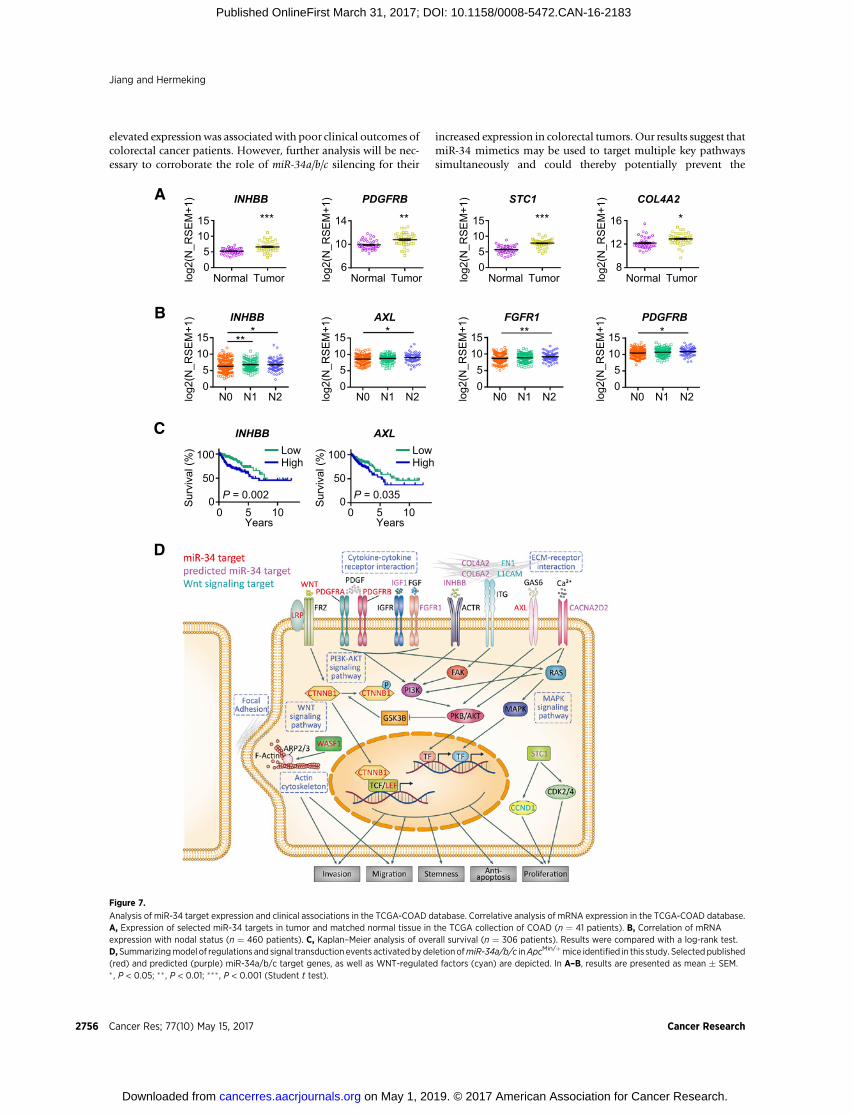
Increased expression of miR-34a/b/c targets in humancolorectal cancers is associated with poor survival
In order to determine whether the miR-34 targets describedabove are also clinically relevant, we analyzed their expressionwithin the expression profiles of 460 human colorectal adenocar-cinomas deposited in TCGA database (30). Interestingly, INHBB,PDGFRB, STC1, and COL4A2 showed a significantly elevatedexpression in primary colorectal cancers (Fig. 7A). Furthermore,a significantly increased expression of INHBB, AXL, FGFR1, andPDGFRB was detected in primary colorectal cancers from patientswith nodal status N2 when compared with samples from patientswith lower nodal status (Fig. 7B). Except for AXL, a significantlyincreased expression of these mRNAs was associated with anincreased tumor stage (Supplementary Fig. S6A). In addition,increased expression of INHBB and AXL in primary colorectalcancers was significantly associatedwith decreased patient survival(Fig. 7C; Supplementary Table S5). Elevated expression ofCOL4A2, WASF1, STC1, and PDGFRB was also associated withpoor survival, althoughnot significantly (SupplementaryFig. S6B).Because poor survival is mostly due to metastatic spread, theseresults suggest that the elevated expression of thesemRNAs, whichmay be due to loss of miR-34a/b/c, promotes metastasis.
Finally, we compiled the regulations identified here and com-bined them with previously published results on miR-34a/b/c-regulated pathways in a summarizing model shown in Fig. 7D.Notably, several of the miR-34 targets displaying elevated expres-sion after miR-34a/b/c deletion are involved in transmembranesignal transductions, which also activate the Wnt signaling path-way by inhibition of GSK-3b. Taken together, miR-34a/b/c pre-sumably suppresses intestinal tumorigenesis caused by loss ofApcby downregulating the expression of a large number of pro-tumorigenic factors. In case miR-34a/b/c expression is lost orsilencedduring tumor progression, these pathwaysmaybe furtheractivated and thereby contribute to colorectal cancer progression.
DiscussionThe increase in the pool of intestinal stem cells (ISC) in themiR-
34a/b/c-deficient mice observed here may underlie the increasedrate of tumor formation in ApcMin/þ mice, because ISC wereshown to serve as efficient tumor initiating cells during intestinaltumorigenesis (38). Paneth cells provide a niche for ISCs andcommunicate with these via multiple signaling pathways, amongthem the Delta/NOTCH and Wnt/APC/b-catenin pathways,which are under negative control by miR-34a/b/c (15, 37): thatis, several key components of theWnt signaling pathway are miR-34a/b/c targets (WNT1, WNT3, LRP6, LEF1, and b-catenin (37,
40). Moreover, GSK-3b, which phosphorylates b-catenin andthereby leads to its poly-ubiquitination and proteasomal degra-dation, is inhibited by the PKB/AKT and PI3K pathways, whichare also under control of miR-34a/b/c. Therefore, the concertedderegulation of the NOTCH and WNT pathways caused by theloss of miR-34a/b/c is a likely cause for the increased numberof Paneth and stem cells. In support of this scenario, wealso noticed an increased tumor organoid formation rate inmiR-34a/b/c�/�; ApcMin/þ adenomas as well as an increased accu-mulation of nuclear b-catenin in the untransformed, epithelialcells of intestinal crypts in miR-34a/b/c-deficient ApcMin/þ mice.In addition, a recent study suggests that activation of the Wnt/b-catenin pathway correlates with T-cell exclusion (42). There-fore, miR-34 may regulate immune responses through Wntsignaling. In addition, the expression of barrier proteins wasdecreased in miR-34a/b/c�/�; ApcMin/þ mice (e.g., Muc1, Tff3,and Retnlb). In combination with the decreased presence ofimmune cells, these effects of miR-34a/b/c loss may contribute,at least in part, to the increased bacterial infiltration observed inmiR-34a/b/c�/�; ApcMin/þ adenomas.
In addition, we found that the deletion of miR-34a/b/c inadenomas leads to the upregulation of previously describedmiR-34a/b/c target mRNAs, such as Pdgfra, Pdgfrb, and Axl, whichare known to enhance colorectal cancer formation (43). Further-more, a set of novel, putative miR-34a/b/c target mRNAs (Wasf1,Fgfr1, Igf1, Stc1, Cacna2d2, Col6a2, Col4a2, and Inhbb), whichwerereported to be involved in tumorigenesis (41, 44–49), was upre-gulated inmiR-34a/b/c-deficient adenomas. The factors encodedbythese genes form complex signaling and functional networks (seealsoFig. 7D): PDGFRA,PDGFRB, FGFR1,AXL, and IGF1,whichareeither ligands or receptors of the tyrosine kinase family, activate thePI3K and MAPK signaling pathway, and thereby promote cellgrowth, survival, EMT, and metastasis. Notably, CACNA2D2, avoltage-dependent calcium channel, deregulates calcium homeo-stasis when ectopically expressed (47). The resulting calciumreleasemay induce the activation of PKB/AKT andRAS to promotecell proliferation and angiogenesis (47). INHBB assembles intoactivins, which are criticalmodulators of growth and survival (49).COL4A2 is a major structural protein of the basement membraneand COL6A2 has an anchoring function. Both collagens stimulateintegrin signaling and are involved in cell growth, angiogenesis,and tumor metastasis (48). WASF1, a new miR-34a/b/c targetcharacterized here, mediates actin polymerization, lamellipodiaformation, and plays a critical role in cancer cell migration andinvasion (41). STC1 is an endocrine regulator of calcium andphosphate homeostasis, which promotes cell proliferation andinhibits apoptosis through CCND1 and CDK2/4 (50). Takentogether, miR-34a/b/c presumably act as global regulators to finetune multiple cellular functions, which are necessary for thehomeostasis of intestinal epithelia. Because miR-34a has beenshown to form bimodal switches with at least some of its targets(15), the loss of miR-34a/b/c during colorectal cancer progressionmayhave significant effects on the regulationof these signalingandexpression networks, which ultimately promote intestinal tumor-igenesis. miR-34a/b/c have a multitude of targets in intestinalepithelial and colon cancer cells as shown here and in previousstudies (10). Therefore, it seems plausible that the effects ofmiR-34a/b/c loss are mediated by the combined activity of mul-tiple upregulated targets and not by one or a few targets.
A subset of the direct targets of miR-34a/b/c may significantlycontribute to human colorectal tumorigenesis because their
Genetic Analysis of miR-34a/b/c in Intestinal Tumorigenesis
www.aacrjournals.org Cancer Res; 77(10) May 15, 2017 2755
on May 1, 2019. © 2017 American Association for Cancer Research. cancerres.aacrjournals.org Downloaded from
Published OnlineFirst March 31, 2017; DOI: 10.1158/0008-5472.CAN-16-2183
elevated expressionwas associatedwith poor clinical outcomes ofcolorectal cancer patients. However, further analysis will be nec-essary to corroborate the role of miR-34a/b/c silencing for their
increased expression in colorectal tumors. Our results suggest thatmiR-34 mimetics may be used to target multiple key pathwayssimultaneously and could thereby potentially prevent the
A
B
log2
(N_R
SE
M+1
)
15
05
N2N1N0
PDGFRB
10
*lo
g2(N
_RS
EM
+1)
15
05
N2N1N0
AXL
10
*
log2
(N_R
SE
M+1
)
15
05
N2N1N0
INHBB
10**
*
log2
(N_R
SE
M+1
)
15
05
N2N1N0
FGFR1
10
**
C
Sur
viva
l (%
)
0
50
100 LowHigh
P = 0.0351050
AXL
YearsYears
Sur
viva
l (%
)
0
50
100 LowHigh
P = 0.0021050
INHBB
**
10
6TumorNormallo
g2(N
_RS
EM
+1)
14
PDGFRB
***10
0TumorNormallo
g2(N
_RS
EM
+1)
15
STC1
5
***10
0TumorNormallo
g2(N
_RS
EM
+1)
15
INHBB
5
*
12
8TumorNormallo
g2(N
_RS
EM
+1)
16
COL4A2
D
Figure 7.
Analysis of miR-34 target expression and clinical associations in the TCGA-COAD database. Correlative analysis of mRNA expression in the TCGA-COAD database.A, Expression of selected miR-34 targets in tumor and matched normal tissue in the TCGA collection of COAD (n ¼ 41 patients). B, Correlation of mRNAexpression with nodal status (n ¼ 460 patients). C, Kaplan–Meier analysis of overall survival (n ¼ 306 patients). Results were compared with a log-rank test.D,Summarizingmodel of regulations and signal transduction events activatedbydeletionofmiR-34a/b/c inApcMin/þmice identified in this study. Selected published(red) and predicted (purple) miR-34a/b/c target genes, as well as WNT-regulated factors (cyan) are depicted. In A–B, results are presented as mean � SEM.� , P < 0.05; �� , P < 0.01; ��� , P < 0.001 (Student t test).
Jiang and Hermeking
Cancer Res; 77(10) May 15, 2017 Cancer Research2756
on May 1, 2019. © 2017 American Association for Cancer Research. cancerres.aacrjournals.org Downloaded from
Published OnlineFirst March 31, 2017; DOI: 10.1158/0008-5472.CAN-16-2183

emergence of resistance caused by mutations of single pathways.Therefore, miR-34a/b/c replacement therapy may represent apotential option for the treatment of colorectal cancer.
Disclosure of Potential Conflicts of InterestNo potential conflicts of interest were disclosed.
Authors' ContributionsConception and design: H. HermekingAcquisition of data (provided animals, acquired and managed patients,provided facilities, etc.): L. Jiang, H. HermekingAnalysis and interpretation of data (e.g., statistical analysis, biostatistics,computational analysis): L. Jiang, H. HermekingWriting, review, and/or revision of the manuscript: L. Jiang, H. HermekingStudy supervision: H. HermekingOther (experimental design): L. Jiang
AcknowledgmentsWe are grateful to Alexander Nikitin for providingmiR-34b/cfl/flmice,Marlon
Schneider for ApcMin/þ mice, Hans Clevers for the Olfm4 plasmid, MatjazRokavec for initial help with setting up theApcMin/þmouse cohorts, andMarkusKaller for advice on bioinformatics analyses. We would also like to thank PeterJung for discussions and Ursula G€otz for technical assistance.
Grant SupportH. Hermeking received support from a Deutsche Krebshilfe Grant
(No. 109531) and the Rudolf Bartling Stiftung (V/124). L. Jiang received afellowship from the China Scholarship Council.
The costs of publication of this articlewere defrayed inpart by the payment ofpage charges. This article must therefore be hereby marked advertisement inaccordance with 18 U.S.C. Section 1734 solely to indicate this fact.
Received August 8, 2016; revised October 4, 2016; accepted March 23, 2017;published OnlineFirst March 31, 2017.
References1. Siegel RL, Miller KD, Jemal A. Cancer statistics, 2016. CA Cancer J Clin
2016;66:7–30.2. Kinzler KW, Vogelstein B. Lessons from hereditary colorectal cancer. Cell
1996;87:159–70.3. Bienz M, Clevers H. Linking colorectal cancer to Wnt signaling. Cell
2000;103:311–20.4. Moser AR, Mattes EM, Dove WF, Lindstrom MJ, Haag JD, Gould MN.
ApcMin, a mutation in the murine Apc gene, predisposes to mammarycarcinomas and focal alveolar hyperplasias. Proc Natl Acad Sci U S A1993;90:8977–81.
5. Luongo C, Moser AR, Gledhill S, Dove WF. Loss of Apcþ in intestinaladenomas from Min mice. Cancer Res 1994;54:5947–52.
6. Moser AR, Luongo C, Gould KA, McNeley MK, Shoemaker AR, Dove WF.ApcMin: a mouse model for intestinal and mammary tumorigenesis. Eur JCancer 1995;31A:1061–4.
7. Vogelstein B, Lane D, Levine AJ. Surfing the p53 network. Nature 2000;408:307–10.
8. Hermeking H.MicroRNAs in the p53 network: micromanagement oftumour suppression. Nat Rev Cancer 2012;12:613–26.
9. Hunten S, Kaller M, Drepper F, Oeljeklaus S, Bonfert T, Erhard F, et al. p53-Regulated networks of protein, mRNA, miRNA, and lncRNA expressionrevealed by integrated pulsed stable isotope labeling with amino acids incell culture (pSILAC) and next generation sequencing (NGS) analyses. MolCell Proteomics 2015;14:2609–29.
10. Rokavec M, Li H, Jiang L, Hermeking H. The p53/miR-34 axis in devel-opment and disease. J Mol Cell Biol 2014;6:214–30.
11. Hermeking H.p53 enters the microRNA world. Cancer Cell 2007;12:414–8.
12. TarasovV, JungP, Verdoodt B, LodyginD, Epanchintsev A,MenssenA, et al.Differential regulation ofmicroRNAs by p53 revealed bymassively parallelsequencing: miR-34a is a p53 target that induces apoptosis and G1-arrest.Cell Cycle 2007;6:1586–93.
13. Siemens H, Jackstadt R, Hunten S, Kaller M,Menssen A, Gotz U, et al. miR-34 and SNAIL form a double-negative feedback loop to regulate epithelial-mesenchymal transitions. Cell Cycle 2011;10:4256–71.
14. Liu C, Kelnar K, Liu B, Chen X, Calhoun-Davis T, Li H, et al. ThemicroRNAmiR-34a inhibits prostate cancer stem cells and metastasis by directlyrepressing CD44. Nat Med 2011;17:211–5.
15. Bu P,Wang L, Chen KY, Srinivasan T,Murthy PK, Tung KL, et al. AmiR-34a-numb feedforward loop triggered by inflammation regulates asymmetricstem cell division in intestine and colon cancer. Cell Stem Cell 2016;18:189–202.
16. Cheng CY, Hwang CI, Corney DC, Flesken-Nikitin A, Jiang L, Oner GM,et al.miR-34 cooperates with p53 in suppression of prostate cancer by jointregulation of stem cell compartment. Cell Rep 2014;6:1000–7.
17. Choi YJ, LinCP,Ho JJ,HeX,OkadaN, BuP, et al.miR-34miRNAsprovide abarrier for somatic cell reprogramming. Nat Cell Biol 2011;13:1353–60.
18. Lodygin D, Tarasov V, Epanchintsev A, Berking C, Knyazeva T, Korner H,et al. Inactivation of miR-34a by aberrant CpG methylation in multipletypes of cancer. Cell cycle 2008;7:2591–600.
19. Siemens H, Neumann J, Jackstadt R, Mansmann U, Horst D, Kirchner T,et al. Detection of miR-34a promoter methylation in combination withelevated expression of c-Met and b-catenin predicts distant metastasis ofcolon cancer. Clin Cancer Res 2013;19:710–20.
20. Adams BD, Parsons C, Slack FJ. The tumor-suppressive and potentialtherapeutic functions of miR-34a in epithelial carcinomas. Expert OpinTher Targets 2016;20:737–53.
21. Rokavec M, Oner MG, Li H, Jackstadt R, Jiang L, Lodygin D, et al. IL-6R/STAT3/miR-34a feedback loop promotes EMT-mediated colorectal cancerinvasion and metastasis. J Clin Invest 2014;124:1853–67.
22. Gregorieff A, CleversH. In situ hybridization to identify gut stem cells. CurrProtoc Stem Cell Biol 2010;Chapter 2:Unit 2F 1.
23. Risso D, Ngai J, Speed TP, Dudoit S. Normalization of RNA-seq datausing factor analysis of control genes or samples. Nat Biotechnol2014;32:896–902.
24. KallioMA, Tuimala JT,Hupponen T, Klemela P, GentileM, Scheinin I, et al.Chipster: user-friendly analysis software for microarray and other high-throughput data. BMC Genom 2011;12:507.
25. Huang da W, Sherman BT, Lempicki RA. Bioinformatics enrichment tools:paths toward the comprehensive functional analysis of large gene lists.Nucleic Acids Res 2009;37:1–13.
26. SubramanianA, TamayoP,Mootha VK,Mukherjee S, Ebert BL,GilletteMA,et al. Gene set enrichment analysis: a knowledge-based approach forinterpreting genome-wide expression profiles. Proc Natl Acad Sci U S A2005;102:15545–50.
27. Lewis BP, Burge CB, Bartel DP. Conserved seed pairing, often flanked byadenosines, indicates that thousands of human genes are microRNAtargets. Cell 2005;120:15–20.
28. Sato T, Stange DE, Ferrante M, Vries RG, Van Es JH, Van den Brink S, et al.Long-term expansion of epithelial organoids from human colon, adeno-ma, adenocarcinoma, and Barrett's epithelium. Gastroenterology2011;141:1762–72.
29. Livak KJ, Schmittgen TD. Analysis of relative gene expression data usingreal-time quantitative PCR and the 2(-Delta Delta C(T)) Method. Methods2001;25:402–8.
30. Cancer Genome Atlas N. Comprehensive molecular characterization ofhuman colon and rectal cancer. Nature 2012;487:330–7.
31. Vogt M, Munding J, Gruner M, Liffers ST, Verdoodt B, Hauk J, et al.Frequent concomitant inactivation of miR-34a and miR-34b/c by CpGmethylation in colorectal, pancreatic, mammary, ovarian, urothelial,and renal cell carcinomas and soft tissue sarcomas. Virchows Arch2011;458:313–22.
32. Lee SH, Hu LL, Gonzalez-Navajas J, Seo GS, Shen C, Brick J, et al. ERKactivation drives intestinal tumorigenesis in Apc(min/þ) mice. Nat Med2010;16:665–70.
33. Love MI, Huber W, Anders S. Moderated estimation of fold change anddispersion for RNA-seq data with DESeq2. Genome Biol 2014;15:550.
34. RobinsonMD,McCarthyDJ, SmythGK. edgeR: a Bioconductor package fordifferential expression analysis of digital gene expression data. Bioinfor-matics 2010;26:139–40.
www.aacrjournals.org Cancer Res; 77(10) May 15, 2017 2757
Genetic Analysis of miR-34a/b/c in Intestinal Tumorigenesis
on May 1, 2019. © 2017 American Association for Cancer Research. cancerres.aacrjournals.org Downloaded from
Published OnlineFirst March 31, 2017; DOI: 10.1158/0008-5472.CAN-16-2183

35. Munoz J, Stange DE, Schepers AG, van de Wetering M, Koo BK, Itzkovitz S,et al. The Lgr5 intestinal stem cell signature: robust expression of proposedquiescent `þ4' cell markers. EMBO J 2012;31:3079–91.
36. Merlos-Suarez A, Barriga FM, Jung P, Iglesias M, Cespedes MV, Rossell D,et al. The intestinal stem cell signature identifies colorectal cancer stem cellsand predicts disease relapse. Cell Stem Cell 2011;8:511–24.
37. Kim NH, KimHS, Kim N-G, Lee I, Choi H-S, Li X-Y, et al. p53 andmiRNA-34 are Suppressors of Canonical Wnt Signaling. Sci Signal 2011;4:ra71.
38. Barker N, Ridgway RA, van Es JH, van de Wetering M, Begthel H, van denBorn M, et al. Crypt stem cells as the cells-of-origin of intestinal cancer.Nature 2009;457:608–11.
39. GarofaloM, JeonYJ,NuovoGJ,Middleton J, Secchiero P, Joshi P, et al.MiR-34a/c-Dependent PDGFR-alpha/beta downregulation inhibits tumorigen-esis and enhances TRAIL-Induced apoptosis in lung cancer. PloS One2013;8:e67581.
40. Kaller M, Liffers S-T, Oeljeklaus S, Kuhlmann K, R€oh S, Hoffmann R, et al.Genome-wide characterization ofmiR-34a induced changes in protein andmRNA expression by a combined pulsed SILAC and microarray analysis.Mol Cell Proteo 2011;10:M111. 010462.
41. Zhang J, Zhou S, Tang L, Shen L, Xiao L,DuanZ, et al.WAVE1 gene silencingvia RNA interference reduces ovarian cancer cell invasion, migration andproliferation. Gynecol Oncol 2013;130:354–61.
42. Corrales L, Matson V, Flood B, Spranger S, Gajewski TF. Innate immunesignaling and regulation in cancer immunotherapy. Cell Res 2016;27:96–108.
43. Craven RJ, Xu LH, Weiner TM, Fridell YW, Dent GA, Srivastava S, et al.Receptor tyrosine kinases expressed inmetastatic colon cancer. Int J Cancer1995;60:791–7.
44. Grose R, Dickson C. Fibroblast growth factor signaling in tumorigenesis.Cytokine Growth Factor Rev 2005;16:179–86.
45. OlivoMarston SE, Hursting SD, Lavigne J, Perkins SN,Maarouf RS, Yakar S,et al. Genetic reduction of circulating insulinlike growth factor1 inhibitsazoxymethaneinduced colon tumorigenesis in mice. Mol Carcinogen2009;48:1071–6.
46. Tamura S, Oshima T, Yoshihara K, Kanazawa A, Yamada T, Inagaki D, et al.Clinical significance of STC1 gene expression in patients with colorectalcancer. Anticancer Res 2011;31:325–9.
47. Warnier M, Roudbaraki M, Derouiche S, Delcourt P, Bokhobza A, Pre-varskaya N, et al. CACNA2D2 promotes tumorigenesis by stimulating cellproliferation and angiogenesis. Oncogene 2015;34:5383–94.
48. Worthley DL, Giraud AS, Wang TC. The extracellular matrix in digestivecancer. Cancer Microenviron 2010;3:177–85.
49. Togashi Y, Kogita A, Sakamoto H, Hayashi H, Terashima M, deVelasco MA, et al. Activin signal promotes cancer progression andis involved in cachexia in a subset of pancreatic cancer. Cancer Lett2015;356:819–27.
50. DuY-Z,GuX-H,Cheng S-F, Li L, LiuH,HuL-P, et al. Theoncogenetic role ofstanniocalcin 1 in lung adenocarcinoma: a promising serum candidatebiomarker for tracking lung adenocarcinoma progression. Tumor Biol2015:1–12.
Cancer Res; 77(10) May 15, 2017 Cancer Research2758
Jiang and Hermeking
on May 1, 2019. © 2017 American Association for Cancer Research. cancerres.aacrjournals.org Downloaded from
Published OnlineFirst March 31, 2017; DOI: 10.1158/0008-5472.CAN-16-2183

2017;77:2746-2758. Published OnlineFirst March 31, 2017.Cancer Res Longchang Jiang and Heiko Hermeking
Suppress Intestinal TumorigenesismiR-34b/c and miR-34a
Updated version
10.1158/0008-5472.CAN-16-2183doi:
Access the most recent version of this article at:
Material
Supplementary
http://cancerres.aacrjournals.org/content/suppl/2017/03/31/0008-5472.CAN-16-2183.DC1
Access the most recent supplemental material at:
Cited articles
http://cancerres.aacrjournals.org/content/77/10/2746.full#ref-list-1
This article cites 48 articles, 8 of which you can access for free at:
Citing articles
http://cancerres.aacrjournals.org/content/77/10/2746.full#related-urls
This article has been cited by 1 HighWire-hosted articles. Access the articles at:
E-mail alerts related to this article or journal.Sign up to receive free email-alerts
Subscriptions
Reprints and
To order reprints of this article or to subscribe to the journal, contact the AACR Publications Department at
Permissions
Rightslink site. Click on "Request Permissions" which will take you to the Copyright Clearance Center's (CCC)
.http://cancerres.aacrjournals.org/content/77/10/2746To request permission to re-use all or part of this article, use this link
on May 1, 2019. © 2017 American Association for Cancer Research. cancerres.aacrjournals.org Downloaded from
Published OnlineFirst March 31, 2017; DOI: 10.1158/0008-5472.CAN-16-2183


