Download - Mechanisms of Mucosal Defense

Mechanisms of Mucosal Defense
Soma Jyonouchi, M.D.
January 24, 2008

Mucosal Surfaces
The GI mucosal surfaces cover 400 m²
Thin – facilitate nutrient absorption.
The Gut Associated Lymphoid Tissue (GALT)
- Organized T and B cell areas
- where antigen is collected and adaptive immune response is generated.
- Tonsils, Peyer’s patches, appendix, solitary lymphoid follicles in the large intestine and rectum.

GALT Architecture
** Dome structures extend into the lumen of the intestine.
Lamina Propria

Lamina Propria – effector site
Inductive Site Effector Site

Enormous Antigen Load Systemic Immune System –
largely sterile environment. Vigorous response to microbial invasion.
Mucosal Immune System – Constant exposure to foreign matter
Human gut is exposed to an enormous amount commensal microorganisms (1 x 10 14)
Constant exposure to food matter

Innate Defense – I. Barrier Fxn1. Glycocalyx – Goblet cells produce mucous
to create a thick barrier that covers the GI epithelium and prevents easy access.
- Pathogens become trapped in the mucous and are expelled via peristalsis.
- Mucous also acts as a reservoir for secretory IgA.

I. Barrier Fxn Epithelial Cell Tight Junctions - prevent
the passages of macromolecules.
** Zonulin – Homology to Vibrio cholera toxin.
upregulated during the acute phase of celiac disease.
- Induces tight junction disassembly and increased intestinal permeability.
Drago et al. Scand J Gastroenterol. 2006 Fasano et al. Lancet 2000

II. Proteolytic Enzymes
Enzymes in the stomach (pepsin) and small bowel (trypsin, chymotrypsin, pancreatic proteases).
Break down large polypeptides into di-peptides and tri-peptides.
Peptides < 8-10 aa are poor immunogens.
Enzymes cytotoxic to pathogens.

III. Antimicrobial Molecules
1. Lactoferrin – binds iron and inhibits bacterial growth.
2. Lysozyme – cleaves cell wall of gram positive bacteria.
3. Defensins – 30-40 aa peptides that disrupts the cell memebranes of bacteria and fungi causing lysis.

IV. Commensal Organisms
>400 species of commensal bacteria
Provide enzymatic breakdown of food
Competes with pathogenic bacteria for space and nutrients
Prevents colonization of the gut
Antibiotics disrupt homeostasis

IV. Commensal Organisms
Germ free mice – no commensal microflora.
- Pups delivered by C-section and raised in sterile conditions.
Hypoplastic peyer’s patches with scant germinal centers.
- decreased IgA plasma cells - decreased lamina propria CD4+ cells - Abnormalities reversed by placing non-germ
free mice in same cage.

Mucosal Immune System: Adaptive Response

“Common Mucosal Immune System”
Peyer’s Patch
Mesenteric Lymph Node
Blood Stream
Resp Tract
Breast Intestinal Mucosa GU Tract
Salivary/Lacrimal Gland
*Antigen Presentation*
Thoracic Duct
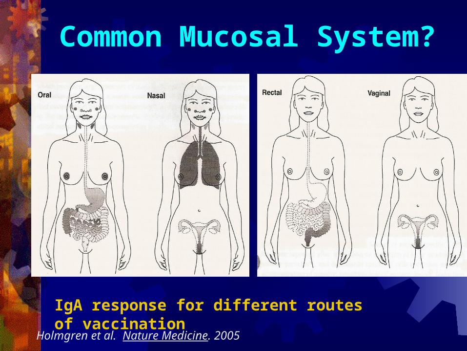
Common Mucosal System?
IgA response for different routes of vaccination
Holmgren et al. Nature Medicine. 2005

GALT vs peripheral Lymphoid tissue
1) Unique epithelium for antigen uptake
2) Unique lymphocyte repertoire
3) IgA dominated humoral response
4) A need to minimize injury to the mucosal tissue while providing protection.

GALT – Unique Epithelium
The epithelium overlying the peyer’s patches is composed of cells that differ from the surrounding enterocytes.

M-Cells (microfold cells)
M-cells lack microvilli
No glycocalyx coating
Designed to to interact directly with antigens in the gut – portal of entry into GALT.
– some pathogens gain entry via M-cells
(salmonella, shigella)

M-Cells (microfold cells) Basolateral aspects are
invaginated.
They contain T-cells, B-cells, Dendritic cells, and Macrophages.
Antigens from the lumen are taken up by endocytosis and presented directly to APCs
APCs migrate to germinal center
Germinal Center

GALT vs peripheral Lymphoid tissue 1) Unique epithelium for antigen uptake
2) Unique Lymphocyte Repertoire
3) IgA dominated humoral response
4) A need to minimize injury to the mucosal tissue as well as development of tolerance.
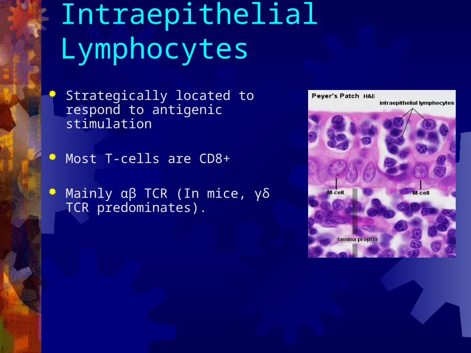
Intraepithelial Lymphocytes Strategically located to respond to
antigenic stimulation
Most T-cells are CD8+
Mainly αβ TCR (In mice, γδ TCR predominates).
IEL: CD8 + T-Cells Limited Repertoire of TCR - marked difference compared to peripheral T-
cells.
Recognize a limited # of antigens
Prevents indiscriminate inflammation
Recognition of self-stress antigens (MIC-A, MIC-B)
- T-cells induce apoptosis of injured epithelial cells.

Van Kerckhove et al: 1992 Analysis of T-cell receptor Vβ gene usage in
IEL vs Peripheral lymphocytes
Quantitative PCR
Results: PBL - fairly even distribution of Vβ gene
usage IEL - 1-3 Vβ families made up more than
43% of total Vβ transcripts detected in each individual

Vβ1, Vβ2, Vβ3, and Vβ6 families frequently shared among IEL from different individuals

Lamina Propria Lymphocytes
T-cells are predominantely CD4 + (95% CD45RO+)
Limited capacity to proliferate
Weak proliferative responses to mitogens or specific antigens.
Still act as helpers for B-cells

MALT vs peripheral Lymphoid tissue
1) Unique epithelium for antigen uptake
2) Unique Lymphocyte Repertoire
3) IgA dominated humoral response
4) A need to minimize injury to the mucosal tissue

B-Cell Response: S-IgA Secretory IgA is the predominant Ig isotype in
the gut. Blood IgA exists mainly as a monomer In the mucosa, IgA is exclusively dimeric
J-Chain

Secretory IgA Function
Inhibits microbial adherence
Neutralizes viruses and toxins
Neutralizes catalytic activity of microbial enzymes.

Secretory IgA Transport
S-IgA is produced by plasma cells in the lamina propria.
S-IgA binds to polymeric Ig receptor on the basolateral surface of intestinal epithelial cells
It is transported to the intestinal lumen by transcytosis.
LumenLaminaPropria

Secretory IgA transport
**Secretory Component (SC) of the receptor remains associated with IgA
SC protects IgA from proteolytic cleavage.
SC also acts as a “glue” to bind IgA to the glycocalyx.

IgA Subtypes
IgA 1 and IgA 2 mainly differ in their hinge regions
IgA 1 ab contain 13 additional aa in the hinge region.
- More flexible
- More susceptible to IgA1 specific proteases made by bacteria.
IgA 2 is resistant to proteases
- Serum ratio 4:1
- Mucosal ratio 3:2 (even higher in colon)
B-Cell Isotype Switching: Cytokine Stimulation IgA response is likely the result of the unique
micorenvironment in the gut.
TGF-β + IL-10 induces sIGM+ B-cells to switch to sIgA+ B-cells
Addition of TGF-β to LPS triggered mouse B-cell cultures leads to increased IgA synthesis.
Mucosal epithelial cells are a major source of TGF-β and IL-10

Van Ginkel et al: 1999 TGF-β knockout mice (-/-) Significantly decreased IgA-committed B-cells in the
gut and secretory IgA
WT TGF-β -/-
Blue stain - IgA
Green stain - IgM
Red stain - IgG
Enhanced IgG and IgM response in the gut (fixes complement)
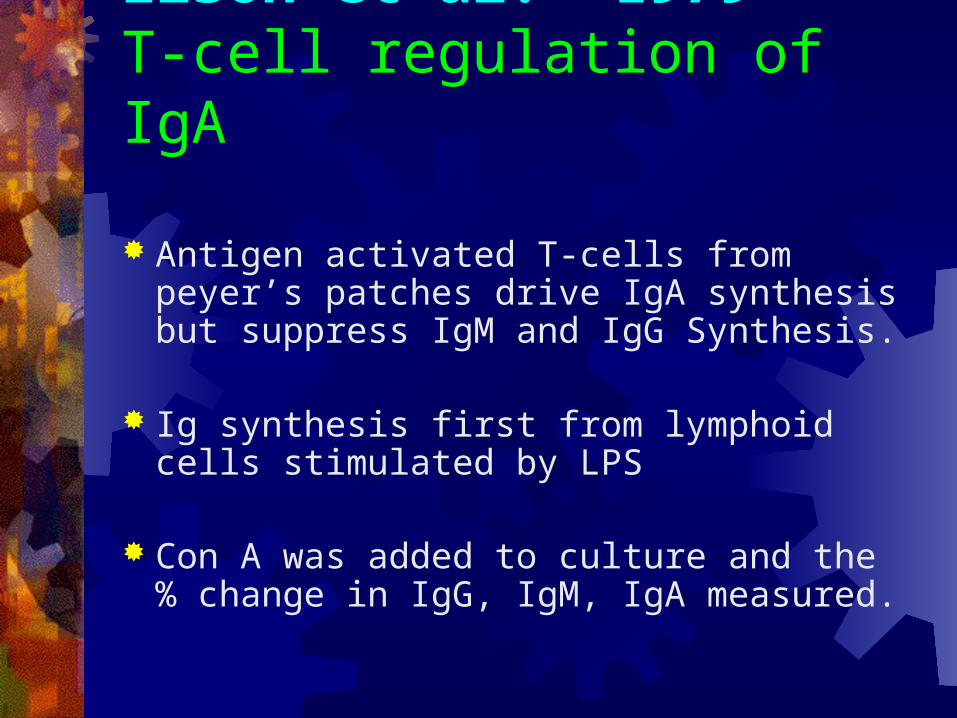
Elson et al. 1979T-cell regulation of IgA
Antigen activated T-cells from peyer’s patches drive IgA synthesis but suppress IgM and IgG Synthesis.
Ig synthesis first from lymphoid cells stimulated by LPS
Con A was added to culture and the % change in IgG, IgM, IgA measured.

Elson et al:
Baseline Addition of Con A
IgM IgG IgA IgA

Elson et al: Unique environment vs. Unique T-cell Subset
T-cells from spleen or PP stimulated with con A then added back into tissue.
Spleen T-cells added to PP cell cx
PP T-cells added to spleen cell cx
IgG IgM IgA
IgA

GALT vs peripheral Lymphoid tissue1) Unique epithelium for antigen uptake
2) Unique Lymphocyte Repertoire
3) IgA dominated humoral response
4) A need to minimize injury to the mucosal tissue.

Gut Anti-Inflammatory Mechanisms: Secretory IgA
IgA is unable to activate complement by classical or alternative pathways.
S-IgA can inhibit phagocytosis and chemotaxis of neutrophils, macrophages
Can down regulate synthesis of
TNF-α and IL-6

Wolf et al: IgA induces IL-1 Receptor antagonist
IgA induces IL-1 R antagonist from monocytes.
IL-1 IL-1 Ra
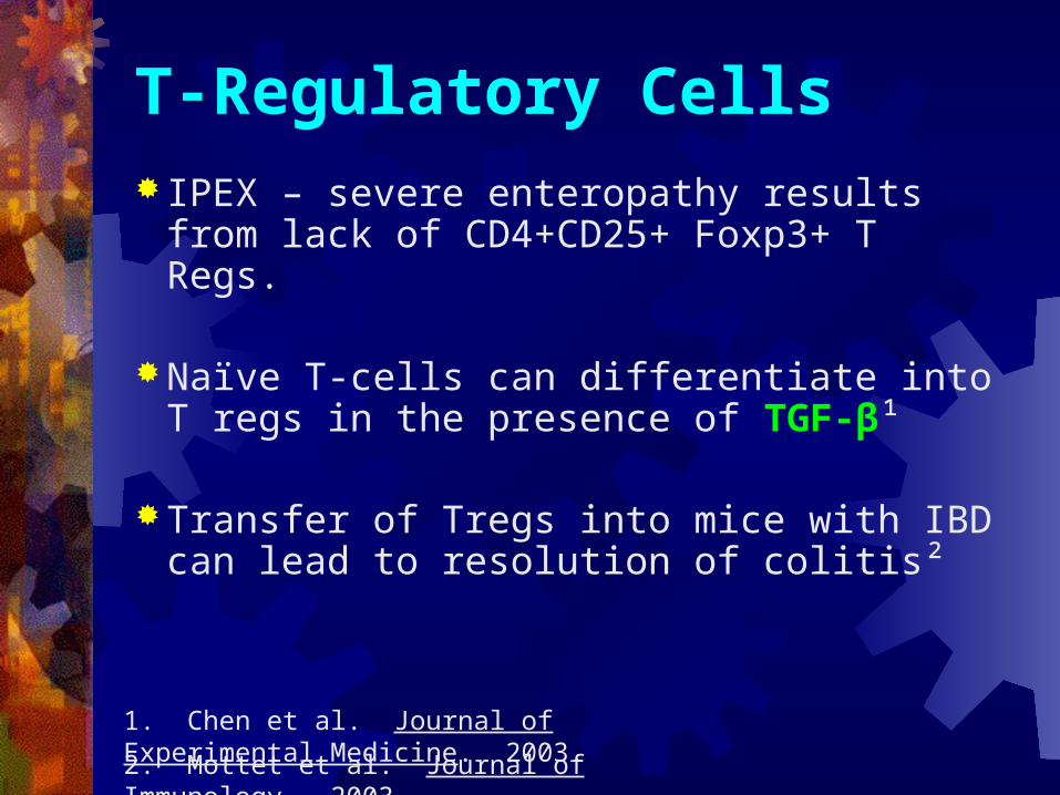
T-Regulatory Cells
IPEX – severe enteropathy results from lack of CD4+CD25+ Foxp3+ T Regs.
Naïve T-cells can differentiate into T regs in the presence of TGF-β¹
Transfer of Tregs into mice with IBD can lead to resolution of colitis²
2. Mottet et al. Journal of Immunology. 2003.
1. Chen et al. Journal of Experimental Medicine. 2003.
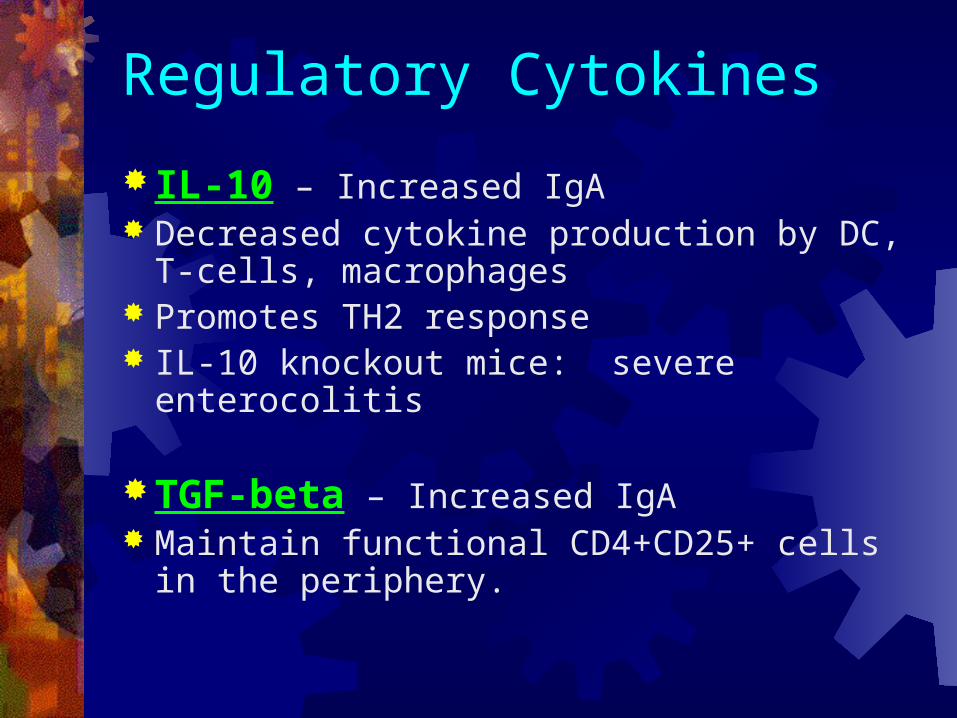
Regulatory Cytokines
IL-10 – Increased IgA Decreased cytokine production by DC, T-
cells, macrophages Promotes TH2 response IL-10 knockout mice: severe enterocolitis
TGF-beta – Increased IgA Maintain functional CD4+CD25+ cells in the
periphery.

Antigen Response Pathogen vs. Commensal response
Both pathogens and commensals often share similar PAMPs
Commensals may be contained by IgA and innate barriers.
- Pathogens have additional virulence factors
(adhesion molecules, toxins)
- commensals also endocytoced by M-cells and engage TLRs

Shigella Infection
Nod 1 (aka CARD 4) – Binds shigella endotoxin Nod 1 dimerization allows binding to RICK
protein kinase Activation of NF-κB Pathway
Release ofIL-8 attractsNeutrophils

Tien et al: Lactobacillus Mucosal Epithelial cells challenged
with shigella then infected with lactobacillus
Macroarray DNA chips used to compare gene expression vs. control
Proteins involved in degradation of I-κBα down-regulated - Result: Inhibition of the NF-κB pathway

Kelly et al: Bacteriodes Rel A: member of NF-κB complex Intestinal cells cultured with Salmonella Bacteriodes induced nuclear clearance of Rel A
limiting the duration of NF-κB action
Kelly, D. Nature Immunology. 2004.
Immunoflourescence at 2 hrs
Salm Salm + BactMedium Bact

Summary Mucosal immune system needs to
selectively respond to pathogens
Humoral immune response is IgA dominated.
Unique lymphocyte repertoire and cytokine environment limit inflammation
Commensal organisms act to maintain the mucosal immune system and have mechanisms to limit inflammation.

The End!

References1. Mayer, L. Mucosal Immunity. Pediatrics. 111, 1595-1600. 2003.
2. Janeway. Immunobiology. 2005
3. Macpherson, A. Interactions between commensal intestinal bacteria and the immune system. Nature Reviews Immunology. 4; 478-485. 2004.
4. Fasano, A. Zonulin, a newly discovered modulator of intestinal permeability, and its expression in coeliac disease. Lancet. 355; 1518 – 1519. 2000.
5. Drago, S. Gliadin, zonulin and gut permeability: Effects on celiac and non-celiac intestinal mucosa and intestinal cell lines. Scandinavian Journal of Gastroenterology. 41; 408 – 419. 2006.
6. Van Ginkel, F. Partial IgA deficiency with increased Th-2 Type Cytokines in TGF-β1 knockout mice. Journal of Immunology. 163; 4. 1999.
7. Wolf, H.M. Anti-inflammatory proterties of human IgA. Clinical Experimental Immunology. 105; 537-543. 1996.

References Macpherson, A. Interactions between commensal intestinal bacteria and the
immune system. Nature Reviews Immunology. Vol 4. June 2004.
Tien, MT. Anti-Inflammatory Effect of Lactobacillus casei on Shigella-Infected Human Intestinal Epithelial Cells. The Journal of Immunology. 176; 1228. 2006.
Coombes, Janine. Control of Intestinal Homeostasis by regulatory T-cells and dendritic cells. Seminars in Immunology. 19; 116-126. 2007.
Van Kerckhove, Catherine. Oligclonality of Human Intestinal Intraepithelial T-cells. Journal of Experimental Medicine. 175; 57-63. 1992.

Antigen Load
GALT must selectively respond to certain pathogens while ignoring other antigens.
Food Proteins – DCs produce IL-10 to produce a TH2 response and suppression of inflammatory response.
Pathogens – TLR ligands sensed by APCs favor pro-inflammatory response.
- Humoral and cellular immune response.


