Mechanisms ofMechanisms of
Cell DeathCell Death

Etiology of cell deathEtiology of cell death
Major Factors
Accidental Genetic
Necrosis Apoptosis
Necrosis: Necrosis: The sum of the morphologic changes that follow cell death in a living tissue or organ
Apoptosis:a physiological process that includes specific suicide signals leading to cell death
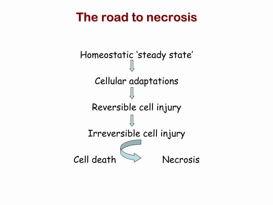
The road to necrosisThe road to necrosis
Homeostatic ‘steady state’
Cellular adaptations
Reversible cell injury
Irreversible cell injury
Cell death Necrosis

Pathogenesis of necrosisPathogenesis of necrosis
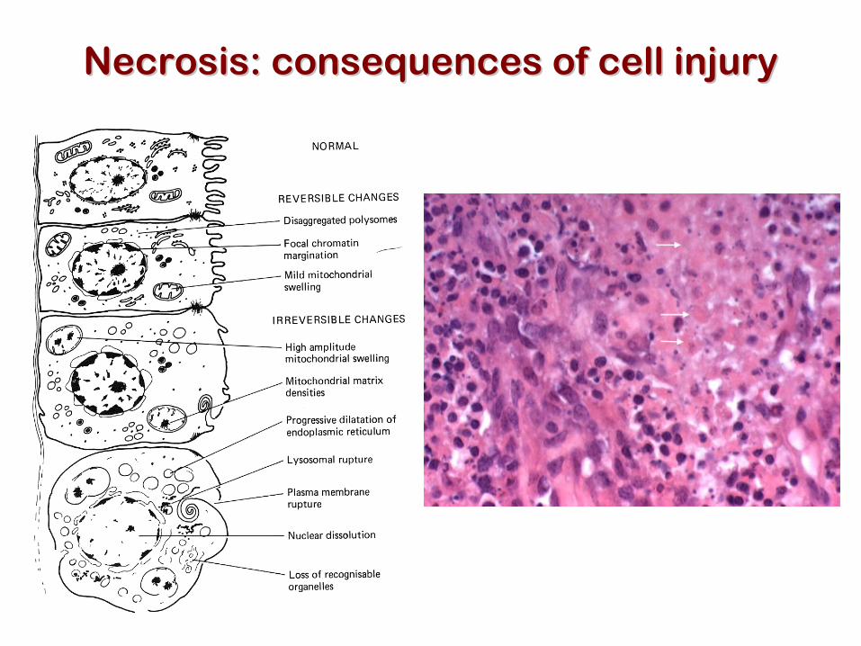
Necrosis: consequences of cell injuryNecrosis: consequences of cell injury
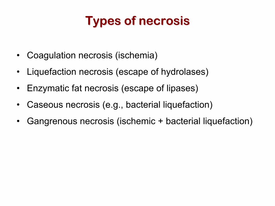
Types of necrosisTypes of necrosis
• Coagulation necrosis (ischemia)
• Liquefaction necrosis (escape of hydrolases)
• Enzymatic fat necrosis (escape of lipases)
• Caseous necrosis (e.g., bacterial liquefaction)
• Gangrenous necrosis (ischemic + bacterial liquefaction)

Necrosis: a pathological responseto cellular injury
Apoptosis: a physiological response to specific suicide signals,
or lack of survival signals
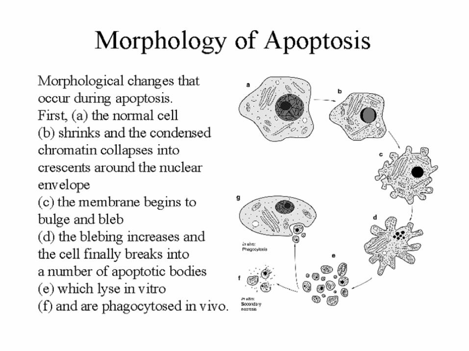
Epitopes appear on plasma membrane marking cell as a phagocytic target.
No spillage, no inflammationGeneral inflammatory response is
triggered
Cell contents are packaged in membrane bounded bodies, internal organelles still
functioning, to be engulfed by neighbours. Cell contents spill out
Blebbing of plasma and nuclear membranesPlasma membrane lyses
Cytoplasm shrinks without membrane ruptureMitochondria swell and rupture
Chromatin condenses and migrates to nuclear membrane. Internucleosomal cleavage leads
to laddering of DNA at the nucleosomalrepeat length, ca. 200 bp.
Chromatin clumps
www.chembio.uoguelph.ca
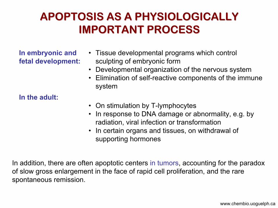
• On stimulation by T-lymphocytes• In response to DNA damage or abnormality, e.g. by
radiation, viral infection or transformation• In certain organs and tissues, on withdrawal of
supporting hormones
In the adult:
• Tissue developmental programs which control sculpting of embryonic form
• Developmental organization of the nervous system• Elimination of self-reactive components of the immune
system
In embryonic and fetal development:
In addition, there are often apoptotic centers in tumors, accounting for the paradox of slow gross enlargement in the face of rapid cell proliferation, and the rare spontaneous remission.
APOPTOSIS AS A PHYSIOLOGICALLYAPOPTOSIS AS A PHYSIOLOGICALLYIMPORTANT PROCESSIMPORTANT PROCESS
www.chembio.uoguelph.ca

APOPTOSIS in APOPTOSIS in C.elegansC.elegans
C.elegans genome: 19099 genes (790 seven-pass transmembrane receptors, 480 zinc finger proteins, and 410 protein kinases)The life cycle of C. elegans from egg to sexual maturity (and new eggs) is about 3 daysced-1, -3, -4, and -9 (Cell death determining) proteins in C.elegans are closely related to mammalian apoptosis-regulating genes
The adult hermaphrodite consists of exactly 959 somatic cells of precisely determined lineage and function. Individual cells are named and their relationships to their neighbors are known
Overall, the 959 somatic cells of adult C.elegans arise from 1090 original cells; exactly 131 somatic cells undergo programmed cell death in the wild type worm
Of the 1090 cells, 302 are neurons, and many of the programmed deaths also lie in the neuronal lineage
www.chembio.uoguelph.ca
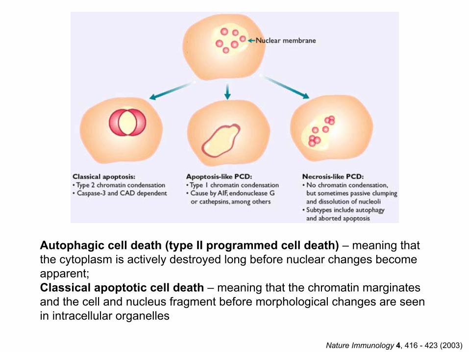
Nature Immunology 4, 416 - 423 (2003)
Autophagic cell death (type II programmed cell death) – meaning that the cytoplasm is actively destroyed long before nuclear changes become apparent;Classical apoptotic cell death – meaning that the chromatin marginatesand the cell and nucleus fragment before morphological changes are seen in intracellular organelles

Nuclear alterations in different forms of programmed cell deathThe use of chromatin condensation as a criterion to distinguish apoptosis from apoptosis-like PCD has been inconsistent in the scientific literature, and the potential for overlapping definitions and errors is large. The following examples of classical apoptosis (c,e) and apoptosis-like PCD (b,d,f,g–i) might provide a general guideline. Examples of control chromatin (a), and caspase-independent chromatin margination triggered directly by microinjection of AIF (b). Caspase-dependent strong chromatin compaction (c) versus caspase-independent, AIF-driven lumpy chromatin condensation (d) in PCD of mouse embryonic stem cells. (e) Caspase-dependent chromatin compaction to crescent shaped masses at the nuclear periphery and chromatin fragmentation to two compact spheres (f) or caspase-independent lumpy chromatin condensation without nuclear fragmentation in colchicine-induced neuronal cell death. Incomplete, lumpy chromatin condensation (compare with b,d) in caspase-independent apoptosis-like PCD triggered by Hsp70 depletion (g) or the active form of vitamin D (i), and in caspase-dependent TNF-induced apoptosis-like PCD (i) in caspase-3 deficient MCF-7 cells.
Nat
ure
Rev
iew
s M
olec
ular
Cel
l Bio
logy
2, 5
89 -5
98 (2
001)
Nature Reviews Cancer 2; 647-656 (2002)
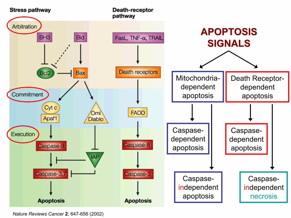
APOPTOSISAPOPTOSISSIGNALSSIGNALS
Mitochondria-dependentapoptosis
Caspase-dependentapoptosis
Death Receptor-dependentapoptosis
Caspase-dependentapoptosis
Caspase-independent
apoptosis
Caspase-independent
necrosis
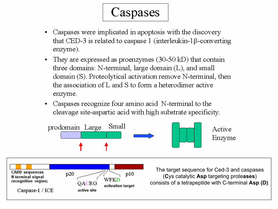
The target sequence for Ced-3 and caspases (Cys catalytic Asp targeting proteases)
consists of a tetrapeptide with C-terminal Asp (D).
• Procaspase-1 can be a substrate for Caspase-1, and autocatalytic activation is common among caspases. Thus activation shows positive feedback characteristics consistent with a binary on-off regulation.
• Ectopic expression of caspases in mammalian cells induces apoptosis. This is the strongest evidence for proteolytic mediation of apoptosis. The key intracellular event appears to be caspase activation by proteolytic cascade.
• Multiple caspases control different apoptotic pathways and provide functional redundancy. Caspase-1/ICE is one of a family of related proteases, which are coexpressed, and cDNAs of all caspases can be recovered from a single cell line. This appears to provide redundancy for an important function and circumvents what was once an embarrassment, that caspase-1 knockout mice still undergo apoptosis.
• The full range of apoptotic processes in mammalian cells involve several caspases, some of which have specialized purposes; a whole subgroup is involved in cytokine activation rather than apoptosis. Some caspases are initiators, i.e. their targets are downstream effector caspases. The target sequences of several caspases resemble the activating sequences of other caspases. In many cases, caspases can target their own activating sequences, suggesting autocatalytic positive feedback.
www.chembio.uoguelph.ca
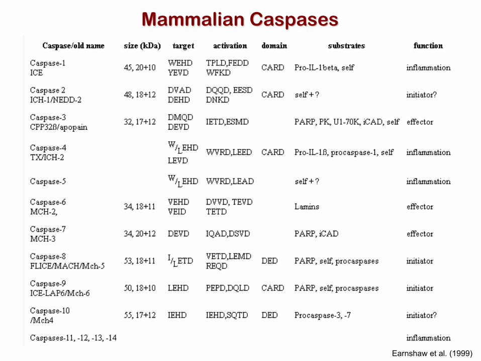
Earnshaw et al. (1999)
Mammalian CaspasesMammalian Caspases
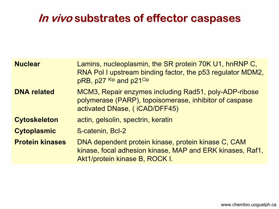
DNA dependent protein kinase, protein kinase C, CAM kinase, focal adhesion kinase, MAP and ERK kinases, Raf1, Akt1/protein kinase B, ROCK I.
Protein kinasesß-catenin, Bcl-2Cytoplasmicactin, gelsolin, spectrin, keratinCytoskeleton
MCM3, Repair enzymes including Rad51, poly-ADP-ribose polymerase (PARP), topoisomerase, inhibitor of caspase activated DNase, ( iCAD/DFF45)
DNA related
Lamins, nucleoplasmin, the SR protein 70K U1, hnRNP C, RNA Pol I upstream binding factor, the p53 regulator MDM2, pRB, p27 Kip and p21Cip
Nuclear
In vivo In vivo substrates of effector caspasessubstrates of effector caspases
www.chembio.uoguelph.ca
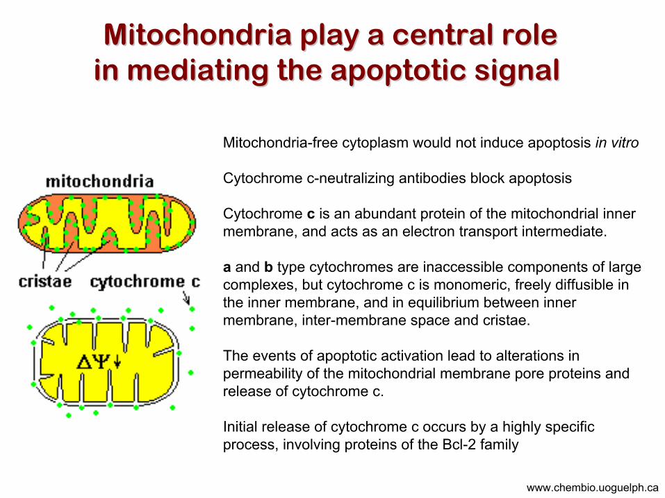
Mitochondria play a central roleMitochondria play a central rolein mediating the apoptotic signal in mediating the apoptotic signal
Mitochondria-free cytoplasm would not induce apoptosis in vitro
Cytochrome c-neutralizing antibodies block apoptosis
Cytochrome c is an abundant protein of the mitochondrial inner membrane, and acts as an electron transport intermediate.
a and b type cytochromes are inaccessible components of large complexes, but cytochrome c is monomeric, freely diffusible in the inner membrane, and in equilibrium between inner membrane, inter-membrane space and cristae.
The events of apoptotic activation lead to alterations in permeability of the mitochondrial membrane pore proteins and release of cytochrome c.
Initial release of cytochrome c occurs by a highly specific process, involving proteins of the Bcl-2 family
www.chembio.uoguelph.ca
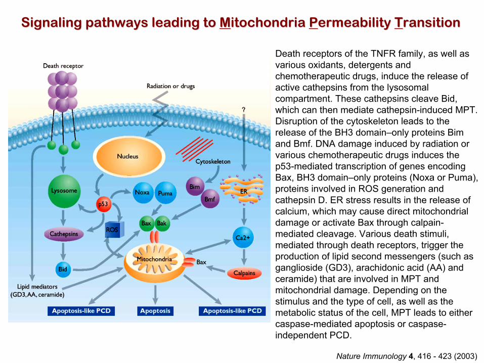
Death receptors of the TNFR family, as well as various oxidants, detergents and chemotherapeutic drugs, induce the release of active cathepsins from the lysosomalcompartment. These cathepsins cleave Bid, which can then mediate cathepsin-induced MPT. Disruption of the cytoskeleton leads to the release of the BH3 domain–only proteins Bimand Bmf. DNA damage induced by radiation or various chemotherapeutic drugs induces the p53-mediated transcription of genes encoding Bax, BH3 domain–only proteins (Noxa or Puma), proteins involved in ROS generation and cathepsin D. ER stress results in the release of calcium, which may cause direct mitochondrial damage or activate Bax through calpain-mediated cleavage. Various death stimuli, mediated through death receptors, trigger the production of lipid second messengers (such as ganglioside (GD3), arachidonic acid (AA) and ceramide) that are involved in MPT and mitochondrial damage. Depending on the stimulus and the type of cell, as well as the metabolic status of the cell, MPT leads to either caspase-mediated apoptosis or caspase-independent PCD.
Nature Immunology 4, 416 - 423 (2003)
Signaling pathways leading to Signaling pathways leading to MMitochondria itochondria PPermeability ermeability TTransitionransition

BclBcl--2 family: Pro2 family: Pro--Life and ProLife and Pro--Death factionsDeath factions
Bcl-2 and its closest relatives Bcl-XL, Bcl-w and Ced-9 are a-helical proteins having all four BH domains and are pro-survival. They suppress cytochrome c release, and are oncogenic when overexpressed. However, Bcl-XS, a splice variant of Bcl-XL having BH4 but lacking BH1 and BH2 is pro-apoptotic.
Bax and Bak lack the BH4 domain, and are pro-apoptotic. Bax expression is stimulated by p53, a mechanism for pro-apoptotic action of p53. Ectopic or overexpression of Bax induces cytochrome c release and apoptosis, and addition of Bax to mitochondria in vitro induces cytochrome c release.
The BH3-only sub group are strongly pro-apoptotic, and include Bim, Bik and Egl-1, which only have the 18-residue BH3 and the transmembrane region, while Bad and Bid only have BH3. The helical BH3 element allows for homo- and heterodimerization between family members. The non-homologous regions of BH3-only proteins could provide links to apoptotic signaling systems.
www.chembio.uoguelph.ca

Caspase-9
Effector caspases
www.chembio.uoguelph.ca
Vertebrate Apaf-1 activation occurs through cytochrome c binding. Bcl-2 and Bcl-XL appears to act by dimerizing with pro-apoptotic agonists such as Bax or Bak.
Normally, the balance is in favor of Bcl-2 or Bcl-XL, but the BH3-only factors appear to act to titrate out the Bcl2/Bcl-XL, tipping the balance in favor of Bax/Bak.
Bax can oligomerize in the membrane to form a permeability channel able to transport cytochrome c.
BH3-only factors have been reported to induce reorganization of the cristae. Alternative models suggest that Bid/Bad/Bak like factors act to open permeability channels such as the permeability transition pore, by disrupting the membrane potential, and affecting the voltage-dependent anion channel VDAC and ATP/ADP exchange transporter.
BclBcl--2 family: Pro2 family: Pro--Life and ProLife and Pro--Death factionsDeath factions

Survival mechanisms downstream of cytochrome c Survival mechanisms downstream of cytochrome c
1. Sequestration by heat shock proteins: Apaf1 interacts with heat shock proteins hsp70 and hsp90. Hsp70 directly sequesters CARD, and blocks caspase-9 recruitment, and possibly assembly of the oligomeric apoptosome as well. Hsp90 also associates with the monomeric Apaf1, and may represent a significant fraction of the normal autoinhibitedstate. Hsp90 appears to compete with cytochrome c for binding, suggesting action at an earlier step than hsp70.
2. Direct inhibition of the caspase catalysis by Inhibitor of Apoptosis Proteins (IAPs):
Inhibitor of apoptosis proteins (IAPs) represent the final line of defense against apoptosis, and act by binding directly to the substrate site of caspases
Smac/DIABLO: the mitochondrial answer to IAPs:
Mitochondria initiate the apoptosis cascade by releasing cytochrome c, but this effect could be nullified if IAP were allowed to maintain their inhibition of caspases. The apoptotic signal is instead sustained by the release of Smac/DIABLO (second mitochon-drial activator of caspase/direct IAP binding protein of low pI), which binds to and antagonizes the IAPs.
www.chembio.uoguelph.ca

Mitochondrial damage leads to the release of numerous mitochondrial proteins that mediate PCD.
• Release of cytochrome c triggers caspase activation and classic apoptosis.
• Smac (also known as Diablo) and Omi assist cytochrome c–induced caspase activation by counteracting caspase inhibitory factors (IAPs).
• AIF triggers a caspase-independent death pathway that culminates in DNA fragmentation and chromatin condensation characteristic of apoptosis-like PCD.
• EndoG cleaves DNA and induces chromatin condensation
• The serine protease activity of Omi can mediate caspase-independent cellular rounding and shrinkage without changes in the nuclear morphology
• Calcium and ROS can lead to severe mitochondrial dysfunction and necrosis-like PCD either directly or through autophagy of damaged mitochondria. Autophagy also may be associated with cathepsin activation and so can result in apoptosis-like PCD.
Nature Immunology 4, 416 - 423 (2003)
Mitochondria permeability transition can trigger caspaseMitochondria permeability transition can trigger caspase--dependentdependentand caspaseand caspase--independent programmed cell death (PCD):independent programmed cell death (PCD):

DEATH RECEPTORS:DEATH RECEPTORS:Pathways linking external signal receptors to caspasePathways linking external signal receptors to caspase--8 8
A variety of cell surface receptors related to TNF-R (tumor necrosis factor receptor) interact with the apoptotic activation system. The intracellular portion of the receptor carries a specific proteininteraction domain called the death domain, DD. The DD is activated by proximity, brought about when bound extracellular ligand induces receptor oligomerization. Activation can also be induced in absence of ligand by artifical cross-linking of the receptor.
Clustered receptor DDs recruit a variety of DD-containing adapters, of which FADD, Fas-associated death domain protein (also known as MORT1) bridges to a second protein interaction domain, DED, or death effector domain. The cluster of FADD-DEDs recruits procaspase-8, which also carries DEDs at its N-terminus (corresponding to the CARDs on Procaspase-9).
Procaspase-8 is activated to Caspase-8 by proximity-induced self-cleavage. Procaspase-10 is the only other caspase with DED boxes, and may substitute for Caspase-8 in some cases.
In some cells, TNF receptors associate with adaptors linked to cell proliferation or inflammatory signalling pathways, and may induceanti-apoptotic c-IAPs.
www.chembio.uoguelph.ca
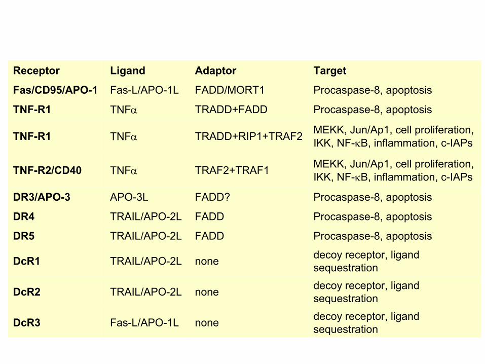
decoy receptor, ligand sequestrationnoneFas-L/APO-1LDcR3
decoy receptor, ligand sequestrationnoneTRAIL/APO-2LDcR2
decoy receptor, ligand sequestrationnoneTRAIL/APO-2LDcR1
Procaspase-8, apoptosisFADDTRAIL/APO-2LDR5
Procaspase-8, apoptosisFADDTRAIL/APO-2LDR4
Procaspase-8, apoptosisFADD?APO-3LDR3/APO-3
MEKK, Jun/Ap1, cell proliferation,IKK, NF-κB, inflammation, c-IAPsTRAF2+TRAF1TNFαTNF-R2/CD40
MEKK, Jun/Ap1, cell proliferation,IKK, NF-κB, inflammation, c-IAPsTRADD+RIP1+TRAF2 TNFαTNF-R1
Procaspase-8, apoptosisTRADD+FADD TNFαTNF-R1
Procaspase-8, apoptosisFADD/MORT1Fas-L/APO-1LFas/CD95/APO-1
TargetAdaptorLigandReceptor

Nature Immunology 4, 416 - 423 (2003)
The death receptor is stimulated by ligand-induced activation of the receptor trimer. The receptor death domains (DDs) of Fas then recruit FADD and RIP1 to the receptor complex. After recruitment to FADD through interactions between their death effector domains (DEDs), caspase-8 and caspase-10 are activated and trigger effector caspases, either directly or through a Bid-mediated mitochondrial pathway (activation of Apaf-1 and caspase-9). FADD and RIP initiate a caspase-independent necrotic pathwaymediated by the formation of, most probably, mitochondrion- or cPLA2-derived ROS. TNFR1 signaling differs from Fas signaling in the following steps: first, binding of FADD and RIP to the receptor complex requires the adaptor protein TRADD; and second, the RIP1-mediated necrotic pathway is inhibited by FADD and activated caspase-8
Death receptorDeath receptor––triggered caspasetriggered caspase--dependentdependentand caspaseand caspase--independent pathwaysindependent pathways
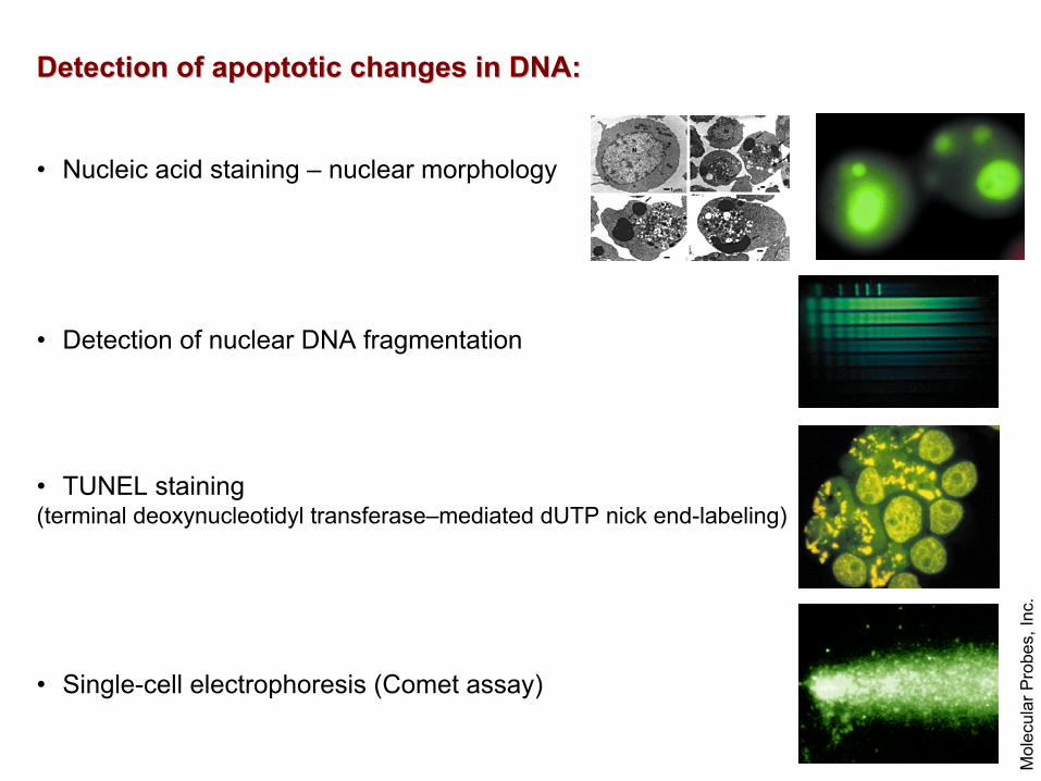
Detection of apoptotic changes in DNA:Detection of apoptotic changes in DNA:
• Nucleic acid staining – nuclear morphology
• Detection of nuclear DNA fragmentation
• TUNEL staining(terminal deoxynucleotidyl transferase–mediated dUTP nick end-labeling)
• Single-cell electrophoresis (Comet assay)
Mol
ecul
ar P
robe
s, In
c.

Detection of changes in membrane integrity:Detection of changes in membrane integrity:
• Membrane permeability
• Phospholipid symmetry (Annexin V staining)
Mol
ecul
ar P
robe
s, In
c.
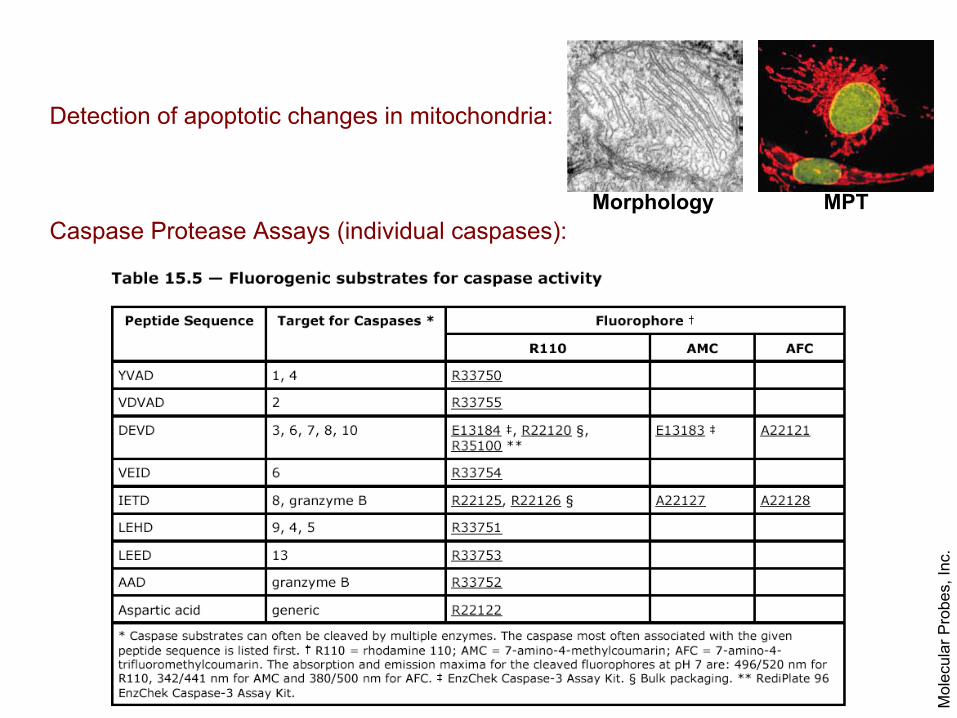
Detection of apoptotic changes in mitochondria:
Caspase Protease Assays (individual caspases):
Mol
ecul
ar P
robe
s, In
c.
MPTMorphology
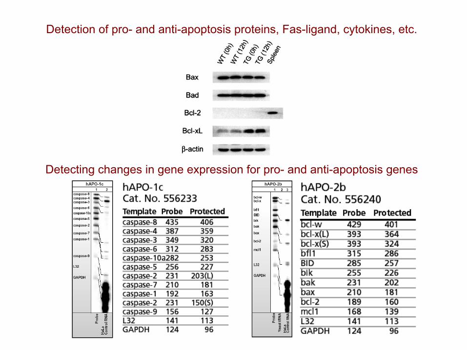
Detection of pro- and anti-apoptosis proteins, Fas-ligand, cytokines, etc.
Detecting changes in gene expression for pro- and anti-apoptosis genes


