MCD Cell Pathology Alexandra Burke-Smith
1
1. Cell Injury Rob Goldin ([email protected])
1. To list the causes of cell injury
2. To list the mechanisms of cell injury.
3. To define (and give examples of) hyperplasia, hypertrophy, atrophy, metaplasia and dysplasia
4. To describe the morphological changes associated with reversible and irreversible injury.
5. To describe the differences between apoptosis and necrosis.
A normal cell is in homeostatic equilibrium
Stress/increased demand on the cell adaptation of the cell
Injury/stimulus with an inability to adapt cell injury/death
Causes of cell injury
Oxygen deprivation
Chemical agents
Infectious agents
Immunological reactions
Genetic defects
Nutritional imbalances
Physical agents
Aging
Example - Oxygen deprivation MI (blockage in coronary artery – oxygen supply decrease – cell death)
- Normal myocyte – increased oxygen demand – HYPERTROPHY (increased cell size) = adaptation
- Normal myocyte – increased oxygen demand – cell injury – cell death (MI)
Injurious Stimuli
Cellular Response Depends on:
Type of injury
Duration
severity
Consequence Depends on:
Type of cell
Cell status
Adaptability
Genetic make up
Four intracellular systems are particularly vulnerable and often interlinked: o Cell membrane integrity o ATP generation o Protein synthesis o Integrity of genetic apparatus
structural and biochemical components of a cell very integrally related
multiple secondary effects rapidly occur
cellular function is lost before cell death which in turn is before morphological changes are seen

MCD Cell Pathology Alexandra Burke-Smith
2
Mechanisms of Cell Injury Adaptation- reversible injury
Atrophy
- Shrinkage in the size of the cell (or organ) by the loss of cell substance
- Same number of cells
- E.g. pernicious anemia is associated with gastric atrophy
- Dementia is associated with brain atrophy
Hypertrophy
- Increase in size of cells and consequently an increase in the size of the organ
- Can be physiological e.g. athletes, or pathological e.g. hypertension
- Caused by increased functional demand or hormone stimulation
Hyperplasia
- An increase in the number of cells in an organ
- Can be physiological or pathological
- Physiological hyperplasia can be either hormonal or compensatory
- Pathological hyperplasia is usually due to excessive hormonal or growth factor stimulation
- E.g. proliferative endometrium, carcinoma
Metaplasia
- A reversible change in which one adult cell type is replaced by another
- E.g. physiological- change in cervix during puberty
- Pathological- acid reflux through oesophagus (Barrett’s columnar lined oesophagus)
Dysplasia
- One meaning: abnormal in form e.g. retinal dysplasia (not what we are talking about)
- Precancerous cells which show the genetic and cytological features or malignancy but not invading the
underlying tissue
- Screening programmes try to diagnose cancer at this stage, as leads to much more successful treatment
- Associated with Barrett’s oesophagus
Associated Light Microscope changes:
fatty change- e.g. alcoholic fatty change during excess consumption
cellular swelling- e.g. ballooning degeneration due to damage of cell membrane
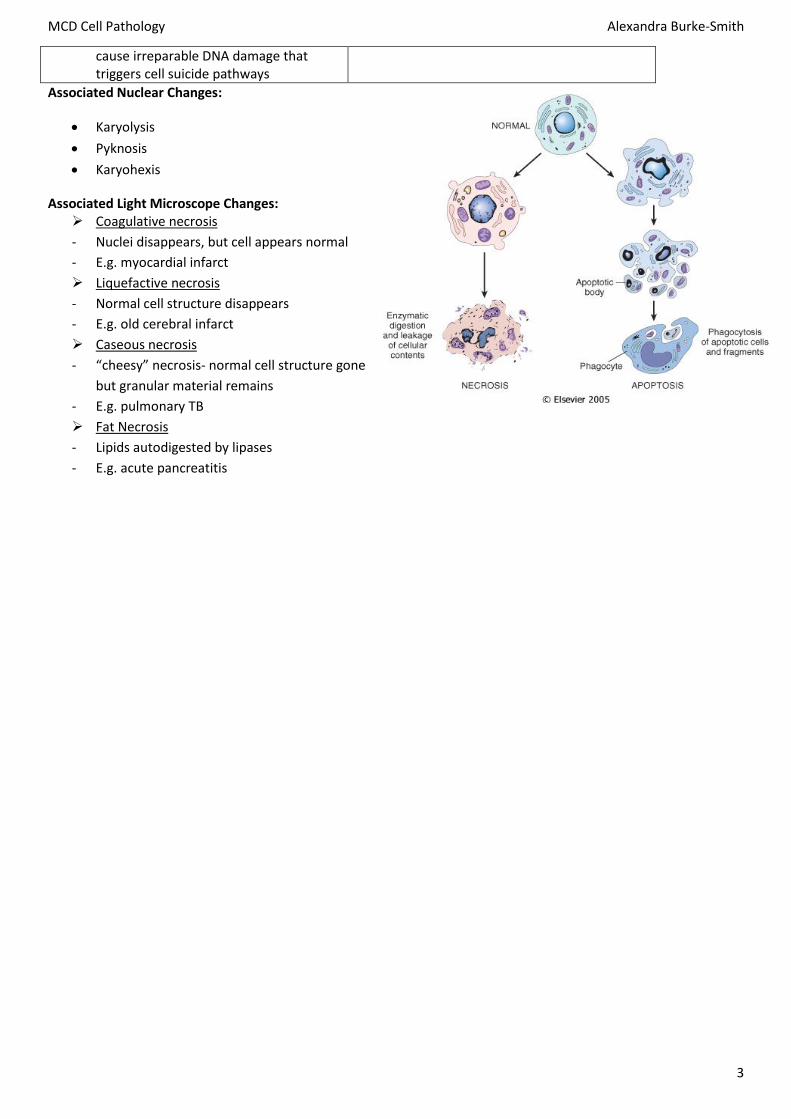
Cell Death- irreversible injury
Apoptosis Necrosis
Programmed cell death
Usually single/small number of cells
No leakage of intracellular components- therefore no associated inflammation
Physiological
Active energy dependent Causes:
1. Embryogenesis 2. Deletion of auto-reactive T cells in the
thymus 3. Hormone-dependent physiological
involution 4. Cell deletion in proliferating populations 5. A variety of mild injurious stimuli that
Programmed cell death
Sheets of cells
Marked inflammatory infiltrate- damage to surrounding tissue
Enzymatic digestion and leakage of cellular components- lack of energy maintaining cell membrane
MCD Cell Pathology Alexandra Burke-Smith
3
cause irreparable DNA damage that triggers cell suicide pathways
Associated Nuclear Changes:
Karyolysis
Pyknosis
Karyohexis
Associated Light Microscope Changes: Coagulative necrosis
- Nuclei disappears, but cell appears normal
- E.g. myocardial infarct
Liquefactive necrosis
- Normal cell structure disappears
- E.g. old cerebral infarct
Caseous necrosis
- “cheesy” necrosis- normal cell structure gone
but granular material remains
- E.g. pulmonary TB
Fat Necrosis
- Lipids autodigested by lipases
- E.g. acute pancreatitis

MCD Cell Pathology Alexandra Burke-Smith
4
2. Haemodynamic Disorders Dr James Carton ([email protected])
1. Describe the causes and consequences of oedema at different sites.
2. Define thrombosis & give causes and potential consequences of such an event.
3. Define embolism & know the importance of pulmonary embolism in clinical practice.
4. Describe possible causes of haemorrhage and potential outcomes.
5. Define shock and identify the possible causes and mechanisms.
6. Define infarction and describe possible causes, including atherosclerosis.
Oedema
Definition: abnormal increase in intersistial fluid. The volume of IF carefully controlled by osmotic pressure,
hydrostatic pressure and lymphatic drainage
Pulmonary oedema
- Caused by raised hydrostatic pressure in the pulmonary capillary bed.
- Most common cause = left ventricular failure.
- Fluid accumulates first in the interstitial space and then eventually spills into the alveolar spaces.
- Breathlessness (dyspnoea) is the main symptom.
- Breathlessness is typically worse on lying flat (orthopnoea).
- Fluid in the alveolar spaces predisposes to bacterial infection in the lung (pneumonia)
Cerebral oedema
- Usually caused by disruption to the cerebral capillaries (and the blood brain barrier)
- Seen in brain tissue surrounding intracranial lesions such as contusions, haemorrhages, infarcts and
tumours.
- Cerebral oedema contributes to a rise in intracranial pressure (ICP).
- High ICP risks brain herniation and death.
- Strategies to reduce ICP include raising the head, infusing isotonic fluids, steroids and osmotic diuretics such
as mannitol.
Generalised Oedema
- Widespread accumulation of fluid in subcutaneous tissues and serous cavities.
- Complex and multifactorial pathophysiology.
- Activation of renin-angiotensin-aldosterone pathway thought to be a key factor.
- Common causes include left ventricular failure, nephrotic syndrome, hepatic failure.
Thrombosis
Definition: abnormal blood clot formation in the circulatory system
3 contributing factors:
- stasis of blood
- injury to vessel wall
- hypercoagulability
Venous thrombosis
- Stasis and hypercoagulability key factors.
- Most form in deep leg veins (deep venous thrombosis or DVT).
MCD Cell Pathology Alexandra Burke-Smith
5
- Pulmonary embolism is the most important potential complication
Arterial thrombosis
- Almost always related to vessel wall injury caused by atherosclerotic plaques.
- Narrowing (stenosis) of the artery by thrombus causes ischaemia of the tissue supplied by the artery.
- Complete blockage (occlusion) of the artery by thrombus causes infarction of the tissue supplied by the
artery.
Cardiac thrombosis
- Stasis is the key factor.
- Left atrial thrombosis usually related to atrial fibrillation.
- Left ventricular thrombosis usually related to prior myocardial infarction.
- Systemic embolisation is the most important potential complication.
Embolism
An embolus is a detached mass within the circulatory system that is carried in the blood to a site distant
from its point of origin.
Most emboli are fragments of dislodged thrombus (thromboemboli).
Emboli are important because they can lodge in vessels and block them off.
Venous Thromboemboli
- Blood clot forms in vein, breaks free, and travels to the heart
- Embolus travels through the heart and blocks a blood vessel in the lung
- Emboli lodging in a major pulmonary artery cause instantaneous death.
- Emboli lodging in medium sized arteries present with breathlessness.
- Emboli lodging in small arteries cause subtle symptoms of breathlessness, chest pain, and dizziness – these
are the hardest to diagnose.
- ~30% of patients with pulmonary embolism will die from it.
- The risk of death increases the longer it takes to make the diagnosis.
Arterial thromboemboli
- Most originate in the heart or carotid arteries.
- May impact in cerebral arteries (stroke), mesenteric arteries (bowel infarction) or the lower limbs (acute
lower limb ischaemia).
Haemorrhage
Definition: Extravasation of blood due to vessel rupture.
May be due to trauma or an intrinsic disease of the vessel.
Rupture of a major vessel causes acute haemorrhage with risk of hypovolaemia, shock and death e.g.
ruptured abdominal aortic aneurysm.
Rupture of a small vessel can still be rapidly fatal if it occurs at a vital site e.g. brainstem haemorrhage.
Formation of a solid haematoma within the enclosed cranial cavity can also be fatal by causing a rise in
intracranial pressure and tonsillar herniation.
Chronic low grade haemorrhage may present with iron deficiency anaemia e.g. bleeding from a colonic
carcinoma.

MCD Cell Pathology Alexandra Burke-Smith
6
Shock
Definition: A generalised failure of tissue perfusion.
Caused by pump failure (e.g. acute myocardial infarction) or peripheral circulation failure (e.g.
hypovolaemia, sepsis, anaphylaxis).
Circulatory collapse ensues leading to ischaemia of multiple organs.
Most vulnerable organs are kidneys, bowel, brain, lungs, heart.
Rapid treatment is required to restore circulatory status and prevent multiple organ failure- patients often in
ICU
Infarction
Definition: Tissue necrosis due to ischaemia.
Most infarcts are due to obstruction of an artery.
Infarction may also occur due to venous obstruction.
Infarcts heal by repair. Although structural integrity is maintained, there is permanent loss of functional
tissue.
Myocardial infarction
- Obstruction in coronary artery
- Area of infarct represents occluded area
- Common in left anterior descending artery
- Large MI = lots of scar tissue – often leads to other infarctions
Cerebral Infarction
- Emboli from heart or carotid artery
- Area of brain undergoes infarction- most common is the middle cerebral artery
- Right MCA left hemiplegia (paralysis)
Small Bowell Infarction
- Emboli often from heart, migrate through aorta into superior mesenteric artery
- Gives intestines plum-brown colour
Atherosclerosis
Definition: An inflammatory disease of large and medium sized systemic arteries characterised by the formation of
lipid-rich plaques in the vessel wall.
Risk factors include smoking, diabetes, hypertension, hyperlipidaemia.
Endothelial injury is thought to be the key initiator (“response to injury hypothesis”).
Important diseases caused by stable
atherosclerotic plaques: stable angina,
chronic lower limb ischaemia.
Important diseases caused by
thrombosis overlying an unstable
atherosclerotic plaque: unstable
angina, myocardial infarction, cerebral
infarction, acute lower limb ischaemia.
MCD Cell Pathology Alexandra Burke-Smith
7
3. Inflammation Dr Mary Thompson ([email protected])
1. How does understanding basic pathology of acute and chronic inflammation help diagnose and treat the patient?
2. What is the clinical significance of granulomas inflammation?
3. What are the complications and long term effects of inflammation?
What is inflammation?
“Reaction of living vascularised tissue to sub-lethal cellular injury”
Evolutionary development to protect against infection and trauma
Many different cell types/soluble mediators
Generally tightly regulated
Acute (hrs/days) or chronic (weeks/months)
Local or systemic effects
Non-specific (but mediators important)
Unwanted effects
Local
- Excess local tissue damage and scarring
- Secondary effects on nearby tissue
Systemic
- Secondary multi-organ failure e.g. septic shock
- Amyloid
Importance
Process underlies many diseases; excessive, deficiency, chronic
Used to predict sequelae and complications of inflammatory reactions
Intervene to prevent/reduce adverse effects: drugs, surgery, target malignant cells etc
Examples of diseases
Infection- common cold, TB
Autoimmune- rheumatoid arthritis
Allergic- asthma
Metabolic- Gout
Malignant- host reaction
Inherited- chronic granulomatous disease
Idiopathic- pulmonary fibrosis, Crohn’s
Occupational/environmental- silicosis, radiation burns
Components of inflammatory reaction
Cells
- Neutrophils
- Macrophages
- Lymphocytes
- Eosinophils
- Mast cells

MCD Cell Pathology Alexandra Burke-Smith
8
ECM
- Collagen
- Proteoglycans
- Fibroblasts
Soluble factors
- Antibodies
- Cytokines
- Complement system
- Coagulation system
Vessels
- Immediate supply of cells and soluble factors
- Tissue repair
Neutrophils
Produced in BM
Circulate in blood, migrate to damaged tissue
Rapid response- first cell into damaged area
Kill bacteria and recruit more cells; phagocytosis and degranulation (enzymes, free radicals and soluble
mediators)
Monocyte/Macrophages
Kidney shaped nucleus
Originate in BM
Circulate in small numbers in blood as monocytes
Become macrophages in tissues
Role; phagocytosis, control of other cells using cytokines, viral/atypical bacterial infections
Other cells
Eosinophils
Allergic causes
Parasitic infections
Mast cells
Allergic diseases
Inflammatory reactions
Acute
Sub-acute
Chronic
1. Acute Inflammation
Evolution
Acute phase – neutrophils (24hrs)
Chronic phase - monocytes/macrophages (1week)
Resolution/repair – macrophages/fibroblasts
MCD Cell Pathology Alexandra Burke-Smith
9
Resolution
Architecture returns to normal only if;
Tissue cells able to regenerate e.g. liver
There is little structural damage done
E.g. pneumococcal lobar pneumonia
Congestion red hepatisation grey hepatisation resolution (during hepatisation, polymorphs and
neutrophils fill alveolar spaces)
Repair
Occurs if resolution unable
Replace normal tissue with fibrous scar tissue
Fibroblasts- produce collagen
Collagen
Remodelling- reorientation of collagen fibres; cross-links over tissue and faces towards stress; maximal
tensile strength
Laboratory Tests
Full blood count (FBC)
- Increased neutrophils- bacterial infections, acute episodes of chronic disease e.g. Crohn’s
- Increased Eosinophils- parasitic infection, drug reactions
- Increased monocytes- viral infections
Monopod test
- Atypical monocytes- glandular fever
Clinical Features
Local
- Symptoms- painful and hot
- Signs- swelling, erythema, heat, exudates
- Due to release of cytokines
Systemic
- Symptoms- malaise, loss appetite
- Signs- fever. shock
Vascular changes
Vascular calibre and flow increased
- Redness and heat
- Enables rapid delivery of inflammatory cells and mediators
Adhesion molecules expressed on endothelium
- Inflammatory cells stick to vessel wall
Increased vascular permeability
- Swelling
- Leaky capillaries allow cells and mediators to enter tissue- EXUDATE
Chemotaxis
Movement of cells towards injured area
EXOGENOUS and ENDOGENOUS compounds attract cells
Breakdown products of bacteria and damaged cells- all chemotactic
MCD Cell Pathology Alexandra Burke-Smith
10
Phagocytosis
Kills:
Free radicals
Lysozyme
Lactoferrin
Major basic protein
EXUDATE
Fluid, cells and proteins (fibrin, antibodies etc)
Function: dilutes pathogen, spread of soluble mediators, stops pathogen spreading, givens inflammatory
cells substrate to hold and migrate through
SEROUS—fluid e.g. blister
FIBRINOUS—fibrin e.g. viral pericarditis
PURULENT—fibrin + inflammatory cells + debris + fluid (PUS) e.g. peritonitis following bowel perforation
Soluble factors in exudate:
Produced locally by cells
- Preformed:
o mast cells- histamine
o neutrophils & macrophages- lysosomal enzymes
- Newly synthesised
o Macrophages- nitric oxide
o Macrophages & lymphocytes- cytokines
o Prostaglandins
o Leucotrienes
Circulating plasma proteins
- Produced by liver
- Include:
o KININ system
o coagulation/fibrinolysis cascade
o complement system
o C-reactive protein
Antibodies
Vasoactive Amines
Released from mast cells, basophils and platelets
E.g. histamine- allergic responses
Increases vascular permeability and therefore oedema and swelling e.g. acute asthmatic reactions
Complement System
Approx 20 proteins
Activated in cascade sequence
Important in many diseases
Effects: chemotaxis, facilitating phagocytosis, lysing of bacterial membranes
Adverse effects: due to over activation – ANAPHYLAXIS, shock e.g. snake bite

MCD Cell Pathology Alexandra Burke-Smith
11
Interleukins
Proteins
Proinflammatory or anti-inflammatory
Procoagulant or anticoagulant
Induce fever, malaise, weight loss
Promote cell proliferation e.g. fibroblast proliferation
Stimulate collage production healing/scarring
Prostaglandins and Leukotrienes
Effects on smooth muscle in walls of blood vessels or bronchi
Effects on permeability
TARGETING INFLAMMATORY MEDIATORS/STOPPING INFLAMMATION
Vasoactive amines-- Antihistamines
Prostaglandins and leukotrienes-- Aspirin etc
IL-1 and TNF -- Anti-TNF antibodies
Mediators and neutrophils have a short half life
Macrophages release anti-inflammatory products
Mast cells and lymphocytes release anti-inflammatory produces (LIPOXINS)
Stimulus- e.g. bacterium removed#
2. Chronic Inflammation (CI)
“Inflammation of prolonged duration in which active inflammation, tissue destruction and attempts at repair occur
simultaneously”
Causes
Persistent damage
Persistent infection
Prolonged exposure to toxic agent
Autoimmunity
Foreign body, e.g. splinter
Cells involved
Macrophages
Lymphocytes
Plasma cells
NO EXUDATE
NB: special type of chronic inflammation is GRANULOMATOUS
Macrophages
- Key cell
- Longer life than neutrophils
- Control the inflammatory process
Lymphocytes and plasma cells

MCD Cell Pathology Alexandra Burke-Smith
12
- Part of immune response generated by tissue destruction
- T-cells regulate immune reaction; can be cytotoxic
- Plasma cells produce antibodies
Granulomatous Inflammation
Particular form of CI showing granuloma formation; cluster of macrophages, involves specific T cells
Causes:
o Infection
o Foreign material
o Reaction to tumours
o Immune diseases
Defects in Inflammatory Process
Inherited
- Chronic granulomatous disease
- Compliment deficiencies
Acquired
- Poor adhesion- diabetes
- Poor phagocytosis and killing- anaemia, diabetes, malnutrition
- Poor production of soluble factors- poor liver function
Repair
What hinders it?
General
- Poor nutrition
o protein for collagen production
o energy for cell function
- Vitamin deficiency
o Vit C needed by fibroblasts to make collagen
o Vit A required for epithelial regeneration
- Mineral deficiency
o E.g. Zinc
- Suppressed inflammation
o Steroids
o Old age
o Diabetes
Local
- Poor blood supply
o E.g. ischaemic leg ulcers
- Persistent foreign body
o E.g. splinter
- Movement
o E.g. across a fracture site—need for a cast

MCD Cell Pathology Alexandra Burke-Smith
13
Complications
KELOID formation- excess collagen deposition
CONTRACTURES- fibrous scar tissue contracts as it matures. If scarring occurs across a joint, cant cause poor
mobility
Impaired organ function- e.g. fibrous scars in the myocardium after a heart attack
Abnormal Inflammatory Responses
Excessive unchecked inflammation
- Local ARDS (Acute respiratory distress syndrome) in lung
- Systemic—septic shock
Tissue damage and MOF (multiple organ failure)
Deficient inflammatory response
- Inherited; abnormal neutrophil adhesion and function
- Acquired; BM suppression
Recurrent infections
CASE STUDIES
CASE 1
38 yr old
Overweight
Female
C/o (complaining of) right upper quadrant pain
O/e (on examination) febrile, loss of appetite, tender RUQ
Blood tests; FBC- raised neutrophil, CRP ESR- both elevated
DIAGNOSIS: Acute cholecystitis
CASE 2
45 yr old
Repeated episodes of bloating and upper abdominal pain, especially after eating fatty food
DIAGNOSIS: chronic cholecystitis

MCD Cell Pathology Alexandra Burke-Smith
14
4. Autopsy Pathology Dr Michael Osborn ([email protected])
1. List four types of death that must be reported to the Coroner
2. List two reasons for conducting Hospital Autopsies
3. Explain how the need for consent from the deceased’s relatives differs between a Coroners’ and Hospital Autopsy
4. List four natural causes of sudden unexpected death in the community
5. What is a bruise? Give an example of a mechanism of injury that would lead to a bruise
6. What is an abrasion? Give an example of a mechanism of injury that would lead to an abrasion
7. What is a laceration? Give an example of a mechanism of injury that would lead to a laceration
8. What is the difference between a cut and a stab? Other than a knife what might cause such a wound?
9. What is the best generic term to use when describing a wound?
Types of Autopsy, the Coroner, Death Certificates
Coroner: an independent judicial officer of the crown who has a statutory duty to investigate the circumstance of
certain categories of death for the protection of the public
Cases that must be reported to the Coroner:
Cause unknown
Deceased has not seen doctor recently (14 days before or directly after) e.g. people who live on their own
COD violent, unnatural or suspicious
COD accident (even if resulting death is significantly later, e.g. car crash epilepsy death years later
COD due to neglect by self or others, e.g. in nursing homes
COD due to industrial disease or occupation e.g. asbestos exposure in a factory
Death due to abortion
Death during an operation or before/directly after recovery from anaesthetic
Question of suicide, e.g. hanging
Death occurred during/shortly after police detention/prison custody
Death related to poisoning
If any doubt of COD, refer to coroner’s office
Coroners’ Autopsy
Establishes COD, and once complete his remit is over- regardless of whether natural or unnatural
Not allowed to use any body parts for research, unlike a hospital autopsy
Hospital Autopsy
Reasons for:
Very thorough examination of the deceased:
- Extent of disease
- Their treatment
- Effect of treatment and disease
Audit- major discrepancies between stated COD and actual COD
- main diagnosis missed in 15% of autopsied cases
Monitoring effects of new treatments
- E.g. complex CHD treatments

MCD Cell Pathology Alexandra Burke-Smith
15
Teaching
- E.g. unrivalled clinic pathological correlation
Research
- E.g. post mortem brain tissue and variant CJD
Death Certificates
Data used for epidemiology
Accurate morbidity and mortality data needed to monitory nations health
Directs the allocation of resources
Detects environment risks
Hospital Autopsy Coroners Autopsy
- Consent from Next of Kin (NOK) – person in qualifying relationship- required
- With consent, tissue can be taken and used from the body (see Human Tissue Act) for research etc
- No consent needed (wishes of NOK considered)
- Material can only be taken if coroner gives permission- needed to establish COD
Conduction of an Autopsy
In a mortuary
Dissect through organs systematically
Look under microscope at specific tissue samples
Death Certification
Filled in for any death
Taken to registrar by family
Scrutinised- must be filled in correctly before registration
1a) immediate COD (compulsory)
1b) predisposing factor leading to 1a (not compulsory)
1c) predisposing factor leading to 1b (not compulsory)
2) Other contributing factors not directly leading to death
Natural Causes of sudden unexpected death (in the community)
Cardiovascular Disease
Approx 75% of deaths handled by medical examiners in US
Cardiac arrhythmia (diagnosis is one of exclusion- full autopsy must be conducted)
Severe coronary artery atherosclerosis (2 or more vessels)
Myocardial scarring
Coronary artery thrombosis
Acute/sub-acute MI
Ischaemic Heart disease
Hypertensive Heart Disease
Cardiomyopathy
Myocarditis
Structural anomalies (eg bridging)
Floppy mitral valve

MCD Cell Pathology Alexandra Burke-Smith
16
Aortic stenosis (usually calcific)
Conduction abnormalities (eg long QT syndrome)
Vascular System
Ruptured aortic aneurism- associated with atherosclerosis & hypertension
Central Nervous System
Non traumatic subarachnoid haemorrhage- Berry aneurism (high mortality rate)
Intracerebral haemorrhage
Epilepsy
Respiratory System
Pulmonary embolus
Asthma
GI tract (not usually unexpected)
Bleeding Oesophageal Varices
Bleeding Ulcers
Pancreatitis
Not natural
Drugs- approx 1% of all cases reported to coroner
Alcohol- usually only sudden in alcoholics, often associated with GI problems and often goes with drugs
Trauma- self induced, accidental, caused by others
Types of Injury
Bruise/Contusion
- Blunt trauma injury
- Occurs alone (skin intact) or in association with other injury
- An extraversated collection of blood which has leaked from small arteries, venules and veins but not
capillaries
- Laxity of skin, fragility of vessels and coagulation state etc all effect
- Takes hours days to form
- May be patterned or deep (cannot be seen on epidermal surface)
- Cannot age a bruise
- Can bruise after death
Abrasion
- Graze/scratch- superficial blunt trauma
- Confined to epidermis
- May extend to superficial dermis
- Can occur before and after death
- Due to TANGENTIAL (distal skin tag occurs) or VERTICAL force (no distal skin tag)
- E.g. friction burn, car radiator, flooring, whip, stamp
Laceration
- Split to skin

MCD Cell Pathology Alexandra Burke-Smith
17
- Result of blunt force over stretching skin
- Deep (full thickness)
- Bleeds
- Margins ragged and crushed/bruised
- Common where skin can be compressed between force and bone (scalp, elbow, shin)
- Rare over soft fleshy areas (buttocks, breasts)
- Flaying- tangentially applied force horizontal laceration (difficult to distinguish object causing it)
Cut (or slash)
- Length longer than depth
- Edges clean
- Well defined
- Minimal damage to surrounding tissue
Stab
- Depth longer than length
- Edges clean
- Well defined
- Minimal damage to surrounding tissue
Both cut and stab wounds are caused by an object with sharp/cutting edge (Usually knife- can be anything e.g.
broken glass, metal)
NB: EITHER USE THE CORRECT TERM (E.G. LACERATION= BLUNT FORCE INJURY- FULL THICKNESS) OR USE A
GENERIC TERM (INJURY/WOUND) (although technicaly bruises and abrasions are not covered by these terms as
they should involve the full thickness of the skin)
MCD Cell Pathology Alexandra Burke-Smith
18
5. Cancer Dr Rathi Ramakrishnan ([email protected])
1. Define cancer, neoplasia, tumour, metastasis, carcinogen
2. List features which distinguish benign from malignant tumours
3. Give examples of cancers cause by infection, chemical and environmental agents
4. Briefly outline the principals of cancer screening
5. Describe features of pathology which predict the prognosis in cancer
What is cancer?
Important socio-economic problem
Significant cause of morbidity and mortality worldwide
Neoplasm: new growth- “abnormal mass of tissue, the growth of which is virtually autonomous and exceeds that of
normal tissues. The growth is uncoordinated and persists after the cessation of the stimuli that initiated the change”
Cancer: from the latin “crab”- the pathological condition characterized by the proliferation of neoplastic cells that
tend to invade surrounding tissue and metastasize to new body sites
Tumour: swelling, originally for inflammation
Metastasis: the development of secondary malignant growths at a distance from a primary site of cancer.
Carcinogen: a substance capable of causing cancer in living tissue
Dysplasia: disordered growth, limited to epithelium
Loss of uniformity of individual cells
Loss of their architectural orientation
Mild-moderate dysplasia may revert to normal
Severe dysplasia is permanent e.g. carcinoma in situ
Metaplasia: substitution of one mature cell type for another more suited to the environment
Result of a chronic stimulus, when withdrawn may resolve to normal i.e. adaptive not premalignant
E.g. smoking causes metaplasia of glandular bronchial epithelium to squamous epithelium
Nomenclature
All tumours have two basic components:
Parenchyma: proliferating neoplastic cells
Stroma: supportive fluid required for tumour growth
Benign Tumours
- suffix “oma” to cell of origin
o From glandular tissues (epithelial origin)
- Adenoma
- Cystadenoma
- Papilloma
- Polyp
o From stromal tissues (mesenchymal origin)
- Fibroma
- Lipoma
MCD Cell Pathology Alexandra Burke-Smith
19
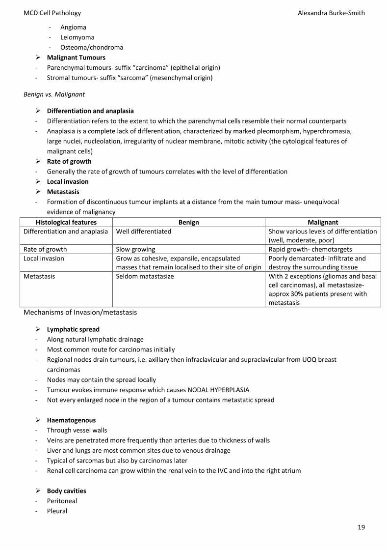
- Angioma
- Leiomyoma
- Osteoma/chondroma
Malignant Tumours
- Parenchymal tumours- suffix “carcinoma” (epithelial origin)
- Stromal tumours- suffix “sarcoma” (mesenchymal origin)
Benign vs. Malignant
Differentiation and anaplasia
- Differentiation refers to the extent to which the parenchymal cells resemble their normal counterparts
- Anaplasia is a complete lack of differentiation, characterized by marked pleomorphism, hyperchromasia,
large nuclei, nucleolation, irregularity of nuclear membrane, mitotic activity (the cytological features of
malignant cells)
Rate of growth
- Generally the rate of growth of tumours correlates with the level of differentiation
Local invasion
Metastasis
- Formation of discontinuous tumour implants at a distance from the main tumour mass- unequivocal
evidence of malignancy
Histological features Benign Malignant
Differentiation and anaplasia Well differentiated Show various levels of differentiation (well, moderate, poor)
Rate of growth Slow growing Rapid growth- chemotargets
Local invasion Grow as cohesive, expansile, encapsulated masses that remain localised to their site of origin
Poorly demarcated- infiltrate and destroy the surrounding tissue
Metastasis Seldom matastasize With 2 exceptions (gliomas and basal cell carcinomas), all metastasize- approx 30% patients present with metastasis
Mechanisms of Invasion/metastasis
Lymphatic spread
- Along natural lymphatic drainage
- Most common route for carcinomas initially
- Regional nodes drain tumours, i.e. axillary then infraclavicular and supraclavicular from UOQ breast
carcinomas
- Nodes may contain the spread locally
- Tumour evokes immune response which causes NODAL HYPERPLASIA
- Not every enlarged node in the region of a tumour contains metastatic spread
Haematogenous
- Through vessel walls
- Veins are penetrated more frequently than arteries due to thickness of walls
- Liver and lungs are most common sites due to venous drainage
- Typical of sarcomas but also by carcinomas later
- Renal cell carcinoma can grow within the renal vein to the IVC and into the right atrium
Body cavities
- Peritoneal
- Pleural

MCD Cell Pathology Alexandra Burke-Smith
20
- Pericardial
- Subarachnoid
- Joint
- Most commonly from ovarian carcinomas which may take the peritoneal surface. Also spread of lung
carcinoma into pleural cavity.
Contiguous
- Spread to adjacent organs etc
Cancer Epidemiology
Age
- Incidence increasing due to aging population
- In general, higher incidence >55 yrs
- Certain cancers specifically affect the young
Geography
- Stomach cancer Japan > USA
- Melanoma NZ & Australia > Iceland
Environmental
- UV light
- Occupational agents like asbestos
- Diet and weight
- Alcohol
- Smoking
- Infections- notably viruses (HPV, HBV, EBV etc)
Genetics
For a large number of cancers there exist some hereditary predispositions. There are 3 categories of hereditary
forms:
Inherited autosomal dominant cancer syndromes
- E.g. Familial retinoblastoma
- FAB (Familial andenomatous polyposis coli)
Familial cancers
- E.g. breast cancers
- Ovarian
- colonic
Autosomal recessive syndromes of defective DNA repair
- E.g. xeroderma pigmentosa
- Bloom syndrome
Non-heredity
Increased risk i.e. liver cirrhosis and HCC
Inflammation & cytokines with growth of transformed cells, promoting genetic instability and carcinogenesis

MCD Cell Pathology Alexandra Burke-Smith
21
The molecular basis of cancer
Carcinogenesis
Multistep process
Genetic hypothesis- tumour mass results from the monoclonal expansion of a single progenitor cell
Accumulation of successive subsequent mutations allows progression and accounts for heterogeneity
INITIATION
DNA damage- genetic mutations
PROMOTION
activation of cancer promoting genes
inhibition of apoptosis
inactivation of cancer suppressor genes
TRANSFORMATION
expression of altered gene products
loss of regulatory gene products
PROGRESSION
malignant neoplasm formed
Targets of genetic damage
4 classes of regulatory genes Oncogenes-- Growth promoting
Anti-oncogenes-- Growth inhibiting/tumour suppressor
Anti-Apoptosis genes—genes regulating programmed cell death
DNA repair genes
Oncogenes
Derived from proto-oncogenes
Genes regulating normal cellular growth
Examples associated with tumours
Myc Burkitt lymphoma
N-Myc neuroblastoma
CyclinD1 mantle cell lymphoma
Tumour suppressor genes
Usually regulate normal cell growth
Classic e.g. is Rb gene in genetically inherited retinoblastoma- requires two hits; inheritance of mutated copy
of one Rb gene followed by acquisition of damage to remaining copy
50% of all tumours contain mutations in the P53 gene; housekeeping gene which prevents genetically
damaged cells from replicating
BRCA-1 & BRCA-2 breast cancers are familial and one of these mutations present in 80% of breast cancers
Anti-apoptosis genes
Genetic defects accumulate causing pro-apoptotic genes (Bax family), which usually cause programmed cell
death
Anti-apoptotic (bcl2) genes prevent this causing unregulated PROLIFERATION
Over-expression of bcl2 and prevention of apoptosis results in indolent growth of lymphocytes found in
many low grade lymphomas and other somatic malignancies

MCD Cell Pathology Alexandra Burke-Smith
22
DNA repair genes
Genomic instability syndromes- inherited mutations of DNA repair proteins
Not directly oncogenic
Act by permitting mutations to occur during normal cell cycles
Carcinogens
Definition: agents that cause genetic damage and induce neoplastic transformation of cells. There are different
classes:
Chemicals
Radiation
Microbial agents (mainly viruses)
Hormones
Miscellaneous
Chemical carcinogens
No common structural features
Some chemicals are inducers (permanent DNA damage)
Others are promoters (reversible DNA damage)
Direct acting agents do not require metabolism for activation e.g. dimethyl sulphate
PROCARCINOGENS need metabolic conversion to active peptide e.g. aromatic amines activated in the liver
causing UB cancer
If enzyme is present within tissues, tumour occurs at the site, e.g. skin and lung cancers with aromatic
hydrocarbons
Major classes- hydrocarbons, amines, nitrosamines, azo dyes, alkylating agents
Promoters include hormones, drugs etc
Microbial Carcinogens
Oncogenic Viruses
- Generally in young
- EBV- Burkitt’s Lymphoma
- HPV- cervical cancers
- HBV & HCV - Hepatic cancers
- HHV8- Kaposi’s sarcoma
- Oncogenic viral DNA genome directly incorporated into host cell DNA
- Oncogenic RNA viral genome transcribed into DNA by enzymes prior to incorporation
Bacterial Carcinogens
- Helicobacter Pylori- stomach and pancreatic cancer
Radiations
Ultraviolet- skin cancer
Ionising EM radiation i.e. X-rays cause an increase in leukaemia and solid tumours

MCD Cell Pathology Alexandra Burke-Smith
23
Clinical Practice
Diagnosis
Laboratory methods
- Cytology FNA (freehand or USS guided)
- Histology (core biopsy, incisional or excisional biopsy)
Other methods
- Tumour typing
- Immunocyto/histochemistry
- Flow cytometry
- Molecular methods (PCR, FISH, DNA microarrays, spectral karyotyping)
- Tumour markers- CEA AFP Ca125 etc
Grading and Staging
Staging
- Main clinical staging system- TNM (WHO), based on
- T – Primary Tumour Size (T1-T4)
- N – Nodal Status (N0, N1-3)
- P -- Presence of metastases (M0,M1-2)
- Others include DUKE’s for colorectal, FIGO for ovarian cancer and Ann Arbor for lymphoma
Grading
- Histological
- low and high grade (Less useful than staging)
- based on the degree of differentiation and the number of mitoses
Effects on Patient
Both benign and malignant tumours affect the host
Anxiety (breast lumps)
Related to location i.e pressure, ulceration, infection, bleeding (hormonal effects)
Metabolic cancer cachexia (increased BMR, reduced fat and muscle bulk) TNF alpha
Paraneoplastic syndromes- endocrinopathies, hypercalcemia, thrombotic diathesis, acanthosis nigricans
Prevention and Screening
Screening
- Detect cancer either at preinvasive stage/early (stage 1) .For successful screening programs:
o Reliable prediction of tumour behaviour
o Treatment available
o Target population has enough people at risk to justify expense
o Cost-effective and reliable screening tool
- In UK, cervical cancer and breast cancer screening with piloting for colorectal cancer
Prevention
- Vaccinations- Human Papilloma virus, Hepatitis B virus to protect against cervical/liver cancers

MCD Cell Pathology Alexandra Burke-Smith
24
6. Cell Pathology Case Studies Dr Marjorie Walker ([email protected])
1. Using the example of Helicobacter Pylori infection of the stomach, discuss the varied outcomes of infection and
why these occur, and how inflammation can lead to cancer or lymphoma in this organ
2. List two major complications of peptic ulcers and describe the consequences of these
3. Using the example of a case of atherosclerosis, list 3 major outcomes of this arterial disease
Helicobacter Pylori
Dyspepsia 1
Male
Age 46 yrs
Dyspepsia for 3 yrs
Worse recently- abdominal pain after meals
Episode of melaena (blood in stool)
Blood test Hb 9.0 (13.0 normal)
Endoscopy- ulcer in duodenum; when acid and food hits ulcer pain after eating
BIOPSY OF GASTIC ANTRUM- helicobacter associated gastritis
o Chronic inflammation; lymphocytes
o Acute inflammation; neutrophils
Helicobacter pylori
20% adults in developed countries by age 50, 80% in UDCs
Majority (70-80%) asymptomatic
2x relative risk for gastric carcinoma
Normal Stomach Helicobacter Pylori Stomach
Lined by gastric mucosa Columnar epithelium mucin secreting glands Acid/pepsinoggen body Lamina propria mucularis mucosa
Glands destroyed by neurtrophils Lamina propria filled with lymphocytes
Topography and Outcome
- D cells of the antrum (in the stomach) produce somatostatin
- Somatostatin inhibits gastrin
- In ANTRAL GASTRITIS (inflammation of a region of the stomach) D cells are reduced
- Principle cause of duodenitis and duodenal ulcer is acid
Gastric Ulcers
- Infection spreads with age
- Pangastritis (inflammation of the entire stomach)
- Atrophy of body mucosa
- Acid decreases
- Increased number of gastric ulcers
Complications
- Haemorrhage
- Perforation
MCD Cell Pathology Alexandra Burke-Smith
25
Dyspepsia 2
Female
Age 76 yrs
C/o vague abdominal pain and nausea
Dyspepsia for years
Blood tests Hb11
Endoscopy performed
INTESTINAL METAPLASIA shown in gastric mucosa
o Response to long term damage
o H. pylori present in bile
o Cancer risk
GASTRIC DYSPLASIA- an abnormal pattern of growth in which some of the histological features of malignancy
are present
o Pre-invasive stage
o Intact BM
Risk of progression to cancer; H. Pylori strong risk in presence of intestinal metaplasia and atrophy;
increased 8x over long time (>15 yrs)
GASTRIC CANCER
o Japan: mass endoscopy programs led to 35% early gastric cancers vs. 10% in US
Dyspepsia 3
Male
Age 76 yrs
Dyspepsia
Early satiety (feeling full)
Blood tests Hb 8
Endoscopy performed
Liver function tests- abnormal
Liver CT scan- multiple lesions
Gastric Cancer
High incidence in Japan, Chile, Italy, China, Portugal, Russia
2:3 ratio women to men
90% of all malignant tumours are carcinomas
asymptomatic until late; weight loss, abdominal pain, nausea, vomiting, altered bowel habit
kills more people worldwide than lung cancer
Dyspepsia 4
Male
Age 76 yrs
Early satiety
Blood tests Hb 8
Endoscopy performed
LYMPHOMA diagnosis

MCD Cell Pathology Alexandra Burke-Smith
26
Atherosclerosis
Occludes arteries slowly
- Angina
- Myocardial scarring
- Dementia
- Claudication
Occludes arteries suddenly
- Plaque rupture
o Thrombosis
o atheroembolization
- Haemorrhages into plaques
o MI
o Stroke
o Gangrene of the bowel
Weakens artery walls
- Aneurysms
Case 1
Male
History of hypertension
Sudden loss of consciousness
Died in A&E
Post mortem (PM) carried out
Case 2
Male
Died in an RTA (road traffic accident)
Post mortem- aneurysm found
Cause of death natural or unnatural?
Case 3
Male
Central chest pain
Died 7 days post admission
These show different examples and outcomes of atherosclerosis.


