Download - M rcc 007 b macular hole - vrmny

Microscopic imaging of a macular hole using the rtx1
R. Spaide Vitreous Retina Macula Consultants of New York, NY, USA
rtx1 adaptive optics retinal cameraCase reportMacular hole

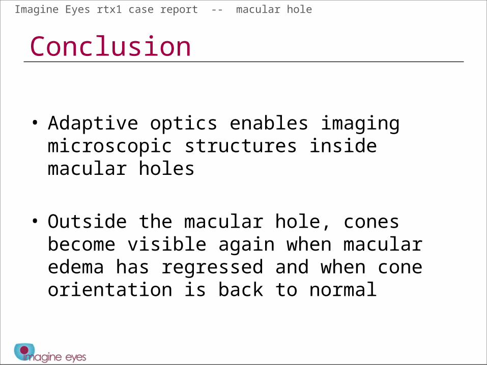
Imagine Eyes rtx1 case report -- macular hole
Combination of several imaging modalitiesMale, 61 yrs old. Epiretinal membrane with pucker and a full-thickness macular hole. Images acquired 2 weeks before operation
Colour fundus camera
rtx1
IR SLO
OCT

Imagine Eyes rtx1 case report -- macular hole
Comparison pre- and post-op
Hyper reflective dots are visible inside the macular hole, not in the periphery
Edema has regressed and cones have become visible in the periphery
2 weeks PRE-OP 2 weeks POST-OP
Imagine Eyes rtx1 case report -- macular hole
Conclusion
• Adaptive optics enables imaging microscopic structures inside macular holes
• Outside the macular hole, cones become visible again when macular edema has regressed and when cone orientation is back to normal


