Download - IRON METABOLISM - cmb.i-learn.unito.it
IRON METABOLISM
Harper’s Illustrated Biochemistry chapter 50
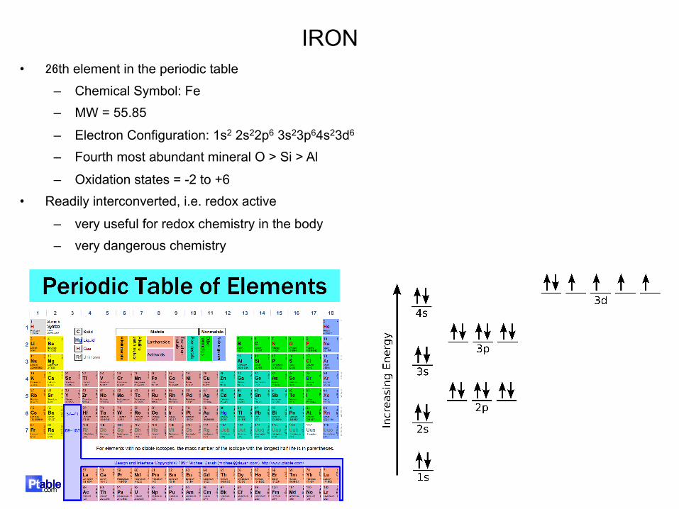
• 26th element in the periodic table
– Chemical Symbol: Fe – MW = 55.85
– Electron Configuration: 1s2 2s22p6 3s23p64s23d6 – Fourth most abundant mineral O > Si > Al
– Oxidation states = -2 to +6 • Readily interconverted, i.e. redox active
– very useful for redox chemistry in the body – very dangerous chemistry
IRON

IRON FUNCTIONS IN BIOLOGY
• Oxygen Transport and Storage – Hemoglobin – Myoglobin
• Electron Transport and Energy Metabolism – Cytochromes – Fe-S proteins
• Substrate Oxidation & Reduction Iron dependent enzyme:
– Ribonucleotide reductase – Amino acid oxidases – Fatty acid desaturases – Nitric oxide synthetase – Peroxidases
• Regulation of intracellular iron
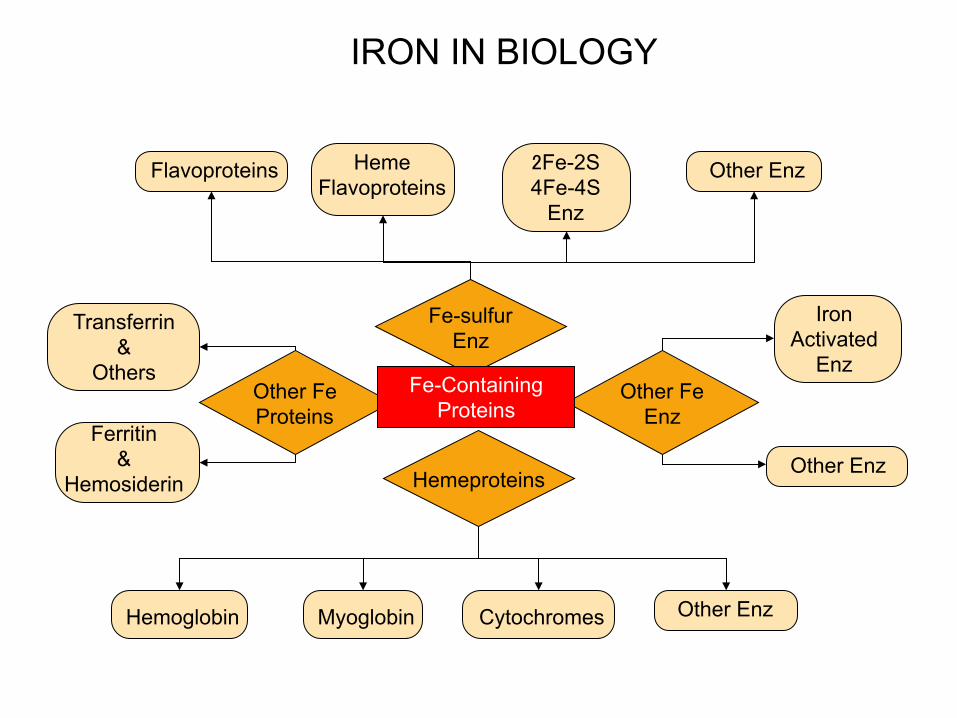
Flavoproteins
Hemeproteins
Fe-sulfur Enz
Other Fe Enz
Other Fe Proteins
Fe-Containing Proteins
Transferrin &
Others
Ferritin &
Hemosiderin
Other Enz Heme Flavoproteins
Hemoglobin
Other Enz
Iron Activated
Enz
2Fe-2S 4Fe-4S
Enz
Myoglobin Cytochromes Other Enz
IRON IN BIOLOGY
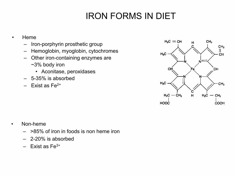
• Heme – Iron-porphyrin prosthetic group – Hemoglobin, myoglobin, cytochromes – Other iron-containing enzymes are
~3% body iron • Aconitase, peroxidases
– 5-35% is absorbed – Exist as Fe2+
• Non-heme – >85% of iron in foods is non heme iron – 2-20% is absorbed – Exist as Fe3+
IRON FORMS IN DIET
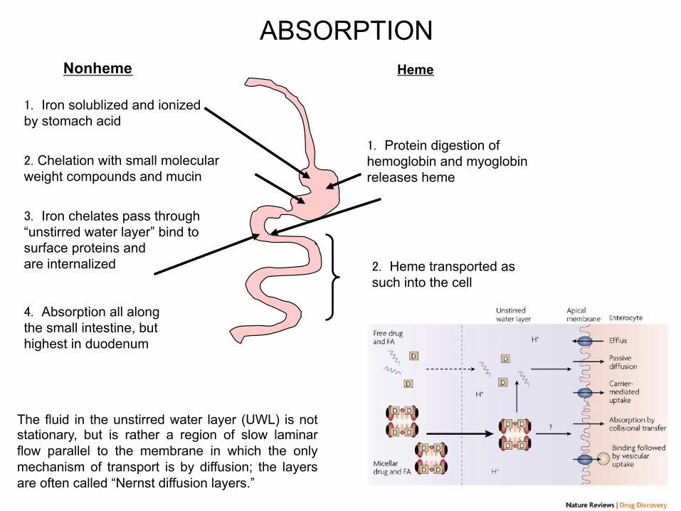
ABSORPTION Nonheme
����Iron solublized and ionized by stomach acid
���Chelation with small molecular weight compounds and mucin
����Iron chelates pass through “unstirred water layer” bind to surface proteins and are internalized
����Absorption all along the small intestine, but highest in duodenum
Heme
����Protein digestion of hemoglobin and myoglobin releases heme
����Heme transported as such into the cell
The fluid in the unstirred water layer (UWL) is not stationary, but is rather a region of slow laminar flow parallel to the membrane in which the only mechanism of transport is by diffusion; the layers are often called “Nernst diffusion layers.”

• Ferrous, Fe2+, most soluble = most absorbable
• Each mechanism has 3 phases – Iron uptake – Intraenterocyte transport – Storage and extraenterocyte transfer
Dietary Iron: • Iron is essential element and must be precisely regulated.
• On the lumen side of small intestine iron is reduced from its ferric form (Fe3+) to ferrous form (Fe2+).
• Ferrous iron is then transported in enterocytes by DMT1 (divalent metal transporter).
ABSORPTION
Intestinal lumen
Duodenal cytochrome b
Divalent metals transporter (DMT1)
Ferroportin Transferrin
Blood
Hephaestin
Enterocyte

• Promotors – Amino Acids – Animal Proteins(for heme) – Ascorbic Acid – Hydrochloric Acid – Organic Acids – Sugars – Mucin
• Inhibitors – Carbonates – Calcium (for heme) – Egg yolk phosvitin – Fiber – Oxalates – Phosphates – Phytates – Plant polyphenols – Soy proteins
• Heme iron is an important dietary sources of iron because it is more effectively absorbed than non-heme iron.
• From 5% to 35% of heme iron is absorbed from a single meal, whereas non-heme iron absorption from a single meal can range 2%-20%, depending on the iron status of the individual and the ratio of enhancers and promotors in the diet. Thus, although it constitutes about 10% of the iron found in the diet, heme iron may provide up to one-third of total absorbed dietary iron.
• The reason ascorbic acid promote iron absorption is ascorbic acid maintain iron in a reduced form and forms a soluble chelate with iron. These actions are shared by organic acids such as citric, lactic acids.
• A number of dietary factors influence iron absorption.
ABSORPTION
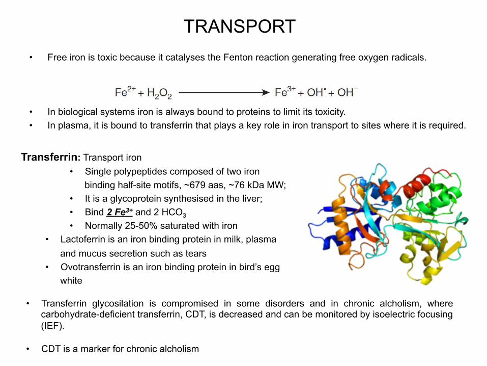
TRANSPORT • Free iron is toxic because it catalyses the Fenton reaction generating free oxygen radicals.
• In biological systems iron is always bound to proteins to limit its toxicity. • In plasma, it is bound to transferrin that plays a key role in iron transport to sites where it is required.
• Transferrin glycosilation is compromised in some disorders and in chronic alcholism, where carbohydrate-deficient transferrin, CDT, is decreased and can be monitored by isoelectric focusing (IEF).
• CDT is a marker for chronic alcholism
Transferrin: Transport iron • Single polypeptides composed of two iron
binding half-site motifs, ~679 aas, ~76 kDa MW; • It is a glycoprotein synthesised in the liver; • Bind 2 Fe3+ and 2 HCO3
• Normally 25-50% saturated with iron • Lactoferrin is an iron binding protein in milk, plasma
and mucus secretion such as tears • Ovotransferrin is an iron binding protein in bird’s egg
white

IRON TRANSPORT: TRANSFERRIN CYCLE
Transferrin cycle: holotransferrin (Tf-Fe) binds to transferrin receptor 1 (TfR1) on cell surface. Clathrin-coated vescicles form and endocitosis occurs forming endosomes where pH is acidic. The acidic pH causes iron release from transferrin. Apotransferrin (Apo.Tf) still binds to TfR1. Ferric iron is converted to ferrous iron by a iron reductase (Step 3). Ferrous iron is transported in the cytosol trhough DMT-1. The TfR1-apo-Tf complex is recycled on cell surface where apo-Tf is released and TfR1 can bind another Tf-Fe.
Clathrin
Early endosome
Apotransferrin is recycled on the cell surface
The low pH of the late endosome causes the Fe3+
release from transferrin
Late endosome
Transferrin receptor (TfR1)
Holotransferrin (Tf-Fe) Apotransferrin (apo-Tf) dissociates from its receptor at neutral pH
External pH 7
IRON TRANSPORT: TRANSFERRIN CYCLE
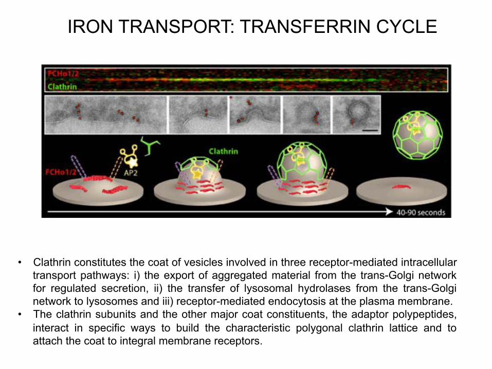
• Clathrin constitutes the coat of vesicles involved in three receptor-mediated intracellular transport pathways: i) the export of aggregated material from the trans-Golgi network for regulated secretion, ii) the transfer of lysosomal hydrolases from the trans-Golgi network to lysosomes and iii) receptor-mediated endocytosis at the plasma membrane.
• The clathrin subunits and the other major coat constituents, the adaptor polypeptides, interact in specific ways to build the characteristic polygonal clathrin lattice and to attach the coat to integral membrane receptors.
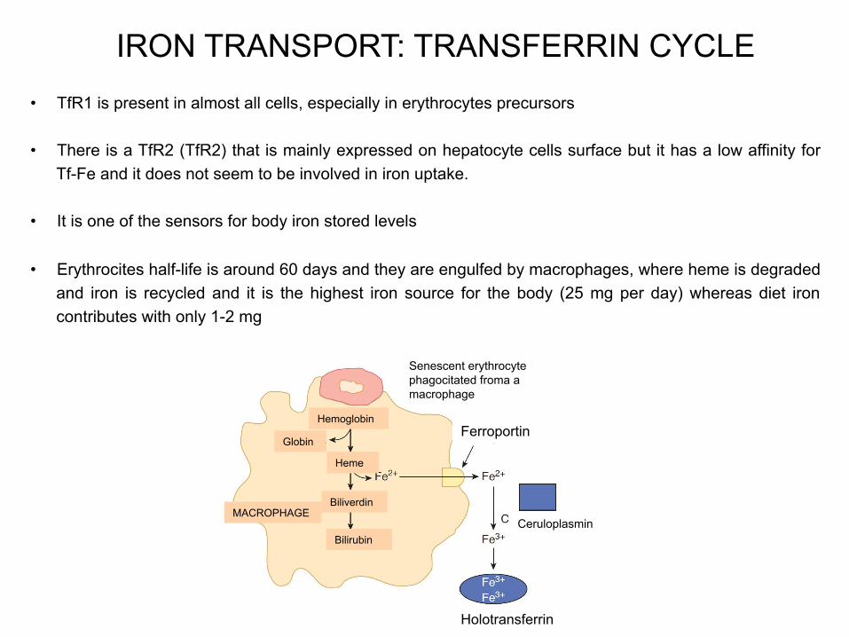
IRON TRANSPORT: TRANSFERRIN CYCLE • TfR1 is present in almost all cells, especially in erythrocytes precursors
• There is a TfR2 (TfR2) that is mainly expressed on hepatocyte cells surface but it has a low affinity for Tf-Fe and it does not seem to be involved in iron uptake.
• It is one of the sensors for body iron stored levels
• Erythrocites half-life is around 60 days and they are engulfed by macrophages, where heme is degraded and iron is recycled and it is the highest iron source for the body (25 mg per day) whereas diet iron contributes with only 1-2 mg
Senescent erythrocyte phagocitated froma a macrophage
Hemoglobin
Globin
Heme
Biliverdin
Bilirubin
MACROPHAGE
Ferroportin
Holotransferrin
Ceruloplasmin
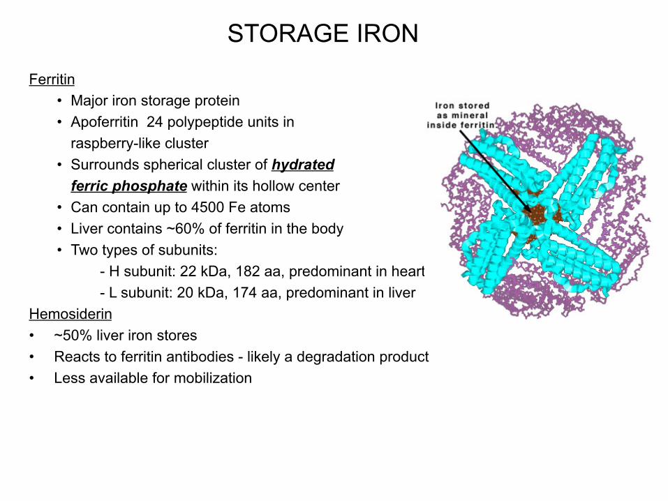
STORAGE IRON
Ferritin • Major iron storage protein • Apoferritin 24 polypeptide units in
raspberry-like cluster • Surrounds spherical cluster of hydrated
ferric phosphate within its hollow center • Can contain up to 4500 Fe atoms • Liver contains ~60% of ferritin in the body • Two types of subunits:
- H subunit: 22 kDa, 182 aa, predominant in heart - L subunit: 20 kDa, 174 aa, predominant in liver
Hemosiderin • ~50% liver iron stores • Reacts to ferritin antibodies - likely a degradation product • Less available for mobilization

REGULATION OF INTRACELLULAR HOMEOSTASIS
• Synthesis of TfR1 and ferritin are linked to the intracellular iron content
• When iron levels are high, ferritin is synthesised for iron storage and TfR1 synthesis is inhibited
• When iron levels are low, ferritin synthesis is blocked whereas TfR1 is active
• Regulation of mRNA stability is involved
• mRNA for ferritin and TfR1 contain iron response elements (IREs) forming hairpins in the untranslated regions at the 5’ and 3’, respectively
• IREs are linked to iron regulatory proteins (IRPs) that are sensitive to intracellular iron levels and induced by low levels of the metal
• IRPs bind to IREs when iron levels are low
• IRPs binding to 5’ UTR mRNA blocks ferritin translation
• IRPs binding to 3’ UTR mRNA stabilises mRNA and increases the synthesis of TfR1.

REGULATION OF INTRACELLULAR HOMEOSTASIS
• Aconitaseisa“moonligh0ng”proteinsinceithasmorethanoneroleinthecell;
• Eukaryoteshave2isoforms:themithochondrialoneispartoftheTCAcycleconver0ngcitrateintoisocitrate
• Thecytosolicisoformhas2roles:1-convertscitrateintoisocitrateprovidingthesubstrateforisocitratedehydrogenasethatgeneratesNADPH2-itpar0cipatedtoironhomeostasis
• AconitasehasaFe-Sclusetrthatdetaches
whenironlevelsarelow
• Theapoenzymehasanewac0vity,sinceitcanbindtomRNAandregulatestheexpressionofofferri0nandandTfr
• Aconitaseisstructurallyiden0caltoIRP1andsimilartoIRP2
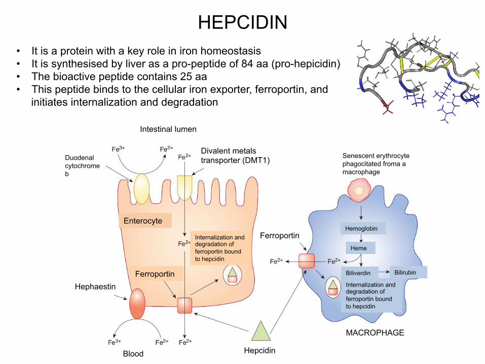
HEPCIDIN • It is a protein with a key role in iron homeostasis • It is synthesised by liver as a pro-peptide of 84 aa (pro-hepicidin) • The bioactive peptide contains 25 aa • This peptide binds to the cellular iron exporter, ferroportin, and
initiates internalization and degradation
Intestinal lumen
Duodenal cytochrome b
Divalent metals transporter (DMT1)
Enterocyte
Ferroportin
Blood
Hephaestin
Hepcidin
Ferroportin
Senescent erythrocyte phagocitated froma a macrophage
Hemoglobin
Heme
Biliverdin Bilirubin
Internalization and degradation of ferroportin bound to hepcidin
MACROPHAGE
Internalization and degradation of ferroportin bound to hepcidin
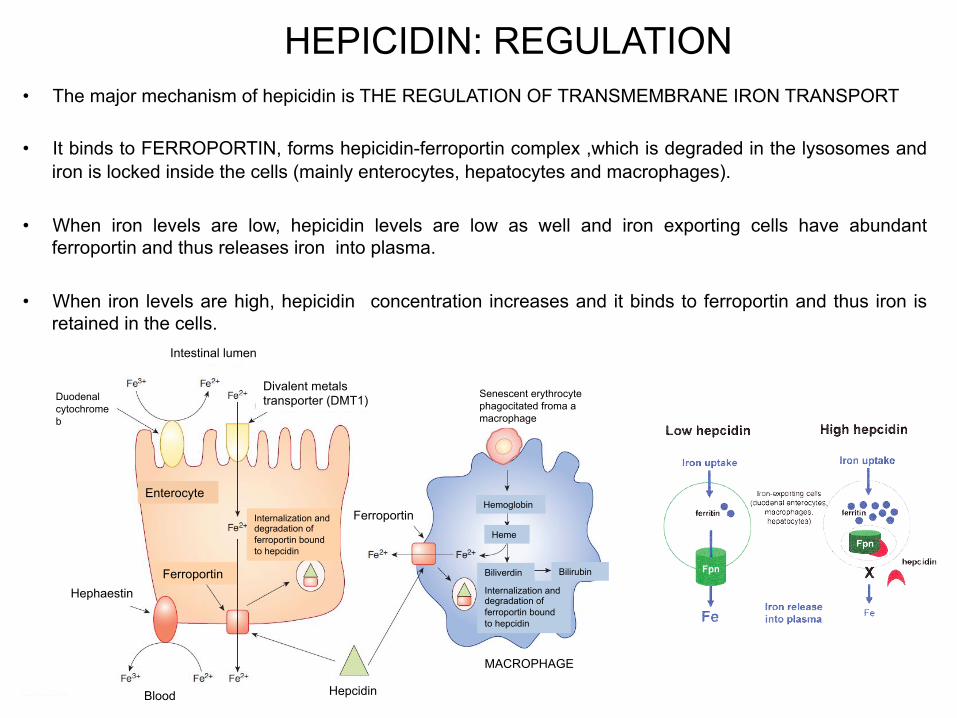
HEPICIDIN: REGULATION • The major mechanism of hepicidin is THE REGULATION OF TRANSMEMBRANE IRON TRANSPORT
• It binds to FERROPORTIN, forms hepicidin-ferroportin complex ,which is degraded in the lysosomes and iron is locked inside the cells (mainly enterocytes, hepatocytes and macrophages).
• When iron levels are low, hepicidin levels are low as well and iron exporting cells have abundant ferroportin and thus releases iron into plasma.
• When iron levels are high, hepicidin concentration increases and it binds to ferroportin and thus iron is retained in the cells.
Intestinal lumen
Duodenal cytochrome b
Divalent metals transporter (DMT1)
Enterocyte
Ferroportin
Blood
Hephaestin
Hepcidin
Ferroportin
Senescent erythrocyte phagocitated froma a macrophage
Hemoglobin
Heme
Biliverdin Bilirubin
Internalization and degradation of ferroportin bound to hepcidin
MACROPHAGE
Internalization and degradation of ferroportin bound to hepcidin


