
1
© Copyright 2012, John P. Fisher, All Rights Reserved
Introduction to Physiology: The Human Body
Adapted From:
Textbook Of Medical Physiology, 11th Ed. Arthur C. Guyton, John E. Hall
Chapter 1
John P. Fisher
© Copyright 2012, John P. Fisher, All Rights Reserved
Organization of the Body Anatomical Directions
Sagittal plane
Transverse plane
Inferior
Superior
Posterior
Frontal plane
Anterior
Lateral
Medial
Lateral
Inferior
Superior
Anterior Posterior

2
© Copyright 2012, John P. Fisher, All Rights Reserved
Organization of the Body Anatomical Directions
• Left To the left of the body
• Right To the right of the body
• Lateral Toward the side, away from the midsagittal plane
• Medial Toward the midsagittal plane, away from the side
• Anterior Toward the front of the body
• Posterior Toward the back (rear) of the body
• Superior Toward the top of the body
• Inferior Toward the bottom of the body
© Copyright 2012, John P. Fisher, All Rights Reserved
Organization of the Body Anatomical Directions
• Dorsal Along or toward the vertebral surface of the body
• Ventral Along or toward the belly surface of the body
• Caudal Toward the tail
• Cephalad Toward the head
• Proximal Toward the trunk
• Distal Away from the trunk
• Visceral Toward an internal organ
• Parietal Toward the wall

3
© Copyright 2012, John P. Fisher, All Rights Reserved
Organization of the Body Structural Hierarchy • Atoms: oxygen, carbon, hydrogen
• Molecules: water, sugar, peptides
• Macromolecules: proteins, polysaccharides
• Organelles: compartments within cell that cannot survive in isolation
• Cells: structural and functional unit of organisms
• Tissues: organization of cells with a common function
• Organs: structures composed of at least 2 tissues
• Organ Systems: organs operating to accomplish a common function
© Copyright 2012, John P. Fisher, All Rights Reserved
Physiology Definition • Study of the characteristics and mechanisms of the human body
• Cells are the basic unit of life within the human body
• Approximately 100 trillion cells make up the typical human, each specially adapted to perform one or a few particular functions • 25 trillion red blood cells act to transport oxygen from the lungs to all tissues in
the body
• All cells have some basic commonalities • Oxygen reacts with carbohydrates, fat, and protein to release energy • Nutrient consumption and energy production mechanisms are similar • Nearly all cells have the ability to reproduce additional, similar cells

4
© Copyright 2012, John P. Fisher, All Rights Reserved
Physiology Extracellular and Intracellular Fluids
• Approximately 60% of the human body is fluid • An aqueous solution containing ions, small molecules, proteins, sugars, and
macromolecules
• Two thirds of the fluid is retained within cells – Intracellular • Intracellular fluid contains large amounts of potassium, magnesium, and
phosphate ions
• One third of the fluid is outside cells – Extracellular • As all cells exist within a similarly constituted extracellular fluid, this space is
sometimes referred to as the internal environment or the milieu interieur • Extracellular fluid contains large amounts of sodium, chloride, and bicarbonate
ions as well as nutrients including oxygen, glucose, fatty acids, and amino acids
© Copyright 2012, John P. Fisher, All Rights Reserved
Homeostatic Mechanisms Homeostasis • Homeostasis describes the active maintenance of an equilibrium state despite
external disturbances
• Homeostasis can be considered in regards to a cell, tissue, organ, biological system, or environmental system
• In physiology, homeostasis implies the maintenance of nearly constant conditions in the internal environment • Actively maintained by organs and tissues
• Lungs provide oxygen consumed by cells, and remove carbon dioxide produced by cells
• Kidneys regulate ion concentrations by augmenting waste composition

5
© Copyright 2012, John P. Fisher, All Rights Reserved
Homeostatic Mechanisms Homeostasis • Extracellular fluid is constantly in motion,
transported in the circulating blood and then mixed between the blood and the tissue fluids by diffusion through the capillary walls
• Blood movement through the circulatory system • 1 circuit per minute at rest • 6 circuits per minute during activity
• Fluid movement between blood capillaries and cells • Few cells are located more than 50
microns away from a capillary
Guyton & Hall. Textbook of Medical Physiology, 11th Edition
© Copyright 2012, John P. Fisher, All Rights Reserved
Organ Systems Nutrient Supply Systems • Respiratory System
• Blood is concentrated with oxygen which is transported through the alveoli in the lungs
• Oxygen diffuses through the alveolar membrane (0.4 - 2.0 µm), a porous membrane that facilitates diffusion
• Gastrointestinal System • Nutrients, including carbohydrates, fatty acids, and amino acids, are
absorbed from ingested food, through the walls of the gastrointestinal tract, and into the extracellular fluid of circulating blood
• Hepatic System • The liver acts to chemically modify ingested, but difficult to absorb, nutrients
into usable forms - other bodily tissues help modify these nutrients or store them until their future use
• Musculoskeletal System • Provides structure and movement that allows the gathering of nutrients as
well as protection from adverse surroundings

6
© Copyright 2012, John P. Fisher, All Rights Reserved
Organ Systems Waste Removal Systems • Respiratory System
• Carbon dioxide is released from the blood stream and into the lung alveoli, where it is then ultimately expired into the atmosphere
• Kidney System • Allows for the removal of waste substances
• Urea and uric acid • Excess ions and water
• Filtration occurs in the kidney by first absorbing large quantities of plasma, and then returning to the blood those substances that are of nutritional value (glucose, amino acids, water, and ions), while excess nutrients or waste products are left behind and ultimately excreted in the urine
© Copyright 2012, John P. Fisher, All Rights Reserved
Organ Systems Regulatory Systems • Nervous System
• Contains three major constituents • Sensory input system: detects the state of the body and surrounding
environment • Touch, sight, hearing
• Central nervous system: stores information, generates thoughts, and determines reactions in response to the sensory input system
• Composed of the brain and spinal cord • Motor output portion: allows for the generation of actions based upon
the signals provided by the central nervous system • Motor neurons which drive muscle actions
• Autonomic System • Operates at a subconscious level to control the function of internal
organs, including heart, gastrointestinal tract, and glandular secretions • Hormonal System
• Eight major endocrine glands secrete hormones that are transported throughout the body to help regulate cellular function
7
© Copyright 2012, John P. Fisher, All Rights Reserved
Organ Systems Reproductive System • Provides a mechanism to maintain the overall population of the species, thus
allowing for population homeostasis
© Copyright 2012, John P. Fisher, All Rights Reserved
Control Systems Maintaining Homeostasis • The maintenance of homeostasis requires the activity of a number of different control
systems • React to the surrounding environment • Initiate actions to maintain cell, tissue, organ, and organ system function
8
© Copyright 2012, John P. Fisher, All Rights Reserved
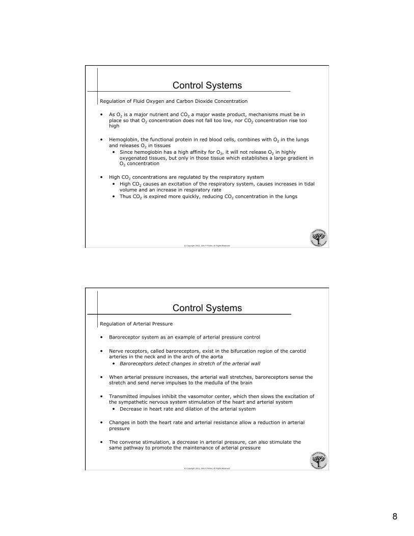
Control Systems Regulation of Fluid Oxygen and Carbon Dioxide Concentration • As O2 is a major nutrient and CO2 a major waste product, mechanisms must be in
place so that O2 concentration does not fall too low, nor CO2 concentration rise too high
• Hemoglobin, the functional protein in red blood cells, combines with O2 in the lungs and releases O2 in tissues • Since hemoglobin has a high affinity for O2, it will not release O2 in highly
oxygenated tissues, but only in those tissue which establishes a large gradient in O2 concentration
• High CO2 concentrations are regulated by the respiratory system • High CO2 causes an excitation of the respiratory system, causes increases in tidal
volume and an increase in respiratory rate • Thus CO2 is expired more quickly, reducing CO2 concentration in the lungs
© Copyright 2012, John P. Fisher, All Rights Reserved
Control Systems Regulation of Arterial Pressure • Baroreceptor system as an example of arterial pressure control
• Nerve receptors, called baroreceptors, exist in the bifurcation region of the carotid arteries in the neck and in the arch of the aorta • Baroreceptors detect changes in stretch of the arterial wall
• When arterial pressure increases, the arterial wall stretches, baroreceptors sense the stretch and send nerve impulses to the medulla of the brain
• Transmitted impulses inhibit the vasomotor center, which then slows the excitation of the sympathetic nervous system stimulation of the heart and arterial system • Decrease in heart rate and dilation of the arterial system
• Changes in both the heart rate and arterial resistance allow a reduction in arterial pressure
• The converse stimulation, a decrease in arterial pressure, can also stimulate the same pathway to promote the maintenance of arterial pressure

9
© Copyright 2012, John P. Fisher, All Rights Reserved
Control Systems Regulation of Extracellular Environment
• Normal ranges are small, typically caused by illness
• Death can occur by persistent, larger deviations • Temperature, acidity, potassium, calcium, and glucose
Normal Value
Normal Range
Short Term Non-Lethal Limit
Unit
Oxygen 40 35 - 45 10 - 1000 mmHg
Carbon Dioxide 40 35 - 45 5 - 80 mmHg
Sodium Ion 142 138 - 146 115 - 175 mmol/L
Potassium Ion 4.2 3.8 - 5.0 1.5 - 9.0 mmol/L
Calcium Ion 1.2 1.0 - 1.4 0.5 - 2.0 mmol/L
Chloride Ion 108 103 - 112 70 - 130 mmol/L
Bicarbonate Ion 28 24 - 32 8 - 45 mmol/L
Glucose 85 75 - 95 20 - 1500 mmol/L
Temperature 37.0 36.7 - 37.1 18.3 - 43.3 °C
[H+] 7.4 7.3 - 7.5 6.9 - 8.0 pH
Normal Value
© Copyright 2012, John P. Fisher, All Rights Reserved
Control Systems Negative Feedback Systems • Most control systems of the body act by negative feedback
• A stimulus causes a reaction that opposes the acting stimulus • Increased CO2 causes increased pulmonary ventilation, which decreases CO2 • Decreased arterial pressure activates the baroreceptor system which acts
increase heart rate and arterial constriction, which increases arterial pressure
• The negative feedback system acts to maintain homeostasis
homeostasis
stimulus
response
negative feedback response

10
© Copyright 2012, John P. Fisher, All Rights Reserved
Control Systems Gain of Control Systems • The gain of a control system is a parameter which describes the degree of
effectiveness with which a control system can maintain constant conditions • Gain = Correction / Error
• Example • In a normal person with a functioning baroreceptor control system, a defined
stimulus causes arterial pressure to increase from 100 mmHg to 125 mmHg • Error is +25 mmHg - if the baroreceptor system provided perfect control, there
would be no change in arterial pressure • A person with a non-functioning baroreceptor control system, the same stimulus
causes arterial pressure to increase from 100 mmHg to 175 mmHg • Difference from the normal response is 50 mmHg • Thus, the baroreceptor system provides a correction of -50 mmHg
• Gain = -50 mmHg / +25 mmHg = -2 • The baroreceptor system reduces the effect of the stimulus by two thirds
© Copyright 2012, John P. Fisher, All Rights Reserved
Control Systems of the Body Positive Feedback • In a positive feedback control system, a stimulus causes a responses that promotes
the stimulus
• In general, positive feedback systems lead to instability and therefore are not utilized as often as negative feedback systems
homeostasis
stimulus
response
positive feedback response

11
© Copyright 2012, John P. Fisher, All Rights Reserved
Control Systems of the Body Positive Feedback • Positive feedback systems do occur in the body
• Blood clotting • A rupture in a blood vessel initiates a clot formation, and enzyme activation
within the clot causes other enzymes in the blood to clot • The cycle continues until the vessel in plugged and bleeding stops
• Uterine contractions in childbirth • Sodium ion flux in nerve signal propagation
• Typically, positive feedback control systems work within a larger negative feedback control system • For example, the blood clotting cycle works within the maintenance of blood
volume negative feedback cycle
© Copyright 2012, John P. Fisher, All Rights Reserved
Control Systems of the Body Adaptive Control
• Many of the control systems developed to maintain homeostasis are not simple negative feedback systems, but more complex communicating networks
• Adaptive control systems change their response each time a stimulus is presented until the proper response is determined • Some body movements require rapid responses that cannot wait for signal
transmission to the central nervous system and the subsequent response • Sensory nerve signals from the moving parts transmit signals to the brain as
to whether the movement was properly completed • If not, the brain sends signals so that the next response is altered • This continues until the proper response is obtained
12
© Copyright 2012, John P. Fisher, All Rights Reserved
Introduction to Physiology: The Cell
Adapted From:
Textbook Of Medical Physiology, 11th Ed. Arthur C. Guyton, John E. Hall
Chapter 2
John P. Fisher
© Copyright 2012, John P. Fisher, All Rights Reserved
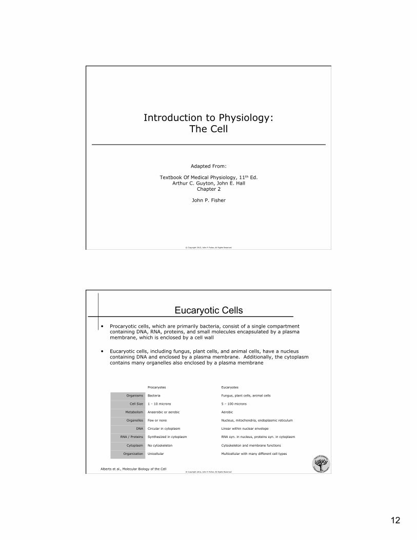
Eucaryotic Cells • Procaryotic cells, which are primarily bacteria, consist of a single compartment
containing DNA, RNA, proteins, and small molecules encapsulated by a plasma membrane, which is enclosed by a cell wall
• Eucaryotic cells, including fungus, plant cells, and animal cells, have a nucleus containing DNA and enclosed by a plasma membrane. Additionally, the cytoplasm contains many organelles also enclosed by a plasma membrane
Procaryotes Eucaryotes
Organisms Bacteria Fungus, plant cells, animal cells
Cell Size 1 – 10 microns 5 – 100 microns
Metabolism Anaerobic or aerobic Aerobic
Organelles Few or none Nucleus, mitochondria, endoplasmic reticulum
DNA Circular in cytoplasm Linear within nuclear envelope
RNA / Proteins Synthesized in cytoplasm RNA syn. in nucleus, proteins syn. in cytoplasm
Cytoplasm No cytoskeleton Cytoskeleton and membrane functions
Organization Unicellular Multicellular with many different cell types
Alberts et al., Molecular Biology of the Cell
13
© Copyright 2012, John P. Fisher, All Rights Reserved
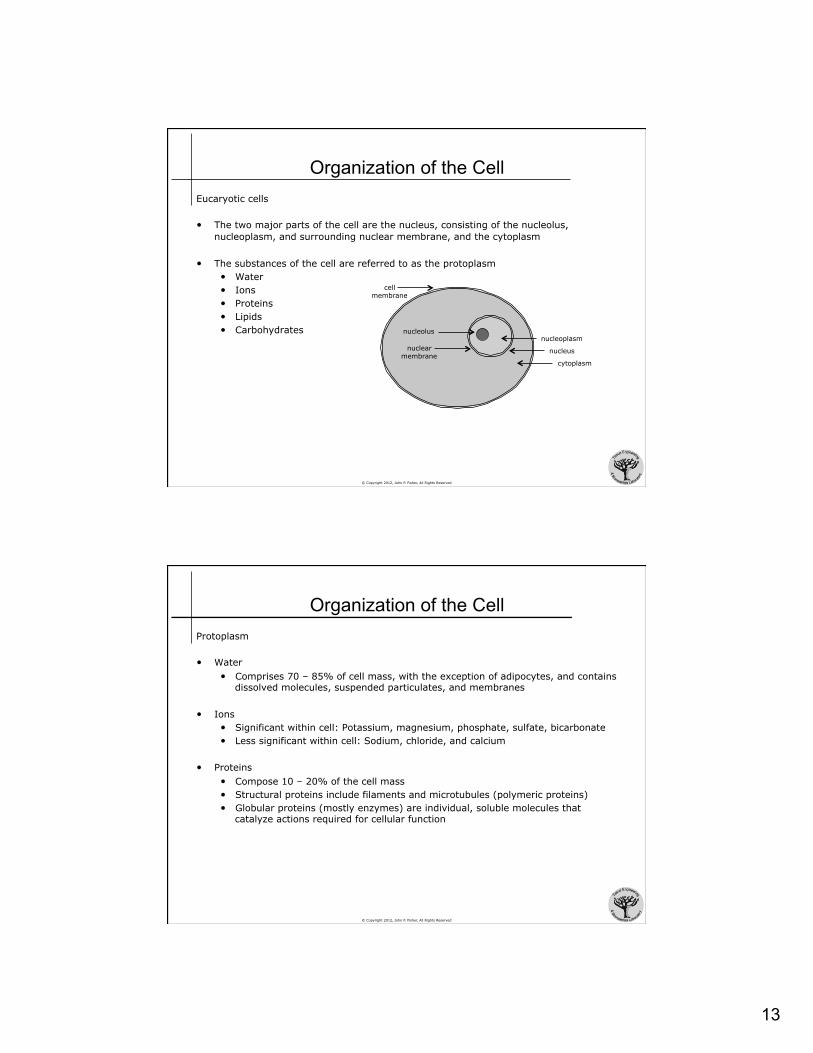
Organization of the Cell Eucaryotic cells
• The two major parts of the cell are the nucleus, consisting of the nucleolus, nucleoplasm, and surrounding nuclear membrane, and the cytoplasm
• The substances of the cell are referred to as the protoplasm • Water • Ions • Proteins • Lipids • Carbohydrates
cell membrane
nucleolus
nuclear membrane
cytoplasm
nucleoplasm
nucleus
© Copyright 2012, John P. Fisher, All Rights Reserved
Organization of the Cell Protoplasm
• Water • Comprises 70 – 85% of cell mass, with the exception of adipocytes, and contains
dissolved molecules, suspended particulates, and membranes
• Ions • Significant within cell: Potassium, magnesium, phosphate, sulfate, bicarbonate • Less significant within cell: Sodium, chloride, and calcium
• Proteins • Compose 10 – 20% of the cell mass • Structural proteins include filaments and microtubules (polymeric proteins) • Globular proteins (mostly enzymes) are individual, soluble molecules that
catalyze actions required for cellular function

14
© Copyright 2012, John P. Fisher, All Rights Reserved
Organization of the Cell Protoplasm • Lipids
• Molecules that are hydrophobic / “fat soluble” / lipophilic • Phospholipids and cholesterol are the most significant (2% of cell mass)
• Form cell membranes and intracellular membrane barriers • Triglycerides are the fat stored by adipocytes as an energy source
• Carbohydrates • Lesser in content, comprising 1 – 6% of cell mass, depending upon cell type • Dissolved glucose is readily available in extracellular fluid • An insoluble glucose polymer, glycogen, is found intracellularly
© Copyright 2012, John P. Fisher, All Rights Reserved
Eucaryotic Cell
Guyton & Hall. Textbook of Medical Physiology, 11th Edition

15
© Copyright 2012, John P. Fisher, All Rights Reserved
Eucaryotic Cell Cell Membrane • Thin, elastic structure approximately 7.5 – 10 nm thick • Composed of proteins and lipids
• 55% proteins • 25% phospholipids • 13% cholesterol • 4% other lipids • 3% carbohydrates
• Basic structure is a lipid bilayer • Composed of two opposing layers of lipids • Hydrophilic head adjacent to the extracellular and
intracellular water • Hydrophilic head is made up of phosphates
• Hydrophobic (lipophilic) tail pointed inwards • Hydrophobic tail is made up of fatty acids
• Interspersed within the lipid bilayer are large globular proteins
hydrophilic head group hydrophobic tail group
lipid micelle
lipid bilayer
lipid vesicle
hydrophilic head group hydrophobic tail group
© Copyright 2012, John P. Fisher, All Rights Reserved
Eucaryotic Cell Cell Membrane • The membrane has selective barrier properties
• Water soluble molecules are excluded • Ions, glucose, and urea are excluded
• Fat soluble molecules are permitted • Oxygen, carbon dioxide, and alcohol
can pass though the membrane easily
• The membrane is not static, but fluid – allowing lipid molecules and proteins to move within the two dimensional surface of the cell
• Cholesterol contains a highly fat soluble core, thus storing the molecule in the lipid bilayer • Cholesterol augments the permeability and
fluidity of the bilayer Guyton & Hall. Textbook of Medical Physiology, 11th Edition

16
© Copyright 2012, John P. Fisher, All Rights Reserved
Eucaryotic Cell Cell Membrane Proteins • The membrane contains many globular proteins, most of which are glycoproteins
• Integral proteins protrude both side of the lipid bilayer • Peripheral proteins exist only on one side
• Typically attached to integral proteins and act to regulate their function
• Many integral proteins provide structural channels • Allow for the selective transport of water soluble molecules, especially ions • Transport may be actively facilitated by carrier proteins, or passively allowed by
pores or channels
• Other integral proteins acts as receptors for water soluble chemicals • Interaction of the cell surface bound receptor with specific ligands cause the
protein to undergo a conformation change that enzymatically activates an intracellular portion of the protein
• Second messengers in the cytoplasm then relay this activated state onto other participants within the cell
© Copyright 2012, John P. Fisher, All Rights Reserved
Eucaryotic Cell Cell Membrane Carbohydrates • Membrane carbohydrates generally occur in combination with proteins or lipids, in
the form of glycoproteins or glycolipids • Most integral proteins are glyoproteins • On tenth of the membrane lipids are glycolipids • Proteoglycans, carbohydrates with protein cores, are also present
• The entire surface of the cell therefore has a loose carbohydrate coat called glycocalyx • Typically have a net negative charge that repels other negatively charged
objects, including other cells, at moderate distances • Glycocalyx is “sticky” allowing close contact (attachment) between cells • Acts as receptors substances for hormones • Augment immune responses

17
© Copyright 2012, John P. Fisher, All Rights Reserved
Eucaryotic Cell Nucleus
• The nucleus is the container of DNA in eukaryotic cells
• DNA condenses into chromosomes • Humans have 6x10⁹ nucleotide
pairs organized into 46 chromosomes, 2 of which are sex chromosomes
• The 46 chromosome molecules vary in size from 50x106 to 250x106 nucleotide pairs
• Humans, and all diploid organisms, have two copies of each chromosome (maternal and paternal) except for the sex chromosomes, thus humans have 24 different chromosomes
Guyton & Hall. Textbook of Medical Physiology, 11th Edition
© Copyright 2012, John P. Fisher, All Rights Reserved
Eucaryotic Cell Nucleus • The nucleus is enveloped by a nuclear
membrane, which consists of two lipid bilayers
• The outer membrane is continuous with the ER, and the space within the two lipid bilayers of the nuclear membrane is continuous with the space within the ER
Alberts et al., Molecular Biology of the Cell

18
© Copyright 2012, John P. Fisher, All Rights Reserved
Eucaryotic Cell Nuclear Membrane • Several thousand nuclear pores
penetrate both membranes • Pores consist of protein complexes
> 100 nm in diameter, but with an effective pore size of about 9 nm
• Molecules < 44 kDa can pass through the pore, molecules < 15 kDa pass into the nucleus extremely rapidly
• Most nuclei contain one or highly staining structures called the nucleoli • Is not defined by a lipid membrane • Contains large amounts of RNA and
proteins, similar to those found in ribosomes
Alberts et al., Molecular Biology of the Cell
© Copyright 2012, John P. Fisher, All Rights Reserved
Eucaryotic Cell Endoplasmic Reticulum
• Interconnected tubular and flat vesicular structures, with extremely high surface area made up of a protein laden lipid bilayer membrane • Up to 30 - 40 times the surface area of the cell membrane
• The space within the tubules and vesicles is filled with endoplasmic matrix • Space is continuous with the space within the nuclear membrane
• Functions in protein synthesis, as some substances enter the ER
• Functions in metabolism, as the surface is covered metabolic proteins
Alberts et al. Molecular Biology of the Cell

19
© Copyright 2012, John P. Fisher, All Rights Reserved
Eucaryotic Cell Endoplasmic Reticulum • Granular endoplasmic reticulum has a surface packed with attached ribosomes
• Ribosomes, formed from RNA and proteins, are responsible for synthesizing new proteins
• Smooth endoplasmic reticulum (SER) has no attached ribosomes • SER functions in the synthesis of lipid substances as well as other enzymatic
processes of the cell
Guyton & Hall. Textbook of Medical Physiology, 11th Edition
© Copyright 2012, John P. Fisher, All Rights Reserved
Eucaryotic Cell Functions of the Endoplasmic Reticulum
• Protein Formation • Proteins are formed by the granular endoplasmic reticulum • Proteins are formed by ribosomes, where they are extruded by ribosomes
through the walls of the endoplasmic reticulum to the interior of endoplasmic vesicles and tubules, known as the endoplasmic matrix
• Enzymes in the endoplasmic matrix rapidly reconfigure the proteins by conjugation with carbohydrate molecules (glycosylation) as well as crosslinking, folding, and cleaving
• Free proteins (not glycoproteins) are formed in the cytosol by ribosomes not bound to the endoplasmic reticulum
• Lipid Formation • Lipids, especially phospholipids and cholesterol, are formed by the smooth
endoplasmic reticulum • These molecules generally are incorporated into the wall of the endoplasmic
reticulum, so that the organelle is continually increasing in size • Endoplasmic reticulum vesicles break away from the SER and mostly migrate to
the Golgi apparatus

20
© Copyright 2012, John P. Fisher, All Rights Reserved
Eucaryotic Cell Golgi Apparatus • Composed of four or more stacked layers of thin, flat enclosed vesicles lying near the
nucleus
• Prominent in secretory cells where it is located on the side of the cell from which secretory substances are released
• Golgi apparatus functions with the ER, where transport vesicles bud off the ER and diffuse to the Golgi apparatus – the Golgi apparatus processes ER vesicles to form lysosomes, secretory vesicles, or other cytoplasmic components
Guyton & Hall. Textbook of Medical Physiology, 11th Edition Alberts et al., Molecular Biology of the Cell
© Copyright 2012, John P. Fisher, All Rights Reserved
Eucaryotic Cell
Guyton & Hall. Textbook of Medical Physiology, 11th Edition
Functions of the Golgi Apparatus
• Involved in the formation of some large saccharide polymers bound with a small amount of protein, such as hyaluronic acid and chondroitin sulfate
• Vesicle formation • Vesicles from the ER, containing newly
synthesized proteins, fuse with the proximal portion of the Golgi, releasing their proteins into the vesicular space
• The Golgi glycosylates and concentrates the proteins as they move distally
• Finally vesicles, containing functional proteins, bud off the Golgi and diffuse throughout the cell
• Time Line: Nutrient amino acids are taken up and synthesized into proteins in the granular ER in 3-5 min, present in the Golgi in 20 min, and secreted from the cell in 1 -2 hr

21
© Copyright 2012, John P. Fisher, All Rights Reserved
Eucaryotic Cell Vesicles: Lysosomes • Small, 250 – 750 nm in diameter
• Surrounded by a lipid bilayer
• Filled with 5 to 9 nm granules • Granules are aggregates of hydrolases • Provide an intracellular digestive system • Digests damaged cellular structures, food particles that have been ingested by
the cell, and unwanted matter (bacteria)
© Copyright 2012, John P. Fisher, All Rights Reserved
Eucaryotic Cell Vesicles: Peroxisomes • Similar to lysosomes, except
• Formed from the SER or self replication - not from the Golgi apparatus • Contain oxidases, rather than hydrolases
• Combine oxygen with hydrogen ions to for hydrogen peroxide (H2O2) • Hydrogen peroxide, in conjucntion with catalase, oxidizes many substances
that may be cytotoxic
Vesicles: Secretory Vesicles • Almost all secretory substances are formed from the ER-Golgi apparatus system and
then released from the Golgi into the cytoplasm in the form of storage vesicles known as secretory vesicles or secretory granules
Guyton & Hall. Textbook of Medical Physiology, 11th Edition

22
© Copyright 2012, John P. Fisher, All Rights Reserved
Eucaryotic Cell Endocytosis
• Most substances pass through the cell membrane by diffusion and active transport
• Very large particles enter the cell by endocytosis
• Two forms of endocytosis exist: pinocytosis and phagocytosis
© Copyright 2012, John P. Fisher, All Rights Reserved
Eucaryotic Cell Endocytosis: Pinocytosis
• Ingestion of extremely small vesicles (100 - 200 nm) that contain extracellular fluid
• Occurs continually at the cell membrane
• Mechanism • Target protein binds to receptors,
concentrated in coated pits • Clathrin molecules, intracellular to the
receptors, initiate an invagination process utilizing actin and myosin contractile filaments
• Finally, a pinocytic vesicle buds off into the cytoplasm of the cell
• Consumes ATP and requires extracellular calcium ions
Guyton & Hall. Textbook of Medical Physiology, 11th Edition

23
© Copyright 2012, John P. Fisher, All Rights Reserved
Eucaryotic Cell Endocytosis: Phagocytosis
• Largely similar, though it involves the consumption of large particles (rather than molecules) and utilizes a separate system of receptor and signaling proteins
• Occurs only in a few cells • Tissue macrophages and white blood cells
• Initiated by particulate binding on the cell surface • In the immune response, particulates are typically covered by antibodies and the
antibodies initiate cell surface binding • The intermediation of antibodies is called opsonization
© Copyright 2012, John P. Fisher, All Rights Reserved
Eucaryotic Cell Endocytosis: Cell Digestion
• The entry of nutrients or particles into the cell by pinocytosis or phagocytosis is followed by an accumulation as well as attachment of lysosomes
• Lysosomes release hydrolases into the vesicle, forming a digestive vesicle
• The hydrolytic products (amino acids, glucose, phosphates) then diffuse through the membrane of the vesicle into the cytoplasm of the cell
• The remaining byproducts are not digestible, and are excreted by the vesicle out of the cell by a process known as exocytosis – essentially the opposite of endocytosis
Guyton & Hall. Textbook of Medical Physiology, 11th Edition

24
© Copyright 2012, John P. Fisher, All Rights Reserved
Eucaryotic Cell Endocytosis: Cell Digestion
• Digestive processes are utilized to reduce a tissues size • Uterus after pregnancy • Skeletal muscles after inactivity
• Digestive processes are also used to eliminate a portion or entire cell after damage • Lysosomes rupture in response to damage,
releasing hydrolases into the cytoplasm of the damaged cell (autolysis)
Guyton & Hall. Textbook of Medical Physiology, 11th Edition
© Copyright 2012, John P. Fisher, All Rights Reserved
Eucaryotic Cell Mitochondria • Powerhouse of the cell, extracting energy from nutrients and oxygen • Present in all portions of the cytoplasm, varying in number from less than 100 to
1000s, depending upon cell type • Enveloped by two lipid bilayers – an outer membrane and an inner membrane • Oxidative enzymes within and on the inner membrane oxidize nutrients to form
carbon dioxide and water as well as energy in the form of adenosine triphosphate (ATP) • ATP then provides energy throughout the cell by phosphate based reactions
• Mitochondria contain their own DNA and are self replicative, forming additional mitochondria as the energy needs of the cell increase
Guyton & Hall. Textbook of Medical Physiology, 11th Edition

25
© Copyright 2012, John P. Fisher, All Rights Reserved
Eucaryotic Cell
Guyton & Hall. Textbook of Medical Physiology, 11th Edition
Functions of the Mitochondria
• Extraction of energy from nutrients • Sources of nutrients are oxygen,
carbohydrates, fats, and proteins • Carbohydrates are converted to glucose • Fats are converted to fatty acids • Proteins are converted to amino acids
• These broken down nutrients can then be absorbed by the cell
• Nutrients are reacted with oxygen under the influence of various enzymes (oxidative reactions) to produce energy
• Oxidative reactions occur within the mitochondrion
© Copyright 2012, John P. Fisher, All Rights Reserved
Eucaryotic Cell Functions of the Mitochondria
• Energy released is captured in the form of the nucleotide adenosine triphosphate (ATP) • Nitrogenous base adenine • Pentose sugar ribose • Three phosphate radicals
• Last two phosphates are held by a high energy phosphate bond
• Each of these bonds contains about 12 Kcal per mol of ATP
• When ATP releases energy, it releases a phosphate group and becomes adenosine diphosphate (ADP)
• When energy is stored by the cell, it does so by reconstituting ATP from ADP
N
NN
N
H2N
O
OP
OP
OP
O-
O
O-
O
O-
O
O-
OHOH
adenine
ribose
phosphate
Adenosine Triphosphate (ATP)

26
© Copyright 2012, John P. Fisher, All Rights Reserved
Eucaryotic Cell Functions of the Mitochondria • Glycolysis
• Glucose enters the cell cytoplasm and then is converted to pyruvic acid by glycolysis
• As a product of this reaction, ADP is converted to ATP
• However, glycolysis accounts for only 5 percent of the cell’s energy needs
• Citric Acid Cycle
• Pyruvic acid, from all nutrients, is converted into acetyl-CoA in the mitochondrion
• As a byproduct, hydrogen atoms are released
• These hydrogen atoms bind with dissolved oxygen, releasing energy that converts ADP to ATP
• This mitochondrial process forms the major portion of ATP
Guyton & Hall. Textbook of Medical Physiology, 11th Edition
© Copyright 2012, John P. Fisher, All Rights Reserved
Eucaryotic Cell Functions of the Mitochondria • ATP is utilized in three general classed of
cellular functions • Membrane transport of substances
• Sodium, potassium, calcium magnesium, phosphate, cholride urate and hydrogen ions
• Synthesis of chemical compounds • Proteins, phospholipids, cholesterol,
purines, pyrimidines • Can reach up to 75% of ATP utilization
• Mechanical work • Muscle fiber contraction, ciliary motion
Guyton & Hall. Textbook of Medical Physiology, 11th Edition
27
© Copyright 2012, John P. Fisher, All Rights Reserved
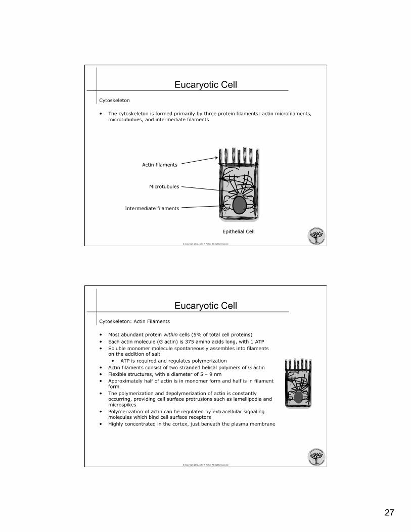
Eucaryotic Cell Cytoskeleton
• The cytoskeleton is formed primarily by three protein filaments: actin microfilaments, microtubulues, and intermediate filaments
Actin filaments
Microtubules
Intermediate filaments
Epithelial Cell
© Copyright 2012, John P. Fisher, All Rights Reserved
Eucaryotic Cell Cytoskeleton: Actin Filaments
• Most abundant protein within cells (5% of total cell proteins) • Each actin molecule (G actin) is 375 amino acids long, with 1 ATP • Soluble monomer molecule spontaneously assembles into filaments
on the addition of salt • ATP is required and regulates polymerization
• Actin filaments consist of two stranded helical polymers of G actin • Flexible structures, with a diameter of 5 – 9 nm • Approximately half of actin is in monomer form and half is in filament
form • The polymerization and depolymerization of actin is constantly
occurring, providing cell surface protrusions such as lamellipodia and microspikes
• Polymerization of actin can be regulated by extracellular signaling molecules which bind cell surface receptors
• Highly concentrated in the cortex, just beneath the plasma membrane

28
© Copyright 2012, John P. Fisher, All Rights Reserved
Eucaryotic Cell Cytoskeleton: Microtubules
• Long hollow cylinders made of the protein tubulin • Outer diameter of approximately 25 nm • Stiff structures that usually have one end anchored in the microtubule
organizing center (centrosome) and the other end free in the cytoplasm
• More rigid than actin • Highly dynamic structures that alternately grow and shrink by the
addition and loss of tubulin subunits • Motor proteins move along microtubules to carry specific membrane
bounded organelles to desired locations in the cell • Kinesins move toward the + end (cytoplasm) of the microtubule • Dyneins move toward the – end (centrosome) of the microtubule
© Copyright 2012, John P. Fisher, All Rights Reserved
Eucaryotic Cell Cytoskeleton: Intermediate Filaments
• Rope like fibers with a diameter of approximately 10 nm • Resist tensional forces • Monomers of different intermediate filaments differ in amino acid
sequence and molecular weight • All contain a dimer-forming, extended coiled coil structure
• Monomer pairs to form coiled coil dimer • Coiled coil dimers align to form filaments
• Major Types of Intermediate Filaments • Nuclear Lamins Found in nuclear lamina • Vimentinlike Proteins Found in mesenchymal cells • Keratins Found in epithelial cells • Neuronal I.F. Found in neurons

29
© Copyright 2012, John P. Fisher, All Rights Reserved
Eucaryotic Cell Locomotion of Cells
• Ameboid movement • Movement of entire cell relative to the
surroundings, involving • Continual formation of new cell
membrane at the pseudopodium • Receptor mediated attachment • Force generation, perhaps by actin
polymerization
• Ciliary Movement • Whip-like movement of cilia (small hairs)
on the surface of cells • In humans, it occurs only in the
respiratory airways and fallopian tubes • Requires ATP, calcium, and magnesium
Guyton & Hall. Textbook of Medical Physiology, 11th Edition
Guyton & Hall. Textbook of Medical Physiology, 11th Edition
© Copyright 2012, John P. Fisher, All Rights Reserved
Genetic Control of Cell Functions
Adapted From:
Textbook Of Medical Physiology, 11th Ed. Arthur C. Guyton, John E. Hall
Chapter 3
John P. Fisher

30
© Copyright 2012, John P. Fisher, All Rights Reserved
Cell Function What do cells do?
• The predominate task of a cell is to synthesize compounds, and largely proteins
• Cells are classified by the proteins they synthesize • Osteoblasts synthesize proteins that makeup the organic phase of bone • Macrophages synthesize proteins that assist in fighting foreign bodies
• A cell’s phenotype is often described by its protein expression
• In principle, all the cells of an organism have the same genetic information, but what differs between cells is the proteins they synthesize
© Copyright 2012, John P. Fisher, All Rights Reserved
Cell Function
DNA
RNA
transcription
cellular membrane
cellular functions
extracellular functions
transcriptional control
RNA processing control
RNA transport control
mRNA degradation & translation control
protein activity control
mRNA AAAAAA
nuclear membrane
protein
ribosomes translation
AAAAAA
31
© Copyright 2012, John P. Fisher, All Rights Reserved
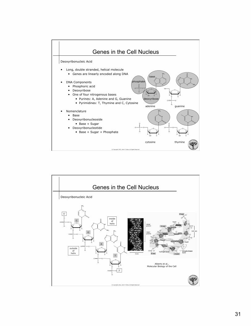
Genes in the Cell Nucleus Deoxyribonucleic Acid
• Long, double stranded, helical molecule • Genes are linearly encoded along DNA
• DNA Components • Phosphoric acid • Deoxyribose • One of four nitrogenous bases
• Purines: A, Adenine and G, Guanine • Pyrimidines: T, Thymine and C, Cytosine
• Nomenclature • Base • Deoxyribonucleoside
• Base + Sugar • Deoxyribonucleotide
• Base + Sugar + Phosphate
-O
N
NN
N
NH2
O
HOH
HH
HH
OP
O-
O
adenine
HO
NH
N
N
O
NH2N
O
H
HH
HHO
PO
O-
O-
guanine
-O
N
NH2
ON
O
HOH
HH
HH
OP
O-
O
cytosine
-O
NH
O
ON
O
HOH
HH
HH
OP
O-
O
thymine
phosphate
deoxyribose
base
© Copyright 2012, John P. Fisher, All Rights Reserved
Genes in the Cell Nucleus Deoxyribonucleic Acid
N
NN
N
NH2
O
HO
HH
HH
PO
O-
O
NH
N
N
O
NH2N
O
H
HH
HHO
PO
O-
O
N
NH2
ON
O
HO
HH
HH
PO
O-
O
O
PO
O-
O
5'
O
HO
HH
HH
PO
O-
O 3"
NH
O
ON
5’
3’
C
G
A
T
outside of
helix
inside of
helix
Alberts et al., Molecular Biology of the Cell

32
© Copyright 2012, John P. Fisher, All Rights Reserved
© Copyright 2012, John P. Fisher, All Rights Reserved
Genes in the Cell Nucleus Deoxyribonucleic Acid • The two strands of the DNA double helix are held
together by specific hydrogen bonding between complementary bases, a purine with a pyrimidine • Adenine binds only with Thymine with 2 H bonds • Guanine binds only with Cytosine with 3 H bonds
• Due to the loose hydrogen bonding of DNA, the double helix can separate into two strands easily
• The double helix is formed by a rotational form resulting from the base bonding • 10 bases are included within one complete turn of
the double helix • Major and minor grooves are created in the helix
due to the unequal spacing of base pairs about the phosphate – sugar backbone
Alberts et al., Molecular Biology of the Cell
33
© Copyright 2012, John P. Fisher, All Rights Reserved
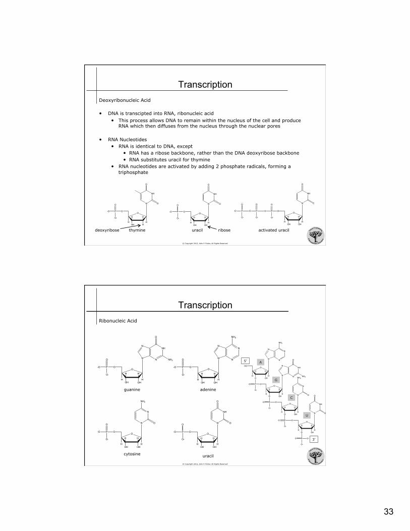
Transcription Deoxyribonucleic Acid
• DNA is transcipted into RNA, ribonucleic acid • This process allows DNA to remain within the nucleus of the cell and produce
RNA which then diffuses from the nucleus through the nuclear pores
• RNA Nucleotides • RNA is identical to DNA, except
• RNA has a ribose backbone, rather than the DNA deoxyribose backbone • RNA substitutes uracil for thymine
• RNA nucleotides are activated by adding 2 phosphate radicals, forming a triphosphate
-O
NH
O
ON
O
HOH
HH
HH
OP
O-
O
-O
NH
O
ON
O
OHOH
HH
HH
OP
O-
O
thymine uracil ribose deoxyribose
O
NH
O
ON
O
OHOH
HH
HH
OP
O-
O
O P
O-
O
-O P
O-
O
activated uracil
© Copyright 2012, John P. Fisher, All Rights Reserved
Transcription Ribonucleic Acid
-O
NH
N
N
O
NH2N
O
OH
HH
HHOH
OP
O
O-
-O
N
NN
N
NH2
O
OHOH
HH
HH
OP
O-
O
-O
N
NH2
ON
O
OHOH
HH
HH
OP
O-
O
-O
NH
O
ON
O
OHOH
HH
HH
OP
O-
O
uracil
adenine guanine
cytosine
HO
N
NN
N
NH2
O
OHO
HH
HH
PO
O-
O
NH
N
N
O
NH2N
O
OH
HH
HHO
PO
O-
O
N
NH2
ON
O
OHO
HH
HH
PO
O-
O
NH
O
ON
O
OHO
HH
HH
PO
O-
O-
5’
3’
C
G
A
U

34
© Copyright 2012, John P. Fisher, All Rights Reserved
Transcription Ribonucleic Acid
• RNA polymerase recognizes a specific DNA sequence, upstream of the gene, known as the promoter
• RNA polymerase binds the DNA promoter • Polymerase binds initiates DNA unwinding for
approximately 20 bases • DNA strands temporarily separate and one
of the strands is then used as a template for RNA synthesis while the other remains inactive
• Polymerase moves down the DNA strand • Unwinding continues • Polymerase adds RNA nucleotides to the
end of the newly forming RNA chain
Alberts et al., Molecular Biology of the Cell
• First, H bonding the DNA base and the complementary RNA base • Second, polymerase breaks 2 of the high energy phosphate bonds on a
RNA nucleotide, utilizing the released energy to cause a covalent linkage of the remaining phosphate with the ribose on the end of the growing RNA chain
© Copyright 2012, John P. Fisher, All Rights Reserved
Transcription Ribonucleic Acid
• At the end of the gene, polymerase recognizes a chain-terminating sequence, causing polymerase to break free from the DNA • Polymerase can then be reused
• The high affinity of the two DNA chains for one another causes the reforming of the DNA helix, releasing the newly synthesized RNA
• The RNA sequence is complementary to the encoding DNA
A C G C TA G TG A C TA ATC G G TC A C C TTA G UGCGAUCACUGAUUAGCCAGUGGAAUC
Encoding DNA
Resulting RNA
35
© Copyright 2012, John P. Fisher, All Rights Reserved
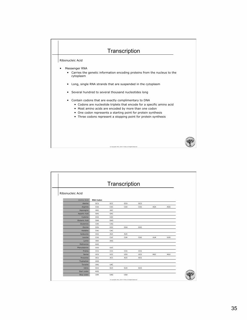
Transcription Ribonucleic Acid
• Messenger RNA • Carries the genetic information encoding proteins from the nucleus to the
cytoplasm
• Long, single RNA strands that are suspended in the cytoplasm
• Several hundred to several thousand nucleotides long
• Contain codons that are exactly complimentary to DNA • Codons are nucleotide triplets that encode for a specific amino acid • Most amino acids are encoded by more than one codon • One codon represents a starting point for protein synthesis • Three codons represent a stopping point for protein synthesis
© Copyright 2012, John P. Fisher, All Rights Reserved
Transcription
Amino Acid RNA Codon
Alanine GCU GCC GCA GCG
Arginine CGU CGC CGA CGG AGA AGG
Asparagine AAU AAC
Aspartic Acid GAU GAC
Cysteine UGU UGC
Glutamic Acid GAA GAG
Glutamine CAA CAG
Glycine GGU GGC GGA GGG
Histidine CAU CAC
Isoleucine AUU AUC AUA
Leucine CUU CUC CUA CUG UUA UUG
Lysine AAA AAG
Methionine AUG
Phenylalanine UUU UUC
Proline CCU CCC CCA CCG
Serine UCU UCC UCA UCG AGC AGU
Threonine ACU ACC ACA ACG
Tryptophan UGG
Tyrosine UAU UAC
Valine GUU GUC GUA GUG
Start codon AUG
Stop codon UAA UAG UGA
Ribonucleic Acid

36
© Copyright 2012, John P. Fisher, All Rights Reserved
Transcription Ribonucleic Acid: Transfer RNA
• Transports activated amino acids to the ribosomes to be used in protein assembly
• Small, approximately 80 nucleotides long • Each type of transfer RNA combines specifically
with 1 of the 20 amino acids that are to be incorporated into proteins
• In ribosomes, each specific transfer RNA recognizes a specific messenger RNA codon, and therefore delivers the appropriate amino acid to the appropriate place in the newly forming amino acid chain, or protein
• Structure: Cloverleaf • Andenylic acid is located at the 3’ (-OH) end
and is the location of amino acid binding • Anticodon is located at the opposite end • Anticodon ensures mRNA codon specificity
transfer RNA
anticodon
T loop D loop
adenylic acid 3’ end
5’ end
anticodon loop
© Copyright 2012, John P. Fisher, All Rights Reserved
Transcription Ribonucleic Acid: Ribosomal RNA
• Forms ribosomes with ~75 proteins • Constitutes ~60% of the ribosome • Structure on which protein are synthesized • Always functions with the other forms of
RNA • mRNA encodes the codon • tRNA delivers the amino acid
• Ribosomes • Composed of 2 units: small subunit
and large submit • Small subunit: 1 RNA molecule
and ~33 proteins • Large subunit: 3 RNA molecules
and ~49 proteins • mRNA and tRNA first bind the small
subunit during protein synthesis • Large subunit provides enzymes for
peptide bonding
Alberts et al. Molecular Biology of the Cell
37
© Copyright 2012, John P. Fisher, All Rights Reserved
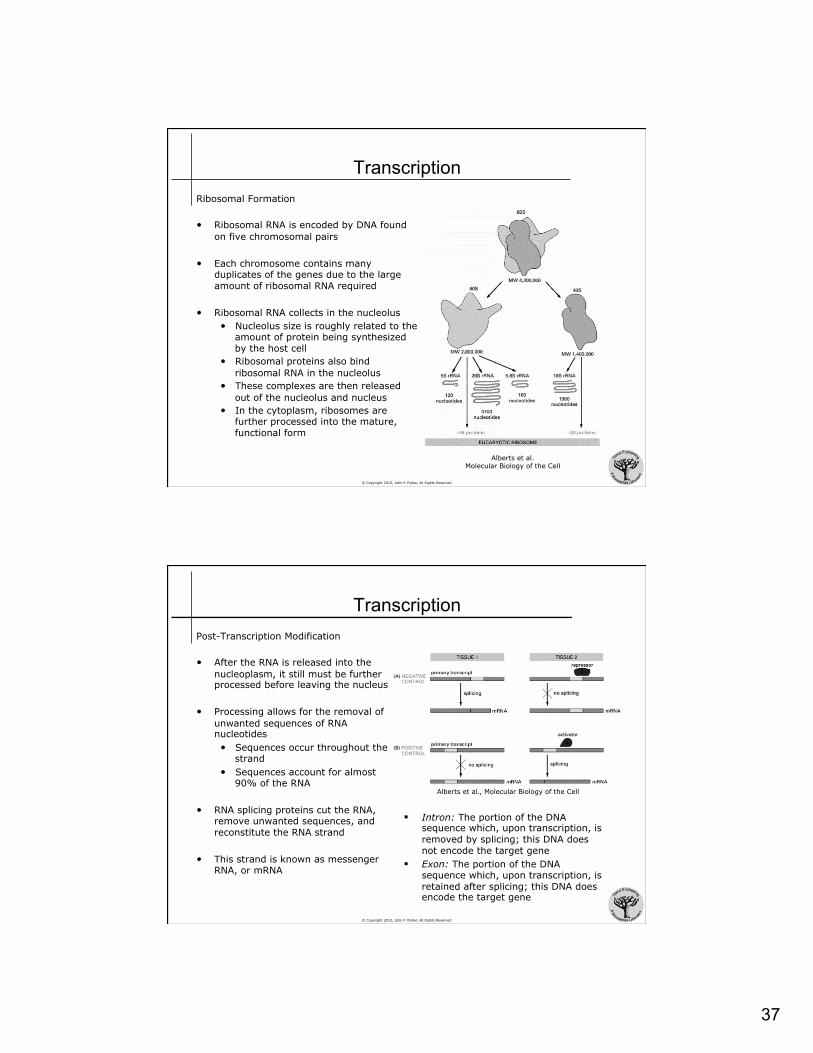
Transcription Ribosomal Formation
• Ribosomal RNA is encoded by DNA found on five chromosomal pairs
• Each chromosome contains many duplicates of the genes due to the large amount of ribosomal RNA required
• Ribosomal RNA collects in the nucleolus • Nucleolus size is roughly related to the
amount of protein being synthesized by the host cell
• Ribosomal proteins also bind ribosomal RNA in the nucleolus
• These complexes are then released out of the nucleolus and nucleus
• In the cytoplasm, ribosomes are further processed into the mature, functional form
Alberts et al. Molecular Biology of the Cell
© Copyright 2012, John P. Fisher, All Rights Reserved
Transcription Post-Transcription Modification
• After the RNA is released into the nucleoplasm, it still must be further processed before leaving the nucleus
• Processing allows for the removal of unwanted sequences of RNA nucleotides • Sequences occur throughout the
strand • Sequences account for almost
90% of the RNA
• RNA splicing proteins cut the RNA, remove unwanted sequences, and reconstitute the RNA strand
• This strand is known as messenger RNA, or mRNA
§ Intron: The portion of the DNA sequence which, upon transcription, is removed by splicing; this DNA does not encode the target gene
§ Exon: The portion of the DNA sequence which, upon transcription, is retained after splicing; this DNA does encode the target gene
Alberts et al., Molecular Biology of the Cell

38
© Copyright 2012, John P. Fisher, All Rights Reserved
Translation Translation is the process of protein synthesis from mRNA
• Messenger RNA travels through the ribosome, starting at a start codon
• The ribosome reads the mRNA, collects the appropriate tRNA for the particular codon, adds the amino acid from the tRNA to the growing amino acid chain, and then releases the tRNA
• When a stop codon is realized, the end of a protein molecule is signaled and the protein is released into the cytoplasm
• Note that there is no specificity for a ribosome for a protein – any ribosome can produce any protein, given the appropriate mRNA
• A few proteins are synthesized directly into the ER – while amino acid assembly is occurring – this pulls ribosomes to the surface of the ER, forming the granular ER
• Polyribosomes • Many ribosomes can translate a mRNA molecule at the
same time • Thus, clusters of 3 to 10 ribosomes – polyribosomes –
can be observed on a single mRNA molecule Alberts et al.
Molecular Biology of the Cell
© Copyright 2012, John P. Fisher, All Rights Reserved
Proteins Protein Structure
• Proteins are polymeric amino acids, where the amino acid sequence determines the protein function
• An amino acid is formed from • A central alpha-carbon, with one lone hydrogen • An amino (NH2) group • A carboxyl (COOH) group • An R side chain group – determines the type of amino acid
• The central carbon atom is asymmetric, allowing the formation of L and D isomers • Proteins consist exclusively of L-amino acids
• At physiological pH (7.4) both the amino group and carboxyl group are ionized (amino group is NH3
+ / carboxyl group is COO-) • Proteins are formed from a condensation reaction between an amino
group and a carboxyl group, releasing water
CCOOH
H R
H2N
C
H
HOOC NH2
R
L isoform
D isoform
HN CH C
H
O
HN CH C
H
OH
O
NH
CH C
H
O
H2N CH C
H
O
peptide bond
N terminus Always written on the left
C terminus
plane formed by rigid C-N bond

39
© Copyright 2012, John P. Fisher, All Rights Reserved
Amino Acids
H2N CH C
CH2
OH
O
C
OH
O
H2N CH C
CH2
OH
O
C
NH2
O
H2N CH C
CH2
OH
O
CH2
CH2
NH
C
NH2
NH
H2N CH C
CH3
OH
O
H2N CH C
CH2
OH
O
SH
H2N CH C
CH
OH
O
CH3
CH2
CH3
H2N CH C
CH2
OH
O
N
NH
H2N CH C
H
OH
O
H2N CH C
CH2
OH
O
CH2
C
OH
O
alanine arginine asparagine aspartic acid cysteine
glutamic acid glutamine glycine histidine isoleucine
H2N CH C
CH2
OH
O
CH2
C
NH2
O
© Copyright 2012, John P. Fisher, All Rights Reserved
H2N CH C
CH2
OH
O
OH
Amino Acids
H2N CH C
CH2
OH
O
CH CH3
CH3
H2N CH C
CH2
OH
O
CH2
CH2
CH2
NH2
H2N CH C
CH2
OH
O
CH2
S
CH3
H2N CH C
CH2
OH
O
HN
C OH
O
H2N CH C
CH2
OH
O
OH
H2N CH C
CH
OH
O
OH
CH3
H2N CH C
CH2
OH
O
HN
H2N CH C
CH
OH
O
CH3
CH3
leucine lysine methionine
phenylalanine
proline
serine tryptophan tyrosine valine threonine
40
© Copyright 2012, John P. Fisher, All Rights Reserved
Amino Acids
AMINO ACID 3L Abbreviation 1L Abbreviation Side Chain Property
Alanine ALA A nonpolar
Arginine ARG R basic
Asparagine ASN N uncharged
Aspartic Acid ASD D acidic
Cysteine CYS C nonpolar
Glutamic Acid GLU E acidic
Glutamine GLN Q uncharged
Glycine GLY G nonpolar
Histidine HIS H basic
Isoleucine ILE I nonpolar
Leucine LEU L nonpolar
Lysine LYS K basic
Methionine MET M nonpolar
Phenylalanine PHE F nonpolar
Proline PRO P nonpolar
Serine SER S uncharged
Threonine THR T uncharged
Tryptophan TRP W nonpolar
Tyrosine TYR Y uncharged
Valine VAL V nonpolar
© Copyright 2012, John P. Fisher, All Rights Reserved
Protein Synthesis
valine
valine
ATP valine-AMP
tRNA CAA
valine
tRNA CAA
GUU mRNA
valine
tRNA CAA GUU
mRNA
GTP
lysine
lysine
ATP lysine-AMP
tRNA TTT
lysine
tRNA TTT
AAA mRNA
lysine
tRNA TTT AAA
mRNA
GTP
alanine
alanine
ATP alanine-AMP
tRNA CGG
alanine
tRNA CGG
GCC mRNA
alanine
tRNA CGG GCC
mRNA
GTP
glycine
glycine
ATP glycine-AMP
tRNA CCC
glycine
tRNA CCC
GGG mRNA
glycine
tRNA CCC GGG
mRNA
GTP
Four high energy bonds are required to synthesize a single peptide bond

41
© Copyright 2012, John P. Fisher, All Rights Reserved
Control of Genetic Functions Regulation • As each cell’s phenotype is in large part described by its protein expression, and all
cells within a single organism are genetically equivalent, there needs to be a strict regulation of protein expression
• Genetic regulation • The degree of gene activation is controlled
• Enzymatic regulation • The activity of formed enzymes (proteins) is controlled
© Copyright 2012, John P. Fisher, All Rights Reserved
Control of Genetic Functions Genetic Regulation • An operon is a sequence of genes located
one after another on the same chromosomal DNA that controls enzyme formation • The constitutive genes are called
structural genes • The expression of the operon is
controlled by an upstream promoter • A repressor operator in the
promoter can be bound by a regulatory protein, controlling RNA polymerase binding to the promoter
• Operon expression is also controlled by a further upstream activator operator
• When bound by an activator protein, operon expression is promoted
Guyton & Hall. Textbook of Medical Physiology, 11th Edition

42
© Copyright 2012, John P. Fisher, All Rights Reserved
Control of Genetic Functions Genetic Regulation • Negative feedback control
• Expression of the structural genes can catalyze substrates to form products that, in turn, regulate operon expression
• Substrate can be a repressor protein that binds the promoter and prevents expression
• Substrate can interfere with activator operator’s promotion of operon expression
• Down regulation of substrate expression, due to lack of enzymer expression, can in promote operon expression
Guyton & Hall. Textbook of Medical Physiology, 11th Edition
© Copyright 2012, John P. Fisher, All Rights Reserved
Control of Genetic Functions Genetic Regulation • Other methods of genetic regulation include
• Regulatory genes • Many operons under same regulatory proteins, regulons • Intermediate operon positioning • RNA regulation • Protein regulation • Histone regulation

43
© Copyright 2012, John P. Fisher, All Rights Reserved
Control of Genetic Functions Enzymatic Regulation • Some cell activities are controlled by intracellular inhibitors or activators that act
directly on specific intracellular enzymes
• Enzyme inhibition • Synthesis of inhibitors that block enzyme function, typically at the beginning of
the signaling cascade
• Enzyme activation • Normally inactive enzymes are activated to induce some signaling cascade
• Lack of ATP induces cAMP levels that promote glucose utilization, and thus the replenishing of ATP levels
• Purine / pyrimidine balance • Purine synthesis inhibits purine synthesis and promotes pyrimidine
synthesis • Pyrimidine synthesis inhibits pyrimidine synthesis and promotes purine
synthesis
© Copyright 2012, John P. Fisher, All Rights Reserved
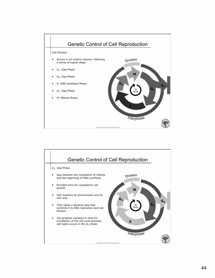
Genetic Control of Cell Reproduction The control of cell and tissue growth is a fundamental application of genetic control
• Cell division is a regulated mechanism, known as mitosis
• Under no inhibition, the life cycle of an eucaryotic cell can be as little as 10 to 30 hours
• Cell division takes approximately 30 min, so most of the cell lifespan is spent outside mitosis in a phase termed interphase
• The lifespan of a cell can be as little as 10 hours and as long as the lifetime of the organism
44
© Copyright 2012, John P. Fisher, All Rights Reserved
Genetic Control of Cell Reproduction Cell Division • Occurs in an orderly manner, following
a series of logical steps
• G1: Gap Phase
• G0: Gap Phase
• S: DNA Synthesis Phase
• G2: Gap Phase
• M: Mitosis Phase
M
G1
S
G2 G0 24
hours
© Copyright 2012, John P. Fisher, All Rights Reserved
Genetic Control of Cell Reproduction G1: Gap Phase
• Gap between the completion of mitosis and the beginning of DNA synthesis
• Provides time for cytoplasmic cell growth
• Cell monitors its environment and its own size
• Then takes a decisive step that commits it to DNA replication and cell division
• The greatest variation in time for completion of the cell cycle between cell types occurs in the G1 phase
M
S
G2 G0 24
hours G1

45
© Copyright 2012, John P. Fisher, All Rights Reserved
Genetic Control of Cell Reproduction G0: Gap Phase
• Pause in cell cycle
• Cells can remain in G0 for days, weeks, or years
• Most mammalian cells are reluctant to proliferate indefinitely • Approximately 50 division limit
• Cells which have reached their limit of divisions enter into G0 and are never released
• Populations have defined endings, but individuals do not • Protect against mutations • Protect against unconstrained
growth (cancer)
M
G1
S
G2 G0 24
hours
© Copyright 2012, John P. Fisher, All Rights Reserved
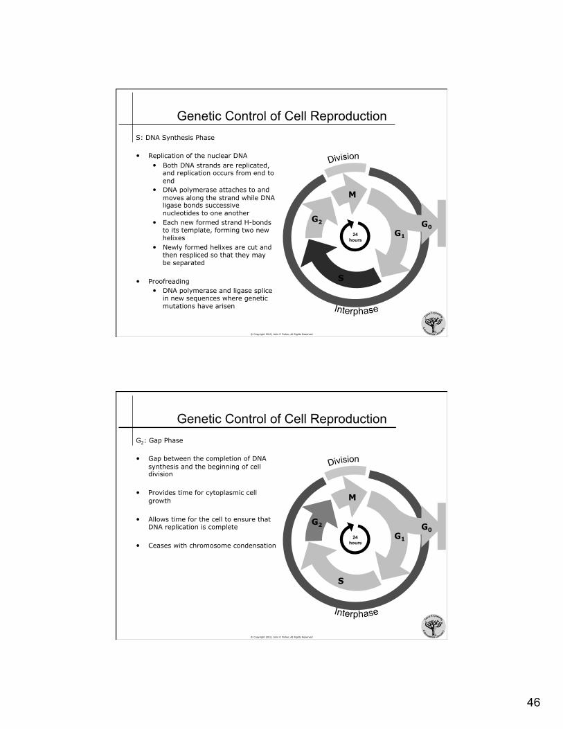
Genetic Control of Cell Reproduction S: DNA Synthesis Phase
• Replication of the nuclear DNA
• About 30% of cells in culture will be in S phase at any given time
• Since DNA doubles in S phase, it can be assayed to identify the state of an individual cell
M
G1
S
G2 G0 24
hours
46
© Copyright 2012, John P. Fisher, All Rights Reserved
Genetic Control of Cell Reproduction S: DNA Synthesis Phase
• Replication of the nuclear DNA • Both DNA strands are replicated,
and replication occurs from end to end
• DNA polymerase attaches to and moves along the strand while DNA ligase bonds successive nucleotides to one another
• Each new formed strand H-bonds to its template, forming two new helixes
• Newly formed helixes are cut and then respliced so that they may be separated
• Proofreading • DNA polymerase and ligase splice
in new sequences where genetic mutations have arisen
M
G1
S
G2 G0 24
hours
© Copyright 2012, John P. Fisher, All Rights Reserved
Genetic Control of Cell Reproduction G2: Gap Phase
• Gap between the completion of DNA synthesis and the beginning of cell division
• Provides time for cytoplasmic cell growth
• Allows time for the cell to ensure that DNA replication is complete
• Ceases with chromosome condensation
M
G1
S
G2 G0 24
hours

47
© Copyright 2012, John P. Fisher, All Rights Reserved
Genetic Control of Cell Reproduction M: Mitosis Phase
• Two parts of the M Phase • Mitosis, or nuclear division
• Prophase • Prometaphase • Metaphase • Anaphase • Telophase
• Cytokinesis, or cell fission
M
G1
S
G2 G0 24
hours
© Copyright 2012, John P. Fisher, All Rights Reserved
Genetic Control of Cell Reproduction Prophase
• Initiation is not a sharply defined event • DNA condenses into chromosomes
• Humans have 6x10⁹ nucleotide pairs organized into 46 chromosomes, 2 of which are sex chromosomes
• The 46 chromosome molecules vary in size from 50x106 to 250x106 nucleotide pairs
• Humans, and all diploid organisms, have two copies of each chromosome (maternal and paternal) except for the sex chromosomes, thus humans have 24 different chromosomes (22 x 2 + 2 = 46)
• Each chromosome is duplicated in the S phase, and now consists of two chromatids
• Two sister chromatids are held together by a DNA sequence known as a centromere
• Mitotic spindle forms • Composed of microtubules and proteins • Formation occurs outside nucleus, between separating
centrosomes (microtubule organizing location)
prophase
chromosome
centromere
mitotic spindle
chromatids

48
© Copyright 2012, John P. Fisher, All Rights Reserved
Genetic Control of Cell Reproduction Prometaphase
• Initiation is a sharply defined event: nuclear membrane disruption • Membranes remnants form nuclear envelope
vesicles • Microtubules enter the nuclear region • Kinetochore microtubules attach to the
centromere on individual chromosomes • Polar microtubules align between the spindle
poles • Astral microtubules are outside the spindle poles
prometaphase
© Copyright 2012, John P. Fisher, All Rights Reserved
Genetic Control of Cell Reproduction Metaphase
• Initiation is not a sharply defined event • Kinetochore microtubules align the chromosomes
in one plane halfway between the spindle poles • Each chromosome is held in tension at the
metaphase plate by paired kinetochores and their associated microtubules, which are attached to opposite poles of the spindle
metaphase

49
© Copyright 2012, John P. Fisher, All Rights Reserved
Genetic Control of Cell Reproduction Anaphase
• Initiation is a sharply defined event: separation of the chromosome complex into two chromatids
• Each chromatid moves toward it spindle pole (1µm / min)
• During the first portion of anaphase (anaphase A), kinetochore molecules shorten as chromatids approach spindle poles
• During the second portion of anaphase (anaphase B), polar microtubules lengthen to move the poles apart
• Anaphase lasts only a few minutes
anaphase
© Copyright 2012, John P. Fisher, All Rights Reserved
Genetic Control of Cell Reproduction Telophase
• Initiation is a sharply defined event: separated chromosomes arrive at the spindle poles
• Polar microtubules continue to elongate • Nuclear envelope begins to reform around each
daughter chromosome Cytokinesis
• Plasma membrane constriction • Cleavage furrow formation • Plasma membrane breakage • Formation of two individual cells
telophase

50
© Copyright 2012, John P. Fisher, All Rights Reserved
Genetic Control of Cell Reproduction
interphase early prophase late prophase prometaphase
metaphase early anaphase late anaphase late telophase
Alberts et al. Molecular Biology of the Cell
© Copyright 2012, John P. Fisher, All Rights Reserved
Genetic Control of Cell Reproduction Duration • The duration of the cell cycle can vary
greatly • Fly embryos: 8 min • Mammalian embryos: 12 hr • Typical mammalian cell: 24 hr • Mammalian liver cell: 1 yr
• Embryonic cell division can occur so rapidly because the resting phases that typically allow for cytoplasmic cell growth are minimized, since division occurs without cell growth (hypertrophy)
M
G1 S
G2
G0
M S G1 G2 M S G1 G2 M S G1 G2
typical cell cycle
embryonic cell cycle
M S M S M S M S M S

51
© Copyright 2012, John P. Fisher, All Rights Reserved
Genetic Control of Cell Reproduction Control of the Cell Cycle • The cell cycle is not a series of
consecutive events that progress due to a set timeline
• The cell cycle is a series of consecutive events that assay cell status and then allow progression
• Check 1 • Cell big enough? • Environment okay?
• Check 2 • DNA replicated?
• Check 3 • Chromosomes aligned?
M
G1
S
G2 G0 24
hours CHECK 1
CHECK 2 C
HEC
K 3
© Copyright 2012, John P. Fisher, All Rights Reserved
The Cell Cycle Control of the Cell Cycle
• The cell cycle is controlled, in large part, by kinase proteins
• Cyclin dependent protein kinases (Cdk) induce downstream processes by phosphorylating selected proteins on serines and threonines
• Cyclins bind Cdk and control their ability to phosphorylate • Cyclins name refers to their ability
to undergo a cycle of synthesis and degradation in each cell cycle
• Mitotic cyclins bind Cdk during G2 and regulate entry into M phase
• G1 cyclins bind Cdk during G1 and regulate entry into S phase
Cyclin dependent
kinase
Mitotic cyclin
G1 cyclin M
G1
S
G2 G0 24
hours

52
© Copyright 2012, John P. Fisher, All Rights Reserved
Genetic Control of Cell Reproduction Cell Death • Apoptosis
• Programmed cell death • Synthesis and release of proteolytic enzymes that induce cell condensation,
cytoskeletal disassembly, and cell surface changes that allow macrophage phagocytosis
• Initiated by caspases
• Neucrosis • Cell death due to injury • Cells swell and burst, spilling their contents into the environment and thus
causing local inflammation and injury


