
arX
iv:1
301.
7716
v1 [
cond
-mat
.mtr
l-sci
] 31
Jan
201
3 Cobalt Substitution in CuFe 2O4 spinel and its
influence on the crystal structure and phonons
M. D. P. Silva,† F. C. Silva,† F. S. M. Sinfrônio,† Alexandre Rocha Paschoal,‡ E. N.
Silva,† and Carlos William de Araujo Paschoal∗,¶,‖
Universidade Federal do Maranhão, Universidade Federal doCeará, Departamento de Física,
CCET, Universidade Federal do Maranhão, 65085-580, São Luís - MA, Brazil, and Department
of Materials Science and Engineering, University of California Berkeley, 94720-1760, Berkeley -
CA, United States
E-mail: [email protected]
Abstract
In this work nanometric spinel Co1−xCuxFe2O4 powders were obtained by polymeric pre-
cursors method at several annealing temperatures between 700 and 1200◦C. The samples were
characterized by means of X-ray powder diffraction, confirming the ideal inverse spinel struc-
ture for CoFe2O4 sample and the tetragonal distorted inverse spinel structure for CuFe2O4
sample. Based on FWHM evaluation, we estimated that crystallite sizes varies between 27 and
37 nm for the non-substituted samples. The optical-active modes were determined by infrared
and Raman spectroscopies. The phonon spectra showed a localtetragonal distortion for mixed
samples.
∗To whom correspondence should be addressed†Departamento de Química, CCET, Universidade Federal do Maranhão, 65085-580, São Luís-MA, Brazil‡Departamento de Física, Universidade Federal do Ceará, Campus do Pici, 60455-760, Fortaleza - CE, Brazil¶Departamento de Física, CCET, Universidade Federal do Maranhão, 65085-580, São Luís - MA, Brazil§Department of Materials Science and Engineering, University of California Berkeley, 94720-1760, Berkeley -
CA, United States‖Author whose correspondence should be addressed: Phone +5598 3301.9209; Fax: +55 98 3301 8204
1

Keywords: Spinel, CoFe2O4, CuFe2O4, Raman, Infrared, X-ray powder diffraction
Introduction
Multiferroics are singular materials that exhibit simultaneously two ferroic orders, usually electric
and magnetic, with a magnetoelectric coupling between themin some cases. These materials have
attracted much attention due to their potential technological applications. As such, multiferroic
compounds bring new functionalities to spintronics and newdevice possibilities, such as memo-
ries, sensors, actuators and tunable filters.1–5 The most investigated multiferroic compound is the
perovskite BiFeO3.6–14 However, the long search for the control of electrical properties by mag-
netic fields has been recently led to in a group of materials known as "frustrated magnets", such as:
the perovskites REMnO315–17 and REMn2O518–21 (with RE being rare earth ions), in the spinels
CuFeO222,23 and CoCr2O4,24 and the Y-type hexagonal ferrites (Ba, Sr)2Zn2Fe12O22,25 among
others.
The restricted number of pure multiferroic compounds is a consequence of the atomic arrange-
ment specificity, since, theoretically, only 13 point groups can lead to the multiferroic behavior.
In addition, by definition, ferroelectrics are insulators (3d transition metal oxides typically have
ions which have an electronic distribution d0), while ferromagnets need conduction electrons. The
difficulties associated with the combination of electric and magnetic responses in a single phase
compound can be solved by making two-phase multiferroic composites consisting of a ferroelectric
component (e.g. PbZr1−xTixO3 - PZT) and a ferromagnetic component (e.g. CoFe2O4).26–38 In
such composites, the magnetoelectric effect is due to the interaction between elastic components of
ferroelectric and ferromagnetic constituents. In such case, an electric field induces a voltage in the
ferroelectric which is transferred to the ferromagnet, that causes the magnetization of the material.
Since the magnetoelectric effect is higher if the coupling at the interface is higher, compounds with
large surface areas (as in multilayer thin films and nanometric powders) and strongly ferroelastic
are particularly interesting. This approach opens new routes to get a magnetoelectric response with
2

the specific choice, relationship and microstructure of theconstituents. In fact, the magnetoelectric
coefficients at room temperature which was achieved exceed those found in low-temperature single
phase samples in three to five orders of magnitude. Thus, magnetoelectric multiferroic composites
are on the boundary of the technological applications.3,4,39,40So, as the spinels have been contin-
uously employed to obtain good multiferroic composites, a careful attention has been given in last
years to the synthesis of spinels in last years for differentmethods.41–61
Besides their extensively investigated ferromagnetic properties, Li-based spinels has been ex-
tensively investigated to be applied as electrode in batteries.? ? ? ? ? Also, binary spinel ferrites like
CdFe2O4, NiFe2O4, ZnFe2O4 and CuFe2O4 also show significant gas-sensing activity.62 More-
over, CuFe2O4 shows high electronic conductivity, high thermal stability and high activity as green
anode for aluminum electrolysis.63 Another important binary spinel-type ferrite is the cobaltferrite
CoFe2O4 due to its high cubic magnetocrystalline anisotropy, high coercivity, and moderate sat-
uration magnetization, being such properties severely affected by to the concentration of divalent
metal cation.59? ? ? ?
Usually CoFe2O4 crystallizes in a spinel structure. The cationic distribution in the spinel struc-
ture can be described by the chemical formula(Fe1−xCox) [Fe1+xCo1−x]O4, where ( ) and [ ] denote
the tetrahedralA and octahedralB sites, respectively. The inversion parameterx is equal to 0 for
inverse spinels and to 1, when the spinel is normal. CoFe2O4 spinel structure is predominantly
inverse(x= 0), with Co2+ ions occupying mainly B sites while Fe3+ ions are distributed almost
equally onA andB sites. The inversion index(1−x) depends on the thermal history of the sam-
ple.64 By the other side, CuFe2O4 has a tetragonally deformed spinel structure, that is stretched
along the< 011> direction.65 Yokoyamaet al.66 observed changes in the crystal structure of
nanosized CuFe2O4 powders obtained by coprecipitation followed by annealing. They observed
that the copper spinel is cubic at temperatures below 300oC and tetrahedral over 400oC. The
formation of considerable quantities of Cu+ in the lattice is the mechanism responsible for the
transition from tetragonal to cubic structure.66 As showed by Kester et al.,67 through reduction
reaction of quenched samples of CuFe2O4, the formation of Cu+takes place, but its fraction in
3

the B-sublattice strongly depends on the synthesis procedures and on the subsequent temperature
treatment. Nanosized particles of CuFe2O4, obtained by a classical ceramic technology, have also
been studied.68 High temperature treatments lead to structural and magnetic surface disorders,
which can be induced by the dispersion of different copper ions in the sublattices, by the arising
of cations and oxygen vacancies, by the structure amorphisation, among others. Also, when the
spinel is synthesized using classical solid state route with accurate stoichiometry(x= 1), it has a
tetragonal structure.69
However, independently of the cation present in the lattice, the physicochemical properties of
such spinel-type ferrites depend on their microstructuralproperties, which are related, in turn, to
the preparation methods of these compounds. Thus, several routes were used to produce these
binary oxides such as hydrolysis,70 ball-milling, solid state reaction, co-precipitation andsol gel
methods, combustion processing etc.71 Since the synthesis route is crucial over the spectroscopic
and structural properties of the spinel-type ferrites, theaim of this work was to evaluate the effect
of cobalt isovalent substitution in CuFe2O4 spinels nanoparticles obtained by polymeric precursors
method.
Materials and Methods
All samples were synthesized using the Polymeric PrecursorMethod, as follow: ferric chloride
hexahydrate (FeCl3.6H2O, Isofar), copper sulphate pentahydrate (CuSO4.5H2O, Isofar) and cobalt
chloride hexahydrate (CoCl2.6H2O, Vetec) were used as purchased without further purification
(pa purity). The precursor solutions of Fe, Cu and Co were prepared by adding the raw solids into
an aqueous solution of citric acid (C6H8O7, Nuclear), using stoichiometric quantities. Therefore,
ethylene glycol (C2H6O2, Nuclear) was added to the metallic solution, according to amolar ratio
1:3. This solution was heated at 110◦C for 5 h in an oven to promote polymerization. Soon after,
the polymerized gel was heated at 300◦C for 1 h, under air atmosphere, to burn the organic matter
and form a black solid mass (primary calcination). Such carbonaceous mass was grounded until
4

its particles were 100 Mesh sized and heat-treated at 300◦C for 12 hours (secondary calcination),
under a high oxygen atmosphere, in order to produce oxygen vacancies in the solids. Finally,
such precursor powders were annealed between 700–1200◦C (ternary calcination) in air for 4 h in
Al2O3 crucibles, and the desired spinel compounds resulted.
Crystal structures of the annealed powders were examined using an X-ray diffractometer Xpert
MPD (Panalytical), with Co Kα radiation (40 kV and 40 mA), speed of 0.02◦ θs−1 and value
ranging from 10 to 100◦. X-ray powder diffraction (XRD) patterns were compared with the Joint
Committee Powder Diffraction Standards (JCPDS) data for the phase evaluation.
Crystallite sizes(D) of the samples were determined from X-ray line broadening analyzes,
employing the Scherrer’s equation:
D =Kλ
βcosθ(1)
whereλ is the X-ray radiation wavelength (λ =1.78896 Å),K is the Scherrer constant,β is the
FWHM of the peak (in radians) andθ is the peak angular position.
The infrared spectra were obtained using an IR prestige-21 infrared spectrometer (Shimadzu),
applying KBr as dispersant agent (1:100 wt./wt.) in the mid range: 400 up to 1000 cm−1.
The confocal Raman spectra were acquired using an alpha 300 system microscope (Witec,
Ulm, Germany), equipped with a highly linear (0.02%) stage,piezo-driven, and an objective lens
from Nikon (20x, NA = 0.40). A Nd:YAG polarized laser (λ = 532 nm) was focused with a
diffraction-limited spot size (0.61λ /NA) and the Raman light was detected by a high sensitivity,
back illuminated spectroscopic CCD behind a 600 g/mm grating. The final power at the end of
the objective lens used to focus on the sample was 3 mW. The spectrometer used was an ultra-
high throughput Witec UHTS 300 with up to 70% throughput, designed specifically for Raman
microscopy. The integration time and number of accumulations were in average 60 s and 3, re-
spectively.
5

Crystalline Structure and Group Theory
Both CoFe2O4 and CuFe2O4 crystallizes according to inverse spinel-based structure. Particularly,
CuFe2O4 assumes a body centered tetragonal distorted inverse-spinel structure belonging to the
space groupI41/amd(D194h), in which there are four molecules per unit cell(Z = 4). In this struc-
ture Fe/Cu, Fe and O atoms occupy 8d (C2h), 4a (D2d) and 16h (Cs) Wyckoff sites, respectively.
On the other hand, CoFe2O4 shows a face centered cubic inverse-spinel structure, belonging to
the space groupFd3m(O7h), with eight molecules per unit cell(Z = 8), in which Fe/Co, Fe and O
atoms occupy 16d (D3d), 8a (Td) and 32e (C3v) Wyckoff sites, respectively.
Since each structure contains 14 atoms in the primitive unitcell, there are 42 degrees of free-
dom and, consequently, 42 phonons are permitted for both structures. Using the Factor Group
Analysis,72 the zone-center vibrational modes distribution was decomposed in terms of the irre-
ducible representations for both Oh and D4h factor groups (Table 1). Due to these site occupations,
for both structures, FeB/M (M = Co or Cu) atoms do not contribute to the Raman-active phonon
spectra. Thus, any Raman assignments can be attributed to (Fe)A and O ions, while the infrared
spectra may be influenced by all constituent ions.
CuFe2O4 and CoFe2O4 spinel Raman and infrared spectra patterns were also analyzed accord-
ing to the quasi-molecular description, using the internalmodes for the FeO4 tetrahedron.73 So, in
this case the structures was described as formed by two different sub-lattice groups: FeB/M atoms
and (FeO4)5− tetrahedron. Tables 2 and 3 summarize the FeO4 group vibrations obtained by the
correlation diagrams for cubic and tetragonal structures,respectively.
According to the FeO4 symmetry,ν1(A1) andν3(F2) are assigned to the Fe–O symmetric and
asymmetric stretchings, respectively; whileν2(E) andν1(F2) vibrational modes are attributed to
the symmetric and asymmetric Fe-O bendings. TheT (F2) vibrational mode is assigned to the
FeO4 tetrahedron translation motion. Finally, the Raman and infrared modes for both cubic and
tetragonal inverse spinels, disregarding the acoustic andsilent modes, are shown in Table 4.
6

Table 1: Factor group analysis from CoFe2O4 (cubic) and CuFe2O4(tetragonal)
Structure Atoms Sites Site symmetry Irreducible representations
Cubic(
O7h
)
Fe/Co 16d D3d A2u⊕Eu⊕2F1u⊕F2u
Fe 8a Td F1u⊕F2g
O 32e C3v A1g⊕A2u⊕Eg⊕Eu⊕F1g⊕2F1u⊕2F2g⊕F2u
Total A1g⊕2A2u⊕Eg⊕2Eu⊕F1g⊕5F1u⊕3F2g⊕2F2u
Acoustic modes F1u
IR modes 4F1u
Raman modes A1g⊕Eg⊕3F2g
Silent modes 2A2u⊕2Eu⊕F1g⊕2F2u
Tetragonal(
D194h
)
Fe/Cu 8d C2h A1u⊕2A2u⊕B1u⊕2B2u⊕3Eu
Fe 4a D2d A2u⊕B1g⊕Eg⊕Eu
O 16h Cvs 2A1g⊕A1u⊕A2g⊕2A2u⊕2B1g⊕B1u⊕B2g
⊕2B2u⊕3Eg⊕3Eu
Total 2A1g⊕2A1u⊕A2g⊕5A2u⊕3B1g⊕2B1u⊕B2g
⊕4B2u⊕4Eg⊕7Eu
Acoustic modes A2u⊕Eu
IR modes 4A2u⊕6Eu
Raman modes 2A1g⊕3B1g⊕B2g⊕4Eg
Silent modes 2A1u⊕A2g⊕2B1u⊕4B2u
Table 2: Correlation charts of the phonon symmetry for AFe2O4 in the(
O7h
)
cubic structure.
7

Table 3: Correlation charts of the phonon symmetry for AFe2O4 in the(
D194h
)
tetragonal phase.
Table 4: Raman and Infrared modes corresponding to the cubicand tetragonal inverse spinels
Spinel structure Activity Assignments
D194h(CuFe2O4)
Raman 2T(B1g⊕Eg)+L(Eg)+ν1(A1g)+2ν2(A1g⊕B2g)+2ν3(B1g⊕Eg)+2ν4(B1g⊕Eg)
IR 5T(2A2u⊕3Eu)+L(Eu)+2ν3(A2u⊕Eu)+2ν4(A2u⊕Eu)
O7h (CoFe2O4)
Raman T(F2g)⊕ν1(A1g)⊕ν2(Eg)⊕ν3(F2g)⊕ν4(F2g)IR 2T(2F1u)⊕ν3(F1u)⊕ν4(F1u)
8

Results and Discussions
Structural properties
Figures 1 and 2 present the XRD patterns for the several Co1−xCuxFe2O4 spinel powders as a func-
tion of the annealing temperature. The results confirm the formation of the single phase spinel,
as indicated by the JCPDS 00-034-0425 (tetragonal CuFe2O4) and JCPDS 00-022-1086 (cubic
CoFe2O4) patterns, excepted for those annealed at 700 and 800◦C Cu-based pure spinels that con-
tain α-Fe2O4 rhombohedral (JCPDS 00-013-0534). It is important to pointout thatα-Fe2O4 is
often found as a secondary phase in spinel synthesis, as previously reported by Sun et al.62 and
Mathew et al.74 In addition, for the cobalt substituted ferrites, it can be noticed than even minor
Co2+ inclusions induce a cubic lattice. The full-width at half maximum (FWHM) (monitoring
the peak (400)) in CoFe2O4 decreases with the increase of the annealing temperature, as shown in
Figure 3, showing that the average crystallite size is becoming larger, while an inverse behavior is
observed in CuFe2O4 (monitoring the peak (211)). Besides, for Co0.25Cu0.75Fe2O4 samples, the
FWHM hardly changes, except when synthesized at 900◦C and above. This behavior could be
associated to minor lattice distortion imposed by the spatial competition between octahedral and
tetrahedral sites. Such fact is confirmed by Figure 4, since Co-substituted ferrites present bigger
average crystallite sizes, as compared to the CuFe2O4 structures. Furthermore, the average crys-
tallite size tends to enlarge with the annealing (26 up to 54 nm). Specifically, CuFe2O4 crystallites
show mean size between 32 and 35 nm, much lower than the value proposed by Sun ıet al.,62 that
estimated the range in between 75 - 110 nm for the ferrites. Inopposition, CoFe2O4 crystallite
sizes were in the range 28 - 37 nm, being also smaller than those reported by Gaikwadet al.,75 i.e.,
60 - 80 nm, but bigger than the one obtained by Valdés-Solíset al.,76 that was 20 nm.
Vibrational properties
Figure 5 shows the Raman spectra obtained for CoFe2O4 and CuFe2O4 pure annealed spinels.
CuFe2O4 spinel, synthesized at 700 and 800◦C (Figure 5b), in fact corresponds to theα-Fe2O4 as
9

20 40 60 80 100
(b)
2θ (degree)
700°C
800°C
900°C
1000°C
1100°C
1200°C
20 40 60 80 100
(a)
2θ (degree)
1200 oC
800 oC
900 oC
1000 oC
1100 oC
700 oC
Figure 1: XRD patterns as function of the annealing temperature for (a) CoFe2O4 and (b) CuFe2O4
spinels.
10

20 40 60 80 100
2θ (degree)
1200 oC
800 oC
900 oC
1000 oC
1100 oC
700 oC
(c)
20 40 60 80 100
2θ (degree)
1200 oC
800 oC
900 oC
1000 oC
1100 oC
700 oC
(b)
20 40 60 80 100
(a)
1200 oC
800 oC
900 oC
1000 oC
1100 oC
2 θ (degree)
700 oC
Figure 2: XRD patterns as function of the annealing temperature for (a) Co0.25Cu0.75Fe2O4. (b)Co0.50Cu0.50Fe2O4 and (c) Co0.75Cu0.25Fe2O4 spinels.
11

700 800 900 1000 1100 12000.14
0.16
0.18
0.20
0.22
0.24
0.26
0.28
0.30
0.32
0.34
FWH
M (d
egre
es)
Annealing temperature (Celsius)
CuFe2O4
Co0,25Cu0,75Fe2O4
Co0,50Cu0,50Fe2O4
Co0,75Cu0,25Fe2O4
CoFe2O4
Figure 3: FWHM as function of the annealing temperature for the Co1−xCuxFe2O4 (x =1.00,0.75,0.50,0.25,0.00) spinels. The lines are guides for the eyes.
700 800 900 1000 1100 120020
25
30
35
40
45
50
55
C
ryst
allit
e si
ze (n
anom
eter
s)
Annealing temperature (Celsius)
CuFe2O4
Co0,25Cu0,75Fe2O4
Co0,50Cu0,50Fe2O4
Co0,75Cu0,25Fe2O4
CoFe2O4
Figure 4: Crystallite size as function of the ternary thermal treatment for the Co1−xCuxFe2O4(x= 1.00,0.75,0.50,0.25,0.00) spinels. The lines are guides for the eyes.
12
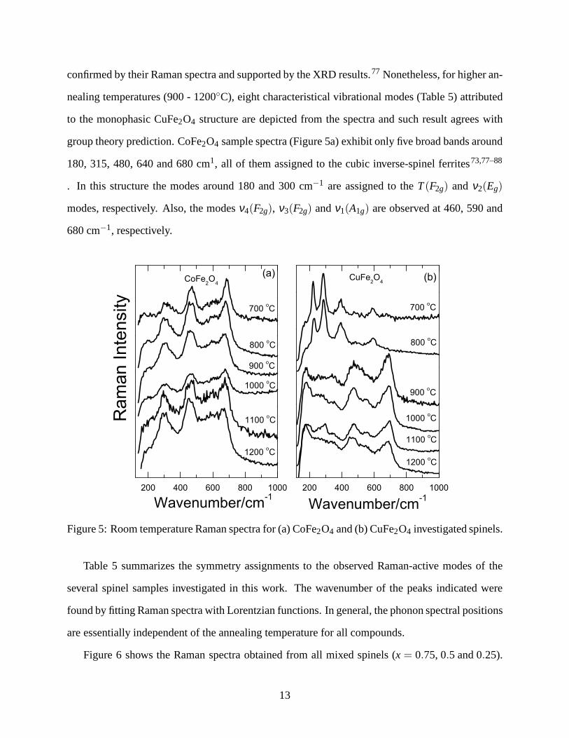
confirmed by their Raman spectra and supported by the XRD results.77 Nonetheless, for higher an-
nealing temperatures (900 - 1200◦C), eight characteristical vibrational modes (Table 5) attributed
to the monophasic CuFe2O4 structure are depicted from the spectra and such result agrees with
group theory prediction. CoFe2O4 sample spectra (Figure 5a) exhibit only five broad bands around
180, 315, 480, 640 and 680 cm1, all of them assigned to the cubic inverse-spinel ferrites73,77–88
. In this structure the modes around 180 and 300 cm−1 are assigned to theT(F2g) andν2(Eg)
modes, respectively. Also, the modesν4(F2g), ν3(F2g) andν1(A1g) are observed at 460, 590 and
680 cm−1, respectively.
200 400 600 800 1000
1200 oC
1100 oC
800 oC
Wavenumber/cm-1
700 oC
900 oC
1000 oC
CuFe2O
4(b)
200 400 600 800 1000
Wavenumber/cm-1
1200 oC
1100 oC
800 oC
700 oC
900 oC
1000 oC
Ram
an Inte
nsity
CoFe2O
4
(a)
Figure 5: Room temperature Raman spectra for (a) CoFe2O4 and (b) CuFe2O4 investigated spinels.
Table 5 summarizes the symmetry assignments to the observedRaman-active modes of the
several spinel samples investigated in this work. The wavenumber of the peaks indicated were
found by fitting Raman spectra with Lorentzian functions. Ingeneral, the phonon spectral positions
are essentially independent of the annealing temperature for all compounds.
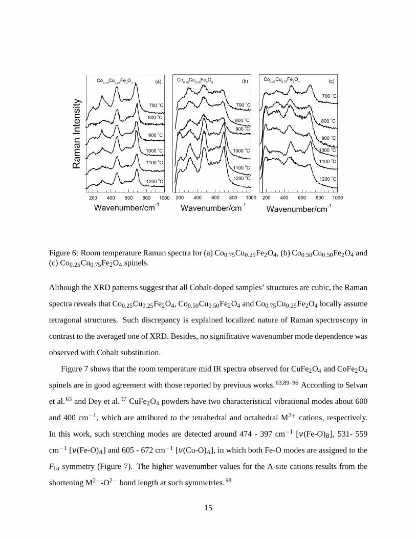
Figure 6 shows the Raman spectra obtained from all mixed spinels (x = 0.75, 0.5 and 0.25).
13

Table 5: Wavenumbers (in cm−1) and Raman modes assignments for Co1−xCuxFe2O4 (x =1.00,0.75,0.50,0.25,0.00) spinels.
Compound modesAnnealing temperature (◦C)
Assignment700 800 900 1000 110 1200
CoFe2O4
1 171 170 169 169 179 188 T(F2g)2 315 300 295 294 296 297 ν2(Eg)3 468 462 459 460 458 456 ν4(F2g)4 599 595 592 593 596 588 ν3(F2g)5 687 682 678 679 680 672 ν1(A1g)
Co0.75Cu0.25Fe2O4
1 192 183 193 189 188 192 T2 300 308 298 303 309 303 ν23 355 355 − 359 − 363 ν4
4 464 469 466 468 468 468 ν45 552 − 563 569 575 570 ν3
6 608 602 619 626 − − ν3
7 − 659 − − 657 641 ν38 681 693 688 690 692 692 ν1
Co0.50Cu0.50Fe2O4
1 196 189 203 180 188 187 T2 − − − 239 239 245 T/L3 303 300 312 309 311 313 ν24 − − − 379 374 368 ν4
5 467 471 469 472 470 473 ν4
6 610 604 605 576 573 592 ν37 − − − 678 662 672 ν3
8 688 689 687 702 696 698 ν1
Co0.25Cu0.75Fe2O4
1 171 170 170 172 174 177 T2 197 194 194 198 200 214 T/L3 238 230 239 245 268 266 ν2
4 320 313 322 321 346 331 ν4
5 482 468 466 460 475 463 ν46 586 582 597 588 577 587 ν3
7 685 651 682 673 667 659 ν3
8 712 693 711 702 701 694 ν1
CuFe2O4
1 − − 177 171 177 168 T2 − − 250 244 245 211 T/L3 − − − − 294 271 ν2
4 − − 343 348 348 346 ν45 − − 467 469 474 462 ν4
6 − − 532 554 554 549 ν3
7 − − 663 658 655 656 ν38 − − 694 696 696 692 ν1
14
200 400 600 800 1000
(c)Co0.25
Cu0.75
Fe2O
4
Wavenumber/cm-1
1200 oC
1100 oC
800 oC
700 oC
900 oC
1000 oC
200 400 600 800 1000
Wavenumber/cm-1
(b)Co0.50
Cu0.50
Fe2O
4
1200 oC
1100 oC
800 oC
700 oC
900 oC
1000 oC
200 400 600 800 1000
Ra
man
In
ten
sity
Wavenumber/cm-1
1200 oC
1100 oC
800 oC
700 oC
900 oC
1000 oC
Co0.75
Cu0.25
Fe2O
4 (a)
Figure 6: Room temperature Raman spectra for (a) Co0.75Cu0.25Fe2O4, (b) Co0.50Cu0.50Fe2O4 and(c) Co0.25Cu0.75Fe2O4 spinels.
Although the XRD patterns suggest that all Cobalt-doped samples’ structures are cubic, the Raman
spectra reveals that Co0.25Cu0.25Fe2O4, Co0.50Cu0.50Fe2O4 and Co0.75Cu0.25Fe2O4 locally assume
tetragonal structures. Such discrepancy is explained localized nature of Raman spectroscopy in
contrast to the averaged one of XRD. Besides, no significative wavenumber mode dependence was
observed with Cobalt substitution.
Figure 7 shows that the room temperature mid IR spectra observed for CuFe2O4 and CoFe2O4
spinels are in good agreement with those reported by previous works.63,89–96According to Selvan
et al.63 and Dey et al.97 CuFe2O4 powders have two characteristical vibrational modes about600
and 400 cm−1, which are attributed to the tetrahedral and octahedral M2+ cations, respectively.
In this work, such stretching modes are detected around 474 -397 cm−1 [ν(Fe-O)B], 531- 559
cm−1 [ν(Fe-O)A] and 605 - 672 cm−1 [ν(Cu-O)A], in which both Fe-O modes are assigned to the
F1u symmetry (Figure 7). The higher wavenumber values for the A-site cations results from the
shortening M2+-O2− bond length at such symmetries.98
15

1000 800 600 400
(a)CoFe2O
4
Tra
nsm
itta
nce
1200oC
1100oC
1000oC
900oC
800oC
700oC
Wavenumber /cm-1
1000 800 600 400
Wavenumber /cm-1
(b)
700 oC
800 oC
900 oC
1000 oC
1100 oC
1200 oC
CuFe2O
4
Figure 7: Room temperature IR spectra of (a) CoFe2O4 and (b) CuFe2O4 investigated spinels.
Once again, theα-Fe2O4 phase is depicted from the spectra of CuFe2O4 samples annealed at
700◦C and 900◦C, as indicated by the strong absorption band at 475 cm−1 [ν(Fe-O)].99 Finally,
the CuFe2O4 samples obtained at 1000, 1100 and 1200◦C present a significant tetragonal distortion
(Jahn-Teller distortion), corroborating to the Raman and XRD data.
Considering the CoFe2O4 sample, similar vibrational modes are observed around 399 -400
[ν(Fe-O)B], 464 - 541 [ν(Fe-O)A] and 576 - 583 [ν(Co-O)A] (Figure 7a). The displacement of the
(Co-O)A modes to lower wavenumbers, as compared to the (Cu-O)A one, results from the smaller
cobalt ionic radius, generating a larger vibrational energy.100–102
The Co-substituted CuFe2O4 IR spectra (Figure 8) show a continuous band narrowing as func-
tion of cobalt contents into the lattice. Nevertheless, thesame tetrahedral and octahedral vibrational
modes are assigned.
16

1000 800 600 400
(c)
Co0.25
Cu0.75
Fe2O
4
Wavenumber /cm-1
1100 oC
1200 oC
1000 oC
900 oC
800 oC
700 oC
1000 800 600 400
Wavenumber /cm-1
465
685
534
(b)
Co0.5
Cu0.5
Fe2O
4
1200 oC
1100 oC
1000 oC
900 oC
800 oC
700 oC
590
1000 800 600 400
Tra
nsm
itta
nce
Wavenumber /cm-1
Co0.75
Cu0.25
Fe2O
4
1200 oC
1100 oC
1000 oC
900 oC
800 oC
700 oC
(a)
Figure 8: Room temperature IR spectra of (a) Co0.75Cu0.25Fe2O4, (b) Co0.50Cu0.50Fe2O4 and (c)Co0.25Cu0.75Fe2O4 spinels.
Conclusions
In this work we have synthesized nanometric Co1−xCuxFe2O4 spinels using a polymeric precursors
method precursors. The crystalline structures were investigated by X-ray powder diffraction, which
confirms that the pure CoFe2O4 and CuFe2O4 samples have cubic and tetragonal inverted spinel
structures, respectively. The crystallite sizes of the mixed samples are bigger than those of the
pure samples. The synthesized samples were investigated byRaman and infrared spectroscopies.
This analysis showed that there is a tetragonal local distortion into the mixed sample. A complete
mode assignment for all the samples was performed based on the internal vibrations of the FeO4
molecular group.
Acknowledgement
The authors are grateful to the Brazilian funding agencies CAPES, CNPq, and FAPEMA.
17
References
(1) Cheong, S.-W.; Mostovoy, M.NATURE MATERIALS2007, 6, 13–20.
(2) Chiba, D.; Sawicki, M.; Nishitani, Y.; Nakatani, Y.; Matsukura, F.; Ohno, H.NATURE2008,
455, 515–518.
(3) Eerenstein, W.; Wiora, M.; Prieto, J. L.; Scott, J. F.; Mathur, N. D.NATURE MATERIALS
2007, 6, 348–351.
(4) Ramesh, R.; Spaldin, N. A.NATURE MATERIALS2007, 6, 21–29.
(5) Scott, J. F.Nature Materials2007, 6, 256–257.
(6) Bea, H.; Paruch, P.NATURE MATERIALS2009, 8, 168–U14.
(7) Chu, Y.-H. et al.NATURE MATERIALS2008, 7, 478–482.
(8) Seidel, J. et al.NATURE MATERIALS2009, 8, 229–234.
(9) Yang, C. H. et al.NATURE MATERIALS2009, 8, 485–493.
(10) Zhao, T.; Scholl, A.; Zavaliche, F.; Lee, K.; Barry, M.;Doran, A.; Cruz, M. P.; Chu, Y. H.;
Ederer, C.; Spaldin, N. A.; Das, R. R.; Kim, D. M.; Baek, S. H.;Eom, C. B.; Ramesh, R.
NATURE MATERIALS2006, 5, 823–829.
(11) Aguilar, R. V.; Mostovoy, M.; Sushkov, A. B.; Zhang, C. L.; Choi, Y. J.; Cheong, S.-W.;
Drew, H. D.PHYSICAL REVIEW LETTERS2009, 102, 047203.
(12) Bea, H.; Bibes, M.; Ott, F.; Dupe, B.; Zhu, X. H.; Petit, S.; Fusil, S.; Deranlot, C.; Bouze-
houane, K.; Barthelemy, A.PHYSICAL REVIEW LETTERS2008, 100, 017204.
(13) Kornev, I. A.; Lisenkov, S.; Haumont, R.; Dkhil, B.; Bellaiche, L.PHYSICAL REVIEW
LETTERS2007, 99, 227602.
18
(14) Wang, J.; Neaton, J.; Zheng, H.; Nagarajan, V.; Ogale, S.; Liu, B.; Viehland, D.;
Vaithyanathan, V.; Schlom, D.; Waghmare, U.; Spaldin, N.; Rabe, K.; Wuttig, M.;
Ramesh, R.SCIENCE2003, 299, 1719–1722.
(15) Kimura, T.; Goto, T.; Shintani, H.; Ishizaka, K.; Arima, T.; Tokura, Y.NATURE2003, 426,
55–58.
(16) Lee, S.; Pirogov, A.; Kang, M.; Jang, K.-H.; Yonemura, M.; Kamiyama, T.; Cheong, S. W.;
Gozzo, F.; Shin, N.; Kimura, H.; Noda, Y.; Park, J. G.NATURE2008, 451, 805–U4.
(17) Pimenov, A.; Shuvaev, A.; Loidl, A.; Schrettle, F.; Mukhin, A. A.; Travkin, V. D.;
Ivanov, V. Y.; Balbashov, A. M.PHYSICAL REVIEW LETTERS2009, 102, 107203.
(18) Bodenthin, Y.; Staub, U.; Garcia-Fernandez, M.; Janoschek, M.; Schlappa, J.; Goloven-
chits, E. I.; Sanina, V. A.; Lushnikov, S. G.PHYSICAL REVIEW LETTERS2008, 100,
027201.
(19) Harris, A. B.; Aharony, A.; Entin-Wohlman, O.PHYSICAL REVIEW LETTERS2008, 100,
217202.
(20) Okamoto, J.; Huang, D. J.; Mou, C. Y.; Chao, K. S.; Lin, H.J.; Park, S.; Cheong, S.-W.;
Chen, C. T.PHYSICAL REVIEW LETTERS2007, 98, 157202.
(21) Sushkov, A. B.; Aguilar, R. V.; Cheong, S.-W.; Drew, H. D. PHYSICAL REVIEW LETTERS
2007, 98, 027202.
(22) Wang, F.; Vishwanath, A.PHYSICAL REVIEW LETTERS2008, 100, 077201.
(23) Ye, F.; Fernandez-Baca, J. A.; Fishman, R. S.; Ren, Y.; Kang, H. J.; Qiu, Y.; Kimura, T.
PHYSICAL REVIEW LETTERS2007, 99, 157201.
(24) Choi, Y. J.; Okamoto, J.; Huang, D. J.; Chao, K. S.; Lin, H. J.; Chen, C. T.; van Veenen-
daal, M.; Kaplan, T. A.; Cheong, S.-W.PHYSICAL REVIEW LETTERS2009, 102, 067601.
19

(25) Kimura, T.; Lawes, G.; Ramirez, A.PHYSICAL REVIEW LETTERS2005, 94, 137201.
(26) Li, L.; Lin, Y. Q.; Chen, X. M.Journal of Applied Physics2007, 102.
(27) Wang, Z. G.; Viswan, R.; Hu, B. L.; Harris, V. G.; Li, J. F.; Viehland, D.Physica Status
Solidi-Rapid Research Letters2012, 6, 92–94.
(28) Yan, L. H.; Liang, W. Z.; Liu, S. H.; Huang, W.; Lin, Y.INTEGRATED FERROELECTRICS
2011, 131, 82–88.
(29) Wang, J.-N.; Li, W.-L.; Li, X.-L.; Fei, W. D.POWDER DIFFRACTION2010, 25, S45–
S47, 10th Chinese National Conference on X-Ray Diffraction/ICDD Workshop, Shanghai,
PEOPLES R CHINA, OCT 12-15, 2009.
(30) Chen, W.; Shannigrahi, S.; Chen, X. F.; Wang, Z. H.; Zhu,W.; Tan, O. K.SOLID STATE
COMMUNICATIONS2010, 150, 271–274.
(31) Zhang, L.; Zhai, J.; Mo, W.; Yao, X.FERROELECTRICS2010, 406, 213–220.
(32) Zhang, J. X.; Dai, J. Y.; Lu, W.; Chan, H. L. W.JOURNAL OF MATERIALS SCIENCE
2009, 44, 5143–5148.
(33) Yan, L.; Xing, Z.; Wang, Z.; Wang, T.; Lei, G.; Li, J.; Viehland, D.APPLIED PHYSICS
LETTERS2009, 94, 192902.
(34) Wu, X.; Cai, W.; Kan, Y.; Yang, P.; Liu, Y.; Bo, H.; Lu, X.;Zhu, J.FERROELECTRICS
2009, 380, 48–55, 6th Asian Meeting on Ferroelectrics, Taipei, TAIWAN, AUG 02-06,
2008.
(35) Pang, L.-H.; Ji, W.-J.; Zhang, Y.; Wang, L.; Zhang, S.-T.; Luo, Z.-L.; Chen, Y.-F.JOURNAL
OF PHYSICS D-APPLIED PHYSICS2009, 42, 045304.
(36) Chen, W.; Wang, Z. H.; Zhu, W.; Tan, O. K.JOURNAL OF PHYSICS D-APPLIED
PHYSICS2009, 42, 075421.
20

(37) Dix, N.; Skumryev, V.; Laukhin, V.; Fabrega, L.; Sanchez, F.; Fontcuberta, J.MATERIALS
SCIENCE AND ENGINEERING B-SOLID STATE MATERIALS FOR ADVANCED TECH-
NOLOGY2007, 144, 127–131, Symposium on Nanoscale Tailoring of Defect Structures for
Optimized Functional and Multifunctional Oxide Films heldat the EMRS 2007, Strasbourg,
FRANCE, 2007.
(38) Thang, P. D.; Pham, M. T. N.; Rijnders, G.; Blank, D. H. A.; Duc, N. H.; Klaasse, J. C. P.;
Bruck, E.JOURNAL OF THE KOREAN PHYSICAL SOCIETY2008, 52, 1406–1409, 1st In-
ternational Workshop on Functional Materials/3rd International Workshop on Nanophysics
and Nanotechnology, Halong City, VIETNAM, DEC 06-09, 2006.
(39) Ederer, C.; Spaldin, N.PHYSICAL REVIEW LETTERS2005, 95, 257601.
(40) Tokunaga, Y.; Furukawa, N.; Sakai, H.; Taguchi, Y.; Arima, T.-h.; Tokura, Y.NATURE
MATERIALS2009, 8, 558–562.
(41) Sun, J. R.; Wang, Z. G.; Wang, Y. Y.; Zhu, Y. B.; Shen, T. L.; Pang, L. L.; Wei, K. F.;
Li, F. S. Materials Science and Engineering B-Advanced Functional Solid-State Materials
2012, 177, 269–273.
(42) Tang, Y.; Wang, X. W.; Zhang, Q. H.; Li, Y. G.; Wang, H. Z.Progress in Natural Science-
Materials International2012, 22, 53–58.
(43) Pui, A.; Gherca, D.; Carja, G.Digest Journal of Nanomaterials and Biostructures2011, 6,
1783–1791.
(44) Girgis, E.; Wahsh, M. M. S.; Othman, A. G. M.; Bandhu, L.;Rao, K. V. NANOSCALE
RESEARCH LETTERS2011, 6, 460.
(45) Fei, C. L.; Zhang, Y.; Yang, Z.; Liu, Y.; Xiong, R.; Shi, J.; Ruan, X. F.Journal of Magnetism
and Magnetic Materials2011, 323, 1811–1816.
21

(46) Naseri, M. G.; Saion, E. B.; Hashim, M.; Shaari, A. H.; Ahangar, H. A.Solid State Commu-
nications2011, 151, 1031–1035.
(47) El-Okr, M. M.; Salem, M. A.; Salim, M. S.; El-Okr, R. M.; Ashoush, M.; Talaat, H. M.
Journal of Magnetism and Magnetic Materials2011, 323, 920–926.
(48) Gatelyte, A.; Jasaitis, D.; Beganskiene, A.; Kareiva,A. Materials Science-Medziagotyra
2011, 17, 302–307.
(49) Yu, B. Y.; Kwak, S. Y.Dalton Transactions2011, 40, 9989–9998.
(50) Gatelyte, A.; Jasaitis, D.; Beganskiene, A.; Kareiva,A. Materials Science-Medziagotyra
2011, 17, 302–307.
(51) Shen, L. M.; Bao, N. Z.; Prevelige, P. E.; Gupta, A.Journal of the American Chemical
Society2010, 132, 17354–17357.
(52) Zhang, Y.; Yang, Z.; Yin, D.; Liu, Y.; Fei, C. L.; Xiong, R.; Shi, J.; Yan, G. L.Journal of
Magnetism and Magnetic Materials2010, 322, 3470–3475.
(53) Ajroudi, L.; Villain, S.; Madigou, V.; Mliki, N.; Leroux, C.Journal of Crystal Growth2010,
312, 2465–2471.
(54) Veverka, M.; Veverka, P.; Jirak, Z.; Kaman, O.; Knizek,K.; Marysko, M.; Pollert, E.; Za-
veta, K.Journal of Magnetism and Magnetic Materials2010, 322, 2386–2389.
(55) Sertkol, M.; Koseoglu, Y.; Baykal, A.; Kavas, H.; Toprak, M. S.Journal of Magnetism and
Magnetic Materials2010, 322, 866–871.
(56) Shahane, G. S.; Kumar, A.; Arora, M.; Pant, R. P.; Lal, K.Journal of Magnetism and Mag-
netic Materials2010, 322, 1015–1019.
(57) Hou, C. Y.; Yu, H.; Zhang, Q. H.; Li, Y. G.; Wang, H. Z.Journal of Alloys and Compounds
2010, 491, 431–435.
22

(58) Shen, X. Q.; Zhou, Z.; Song, F. Z.; Meng, X. F.Journal of Sol-Gel Science and Technology
2010, 53, 405–411.
(59) Manova, E.; Paneva, D.; Kunev, B.; Estournes, C.; Riviere, E.; Tenchev, K.; Leaustic, A.;
Mitov, I. JOURNAL OF ALLOYS AND COMPOUNDS2009, 485, 356–361.
(60) Gozuak, F.; Koseoglu, Y.; Baykal, A.; Kavasa, H.Journal of Magnetism and Magnetic
Materials2009, 321, 2170–2177.
(61) Maaz, K.; Karim, S.; Mumtaz, A.; Hasanain, S. K.; Liu, J.; Duan, J. L.Journal of Magnetism
and Magnetic Materials2009, 321, 1838–1842.
(62) Sun, Z.; Liu, L.; zeng Jia, D.; Pan, W.Sensors and Actuators B: Chemical2007, 125, 144 –
148.
(63) Selvan, R.; Augustin, C.; Berchmans, L.; Saraswathi, R. MATERIALS RESEARCH BUL-
LETIN 2003, 38, 41–54.
(64) Sawatzky, G. A.; Vanderwo, F.; Morrish, A. H.PHYSICAL REVIEW1969, 183, 383–386.
(65) Nedkov, I.; Vandenberghe, R. E.; Marinova, T.; Thailhades, P.; Merodiiska, T.; Avramova, I.
APPLIED SURFACE SCIENCE2006, 253, 2589–2596.
(66) Yokoyama, M.; Nakamura, A.; Sato, T.; Haneda, K.JOURNAL OF THE MAGNETICS
SOCIETY OF JAPAN1998, 22, 243–245.
(67) Kester, E.; Gillot, B.; Villette, P., C. Tailhades; Rousset, A.THERMOCHIMICA ACTA1997,
297, 71–78.
(68) Jiang, J. Z.; Goya, G. F.; Rechenberg, H. R.JOURNAL OF PHYSICS-CONDENSED MAT-
TER1999, 11, 46063–4078.
(69) Hamdeh, H.; Ho, J.; Oliver, S.; Willey, R.; Oliver, G.; Busca, G.JOURNAL OF APPLIED
PHYSICS1997, 81, 1851–1857.
23

(70) Sreekumar, K.; Mathew, T.; Devassy, B.; Rajgopal, R.; Vetrivel, R.; Rao, B.APPLIED
CATALYSIS A-GENERAL2001, 205, 11–18.
(71) Tao, S.; Gao, F.; Liu, X.; Sorensen, O. T.Materials Science and Engineering: B2000, 77,
172 – 176.
(72) Rousseau, D. L.; Bauman, R. P.; Porto, S. P. S.JOURNAL OF RAMAN SPECTROSCOPY
1981, 10, 253–290.
(73) VERBLE, J.PHYSICAL REVIEW B1974, 9, 5236–5248.
(74) Mathew, T.; Shylesh, S.; Devassy, B.; Vijayaraj, M.; Satyanarayana, C.; Rao, B.;
Gopinath, C.APPLIED CATALYSIS A-GENERAL2004, 273, 35–45.
(75) Gaikwad, R. S.; Chae, S.-Y.; Mane, R. S.; Han, S.-H.; Joo, O.-S.International Journal of
Electrochemistry2011, 2011, 729141.
(76) Valdes-Solis, T.; Tartaj, P.; Marban, G.; Fuertes, A. B. NANOTECHNOLOGY2007, 18,
145603.
(77) Shebanova, O.; Lazor, P.JOURNAL OF SOLID STATE CHEMISTRY2003, 174, 424–430.
(78) Gupta, R.; Sood, A.; Metcalf, P.; Honig, J.PHYSICAL REVIEW B2002, 65, 104430.
(79) Gasparov, L.; Tanner, D.; Romero, D.; Berger, H.; Margaritondo, G.; Forro, L.PHYSICAL
REVIEW B2000, 62, 7939–7944.
(80) Li, J.; Huan, A.; Wang, L.; Du, Y.; Feng, D.PHYSICAL REVIEW B2000, 61, 6876–6878.
(81) Bersani, D.; Lottici, P.; Montenero, A.JOURNAL OF RAMAN SPECTROSCOPY1999, 30,
355–360.
(82) deFaria, D.; Silva, S.; deOliveira, M.JOURNAL OF RAMAN SPECTROSCOPY1997, 28,
873–878.
24

(83) THIERRY, D.; PERSSON, D.; LEYGRAF, C.; BOUCHERIT, N.; HUGOTLEGOFF, A.
CORROSION SCIENCE1991, 32, 273–284.
(84) BOUCHERIT, N.; GOFF, A.; JOIRET, S.CORROSION SCIENCE1991, 32, 497–507.
(85) DUNNWALD, J.; OTTO, A.CORROSION SCIENCE1989, 29, 1167–1176.
(86) GRAVES, P.; JOHNSTON, C.; CAMPANIELLO, J.MATERIALS RESEARCH BULLETIN
1988, 23, 1651–1660.
(87) DEGIORGI, L.; BLATTERMORKE, I.; WACHTER, P.PHYSICAL REVIEW B1987, 35,
5421–5424.
(88) OHTSUKA, T.; KUBO, K.; SATO, N.CORROSION1986, 42, 476–481.
(89) Hashim, M.; Alimuddin,; Kumar, S.; Koo, B. H.; Shirsath, S. E.; Mohammed, E. M.;
Shah, J.; Kotnala, R. K.; Choi, H. K.; Chung, H.; Kumar, R.JOURNAL OF ALLOYS AND
COMPOUNDS2012, 518, 11–18.
(90) Eshraghi, M.; Kameli, P.CURRENT APPLIED PHYSICS2011, 11, 476–481.
(91) Cedeno-Mattei, Y.; Perales-Perez, O.MICROELECTRONICS JOURNAL2009, 40, 673–
676, Conference on European Nano Systems, Paris, FRANCE, DEC 03-04, 2007.
(92) Zhao, L.; Zhang, H.; Xing, Y.; Song, S.; Yu, S.; Shi, W.; Guo, X.; Yang, J.; Lei, Y.; Cao, F.
JOURNAL OF SOLID STATE CHEMISTRY2008, 181, 245–252.
(93) Gillot, B.; Nivoix, V.; Kester, E.; Nusillard, O.; Villette, C.; Tailhades, P.; Rousset, A.
MATERIALS CHEMISTRY AND PHYSICS1997, 48, 111–118.
(94) Gillot, B.; Buguet, S.; Kester, E.JOURNAL OF MATERIALS CHEMISTRY1997, 7, 2513–
2517.
(95) Gillot, B.; Laarj, M.; Kacim, S.JOURNAL OF MATERIALS CHEMISTRY1997, 7, 827–
831.
25

(96) WALDRON, R.PHYSICAL REVIEW1955, 99, 1727–1735.
(97) Dey, S.; Ghose, J.MATERIALS RESEARCH BULLETIN2003, 38, 1653–1660.
(98) Costa, A. F.; Pimentel, P. M.; Melo, D. M. A.; Melo, M. A. F.; Aquino, F. M.Cerâmica
2011, 57, 352 – 355.
(99) Gotic, M.; Jurkin, T.; Music, S.COLLOID AND POLYMER SCIENCE2007, 285, 793–800.
(100) Popmintchev, T.; Chen, M.-C.; Bahabad, A.; Gerrity, M.; Sidorenko, P.; Cohen, O.;
Christov, I. P.; Murnane, M. M.; Kapteyn, H. C.PROCEEDINGS OF THE NATIONAL
ACADEMY OF SCIENCES OF THE UNITED STATES OF AMERICA2009, 106, 10516–
10521.
(101) Singhal, S.; Bhukal, S.; Singh, J.; Chandra, K.; Bansal, S.Journal of Nanotechnology2011,
930243.
(102) Zhang, Y.; Wen, D.Journal of the American Ceramic Society2012, cited By (since 1996)
0; Article in Press.
26

200 400 600 800
(b)
Wavenumber/cm-1
200 400 600 800
Wavenumber/cm-1
Ram
an
In
ten
sity
(a)


