
Braz Dent Sci 2018 Oct/Dec;21(4)470
ABSTRACTObjective: The aim of this study was to systematically review the literature to assess static fracture strength tests applied for fixed dental prostheses (FDPs) and analyze the impact of periodontal ligament (PDL) simulation on the fracture strength. Material and Methods: Original scientific papers published in MEDLINE (PubMed) database between 01/01/1981 and 10/06/2018 were included in this systematic review. The following MeSH terms, search terms, and their combinations were used:“Dentistry”, “Fracture Strength”, “Fracture Resistance”, “Fixed Dental Prosthesis”, “Fixed Partial Denture”, “Mechanical Loading”. Two reviewers performed screening and analyzed the data. Only the in vitro studies that reported on load-bearing capacity of only FDP materials where mean or median values reported in Newnton (N) were included. Results: The selection process resulted in the 57 studies. In total, 36 articles were identified related to all-ceramics, 10 were fiber reinforced composite resin (FRC), 8 of composite resin (C) and 5 of metal-ceramic. As for clinical indications, 3 and 4-unit FDPs were more commonly studied (n=32; with PDL=21, without PDL=11), followed by single crowns (n=13; with PDL=3, without PDL=10), and inlay-retained and cantilever FDPs (n=12; with PDL=8, without PDL=4). Conclusion: An inclination for decreased static fracture strength could be observed with the simulation of PDL but due to insufficient data this could not be generalized for all materials used for FDPs.
RESUMOObjetivo: O objetivo deste estudo foi revisar sistematicamente a literatura para avaliar os testes de força de fratura estática aplicados para próteses dentárias fixas (FDPs) e analisar o impacto da simulação do ligamento periodontal (PDL) na resistência à fratura. Material e Métodos: Artigos científicos originais publicados na base de dados MEDLINE (PubMed) entre 01/01/1981 e 10/06/2018 foram incluídos nesta revisão sistemática. Foram utilizados os seguintes termos MeSH, termos de pesquisa e suas combinações: “Dentistry”, “Fracture Strength”, “Fracture Resistance”, “Fixed Dental Prosthesis”, “Fixed Partial Denture”, “Mechanical Loading”. Dois revisores realizaram a triagem e analisaram os dados. Apenas os estudos in vitro que reportaram a capacidade de suporte de carga de FDP, com os valores das médias ou medianas relatados em Newton (N) foram incluídos. Resultados: O processo de seleção resultou em 57 estudos. No total, 36 artigos foram identificados relacionados à restaurações totalmente cerâmicas, 10 em resina composta reforçada com fibra (FRC), 8 em resina composta (C) e 5 em metalocerâmica. Quanto às indicações clínicas, os PDF de 3 e 4 unidades foram mais comumente estudados (n = 32; com PDL = 21, sem PDL = 11), seguidos de coroas isoladas (n = 13; com PDL = 3, sem PDL = 10) e FDPs retidas por inlays e com cantilever (n = 12; com PDL = 8, sem PDL = 4). Conclusão: Uma inclinação para a diminuição da resistência à fratura estática pôde ser observada com a simulação do PDL, mas devido a dados insuficientes, isso não pôde ser generalizado para todos os materiais utilizados para as FDPs.
S Y S T E M AT I C R E V I E W
Influence of testing parameters on the load-bearing capacity of prosthetic materials used for fixed dental prosthesis: A systematic reviewInfluência de parâmetros de testes na capacidade de suporte de carga de materiais protéticos utilizados para prótese dentária fixa: uma revisão sistemática
Mutlu ÖZCAN1, Julia HÖHN1, Gabriela Monteiro de ARAÚJO2, Dayanne Monielle Duarte MOURA2, Rodrigo Othávio Assunção SOUZA2
1 – University of Zurich – Center for Dental and Oral Medicine – Dental Materials Unit – Clinic for Fixed and Removable Prosthodontics and Dental Materials Science – Zurich – Switzerland.
2 – Federal University of Rio Grande do Norte (UFRN) – Department of Dentistry – Natal – RN – Brazil.
doi: 10.14295/bds.2018.v21i4.1652
UNIVERSIDADE ESTADUAL PAULISTA “JÚLIO DE MESQUITA FILHO”
Instituto de Ciência e TecnologiaCampus de São José dos Campos
Ciência Odontológica Brasileira
ANOS
1998-20
18
KEYWORDSCeramics, Dental prosthesis, Periodontal ligament.
PALAVRAS-CHAVECerâmica, Prótese dentária, Ligamento periodontal.

Influence of testing parameters on the load-bearing capacity of prosthetic materials used for fixed dental prosthesis: A systematic review
Ozcan M et al.
Braz Dent Sci 2018 Oct/Dec;21(4)471
1 INTRODUCTION
D urability of restorations is crucial for clinical dentistry since mechanical failures in the
form of fractures have financial consequences both for the patient and the dentist. Removal and repair of restorations may be arduous and have also biological costs. Thus, decision for choosing the best performing material in terms of mechanical durability is often made based on the results of in vitro studies.
Load to fracture test is a common way of testing dental materials used for fixed dental prosthesis (FDP) to assess their mechanical strength for different indications. Today, an increased plethora of metal, all-ceramic or polymeric materials are being offered for clinical use. Neither ethically, nor technically it is possible to test their performance in randomized controlled clinical trials. Therefore, preclinical evaluations help to rank physical and mechanical properties of materials. Ranking prosthetic materials after such tests are generally taken into consideration for clinical indications especially for posterior segments of the mouth where increased chewing forces are experienced. Static load-bearing tests require a controlled environment where the specimen dimensions and the loading conditions are standardized. Besides recording fracture strength values, failure type and fractography analysis after such tests provide additional information on the origins and onset of the failure.
Although there are norms for testing FDP materials (DIN EN ISO 22674) [1], among in vitro tests, a great heterogeneity is being noticed in the dental literature related to load to fracture tests. While some studies were performed on metal abutments [2-9] others used polymers [10-16], or natural tooth [4,9,16] as abutment material. An important other factor is involvement of the periodontal ligament simulation (PDL) for tooth-borne FDPs. In an attempt to simulate the biological conditions and physiologic mobility of the teeth, different types of PDL materials are being used. The lack of PDL simulation could still contain useful information for the durability of implant-borne FDPs. Yet, the consequence of using PDL in static loading tests is not known.
Since the test parameters vary considerably among the available published studies, there is apparent need to develop some guidelines in testing and interpreting the data on load-bearing
capacity of different FDP materials in order to estimate their lifespan more realistically and not to deliver misleading information in terms of ranking materials for durability.
The objective of this systematic review was in particular to analyze the effect of PDL simulation on the load-bearing capacity of different FDP materials for different prosthetic indications.
2 MATERIAL AND METHODS2.1 Search strategyBefore the initiation of the literature
search, a protocol to be followed was agreed upon by the authors. An electronic search at MEDLINE (PubMed) (http://www.ncbi.nlm.nih.gov/entrez/query.fcgi) from 01/01/1981 and 10/06/2018 was conducted for English-language articles published in the dental literature, using the following MeSH terms, search terms and their combinations: ““Dentistry”, “Fracture Strength”, “Fracture Resistance”, “Fixed Dental Prosthesis”, “Fixed Partial Denture”, “Mechanical Loading”. The MEDLINE search are presented in Table I. In addition, hand searches were performed on bibliographies of the selected articles as well as identified narrative reviews to find out whether the search process has missed any relevant article. This did add the new four additional articles to be involved in the review process.
Table I - Search strategy in MEDLINE applied for this review. #: search, MeSH: Medical subjects heading, a thesaurus word.
Search Literature search strategy
1 “Fracture Resistance and Fixed Partial Denture AND Dentistry”
2 “Fracture Resistance and Fixed Dental Prosthesis AND Dentistry”
3 “Fracture Strength AND Fixed Dental Prosthesis AND Dentistry”
4 Fracture Strength AND Fixed Partial Denture AND Dentistry”
5 “Mechanical Loading AND Fixed Dental Prosthesis AND Den-tistry”
6 Mechanical Loading AND Fixed Partial Denture and Dentistrty”
7 “Mechanical Loading AND Fracture Resistance and Fixed Dental Prosthesis”
8 “Mechanical Loading AND Fracture Resistance AND Fixed Partial Denture”
9 Mechanical Loading AND Fracture Strength AND Fixed Dental Prosthesis
10 Mechanical Loading AND Fracture Strength AND Fixed Partial Denture
11 Mechanical Loading AND Fracture Strength AND Fracture Resistance AND Dentistry

Influence of testing parameters on the load-bearing capacity of prosthetic materials used for fixed dental prosthesis: A systematic review
Ozcan M et al.
Braz Dent Sci 2018 Oct/Dec;21(4)472
2.2 PICOs
The population, intervention, comparison and outcomes, i.e. the “PICOs” for this systematic review were defined as follows:
Population: Type of material (metal-ceramic - MC, all ceramic - AC, fibre-reinforced composite - FRC, composite resin – C. Type of restoration (FDPs of 3 units, 4 units, retained by inlay and cantilever);
Intervention: test method (static loading);
Comparison: with periodontal ligament and without periodontal ligament;
Outcomes: static fracture strength;
Study design: in vitro studies.
2.3 Inclusion/Exclusion criteria
In vitro studies reporting on load-bearing capacity of only FDP materials where mean or median values reported were included. Publications were excluded if fatigue loading was performed or data were not presented in Newton (N). Also, studies performed with finite element analysis were excluded.
2.4 Study selection
The search process led to titles of 1559 journal articles reviewed by two independent reviewers for possible inclusion in this systematic review. After title screening, 125 abstracts were considered relevant, and full-text articles were downloaded. Thereafter, from 125 journal articles, 57 were included in this review. The process of identifying the studies included in the review is presented in Figure 1.
Figure 1 - The PRISMA flowchart showing the study selection process.

Influence of testing parameters on the load-bearing capacity of prosthetic materials used for fixed dental prosthesis: A systematic review
Ozcan M et al.
Braz Dent Sci 2018 Oct/Dec;21(4)473
2.5 Data extraction
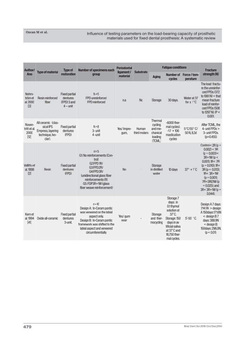
The two reviewer’s extracted data independently using a data extraction form previously agreed upon. Process of identifying the studies included in the review from is presented in Fig. 1. Data on the following parameters were extracted: author(s), year of publication, type of material tested, type of restoration, number of samples per group, periodontal ligament simulation material, substrate, fatigue conditions and fracture resistance in Newton. The data were presented according to the type of restoration: single crowns, 3-unit FDP, 4-unit FDP, inlay-retained and cantilever FDPs (tables II, III and IV). Disagreement regarding data extraction was resolved by discussion and a consensus was reached.
2.6 Risk of bias assessment
The risk-of-bias was assessed based on previous studies [17]. The risk of bias was calculated from 6 criteria: sample size calculation, sample randomization, sample preparation, specified aging, standardization of procedures by ISO and operator. For each parameter values from 0 to 2 were attributed: 0 – if the authors clearly reported the parameter; 1 – if the author reported the execution/respect of the parameter but accuracy of the execution is unclear; 2 – if the author did not specify the parameter or the information is not present. If the total sum of the attributed values ranged between 0 up to 4 it was considered alow risk, between 5up to 9 a medium risk and 10 up to14 a high risk of bias.
3. RESULTS3.1 Characteristics of the included/
excluded studies
Two independent reviewers screened the 1559 titles retrieved from the electronic search for possible inclusion in the review.
After initial elimination, based on the titles and the abstracts, 744 abstracts were accepted for inclusion by both reviewers. The two reviewers independently assessed the 125 full-text articles to determine whether they fulfilled the defined criteria for final inclusion. 72 articles had to be excluded after full text reading and risk of bias. Any disagreement was resolved by discussion. Finally, 57 [2,3,5,7,9,11-15,17-62] studies were found to qualify for inclusion in the review. Among all the studies included, all-ceramics (n=36) were more commonly tested followed by FRC (n=10), composite (n=8) and metal-ceramic (n=5). As for clinical indications, 3 and 4-unit FDPs were more commonly studied (n=32; with PDL=21, without PDL=11), followed by single crowns (n=13; with PDL=3, without PDL=10), and inlay-retained and cantilever FDPs (n=12; with PDL=8, without PDL=4). Table II [2,3,5,7,13, 15,17-41] III [14,42-53] and IV [9,11,16,54-62]. According to the results, from 57 studies included, 32 involved PDL. In all selected subgroups, the search identified the use of wax, silicon, gummy resin, latex, vinyl silicone impression, acrylic resin base and silicone rubber to simulate PDL. The studied also used some kind of substrate, among them vital teeth such as third molars (n=21), pre-molars (n=18) and central incisors (n=4); artificial teeth (n=8) or implants/metal (n=7) to simulate clinical conditions.
3.2. Risk of bias
According to the bias risk assessment, 57 studies included in this systematic review presented a risk of bias medium (between 5 and 9). The rest of the articles presented a low risk of bias (between 0 and 4). The data were described in table V. Most of the studies did not describe the calculation of the sample size, the laboratory procedures by a single operator and standardization of procedures (ISO).

Influence of testing parameters on the load-bearing capacity of prosthetic materials used for fixed dental prosthesis: A systematic review
Ozcan M et al.
Braz Dent Sci 2018 Oct/Dec;21(4)474
Author/Ano Type of material Type of
restorationNumber of specimens
each group
Periodontal ligament /
materialSubstrate
Fatigue conditions Fracture
strength (N)Aging Number
of cycles Force / tem-
perature
Partiyan et al., 2017[17]
Zirconia: manually aided de-
sign–manually aided milling
(MAD/MAM) and Computer assisted design–computer
assisted milling(CAD/CAM)
Three-unit zirconia
fixed partial denture
n=20Group I (MAD/MAM) con-
ventional.Group II: (MAD/MAM) Inno-
vative. Group III
(CAD/CAM).Conventional
Group IV(CAD/CAM). Innovative.
Yes/acrylic resin base
second premolar
and second molar
Stored in distilled water/
thermocy-cling
72hrs / 1000 cycles
37°C / 5°/ 55°C, 30s.
G2>G4>G3>G1 (P<0.0001).
Murase et al., 2014[18]
5% Y-TZP (Aadva Zirconia, GC)
All-ceramicfixed partial
dentures (FPDs)
n=15cross-sectional áreas:
1: 9.0mm2
2: 7.0 mm2
3: 5.0 mm2
Yes/vinyl silicone
impression
Central and lateral
incisors
stored in distilled water
24hrs 37°C 1> 2 > 3. (p<0,001)
Chaar, et al., 2013
[19]
LV (layering techni-que/Vintage ZR); LZ (layering technique/
ZIROX); PP (CAD/CAM and press-over techni-
ques/PressXZr
3-unit posterior
fixed dental prostheses
(FDPs)
n=16G1: LV G2: LZ G3: PP
Yes/gum resin
Human premolars
thermo-mechanica
1 200 000 cycles -
G2> G1>G3. (NON-AGED)G3>G2>G1
(AGED) (P<0,05)
Eroglu and
Gurbulak 2013[20]
zirconia-ceramic (ZC), galvano-ceramic
(GC), and porcelain-fu-sed-to-metal (PFM)
Fixed partial denture 3- unit
n = 10
ZC, GC and PFM with or without thermocycling and mechanical loading (TCM)
No
Metal (maxillary
canine and second
premolar)
Thermocy-cling and mechani-
cal loading
- Thermo-cycling: 10,000 cycles
- Me-chanical loading: 100,000
cicles.
Thermo-cycling: 5º
- 55º;
Mechanical loading: 50 N;
GC (1678.1 ±211.6) > GC/TCM (1475.8 ± 227.9) -
p < 0.05
PFM (1878.5 ±176.5) > PFM/TCM (1687.8 ±
162.2) - p < 0.05
Takuma, Y. et al.,
2013[21]
3% Y-TZP (Everest®Zirconium Soft)
4-unit all-ce-ramic FPDs
Framework connectors cross-sectional áreas: A:9.0
or B: 7.0mm2).Cross-sectional forms:
a circular form (1:1 (Type A); an oval form, (3:4 (type B);
and another oval(2:3 (type
C). Connector types:
mesial/distal connectors (A-A,
B-B, C-C) and central con-nector (-A-,-B-, -C-).
- -stored in distilled water
24hrs 37°C
Cross-sectional área: A>B.
(p<0,01)Mesial and distal
connector’s type: A-A> C-C.
(p<0,01)Central connec-tor’s type: A>C (p<0,05); A>B
(p<0,01)
Preis et al., 2012.
[22]
Yttria-stabilized zirconia (Cercon ht,
Degudent)
Three-unit zirconia-ba-
sed FPDs
n=8G1: AD – sintered; G2: AD – sintered – glazed; G3:
AD – sintered – sandblasted – glazed; G4: AD – sintered – polished – grinded (contact points adjusted); G5: AD –
sintered – polished – grinded – repolished; G6: ARD – sin-
tered – veneered; G7: control: analogous to #3 but without
thermal cycling (TC) and mechanical loading (ML).mechanical loading (ML).
Yes/wax
Artificial identical polyme-thylme-
thacrylat (PMMA) molars
thermal cycling and mechani-
cal loading
TC: 6000
5°/55° ×2 min each
cycle1.2 × 106 ×
50 N; 1.6 Hz)
No statistically significant
differences were found between the groups (p =
0.910)
Table II - Characteristics from the studies included in the systematic review of Fixed Dental Prothesis 3-unit and 4-unit.

Influence of testing parameters on the load-bearing capacity of prosthetic materials used for fixed dental prosthesis: A systematic review
Ozcan M et al.
Braz Dent Sci 2018 Oct/Dec;21(4)475
Author/Ano Type of material Type of
restorationNumber of specimens
each group
Periodontal ligament /
materialSubstrate
Fatigue conditions
Fracturestrength (N)
Aging Number of cycles
Force / tem-perature
Salimi, H. et al.,
2012[23]
Cercon Base ceramic, Degudent, Germany
Zirconium oxide
posterior fixed partial
dentures (FPD)
Group I: copings with 3 × 3 connector dimension and
standard designGroup II: copings with 3 × 3 connector dimension and
modified designGroup III: copings with 4 ×
4 connector dimension and standard design
Group IV: copings with 4 × 4 connector dimension and
modified design.
-Maxillary typodont
model
artificial saliva at
37°C/ thermocy-
cling
2000 cycles
5 and 55°C for 30 s
each, with an intermediate pause of 15 s.
Group IV was significantly higher than group I (P <
0.001) and group II (P < 0.001), but
there was not any significant difference be-
tween group IV and group III (P =
0.156)
Noth-durft et. al 2011
[24]
ZirconiaFixed partial
denture 3- unit
n = 8Implant -
tooth supportedrestorations (IT)
or implant -implant (II)
with:- individualised abutments
(i) or no individualised (ni) - with (TC) or without ther-
mocycling (N)
Yes/ Gum resin
Zirconia abut-
ments and cast metal teeth (First molar and pre-molar)
Thermocy-cling
- Thermo-cycling: 10,000 cycles
Thermocy-cling: 5º - 55º;
IT < II- p < 0.05
iTC < nTC- p < 0.05
Onodera et al., 2011[25]
3 vol% (YTZP:Kavo Everest ® Zirco-
nium Soft, Biberach, Germany)
all-ceramic FPDs molar
region
n=15.Cross-sectional area: A: 9.0,
B: 7.0; C:5.0mm. Conector shape: A: 1:1, B: 3:4, C: 2:3
Yes/Silicone material
Second premolar
andsecond molar
stored in distilled water 24hrs 37°C
Cross-sectional area (mm2):
A>B>C. P<0.05). Conector
shape: A=B=C. (p<0,05)
Rosen-tritt, M.
et al., 2011[26]
Glass-infiltrated, alumina based,
all-ceramic material (Inceram Alumina, Vita
Zahnfabrik)
All-ceramic three-unit
fixed partial dentures
(FPDs)
n=8Group A (control):
in polymethyl methacrylate (PMMA).
Group B: polyether layer (Impregum, 3M ESPE). Group
C: polyether layer during aged.
Yes/ wax bath
human molars
Thermal cycling and mechani-
cal loading
TC: 6000 cycles.
5°/55° × 2 min each
cycle;1.2 × 106 × 50
N; 1.6 Hz)
Group A> Group C (P = .047)= B
(P = .364). Goup C=B. (P =
.961)
Ei-senbur-
ger et. al. 2008[27]
Composite resin. (Protemp, Luxatemp,
Cron-Mix).
Fixed partial denture 4- unit
30 Yes/ Latex varnish
Artificial resin teeth (24 and 27)
Thermocy-cling 10.000 5 – 55 ºC
Luxatemp > CronMix (p=0.014)
Luxatemp - Without fibre
Stick > EverStick (p= 0.004)
CronMix: Without fibre > EverStick (p =
0.015)

Influence of testing parameters on the load-bearing capacity of prosthetic materials used for fixed dental prosthesis: A systematic review
Ozcan M et al.
Braz Dent Sci 2018 Oct/Dec;21(4)476
Author/Ano Type of material Type of
restorationNumber of specimens each
group
Periodontal ligament /
materialSubstrate
Fatigue conditions Fracture
strength (N)Aging Number of
cycles Force / tem-
perature
Att et al. 2007[28]
Zirconia (DCS, Procera and Vita
CerecInlab)
Fixed partial denture 3- unit
n= 8G1: DCS with artificial aging;
G2: DCS without artificial aging;
G3: Procera with artificial aging;
G4: Procera without artificial aging;
G5: Vita with artificial aging; G6: Vita without artificial
aging.
Yes/ Gum resin
Human mandibular premolars
and molars
Termome-chanical fatigue
- 1,200,000 cycles
- Mechanical loading: 49 N;
- Thermo-cycling: 5º
- 55º.
G3 (1297) < G5 (1593) – p= 0.015G3 < G1 (1618) –
p= 0.038
Att et al. 2007*
Zr[29]
Zirconia (DCS, Procera and Vita CerecInlab) ve-
neered using Vita VM9.
Fixed partial denture 3- unit
n= 8G1: DCS with artificial aging;
G2: DCS without artificial aging; G3: Procera with artifi-cial aging; G4: Procera without
artificial aging; G5: Vita with artificial aging;
G6: Vita without artificial aging.aging.
Yes/ Gum resin
Human mandibular premolars
and molars
Termome-chanical fatigue
- 1,200,000 cycles
- Mechanical loading: 49 N;
- Thermo-cycling: 5º
- 55º.
G3 (1094) < G1 (1481) – p= 0.042
Larsson et al. 2007[30]
Zirconia (Procera)Fixed partial
denture 4- unit
8G1: 2.0 mm connector;/ G2: 2.5
mm conector;/ G3: 3.0 mm conector;/
G4: 3.5 mm conector;/ G5: 4.0 mm conector.
No
Artificial acrylic resin
teeth (34 and 37)
Thermocy-cling, and mechani-
cal loading.
- Mechanical loading: 10
000;
- Thermocy-cling: 5000.
- Mechanical loading: 30
-300 N;
- Thermo-cycling: 5º
- 55º.
G1 and G2 fractured during preload (30–300 N, 10 000 cycles);
G5 (897) > G4 (602) > G3 (428).
Kohorst et al. 2007[31]
Zirconia –Partially sintered
(Cercon);Fully sintered
zirconia (Digizon)
Fixed partial denture 4- unit
10G1: Cercon without preliminar echanical damage; G2: Cer-
con with preliminar mechani-cal damage;
G3: Digizon without preliminar mechanical damage;
G4: Digizon with preliminar mechanical damage.
Yes/ Latex
Artificial polyure-
thane resin teeth (24 and 27)
Storage, thermocy-cling and mechani-
cal loading
- Storage: distilled
water at 36 °C for 200
days;- Thermo-
cycling: 104 cycles
- Mechanical loading: 106
cucles.
- Thermo-cycling: 5º
- 55º;
-Mechanical loading: 100 N;
G1 (903.7) < G3 (1262.6);
G2 (921.1) < G4 (1132.4).
Pfeiffer et al. 2006[32]
Thermoplas-tic polymer (Promysan
Star), veneering composite (Vita Zeta or Sinfony),
non-impregnated (Ribbond) and impregnated
polyethylene fiber reinforced resin
(Targis/Vectris);-Conventional poly
methyl metha-crylate (Biodent
K+B).
Fixed partial denture 4- unit
n= 3G1: Biodent – 4.3 pontic height;
G2: Biodent – 5.8 pontic height;
G3: Promysan - 4.3 pontic height;
G4: Promysan - 5.8 pontic height;
G5: Promysan/Vita Zeta - 4.3 pontic height;
G6: Promysan/Vita Zeta - 5.8 pontic height;
G7: Ribbond/Sinfony - 4.3 pontic height;
G8: Ribbond/Sinfony - 5.8 pontic height;
G9: Vectris/Targis - 4.3 pontic height
G10: Vectris/Targis - 5.8 pontic height
No
CoCr-alloy (premolar maxillary
and molar)
Thermocy-cling 5.000 5 – 55 ºC
- G9 and G10 (197.4 – 377.0) >others groups (p
< 0.05);- G6 (97.2) < G1,
G2,G3, G4, G7, G8 ( p < 0.05);
- G1 (197.4) <G2 (377.0) - p <
0.05).

Influence of testing parameters on the load-bearing capacity of prosthetic materials used for fixed dental prosthesis: A systematic review
Ozcan M et al.
Braz Dent Sci 2018 Oct/Dec;21(4)477
Author/Ano Type of material Type of
restorationNumber of specimens each
group
Periodontal ligament /
materialSubstrate
Fatigue conditions Fracture
strength (N)Aging Number of
cycles Force / tem-
perature
Rosen-tritt et al.
2006[7]
Lithium disilicate (Empress 2)
Fixed partial denture 3- unit
n= 8 Yes/ Polye-ther
Human molar or
CoCr-alloy or Liquid
Crystal Polymer
Termome-chanical fatigue
- 1,200,000 cycles
- Mechanical loading: 50 or
150 or 50-100-150 N;
- Thermocy-cling: 25º or
5º - 55º.
Human abutments
and artificial periodontium
(410) < human abutments
and no artificial periodontium
(783)
Stiesch-Scholz
et al. 2006[33]
Fiber-reinforced (EverStick or Vec-
tris), composite resin (Sinfony or
Vita Zeta or Targis)
Fixed partial denture 4- unit
n= 10
G1: Sinfony; G2: Sinfony/ EverStick; G3: Vita Zeta. G4:
Vita Zeta/ EverStick ;G5: Targis; G6: Targis/ EverSti-
ck G7: Targis/ Vectris.
Yes/ Latex
Polyuretha-ne-based resin (24 and 27 teeth)
Thermocy-cling 10.000 5 – 55 ºC
G2, G4, G6, G7 (615 – 1191) >
G1, G3, G5 (178 – 307) – p< 0.05;G2 (1137) > G4 (878), G6 (615)
- p< 0.05;G1 (307), G5 (276) > G3 (178) – p<
0.05;G6 (615) < G7
(1191) – p(1191) – p< 0.05.
Rosen-tritt et al.
2005[34]
metal-based FPDs (gold) with
composite resin veneering
metal-based FPDs with
different composi-te veneering
Fixed partial denture 3- unit
n= 4G1: Adoro LC. G2: Adoro HP.
G3: Adoro Thermo Graud. G4: Belleglass. G5: Sinfony
Yes/ polye-ther
Human molars
Thermocy-cling and mechani-
cal loading
- Thermocy-cling: 6000
cycles
- Mechanical loading: 106
cucles.
- Thermo-cycling: 5º
- 55º;
-Mechanical loading: 100 N;
G1 (1555) > G5 (909) - p = 0.005
G4 (1051) > G5 (909) – p =
0.0029 G3 (1700) >
G5 (909) – p =0.007
Sundh et al 2005
[5]
Yttria-stabilized zirconia
Fixed partial denture 3-
Fixed partial denture 3- unit
n= 5
G1: delivered after machining, G2: delivered after machining, no dynamic loading in water. G3: heat-treatment similar to veneering (HT) with a glass–
ceramic (Eris) G4: HT with feldspar-based ceramic (Vita D) G5; veneered (V) with ERis.
G5: V with Vita D
No
Stainless steel
(second lower molar
- second lower
premolar)
Storage and me-chanical loading
- Storage: distilled
water at 37 °C for 24 h;
- Mechanical loading: 105
cicles.
-Mechanical loading: 50 N;
G2 (2251 ± 120)> G3 (1611 ± 463)
– p < 0.05
G1 ( 3291 ± 444) and G2 ( 3480± 139) > the others groups
– p< 0.05
Pfeiffer, et al., 2003.[35]
Prosthodontic resin materials
Fixed partial dentures
(FPDs)
n=3G1: PMMA material.G2: Promysan Star
G3: Promysan Star/Vita ZetaG4: Ribbond/Sinfony
G5: Vectris/Targis
Yes/Wax -Storagem and ther-
mocycling
24 hou-rs/5000
cycles
at room temperature
(21°C)/5°/55°C, 30s.
G1=G2(p<0,05). G3<G4 and G5
(p<0,05)
Chit-mon-
gkolsuk et al., 2002.[36]
All Ceramic(AL) and Porcelain- fused to metal
(PMF)
FDP 3 - unit
N=48/n=16G1: AL Normal Preparation.
G2: AL Modified preparation.G3: PMF - Control
Yes/gum resin
Human mandibular premolars
and molars
- - - PMF>G1>G2. (p<0,05)
Kolbeck et al.,
2002* FDP[37]
Polyethylene-Fibre-reinforced-
composite system (PFRC)
glass-Fibre-rein-forced-composite
system (GFRC).
FDP 3- unit N=64 Yes/impre-gum
Human third molars - - -
PFRC-FPDs (830 N) = GFRC-FPDs (884 N) (p =0,60)

Influence of testing parameters on the load-bearing capacity of prosthetic materials used for fixed dental prosthesis: A systematic review
Ozcan M et al.
Braz Dent Sci 2018 Oct/Dec;21(4)478
Author/Ano Type of material Type of
restorationNumber of specimens each
group
Periodontal ligament /
materialSubstrate
Fatigue conditions Fracture
strength (N)Aging Number of
cycles Force / tem-
perature
Naka-mura et al., 2002
[15]
Glass–ceramic FPD- 3 unit
N=5G1: Lithium disilicate (Em-press2* Core), G2: layering
dentin porcelain (Empress2 Porcelain),
G3:leucite-based glass-cera-mics(Empress*), G4: castable
glass-ceramics (Dicor†)
No - Storage 24hours At romm G1>G3>G4. (p<0,01)
Ellakwa et al. 2001[13]
Fibre-reinforced composite (Con-
nect andHerculite XRV(-
Dentine).
FDP 3-unit
n=10G1: Connect/Wet.
G2: Connect/Dry. G3: Hercu-lite/Wet.
G4: Herculite/DryG5: Control/Wet.G6: Control/Dry.
No No
distilled water or
dry in air at 37 °C for 2
weeksWet: distil-led water
37 °C. Dry: air at
37 °C for 2 weeks.
- -
The Connect fibre and
Herculite XRV improved the
fexural proper-ties (p<0,05).
Wet =Dry. (P>0,05)
Kherad-mandan
et al., 2001[38]
GC: AGC galvano-ceramic.
CA:Celay In-Ce-ram Alumin. (E2):
IPS Empress 2. CM) ceramo-me-
tal (control).
FDP 3-unit GC: AGC
galvano-ce-ramic.
CA:Celay In-Ceram
Alumin. (E2): IPS Empress
2CM) cera-mo-metal (control).
N=64/n=8 Gum resinHuman
maxillary incisors
- - -
CM (681N) >GC (397N)>-
CA(239N); (p=0,085). E2 (292N) = CA
(p=0,17) and GC. (p=0,14)
El-Mowafy
et al. 2000[39]
Nonprecious metal alloy (Lite-cast B, Ivoclar/
Williams)
Modified resin-bondedfixed partial
denture (RBFPD)
- Cement-It.- Panavia 21
N=70/n=7G1: conventional RBFPDs- Ce-ment it. G2 and G3: modified RBFPDs with retentive-slot
Cement-It G4: RBFPDs with retentive-s-
lot- Panavia 21.G5: similarly to the groups 2 and 3 but with inlay prepa-
rationsinstead of the retentive slots-
Cement-It.
No Premolar and Molar
Load cycling
230,000 cycles
4 Hz under water.
G2 (525 N) and G3(562 N)>G5(417 N>
G1(361 N). (P =0.0022)
Kou-tayas, et al., 2000
[40]
Aluminum-o-xide ceramic
(In-Ceram, Vita, Bad Sackingen,
Germany
All-ceramic, resin-bonded fixed partial
dentures(RBFPDs) – 3
unit.W1- cantile-vered single
-retainerDesign.
W2: con-ventional 2-retainer
Design.
N=48/n=8G1: W1/45 degree long axis
angle.G2: W1/0 degree.
G3: W2/45 degreeG4: W2/0 degree
Yes/gum resin
Maxillary central incisor
Dynamic load/
Thermocy-cling
n.a 50 or 25 N at 1.3 Hz/5’-55’
°C.
45-degree loading, were
between 134 and 174 N
and under 0-degree loading
about 233 N. (p>0,05)
Influence of testing parameters on the load-bearing capacity of prosthetic materials used for fixed dental prosthesis: A systematic review
Ozcan M et al.
Braz Dent Sci 2018 Oct/Dec;21(4)479
Author/Ano Type of material Type of
restorationNumber of specimens each
group
Periodontal ligament /
materialSubstrate
Fatigue conditions Fracture
strength (N)Aging Number of
cycles Force / tem-
perature
Nohrs-tröm et al. 2000
[3]
Resin reinforced fiber
Fixed partial dentures
(FPD) 3 and 4 – unit
N=5FPD unreinforced
FPD reinforced n.a No Storage 30 days. Water at 37 for ± 1°C
The load fractu-re the unreinfor-ced FPDs (372
to 1061 N) < that mean fractureload of reinfor-ced FPDs (508 to 1297 N). (P <
0.001.
Rosen-tritt et al.
2000[12]
All ceramic (clas-sical IPS
Empress, layering technique, Ivo-
clar).
Fixed partial dentures
(FPD)
N=83- unit4 -unit
Yes/ Impre-gum,
Human third molars
Thermal cycling
and me-chanical loading (TCML)
-6000 ther-mal cycles).
-1.2 × 10⁶mastication
cycles
5°C/55° C/ 50 N, 8,3d
After TCML, the 4- unit FPDs > 3- unit FPDs.
(p=0.455)
Vallittu et al. 1998
[2]Resin
Fixed partial dentures
(FPD)
n=5G1: No reinforcements (Con-
trol)G2:FPD 1R/G3:FPD:2R/G4:FPD:3R/
(unidirectional glass fiber reinforcements (R)
G5: FDP3R+1W (glassfiber weave reinforcement)
No -Storage
in distilled water
10 days 37° ± 1°C
Control< 2R (p =0.002) < 3R
(p = 0.003)<3R+1W (p <
0.001); 1R< 2R(p = 0.010); 1R<3R (p = 0.013);
1R< 3R+1W (p = 0.001);
2R<3R+1W (p = 0.025); and
3R< 3R+1W (p =0.044).
Kern et al. 1994
[41]Oxide all-ceramic
Fixed partial dentures
3-unit.
n=10Design A: In-Ceram pontic was veneered on the labial
aspect only. Design B: In-Ceram pontic
framework was shifted to the labial aspect and veneered
circumferentially
Yes/ gum resin -
Storage and ther-mocycling
Storage 7 days: in
0.1 thymol solution at
37’ C. Storage: 150
days in avtificial saliva at 37’ C and 18,750 ther-mal cycles.mal cycles.
5’-55’ °C.
Design A 7 days: 214.5N > design A 150days:171.6N
< design B 7 days: 388.9N < design B:
150days: 296.0N. (p < 0.01).

Influence of testing parameters on the load-bearing capacity of prosthetic materials used for fixed dental prosthesis: A systematic review
Ozcan M et al.
Braz Dent Sci 2018 Oct/Dec;21(4)480
Table III - Characteristics from the studies included in the systematic review of single crowns.
Author/Year Type of material
Number of specimens each group
Periodontal ligament /
materialSubstrate
Fatigue conditions Fracture
strength (N)Aging Number of
cycles Force / tem-
perature
Dogan, et al., 2017 [42]
lithium disilicate glass (LD) IPS e.max CAD, feldspathic glass
ceramic(FEL) Vita Mark II, and resin nano-
ceramic (RNC) Lava Ultimate.Lithium disilicate glass (LD) IPS
e.max CAD, feldspathic glass ceramic(FEL) Vita Mark II, and resin nano-ceramic (RNC) Lava Ultimate.
n=12 Titanium abutments
Titanium abutments
Thermocy-cling/
6,000 ther-mocycles 5°C/55°C
lithium disilicate glass (LD) IPS e.max CAD, feldspathic
glass ceramic(FEL) Vita Mark II, and resin nano-ceramic (RNC) Lava
Ultimate. LD >FEL> RNC for F-initial load value and (LD > RNC) > FEL for F-max load value.
Hussien et al., 2016
[43]
Implant-supported crowns : mono-lithic zircônia (MZ), veneered zircô-nia(VZ), and lithium disilicate(LD)
n=10 - - - - - MZ>LD>VZ. (p<0,05)
Weyhrauch, et al., 2016
[44]
(Vita Mark II, [FSC]; Empress CAD, [LrGC];
Ivoclar e.max CAD, [LiDS]; Vita Suprinity,
[PSZirLS]; Vita Enamic, [PolyFSP]; Lava
Ultimate; [ResNC]; Celtra Duo, [FcZirLS
N=525 implant abutments
37°C for 30 minutes/
5,000 cycles of thermocy-
cling5°C/55°C
LiDS, PSZirLS, PolyFSP, and ResNC > that FSP, FcZirLS,
and LrGC. ThePSZirLS ceramic especially showed significantly better
results. (p<0,05)
Altamimi et. al 2014
[45]
Bilayered zirconia/fluorapatite and monolithic lithium disilicate
n = 10G1: bilayered
zirconia/ stan-dard design
crown copings . G2: bilayered
zirconia/anatomical
design crown copings.G3:
lithium disilica-te monolithic
crowns
- Metal100,000
mastica-tory cycles
250 NG1 (561.87 ± 72.63) < G2
(1,014.16 ± 70.18) < G3 (1,360.63 ± 77.95)
Taguchi., et al 2014
[46]
Porcelain-fused-to-metal crowns (PFM), zirconia-based all-ceramic
crowns (ZAC), zirconia--based indirect composite-layered (ZIC-E),
and zirconia-based indirect compo-site-layered crowns (ZIC)
n=11 - - 37°C for 24 h - - ZIC< PFM, ZAC, ZIC-E. (P
< 0.044)
Nie et. al 2013[47]
Cobalt–chromium
n = 22G1: mechanical
loadingG2: no pre-treatment
- human premolars
37°C/ 3 days
1,200,000 mastica-
tory cycles tory cycles
127.4 N G1 = G2
Abou-Ma-dina, et al.,
2012
[48]
Empress 2
n=16G1: Unprepared
molars.G2: cemented
with Panavia F 2.0.
G3: cemented with Rely X
Unicem
Yes/ silicone rubber
(Imprint II, 3M ESPE)
human maxillary
first molars
Thermo-cycling/stored in distilled water
5,000 ther-mocycles
5°C/55°C60 seconds,
transfer time: 12 seconds./
(37°C ± 1°C).
G1 (1,043 )> G2 and G3. (P < .05). Cement type did not significantly affect fracture
resistance (P > .05)

Influence of testing parameters on the load-bearing capacity of prosthetic materials used for fixed dental prosthesis: A systematic review
Ozcan M et al.
Braz Dent Sci 2018 Oct/Dec;21(4)481
Author/Year
Type of mate-rial
Number of specimens each group
Periodontal ligament /
materialSubstrate
Fatigue conditions Fracture
strength (N)Aging Number of
cycles Force / tem-
perature
Attia et al 2006[49]
Composite resin (CR) or
lithium dissilica-te (LD)
Thermal cycling and mecânica
lloading(TCM)
n = 8G1: CR, RelyX ARC, TCM
G2: CR, RelyX ARC, no TCM. / G3: CR, GC Fuji CEM,
with TCM. /G4: CR, GC Fuji CEM, no TCM./ G5: CR, zinc phosphate, with TCM./ G6:
CR, zinc phosphate, no TCM./ G7:LD, RelyX ARC, TCM. G8: LD, RelyX ARC, no TCM. G9: LD, GC Fuji CEM, with TCM.
G10: LD, GC Fuji CEM, no TCM G11: LD, zinc phosphate,
with TCM. G12: LD, zinc phosphate, no TCM
Gum resin human premo-lars
Storage in distilled: 1 week /
37°C
600,000 mastica-
tory cycles 3500
thermal cycles
58°C - 4°C (for 60
seconds) / 49 N
G4 (914.7 ± 131.7) > G6 (827.1 ± 86.3) – p = 0.12
G10 (923.6 ± 153.5)> G12 (772.3 ± 134.7) – p = 0.12G2 (955.9 ± 130.6) > G6
(827.1 ± 86.3) – p = 0.003G8 (929.1 ± 148.5) > G12
(772.3 ± 134.7) – p = 0.003G3 (706.2 ± 122.8) > G5
(552.5 ± 123.6) – p = 0.002G9 (721.1 ± 141.5) > G11
(571.5 ± 117.9) – p = 0.002 G1 (724.4 ± 117.8) > G5
(552.5 ± 123.6) – p = 0.001 G7 (752.7 ± 99.6) > G11
(571.5 ± 117.9) – p = 0.001
Mitov et. al 2005[50]
Monolithic zirconia crowns
n = 10Groups: shoulderless prepa-
ration (SP)/ no pre-treatment X thermal cycling and me-
chanical loading.
-Acrylic ma-xillary right
molar
3 hours of autoclave
treatment/ 134°C/ 2
bar
1,200,000 mastica-
tory cycles 5,000
thermal cyclescycles
5°C - 55°C / 50 N
Shoulderless preparation > chamfer preparation - p
< 0.001No pre-treatment > arti-ficial aging procedures - p
< 0.001
Attia et al., 2004[51]
All-ceramic crowns: lithium
disilicate glass-ceramic (IPS-Empress 2) and a leuci-te-reinforced
glass ceramic (ProCAD)
n=8IPS- Panavia FIPS Superbond
ProCAD –Panavia FProCAD- Superbond
Yes/gum resin
human premo-lars
Under wet conditions
for 600,000 masticatory cycles and 3500 ther-mal cycles
between 4°C and 58°C
(dwell time 60 seconds
Cyclic loading did not significantly influence
the median fracture load of the natural teeth (con-
trol) (P=.430), Empress 2 (P=.431) and ProCAD
(P=.128) crowns luted using Panavia F.
Ku et al., 2002.
[14]
Metal-ceramic crowns and
three ceromer crowns (Art-
glass, Sculpture, Targis). Targis).
N=40/n=10 No Maxillary central incisor No - -
Metal-ceramic crowns (1317) > Artglass
(575),Sculpture (621) and Targis (602). (p<0,05).
Artglass (575) = Sculpture (621)= Targis (602) (P Targis (602) (P>0,05)
Rosentritt et al. 2000
*single crowns
[52]
All- ceramic (Empress 2,
Ivoclar)N=28 No
Artificial teeth (Vectra, Ivo-
clar)/Metal Alloy
Teeh (Co-Cr-Mo;
Bioseal F, Kulzer)/
Human molars
Thermocy-cling andmechani-
cal loading
6,000 ther-mocycles-1.2 × 10⁶
5°C/55°C50N
Fracture force was higher for crowns
fixed on substitute mate-rials (alloy = 1,838 N; LCP =1,392 N) than for crowns on
humanteeth (888 N). (p<0,05)
Scherrer et al. 1996
[53]
Oxide all-ce-ramic
N=40G1: feldspathic
Porcelain; G2: castable glass-ceramic.; G3: glass-in-
filtrated alumina ceramic.
NoStorage in
distilledwater.
5 days room tempe-rature
G1( 1.28 kN) =G2( 1.56 kN)=G3( 2.06kN). (p=n.a)

Influence of testing parameters on the load-bearing capacity of prosthetic materials used for fixed dental prosthesis: A systematic review
Ozcan M et al.
Braz Dent Sci 2018 Oct/Dec;21(4)482
Author/Ano Type of material Type of
restorationNumber of specimens each
group
Periodontal ligament /
materialSubstrate
Fatigue conditions Fracture
strength (N)Aging Number of
cycles Force / tem-
perature
Özcan et al., 2012
[54]
Inlay-retained FRC FPDs
Resin composite
/natural tooth/acrylic
denture/ porcelain denture
tooth/resin composite.
n=9Material Type: a) resin compo-site; b) natural tooth, c) acrylic
denturetooth, d) porcelain denture too-th and e) resin composite;Oc-clusal morphology: i) ‘circular;
ii) ‘elliptic I’;; iii) ‘elliptic II’
Yes/Silicon Premolar and molar - - -
Group e (1,186 N) > a, b,c,d.
(p<0,05). Groups a=b=c=d
(p>0,05). Group iii (871 N) < ii and
i. (p<0,05)
Mohsen et al., 2010[55]
Ceramic inlay-re-tained fixed partial
dentures
Zircon milled ceramic material.
n=10G1: inlay-shaped (occluso-pro-ximal inlay + proximal box), G2: tub-shaped (occluso-proximal
inlay), G3: proximal box-sha-ped preparations.
Yes/ epoxy resin
artificial teeth
stored and thermocy-
cling
24 hou-rs/6000 cycles.
37 °C (5–55 °C)
G1>G2>G3 (p<0,05)
Xie et al. 2007[56]
Fiber-reinforced composite (FRC)/
fixed partial dentures (FPDs)
3-unit
Composite resin
n = 6G1: unidirectional glass fiber;G2: unidirectional glass fiber with multidirectional fiber in
pontic portion; G3: unidirectional glass fiber with short unidirectional fiber
pieces in pontic portion;G4: unidirectional glass fiber with short unidirectional fiber pieces in pontic portion in 908
angle tothe main framework.
Yes/ Polyether
impression material
Human mandibular premolars
and first molars
Storage and ther-mocycling
- Storage: distilled
water at 37 °C for 24 h
- Thermocy-cling: 6000×
cycles
5–55 °C
G4 (2353.8) >G1 (1497.8) - p =
0.000; G4 > G2 (1563.0)
– p = 0.000; G4 > G3 (1711.2)
– p = 0.005. - Buccal cusp:
G4 (1416.3) > G1 (1205.8) - p =
0.044;G4 = G2 (1106.7)
– p = 0.065;G4 > G3 (1075.2)
– p = 0.010.- Occlusal
Fossa> Buccal cusp – for all groups (p <
0.05).
Dyer et al. 2005
[57]
Fixed partial denture 3- unit
Reinforced composite resin with
glassfibers
n = 5G1: Crown preparation
G2: Slot preparationG3: No tooth preparation
G4: Combination design with a slot preparation and the thin,
broad surface
noMaxillary
human molars
Storage and ther-
mocycling
- Storage: distilled
water at 37 °C for 1 week;- Thermocy-cling: 5000
cycles
- Thermocy-cling: 5º - 55º
- Initial failures:G2 (1284) < G4,
G1 p<0.5
- Final failures:G2 (1313) < G1
(1755), G3 (1758), G4 (1836) –
pp<0.5
Ohlmann et al. 2005
[9]
Fixed partial denture 3- unit or
4 - unit
Zircon fra-mes venee-red with the
polymer glass (G) or zircon frames
veneered with a press ceramic (C)
n= 8Proximal box (P) Occlusal box (O)
Proximal and occlusal box (PO)
no
Cobalt–chromium
alloy (second
premolar, second mo-
lar or frist premolar
and second molar)
Thermocy-cling, and mechani-
cal loading.
- Mechanical loading: 600
000;
- Thermocy-cling: 104.
- Mechanical loading: 50 N;
- Thermo-cycling: 6.5º
- 60º.
Proximal box (P): - 7 mm span
length < 12 mm span length – p
= 0.021 - 12 mm span
length < 19 mm span length – p
= 0.007C > G – p<0.5
Table IV - Characteristics from the studies included in the systematic review of inlay-retained and cantilever FDPs.

Influence of testing parameters on the load-bearing capacity of prosthetic materials used for fixed dental prosthesis: A systematic review
Ozcan M et al.
Braz Dent Sci 2018 Oct/Dec;21(4)483
Author/Ano Type of material Type of
restorationNumber of specimens each
group
Periodontal ligament /
materialSubstrate
Fatigue conditions Fracture
strength (N)Aging Number of
cycles Force / tem-
perature
Ozcan et al. 2005
[58]Fixed partial
denture 3- unit
Reinforced composite resin with
glassfibers
n= 7G1: conventional inlay bursG2: SONICSYS approx tips
(small)G3: SONICSYS approx tips
(large)
no
human mandibular
right first premolars
and first molars
Storage and ther-
mocycling
- Storage: distilled
water at 36 °C for 72 h;
- Thermocy-cling: 6000
cycles.
- Thermocy-cling: 5º - 55º
Initial and final failures:
G1(842 ± 267 N, 1161 ± 428 N) =G2 (1088 ± 381
N, 1320 ± 380 N) = G3 (1070 ± 280 N, 1557 ± 321 N)
p = 0.3
Behr et al., 2003
[16]
Fixed glass fibre-reinforced molar
crowns
Fibre-reinfor-ced system
Vectris/Targis
n=8G1: Inner fibre framework.
G2: Control group;G3: Inner composite layer
Yes/Impre-gum
third hu-man molars
Thermal and me-chanical loading
-6000 ther-mal cycles).
-1.2 × 10⁶mastication
cyclescycles
5°C/55° C50 N, 1.66 Hz
G1 (1896 N)=G3 ( 1754 N) >
G2 (1509 N). p(<0,05).
Rosen-tritt. et
al., 2003[59]
Three-unit FPDs and inlay FPDs.
IPS Vectris/Empress 2, zircon cera-mic (Lava)
and Vectris/
targis
FDP: G1: Vectris/Empress . G2: Zircon. G3: Vectris/targis Inlay FDP: G4: Vectris/Empress .G5:
Zircon. G6: Vectris/targis
Yes/Impre-gum
human molars
Thermoci-clyng 5.000 cycles 5°C/55° C
FDP: G1 (1400N) > G2(800 N) >
G3(350N).Inlay FDP: G5 (1000N) and
G6 (14000N) > G4(500N)
Song et al., 2003.
[60]
Inlay fixed partial dentures
Targis/Vec-tris system
N=10A) a 7-mm tub-shaped B) an
11-mm tub-shaped C) a 7-mm box-shaped D) an 11-mm
box-shaped. box-shaped.
Yes/Impre-gum
Premolars and molars - - -
C (1779N)> A (1368 N)>B (885N)> D (1336N). ( P
<.001)
Kolbeck et al., 2002[61]
Inlay fixed partial dentures (IFPDs)
– 3 unit
Polyethylene fiber–rein-
forced composite.
Glass fiber–reinforced
composites. All-ceramic
material.
n=80G1:Connect/BelleGlass,G2: FibreKor/Conquest
Sculpture, G3:
Vectris/Targis, G4: Everstick/Sinfony,
G5:Empress2
Yes/Impre-gum
Human molars
Thermal and me-chanical loading
-6000 ther-mal cycles).
-1.2 × 10⁶
mastication cycles
5°C/55° C50 N, 1.66 Hz
FibreKor (368N) < Connect/
BelleGlass (898 N), Vectris/Targis (723 N), Eversti-ck/Sinfony (634
N) and Empress2 (520 N).
Behr et al. 1999
[62]
Fixed partial inlay – 3 unit
Fibre-reinfor-ced system
Vectris/Targis
N=60G1: box-shaped G2: tub-sha-
ped No. -
Thermocy-cling and mechani-
cal loading
- 6000 ther-mal cycles
-1.2X10⁶ mastication
cyclescycles
5°C/55°C/50 N, 1.66
Hz
No significant differences (p=
0.065).
Rosen-tritt et al.1998
[11]
Fiber-reinforced composite (FRC)/
fixed partial dentures (FPDs)
3-unit
Composite resin
N=73-Original,
-Repaired A (2400 × 5°C/55° C, 480.000 × 50 N)
Repaired B 6000 × 5° C/55°C, 1.2 × 106 × 50 N)
Yes/ Impre-gum -
Thermal and me-chanical loading
-6000 ther-mal cycles).
-1.2 × 10⁶mastication
cycles
5°C/55°C/50N
Original FPD (1450 N) >
repaired A (1000 N) and B (1190 N).
(p=0,0026)

Influence of testing parameters on the load-bearing capacity of prosthetic materials used for fixed dental prosthesis: A systematic review
Ozcan M et al.
Braz Dent Sci 2018 Oct/Dec;21(4)484
Table V - Risk of Bias of the Studies Considering for the inclusion in the systematic review.
Author / Year Sample size calculation Randomization Preparation of
samples AgingStandardization
of procedures (ISO)
Operator Total
Partiyan et al., 2017[17] 1 1 0 0 2 2 6
Murase et al., 2014[18] 2 1 0 0 2 2 7
Chaar, et al., 2013 [19] 2 1 0 0 2 2 7
Eroglu and Gurbulak 2013 [20] 2 1 0 0 2 2 7
Takuma, Y. et al., 2013[21] 2 1 0 0 2 2 7
Preis et al., 2012.[22] 2 1 0 0 0 2 5
Salimi, H. et al., 2012[23] 2 1 0 0 2 1 6
Nothdurft et. al 2011 [24] 2 2 0 0 2 2 8
Onodera et al., 2011. [25] 2 1 0 0 2 2 7
Rosentritt, M. et al., 2011 [26] 2 1 0 0 0 2 5
Eisenburger et. al. 2008 [27] 2 2 0 0 2 2 8
Att et al. 2007 [28] 2 1 0 0 2 2 7
*Att et al. 2007 [29] 2 1 0 0 2 2 7
Larsson et al. 2007 [30] 2 2 0 0 2 2 8
Kohorst et al. 2007 [31] 2 2 0 0 2 2 8
Pfeiffer et al. 2006 [32] 2 2 0 0 2 2 8
Rosentritt et al. 2006 [7] 2 2 0 0 2 2 8
Stiesch-Scholz et al. 2006 [33] 2 1 0 0 2 2 7
Rosentritt et al. 2005 [34] 2 2 0 0 2 2 8
Sundh et al 2005 [5] 2 2 0 0 2 2 8
Pfeiffer, et al., 2003 [35] 2 2 1 0 2 2 9
Chitmongkolsuk et al., 2002 [36] 2 1 0 2 2 2 9
*Kolbeck et al., 2002 [37] 1 1 0 0 0 1 3
Nakamura et al., 2002 [15] 2 1 0 0 1 2 6
Ellakwa et al. 2001 [13] 1 0 0 1 1 1 4
Kheradmandan et al., 2001 [38] 2 1 0 2 2 2 9
El-Mowafy et al. 2000 [39] 2 1 0 0 0 1 4
Koutayas, et al., 2000 [40] 1 0 0 1 0 1 3
Nohrström et al. 2000 [3] 1 0 0 0 1 1 3
Rosentritt et al. 2000[12] 1 0 0 0 0 1 2
Vallittu et al. 1998[2] 2 1 0 0 1 1 5
Kern et al. 1994[41] 2 2 0 0 1 2 7
Dogan, et al., 2017[42] 2 1 0 0 2 2 7
Hussien et al., 2016[43] 2 1 0 2 2 2 9
Weyhrauch, et al., 2016[44] 2 1 1 1 1 2 8
Altamimi et. al 2014[45] 2 2 0 0 2 2 8
Taguchi., 2014[46] 2 1 0 0 2 2 7
Nie et. al 2013[47] 2 1 0 0 2 2 7
Abou-Madina, et al., 2012[48] 2 1 0 0 2 2 7
Attia et al 2006[49] 2 2 0 0 2 2 8
Mitov et. al 2005[50] 2 1 0 0 2 2 7

Influence of testing parameters on the load-bearing capacity of prosthetic materials used for fixed dental prosthesis: A systematic review
Ozcan M et al.
Braz Dent Sci 2018 Oct/Dec;21(4)485
Author / Year Sample size calculation Randomization Preparation of
samples AgingStandardization
of procedures (ISO)
Operator Total
Ku et al., 2002[51] 2 2 0 0 2 2 8
Rosentritt et al. 2000[52] 1 0 0 0 0 1 2
Scherrer et al. 1996[53] 2 1 1 0 1 1 6
Özcan et al., 2012 [54] 0 0 0 1 1 0 2
Mohsen et al., 2010[55] 2 1 0 0 2 2 7
Xie et al. 2007 [56] 2 1 0 0 2 2 7
Dyer et al. 2005 [57] 2 2 0 0 2 2 8
Ohlmann et al. 2005[9] 2 2 0 0 2 2 8
Ozcan et al. 2005 [58] 2 2 0 0 2 1 7
Behr et al., 2003 [16] 1 2 0 0 1 2 6
Rosentritt. et al., 2003 [59] 2 1 0 1 2 2 8
Song et al., 2003. [60] 2 1 0 2 2 2 9
Kolbeck et al., 2002 [61] 1 1 0 0 0 1 3
Behr et al. 1999 [62] 2 1 0 0 1 1 5
Rosentritt et al.1998 [11] 2 1 0 0 1 1 5
3.3 Characteristics of studies with different materials tested with and without PDL simulation
3.3.1 Metal-ceramic (MC)With MC without PDL simulation for
3-unit, 4-unit, one study was found [39]. With PDL simulation, for 3-unit, 4-unit, two studies [34, 36] reported the use of materials such as polyether and gum resin, respectively, to simulate the PDL. With PDL simulation data were not available for single crowns and for inlay-retained FDPs. Thus, the effect of PDL could not be identified for single crowns and inlay-retained FDPs and cantilever made of MC.
3.3.2 All-ceramic (AC)With AC material without PDL simulation,
five studies were available for 4-unit FDPs. Of these, four studies have used Yttria-stabilized zirconia as a ceramic material [5, 23, 30, 20] and one study using glass-ceramic [15].
For single crowns, only three studies with AC material had PDL simulation [48, 49, 51]. The ceramic materials varied widely among the studies and ceramics such as: Lithium disilicate glass, feldspathic glass ceramic, monolithic zirconia, leucite-reinforced glass ceramic, zirconia-reinforced lithium silicate ceramic (Vita
Suprinity, polymer reinforced finestructure feldspathic ceramic (Vita Enamic).
For inlay-retained FDPs and cantilever the simulation of PDL was observed in all studies with the AC material.
3.3.3 Fiber-reinforced composite (FRC) Five studies of FDP3-unit and 4-unit using
FRC were found. Of these, only one was without PDL. [32]. For single crowns no studies using FRC were found.
Two studies of the FRC material inlay-retained FDPs and cantilever observed the effect of the PDL simulation [61, 62].
3.6 Composite (C)\No FDP3-unit and 4-unit studies were
found with material C. For Single crowns, only one study used this material [49] and simulated the PDL. All five studies with FRC composite material inlay-retained FDPs and cantilever simulated PDL.
4. DISCUSSIONTeeth are surrounded by the periodontal
ligament (PDL) which is a thin membrane consisting of collagen fibers. This ligament provides the attachment of the tooth to the

Influence of testing parameters on the load-bearing capacity of prosthetic materials used for fixed dental prosthesis: A systematic review
Ozcan M et al.
Braz Dent Sci 2018 Oct/Dec;21(4)486
surrounding alveolar bone, and under normal circumstances there is no direct contact between the root and the bone. Forces applied to the crown of the tooth are transmitted to the alveolar bone through this layer, stretching, and compressing the ligament [63]. Different cell types, like fibroblasts, osteocytes and osteoblast, respond to the changes in mechanical environment. This biological environment is tried to be simulated using different materials when testing load-bearing capacity of different materials used for various clinical indications. In this way, an artificial periodontal membrane can be used, as previously described in the literature, to simulate the human periodontal membrane and the physiological mobility of the teeth [41]. In addition, some studies report that the support relationship of the abutments may influence the in vitro evaluation of fracture resistance [64,38], thus when this artificial material is used, for example a polyether, represent the alveolar bone relative to a simulated biological “width” of 2 mm, conditions that approximate the clinical situation. In this sense, the objectives of this review were to identify the materials used for this purpose and to clarify whether such simulation would decrease the ultimate strength of the restorations. Unfortunately, data were missing for some materials and some clinical indications to state whether PDL simulation decreases the results or not. yet, some trend could be observed for decreased results that could not be statistically verified. As for materials interestingly, although metal ceramics are being used for decades, proper number of in vitro tests was not performed with and without PDL. It was also not considered as a control group when comparing AC, FRC or C materials with that of MC.
Some authors preferred to simulate the PDL with polyether [7,12,56,59,62,64,65], others gum resin [19,36,49,65,66] latex [27,31], wax [22,26] or silicone [12,48] presented an analytical way of predicting significant quantities (stresses, strains, strain-energy breakdown, tooth mobility and the position of the centre of resistance) relating to the horizontal translation of a single-rooted tooth [67]. Followed the work of Haack and Haft (1972) [68] in representing the root of a maxillary central incisor as a paraboloid, surrounded by the ligament. However, the shape
of the root can be approximated better by using an elliptical paraboloid. In the analyzed in vitro studies, dipping the roots in these materials simulated the presence of PDL. This simplistic approach considered neither the elastic modulus nor the thickness of the used PDL materials. Certainly, simulation of biological structures in vitro is a challenge. Yet, the arbitrary choice of the PDL materials may not translate the stretching behaviour of this biological structure. Furthermore, since lateral displacement forces are dominated with the thickness of the PDL material, it can be anticipated that the forces would be unfavourable when PDL is thicker. In that respect, failure type analysis could have been an adjunct to the fracture strength values alone in understanding the effect of displacement forces in the presence of PDL. However, although initially intended, no description or the heterogeneous description of failure types and lack of fractography analysis could not allow us to focus on the PDL effect on the failure types.
Overall, regarding to materials for single crowns, fracture strength of FRC was higher than that of AC and MC. This could possibly be attributed to lack of delamination with the FRC as oppsed to AC and MC where bilayered ceramics are used in the latter two. Delamination of the veneering ceramic leads to seizing the further load application and thereby, an early failure of the whole reconstruction. In this review, similar results were observed made for 3-unit FDPs where FRC and C presented comparable results being higher than those of all-ceramic and metal ceramic. In principle, metal tends to prevent the tensile stresses for veneering ceramics but when veneering ceramic is chipped or fractured, ultimate failure of the metal is not measured since the universal testing machine stops further loading. For 4-unit FDPs, AC showed higher fracture strength values than those of FRC and C. In such long span FDPs possibly polymeric materials did not stand the bending forces. For inlay-retained FDPs, FRC and AC showed similar results yet not being identified statistically. This kind of indication is highly governed by the adhesion of the cement to the abutment and the restorative material. Better adhesion of resin-based cements to FRC might have compensated for its low flexural strength as opposed to AC.

Influence of testing parameters on the load-bearing capacity of prosthetic materials used for fixed dental prosthesis: A systematic review
Ozcan M et al.
Braz Dent Sci 2018 Oct/Dec;21(4)487
Ultimate goal in measuring load-bearing capacity of materials is to know clinically whether they could endure chewing forces. Different testing methods and the difficulty in measuring masticatory forces result in a wide range of force values. Stress applied during mastication may range between 441 N and 981 N, 245 N and 491 N, 147 N and 368 N, and 98 N and 270 N in the molar, premolar, canine, and incisor regions, respectively [69]. A restoration should be able to withstand stress to approximately 500 N in the premolar region and 500 N to 900 N in the molar region. The results of this study indicated values lower than 500 N only in C material with PDL simulation (393 N).
Although initially intended, failure type analysis could not be classified in this review due to inconsistency of reporting. In fact, the mode of fracture is a good indicator of the path of crack propagation. In a previous study, the changes in energy levels revealed small failures occurring between 300 N to 500 N and continuing until final failure occurred [58]. Future studies should identify and report failures in a more systematic way perhaps also using acoustic emission (AE) signals from the material [58].
One of the main causes of structural failure in restorative dentistry is often as a consequence of fatigue, although static fracture tests may help to screen the durability of FDPs, cyclic loading could be considered a more clinically relevant testing approach. It has been reported that dental restorations fail more frequently under cyclic loading tests that are well below the ultimate flexural strength of these materials as opposed to the application of a single, relatively higher static load [69]. Repeated stresses can predispose restorations to fail under fatigue. By selecting materials with a lower modulus of elasticity than those of cast metal alloys, stress at the interface can be diminished. However, there is no standard method for cyclic loading tests since the chewing cycles vary in every individual.
The studies on in vitro FDP systems in the dental literature practiced cycling times ranging from 100 to 28x106 [11]. It has been previously reported that 2x10⁶ cycles correspond to approximately four years of normal occlusal and masticatory activity [69]. The load applied
also showed variations between 5 to 100 N. On the other hand, from the technical point of view, the magnitude of the applied load with regard to the highest-level force in a fatigue test, should not exceed 50% of the ultimate strength of the material on trial. Unfortunately, this information was not available in the references that performed static loading after fatigue. For this reason, they were excluded from the selection. Therefore, future studies should incorporate the fatigue component in the study set-up in order to deduce more clinically relevant information considering the ultimate strength of the material to be tested.
The cement plays an important role on the retention of FDPs on the abutment materials. Abutment material let alone, may further affect the ultimate strength of the FDPs. In this study, abutment materials, namely, metals, polymers, ceramics and tooth substance were all pooled in one group in order to increase the number of selected studies. Whether abutment type affects the fracture strength results needs further focus in future studies.
Clinically, sufficient fracture strength values are not known for durable FDPs. The great variation in testing parameters and testing environment would continue to create the confusion in the dental literature. Since in the future new studies are expected to appear in this field, the following items it’s advised be disclosed in in vitro studies:
• The dimensions of the FDP and abutment type, abutment material, cement type and its chemical composition, loading conditions (jig dimensions, type, cross-head speed) should be defined precisely.
• A consensus needs to be made on simulating periodontal ligament material and its thickness.
• The fracture strength data should be presented with confidence intervals, mean, minimum and maximum values.
• At least 6 specimens should be tested in one experimental group.
• Failure types after fracture test should be listed in detail and preferably fractography should be performed.
Influence of testing parameters on the load-bearing capacity of prosthetic materials used for fixed dental prosthesis: A systematic review
Ozcan M et al.
Braz Dent Sci 2018 Oct/Dec;21(4)488
• Fracture strength results before and after fatigue conditions should be reported.
5. CONCLUSIONFrom this study, the following could be
concluded:
1. Current studies regarding the fracture strength of FDPs made of different materials should be evaluated cautiously considering testing conditions. Some more systematic approach especially regarding the simulation conditions is needed when studying fracture strength of FDPs.
2. PDL simulation seems to show some tendency for decreased fracture strength values. Yet, it could not be verified statistically because in vitro data with and without PDL in the same clinical conditions are not sufficient.
ACKNOWLEDGEMENTS The authors acknowledge Mrs. H. Eschle,
University of Zürich, Center for Dental and Oral Medicine, Zürich, Switzerland, for her assistance with the library facilities and Mrs. M. Roos, from the Division of Biostatistics, Institute of Social and Preventive Medicine, University of Zurich, Switzerland for her support with the statistical analysis.
CONFLICT OF INTERESTThe authors declare that they have no
conflict of interest.
REFERENCES1. DIN EN ISO 22674 norm. Metallic materials for fixed and removable restorations
and application, DIN, German Institute for norming.
2. Vallittu P. The effect of glass fiber reinforcement on the fracture resistance of a provisional fixed partial denture. J Prosthet Dent. 1998 Feb;79(2):125-30.
3. Nohrström T, Vallittu P, Yli-Urpo A. The effect of placement and quantity of glass fibers on the fracture resistance of interim fixed partial dentures. Int J Prosthodont. 2000 Jan-Feb;13(1):72-8.
4. Kiliçarslan M, Kedici P, Kücükesmen H, Uludag B. In vitro fracture resistance of posterior metal-ceramic and all-ceramic inlay-retained resin-bonded fixed partial dentures. J Prosthet Dent. 2004 Oct;92(4):365-70.
5. Sundh A, Molin M, Sjögren G. Fracture resistance of yttrium oxide partially-stabilized zirconia all-ceramic bridges after veneering and mechanical fatigue testing. Dent Mater. 2005 May;21(5):476-82.
6. Bhakta S, van Noort R, Cardew G. Improved retention of anterior cantilever
resin-bonded prostheses by design alteration: an experimental and finite element study. J Prosthet Dent. 2006 Mar;95(3):209-17.
7. Rosentritt M, Behr M, Gebhard R, Handel G. Influence of stress simulation parameters on the fracture strength of all-ceramic fixed-partial dentures. Dent Mater. 2006 Feb;22(2):176-82.
8. Garoushi S, Vallittu P, Lassila L. Use of short fiber-reinforced composite with semi-interpenetrating polymer network matrix in fixed partial dentures. J Dent. 2007 May;35(5):403-8.
9. Ohlmann B, Schmitter M, Gabbert O, Rammelsberg P. Fracture load of fixed partial dentures anchored by composite inlays. Am J Dent. 2007 Dec;20(6):405-10.
10. Stewart R, Staab G. Cross-sectional design and fatigue durability of cantilevered sections of fixed implant-supported prostheses. J Prosthodont. 1995 Sep;4(3):188-94.
11. Rosentritt M, Behr M, Leibrock A, Handel G, Friedl K, 1998. Intraoral repair of fiber-reinforced composite fixed partial dentures. J Prosthet Dent. 1998 Apr;79(4):393-8.
12. Rosentritt M, Behr M, Lang R, Handel G. Experimental design of FPD made of all-ceramics and fibre-reinforced composite. Dent Mater. 2000 May;16(3):159-65.
13. Ellakwa A, Shortall A, Shehata M, Marquis P. The influence of fibre placement and position on the efficiency of reinforcement of fibre reinforced composite bridgework. J Oral Rehabil. 2001 Aug;28(8):785-91.
14. Ku C, Park S, Yang H. Comparison of the fracture strengths of metal-ceramic crowns and three ceromer crowns. J Prosthet Dent. 2002 Aug;88(2):170-5.
15. Nakamura T, Ohyama T, Imanishi A, Ishigaki S. Fracture resistance of pressable glass-ceramic fixed partial dentures. J Oral Rehabil. 2002 Oct;29(10):951-5.
16. Behr M, Rosentritt M, Sikora M, Karl P, Handel G. Marginal adaptation and fracture resistance of adhesively luted glass fibre-composite reinforced molar crowns with different inner crown surfaces. J Dent. 2003 Sep;31(7):503-8.
17. Partiyan A, Osman E, Rayyan MM, Aboushelib M, Ibrahim A, Jimbo R.Murase. Fracture resistance of three-unit zirconia fixed partial denture with modified framework. Odontology. 2017 Jan;105(1):62-7.
18. Murase T, Nomoto S, Sato T, Shinya A, Koshihara T, Yasuda H. Effect of connector design on fracture resistance in all-ceramic fixed partial dentures for mandibular incisor region. Bull Tokyo Dent Coll. 2014;55(3):149-55.
19. Chaar MS1, Witkowski S, Strub JR, Att W.Eroglu and Gurbulak. Effect of veneering technique on the fracture resistance of zirconia fixed dental prostheses. J Oral Rehabil. 2013 Jan;40(1):51-9.
20. Eroglu Z, Gurbulak AG. Fatigue behavior of zirconia-ceramic, galvano-ceramic, and porcelain-fused-to-metal fixed partial dentures. J Prosthodont. 2013;22(7):516-22.
21. Takuma Y1, Nomoto S, Sato T, Sugihara N. Effect of framework design on fracture resistance in zirconia 4-unit all-ceramic fixed partial dentures. Bull Tokyo Dent Coll. 2013;54(3):149-56
22. Preis V, Behr M, Hahnel S, Handel G, Rosentritt M. In vitro failure and fracture resistance of veneered and full-contour zirconia restorations. J Dent. 2012 Nov;40(11):921-8.
23. Salimi H, Mosharraf R, Savabi O. Effect of framework design on fracture resistance of zirconium oxide posterior fixed partial dentures. Dent Res J (Isfahan). 2012;9(6):764-9.
24. Nothdurft FP, Merker S, Pospiech PR. Fracture behaviour of implant-implant- and implant-tooth-supported all-ceramic fixed dental prostheses utilising zirconium dioxide implant abutments. Clin Oral Investig. 2011;15(1):89-97.

Influence of testing parameters on the load-bearing capacity of prosthetic materials used for fixed dental prosthesis: A systematic review
Ozcan M et al.
Braz Dent Sci 2018 Oct/Dec;21(4)489
25. Onodera K, Sato T, Nomoto S, Miho O, Yotsuya M. Effect of connector design on fracture resistance of zirconia all-ceramic fixed partial dentures. Bull Tokyo Dent Coll. 2011;52(2):61-7.
26. Rosentritt M, Behr M, Scharnagl P, Handel G, Kolbeck C. Influence of resilient support of abutment teeth on fracture resistance of all-ceramic fixed partial dentures: an in vitro study. Int J Prosthodont. 2011 Sep-Oct;24(5):465-8.
27. Eisenburger M, Riechers J, Borchers L, Stiesch-Scholz M. Load-bearing capacity of direct four unit provisional composite bridges with fibre reinforcement. J Oral Rehabil. 2008 May;35(5):375-81.
28. Att W, Grigoriadou M, Strub J. ZrO2 three-unit fixed partial dentures: comparison of failure load before and after exposure to a mastication simulator. J Oral Rehabil. 2007 Apr;34(4):282-90.
29. *Att W, Stamouli K, Gerds T, Strub J. Fracture resistance of different zirconium dioxide three-unit all-ceramic fixed partial dentures. Acta Odontol Scand. 2007 Feb;65(1):14-21.
30. Larsson C, Holm L, Lovgren N, Kokubo Y, Vult von Steyern P. Fracture strength of four-unit Y-TZP FPD cores designed with varying connector diameter. An in-vitro study. J Oral Rehabil. 2007;34(9):702-9.
31. Kohorst P, Herzog T, Borchers L, Stiesch-Scholz M. Load-bearing capacity of all-ceramic posterior four-unit fixed partial dentures with different zirconia frameworks. Eur J Oral Sci. 2007 Apr;115(2):161-6.
32. Pfeiffer P, Grube L. Effect of pontic height on the fracture strength of reinforced interim fixed partial dentures Dent Mater. 2006 Dec;22(12):1093-7.
33. Stiesch-Scholz M, Schulz K, Borchers L. In vitro fracture resistance of four-unit fiber-reinforced composite fixed partial dentures. Dent Mater. 2006 Apr;22(4):374-81.
34. Rosentritt M, Behr M, Brückner H, Handel G. Composite veneering of metal based fixed partial dentures. J Oral Rehabil. 2005 Aug;32(8):614-9.
35. Pfeiffer P, Grube L. In vitro resistance of reinforced interim fixed partial dentures. J Prosthet Dent. 2003 Feb;89(2):170-4.
36. Chitmongkolsuk S, Heydecke G, Stappert C, Strub J. Fracture strength of all-ceramic lithium disilicate and porcelain-fused-to-metal bridges for molar replacement after dynamic loading. Eur J Prosthodont Restor Dent. 2002 Mar;10(1):15-22.
37. Kolbeck C, Rosentritt M, Behr M, Lang R, Handel G. In vitro study of fracture strength and marginal adaptation of polyethylene-fibre-reinforced-composite versus glass-fibre-reinforced-composite fixed partial dentures. J Oral Rehabil. 2002 Jul;29(7):668-74.
38. Kheradmandan S, Koutayas S, Bernhard M, Strub J. Fracture strength of four different types of anterior 3-unit bridges after thermo-mechanical fatigue in the dual-axis chewing simulator. J Oral Rehabil. 2001 Apr;28(4):361-9.
39. El-Mowafy O, Rubo M. Retention of a posterior resin-bonded fixed partial denture with a modified design: an in vitro study. Int J Prosthodont. 2000 Sep-Oct;13(5):425-31.
40. Koutayas S, Kern M, Ferraresso F, Strub J. Influence of design and mode of loading on the fracture strength of all-ceramic resin-bonded fixed partial dentures: an in vitro study in a dual-axis chewing simulator. J Prosthet Dent. 2000 May;83(5):540-7.
41. Kern M, Fechtig T, Strub J. 1994. Influence of water storage and thermal cycling on the fracture strength of all-porcelain, resin-bonded fixed partial dentures. J Prosthet Dent. 1994 Mar;71(3):251-6.
42. Dogan DO, Gorler O, Mutaf B, Ozcan M, Eyuboglu GB, Ulgey M. Fracture Resistance of Molar Crowns Fabricated with Monolithic All-Ceramic CAD/CAM Materials Cemented on Titanium Abutments: An In Vitro Study. J Prosthodont. 2017 Jun;26(4):309-14.
43. Hussien AN, Rayyan MM, Sayed NM, Segaan LG, Goodacre CJ, Kattadiyil MT. Effect of screw-access channels on the fracture resistance of 3 types of ceramic implant-supported crowns. J Prosthet Dent. 2016 Aug;116(2):214-20.
44. Weyhrauch M, Igiel C, Scheller H, Weibrich G, Lehmann KM. Fracture Strength of Monolithic All-Ceramic Crowns on Titanium Implant Abutments. Int J Oral Maxillofac Implants. 2016;31(2):304-9.
45. Altamimi AM, Tripodakis AP, Eliades G, Hirayama H. Comparison of fracture resistance and fracture characterization of bilayered zirconia/fluorapatite and monolithic lithium disilicate all ceramic crowns. Int J Esthet Dent. 2014;9(1):98-110.
46. Taguchi K, Komine F, Fushiki R, Blatz MB, Kamio S, Matsumura H. Fracture resistance of single-tooth implant-supported zirconia-based indirect composite-layered molar restorations. Clin Oral Implants Res. 2014;25(8):983-91.
47. Nie EM, Chen XY, Zhang CY, Qi LL, Huang YH. Influence of masticatory fatigue on the fracture resistance of the pulpless teeth restored with quartz-fiber post-core and crown. Int J Oral Sci. 2012;4(4):218-20.
48. Abou-Madina MM, Özcan M, Abdelaziz KM.Attia A, Abdelaziz K, Freitag S, Kern M. Influence of resin cements and aging on the fracture resistance of IPS e.max press posterior crowns. Int J Prosthodont. 2012 Jan-Feb;25(1):33-5.
49. Attia A, Abdelaziz KM, Freitag S, Kern M. Fracture load of composite resin and feldspathic all-ceramic CAD/CAM crowns. J Prosthet Dent. 2006;95(2):117-23.
50. Mitov G, Anastassova-Yoshida Y, Nothdurft FP, von See C, Pospiech P. Influence of the preparation design and artificial aging on the fracture resistance of monolithic zirconia crowns. J Adv Prosthodont. 2016;8(1):30-6.
51. Attia A, Kern M. Influence of cyclic loading and luting agents on the fracture load of two all-ceramic crown systems. J Prosthet Dent. 2004 Dec;92(6):551-6.
52. Rosentritt M, Plein T, Kolbeck C, Behr M, Handel G. In vitro fracture force and marginal adaptation of ceramic crowns fixed on natural and artificial teeth. Int J Prosthodont. 2000 Sep-Oct;13(5):387-91.
53. Scherrer S, De Rijk W, Belser U. Fracture resistance of human enamel and three all-ceramic crown systems on extracted teeth. Int J Prosthodont. 1996 Nov-Dec;9(6):580-5.
54. Ozcan M, Breuklander M, Salihoglu-Yener E.Fracture resistance of direct inlay-retained adhesive bridges: effect of pontic material and occlusal morphology. Dent Mater J. 2012;31(4):514-22
55. Mohsen C. Fracture resistance of three ceramic inlay-retained fixed partial denture designs. An in vitro comparative study. J Prosthodont. 2010 Oct;19(7):531-5.
56. Xie Q, Lassila L, Vallittu P. Comparison of load-bearing capacity of direct resin-bonded fiber-reinforced composite FPDs with four framework designs. J Dent. 2007 Jul;35(7):578-82.
57. Dyer S, Lassila L, Alander P, Vallittu P. Static strength of molar region direct technique glass fibre-reinforced composite fixed partial dentures. J Oral Rehabil. 2005 May;32(5):351-7.
58. Özcan M, Breuklander M, Vallittu P. The effect of box preparation on the strength of glass fiber-reinforced composite inlay-retained fixed partial dentures. J Prosthet Dent. 2005 Apr;93(4):337-45.
59. Rosentritt M, Behr M, Handel G. Fixed partial dentures: all-ceramics, fibre-reinforced composites and experimental systems. J Oral Rehabil. 2003 Sep;30(9):873-7.
60. Song H, Yi Y, Cho L, Park D. Effects of two preparation designs and pontic distance on bending and fracture strength of fiber-reinforced composite inlay fixed partial dentures. J Prosthet Dent. 2003 Oct;90(4):347-53.

Influence of testing parameters on the load-bearing capacity of prosthetic materials used for fixed dental prosthesis: A systematic review
Ozcan M et al.
Braz Dent Sci 2018 Oct/Dec;21(4)490
61. Kolbeck C, Rosentritt M, Behr M, Lang R, Handel G. In vitro examination of the fracture strength of 3 different fiber-reinforced composite and 1 all-ceramic posterior inlay fixed partial denture systems. J Prosthodont. 2002 Dec;11(4):248-53.
62. Behr M, Rosentritt M, Leibrock A, Schneider-Feyrer S, Handel G. In-vitro study of fracture strength and marginal adaptation of fibre-reinforced adhesive fixed partial inlay dentures. J Dent. 1999 Feb;27(2):163-8.
63. Van Schepdael A, Geris L, Van der Sloten J. Analytical determination of stress patterns in the periodontal ligament during orthodontic tooth movement. Med Eng Phys. 2013 Mar;35(3):403-10. doi: 10.1016/j.medengphy.2012.09.008. Epub 2012 Oct 6.
64. Rosentritt M, Siavikis G, Behr M, Kolbeck C, Handel G. Approach for valuating the significance of laboratory simulation. J Dent. 2008 Dec;36(12):1048-53.
65. Att W, Grigoriadou M, Strub J. ZrO2 three-unit fixed partial dentures: comparison of failure load before and after exposure to a mastication simulator. J Oral Rehabil. 2007 Apr;34(4):282-90.
66. Att W, Stamouli K, Gerds T, Strub J. Fracture resistance of different zirconium dioxide three-unit all-ceramic fixed partial dentures. Acta Odontol Scand. 2007 Feb;65(1):14-21.
67. Provatidis CG. A comparative FEM-study of tooth mobility using isotropic and anisotropic models of the periodontal ligament. Finite element method. Med Eng Phys. 2000 Jun;22(5):359-70.
68. Haack DC, Haft EE. Ananalysis of stresses in a model of the periodontal ligament. Int J Eng Sci 1972;10(12):1093-106.
69. Baldissara P, Özcan M, Melilli D, Valandro LF. Effect of cyclic loading on fracture strength and microleakage of a quartz fiber dowel with different adhesive, cement and resin core material combinations. Minerva Stomatol. 2010 Jul-Aug;59(7-8):407-14.
Rodrigo O. A. Souza (Corresponding address) DDS, MSc, PhD, Department of Dentistry, Federal University of Rio Grande do Norte (UFRN) Address: Av. Salgado Filho, 1787, Lagoa Nova, Natal/RN. CEP: 59056-000e-mail: [email protected]
Date submitted: 2018 Aug 28
Accept submission: 2018 Oct 04


