
Incontinence Associated Dermatitis: Update 2013
Mikel Gray, PhD, FNP, PNP, CUNP, CCCN, FAANP, FAAN
Professor & Nurse Practitioner
University of Virginia Department of Urology
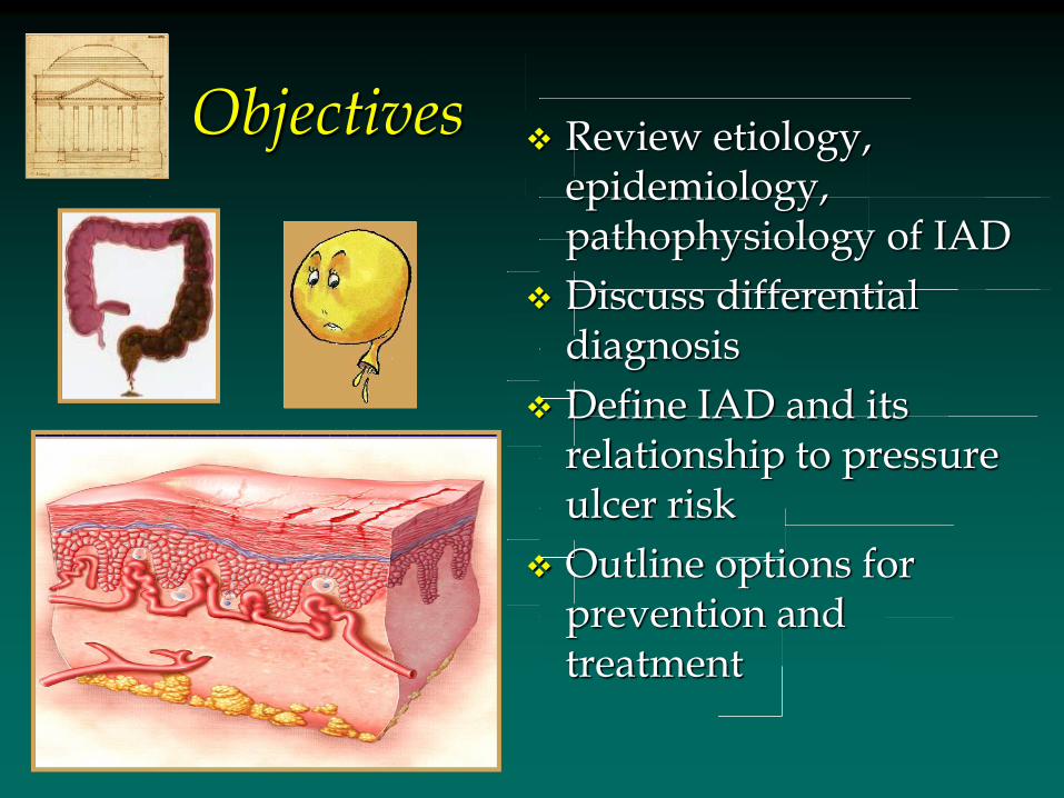
Objectives Review etiology,
epidemiology, pathophysiology of IAD
Discuss differential diagnosis
Define IAD and its relationship to pressure ulcer risk
Outline options for prevention and treatment
Inflammation
Redness
Swelling
Release ofcytokines
Inflammation
Cracking ofskin
Faculty disclosure: none

Functions of the Skin
Thermoregulation
Sensory organ/ communication
Immune functions; acts as a first line of defense
Vitamin D metabolism
Barrier against toxins in external environment and against fluid & electrolyte loss from internal environment
Burns T et al. Textbook of Dermatology, 2004. Mass: Blackwell Science.
Figure: Verdier-Sevrain S, Bonte F. Journal of Cosmetic Dermatology 2007; 6:75.

Definition: Incontinence Associated Dermatitis (IAD)
Irritation and inflammation associated with exposure to stool or urine
Often accompanied by erosion of the skin
Sometimes accompanied by secondary cutaneous infection (ie: candidiasis)
Distinct etiology and pathophysiology
Photograph courtesy Linda Bohacek
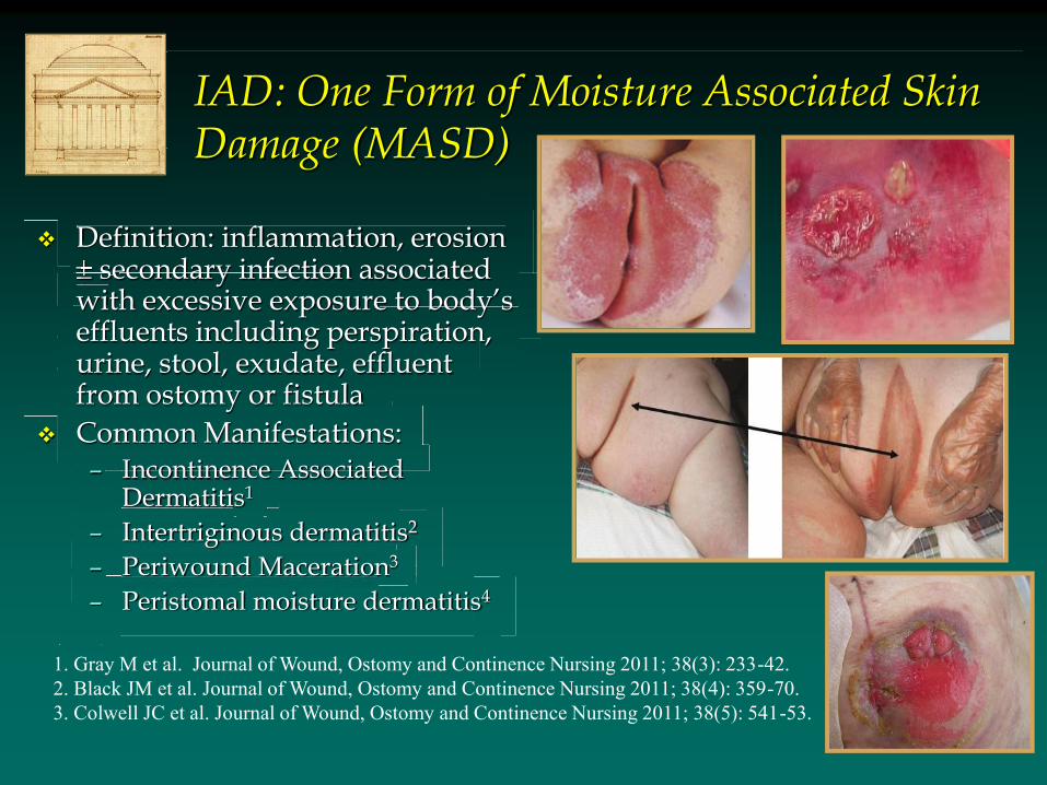
IAD: One Form of Moisture Associated Skin Damage (MASD)
Definition: inflammation, erosion Definition: inflammation, erosion Definition: inflammation, erosion secondary infection associated
with excessive exposure to body’s effluents including perspiration, urine, stool, exudate, effluent from ostomy or fistula
Common Manifestations: – Incontinence Associated
Dermatitis1
– Intertriginous dermatitis2
– Periwound Maceration3
– Peristomal moisture dermatitis4
1. Gray M et al. Journal of Wound, Ostomy and Continence Nursing 2011; 38(3): 233-42. 2. Black JM et al. Journal of Wound, Ostomy and Continence Nursing 2011; 38(4): 359-70. 3. Colwell JC et al. Journal of Wound, Ostomy and Continence Nursing 2011; 38(5): 541-53.

Etiologic Factors: Urine
Water in urine
– skin hardness, rendering it more susceptible to friction and erosion1-3
– Compromises barrier function of skin4
permeability to pathogenic species
permeability to irritants in urine or stool
– Effects exacerbated by presence of occlusive device such as warp around incontinence brief
1. Berg W et al. Pediatric Dermatology 1986; 3: 102. 2. Leyden JJ et al. Archives of Dermatology 1977; 113: 1678. 3. Gray M. Journal of WOC Nursing 2004; 31(1 Suppl):S2-9. 4. Zimmerer RE et al. Pediatric Dermatology 1986; 3: 95.
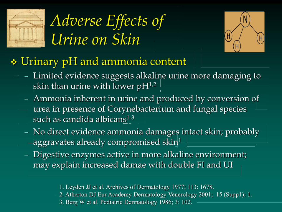
Adverse Effects of Urine on Skin
Urinary pH and ammonia content – Limited evidence suggests alkaline urine more damaging to
skin than urine with lower pH1,2
– Ammonia inherent in urine and produced by conversion of urea in presence of Corynebacterium and fungal species such as candida albicans1-3
– No direct evidence ammonia damages intact skin; probably aggravates already compromised skin1
– Digestive enzymes active in more alkaline environment; may explain increased damae with double FI and UI
1. Leyden JJ et al. Archives of Dermatology 1977; 113: 1678. 2. Atherton DJ Eur Academy Dermatology Venerology 2001; 15 (Supp1): 1. 3. Berg W et al. Pediatric Dermatology 1986; 3: 102.
Adverse Effects of Stool on Skin
Fecal enzymes
– Protease & lipase potentially break down both principal elements of moisture barrier1,2
– In vivo evidence shows that exposure to digestive enzymes in human skin led to3
TEWL
pH
Visible damage only when occlusion present
Evidence of damage present after 12 days
1. Atherton DJ Eur Academy Dermatology Venerology 2001; 15 (Supp1): 1. 2. Gray M. Journal of WOC Nursing 2004; 31(1 Suppl):S2-9. 3. Anderson PH et al. Contact Dermatitis 1994; 30(3): 152.
Associated Factors: Occlusion
Use of absorptive containment devices – Exacerbate overhydration by promoting
perspiration & retaining urine and stool; with padding alone:
TEWL increases 3-4 fold within days
CO2 emission increases > 4 fold
pH increases from 4.4 to 7.1 (without incontinence)
1. Grove GL et al. Clinical Problems in Dermatology 1998; 26:183 2. Zimmerer RE et al. Pediatric Dermatology 1986; 3: 95. 3. Zhai H et al. Skin Research & Technology 2002; 8:13.
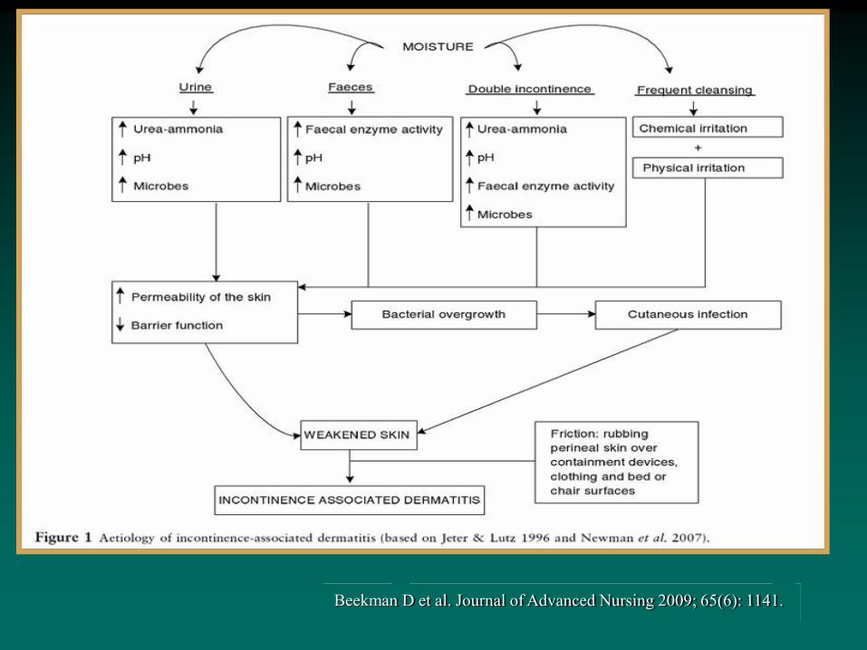
Beekman D et al. Journal of Advanced Nursing 2009; 65(6): 1141.
Associated Factors: IAD & Pressure Ulcers
Association between these conditions is undeniable; nature of relationship remains a mystery
IAD vs Stage II PU may be a problem with differential diagnosis?
IAD impairs skin’s tolerance for pressure/ shear Ongoing debate & controversy about nature of
relationship reflects difficulty differentiating based on visual inspection alone
FI and double incontinence strongly associated with PU risk, mixed evidence concerning UI alone2-6
1. Bates-Jensen BB. Journal of Wound, Ostomy and Continence Nursing, 2009; 36 (3): 277-84. 2. Maklebust J & Magnan MA Advances in Wound Care 1994; 7(6): 25. 3. Gunninberg L. Journal of Wound Care 2004; 13(7): 286. 4. Fader M et al. Journal of Clinical Nursing 2003; 12(3):374. 5. Berlowitz DR et al. Journal of the American Geriatrics Society 2001; 49(7):866-71. 6. Narayan S et al. Journal of WOCN 2005; 32(3): 163.

Epidemiology: Prevalence of IAD
Table from: Gray M et al. Journal of Wound, Ostomy and Continence Nursing 2012; 39(1): 61-74.

Epidemiology of IAD: Incidence
Table from: Gray M et al. Journal of Wound, Ostomy and Continence Nursing 2012; 39(1): 61-74.

IAD: Screening begins with CNA or other non-licensed care providers

IAD: Diagnosis
Primarily based on visual inspection – Inflammation (bright
red) in persons with lighter skin tones
– Located in skin fold or underneath containment device
– Borders are poorly demarcated & irregular
– Surface of skin may “glisten” owing to serous exudate

IAD: Diagnosis in persons with Darker Skin Tones
Inflammation not readily apparent (ie: not bright red); often seen as areas of hyperpigmentation or variable red tones
Hypopigmented areas with chronic inflammation
Pattern of skin damage does not vary
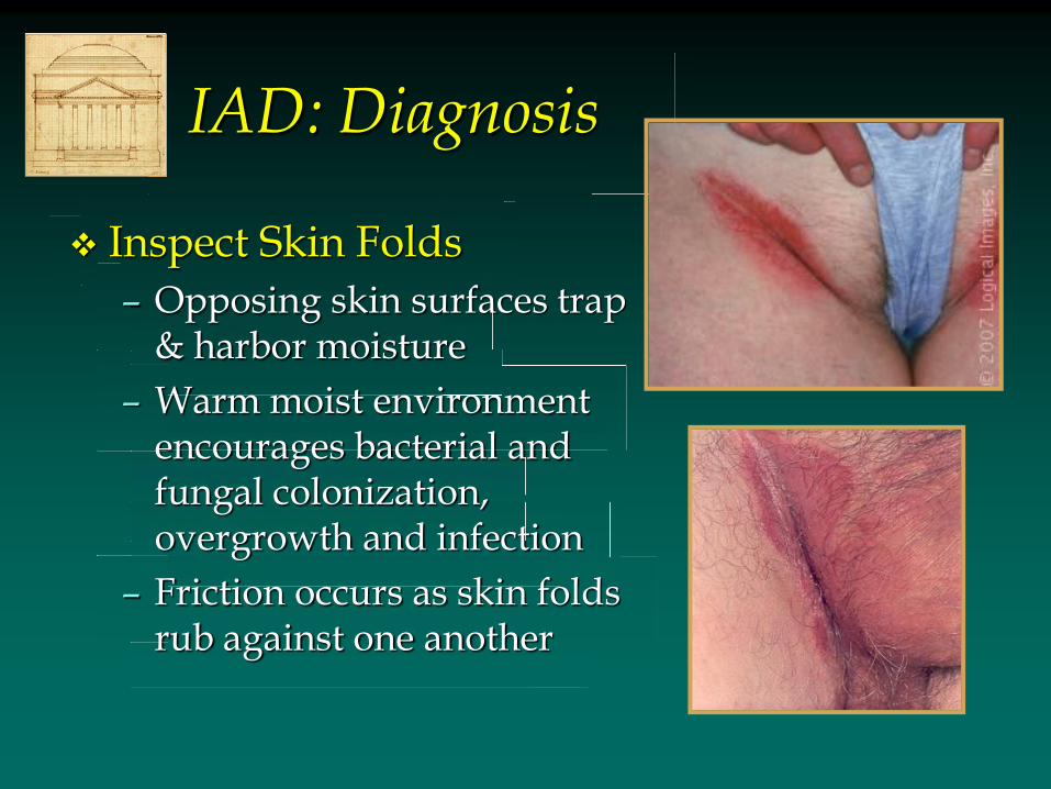
IAD: Diagnosis
Inspect Skin Folds
– Opposing skin surfaces trap & harbor moisture
– Warm moist environment encourages bacterial and fungal colonization, overgrowth and infection
– Friction occurs as skin folds rub against one another
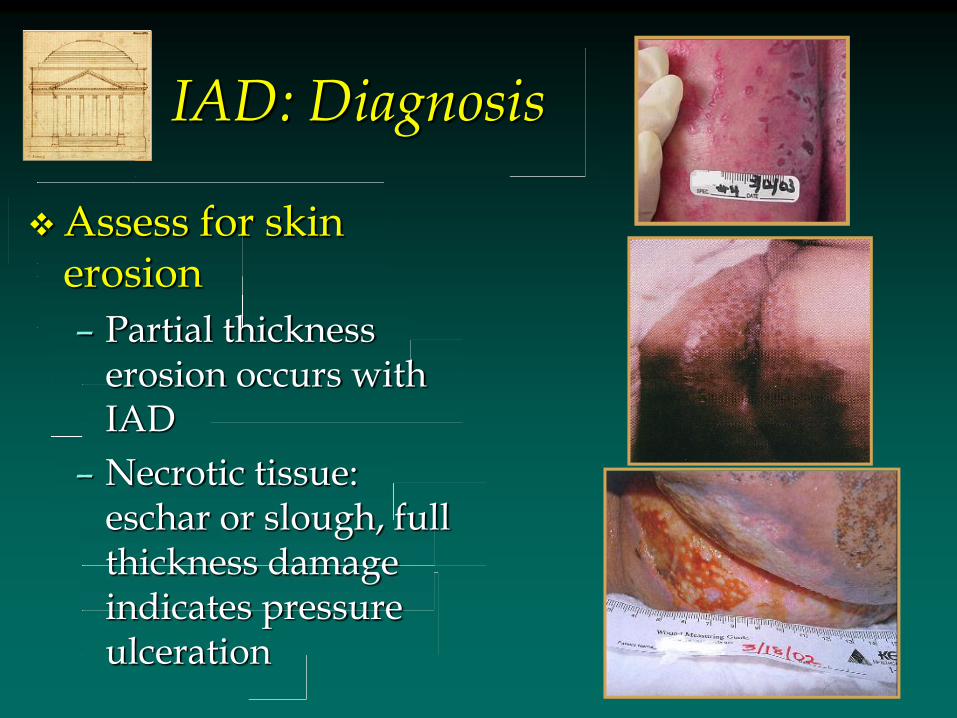
IAD: Diagnosis
Assess for skin erosion
– Partial thickness erosion occurs with IAD
– Necrotic tissue: eschar or slough, full thickness damage indicates pressure ulceration

IAD: Diagnosis
Look for secondary cutaneous infection, especially candidiasis
– Opportunistic infection with candida albicans
– Thrives in warm, moist environment & damages stratum corneum
– Seen in 18% of one group of 976 acute care inpatients1
Junkin J, Selekof J. IAD prevalence in acute care. WOCN National Conference, June 2006 Minneapolis, MN.
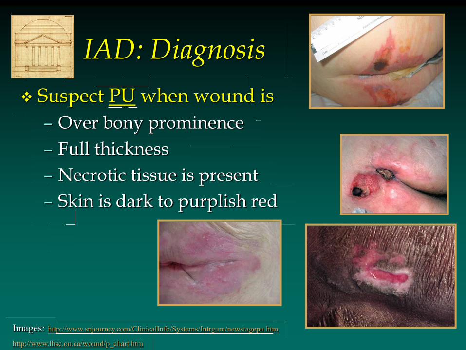
IAD: Diagnosis
Suspect PU when wound is
– Over bony prominence
– Full thickness
– Necrotic tissue is present
– Skin is dark to purplish red
Images: http://www.snjourney.com/ClinicalInfo/Systems/Intrgum/newstagepu.htm
http://www.lhsc.on.ca/wound/p_chart.htm

IAD Diagnosis: Do not Forget the History
Emerging evidence reminds us that isolated photographs are insufficient
The biggest aid in this case is a thorough history
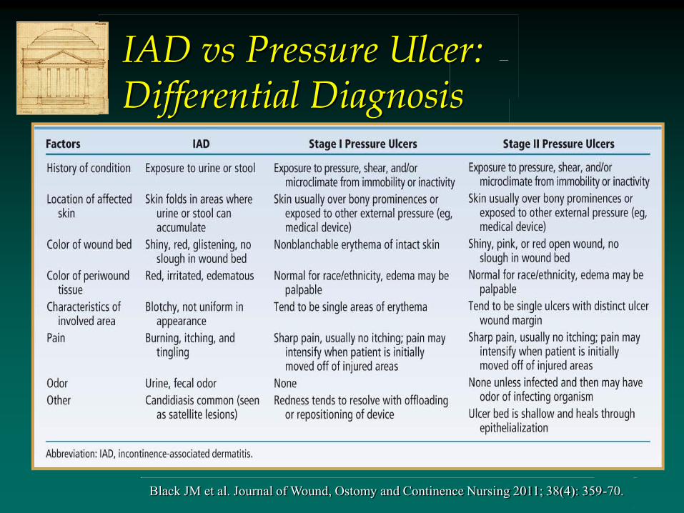
IAD vs Pressure Ulcer: Differential Diagnosis
Black JM et al. Journal of Wound, Ostomy and Continence Nursing 2011; 38(4): 359-70.


