Improved Coomassie Blue Dye-Based Fast StainingProtocol for Proteins Separated by SDS-PAGEPavel Majek*, Zuzana Riedelova-Reicheltova, Klara Pecankova, Jan E. Dyr
Department of Biochemistry, Institute of Hematology and Blood Transfusion, Prague, Czech Republic
Abstract
The time required to visualize proteins using Coomassie Blue dye has been significantly reduced with the introduction offast staining protocols based on staining with a Coomassie Blue dye solution at boiling temperatures. However, faststainings suffer from high gel backgrounds, reducing the signal-to-noise ratio and limiting the number of detectable spotsin the case of 2D SDS-PAGE. The aim of this work was to eliminate the high gel background, and thus improve fast stainingprotocols based on Coomassie Blue dye. We show that merely replacing water with a 4 mM EDTA washing solution atboiling temperatures, results in a transparent gel background within 50 to 60 minutes of destaining. Moreover, when acombination of imidazole-zinc reverse staining and Coomassie Blue-based fast staining is used the sensitivity is improvedsignificantly; nanogram amounts of proteins can be detected using 1D SDS-PAGE, and about 30% to 60% more spots can bedetected with 2D SDS-PAGE in plasma, platelet, and rat brain tissue samples. This work represents an optimized fast stainingprotocol with improved sensitivity, requiring between 60 to 75 minutes to complete protein visualization.
Citation: Majek P, Riedelova-Reicheltova Z, Pecankova K, Dyr JE (2013) Improved Coomassie Blue Dye-Based Fast Staining Protocol for Proteins Separated bySDS-PAGE. PLoS ONE 8(11): e81696. doi:10.1371/journal.pone.0081696
Editor: Andy T. Y. Lau, Shantou University Medical College, China
Received July 10, 2013; Accepted October 15, 2013; Published November 21, 2013
Copyright: � 2013 Majek et al. This is an open-access article distributed under the terms of the Creative Commons Attribution License, which permitsunrestricted use, distribution, and reproduction in any medium, provided the original author and source are credited.
Funding: This study was supported by the Czech Science Foundation P205/12/G118 and by the project (Ministry of Health, Czech Republic) for conceptualdevelopment of research organization (Institute of Hematology and Blood Transfusion). The funders had no role in study design, data collection and analysis,decision to publish, or preparation of the manuscript.
Competing Interests: The authors have declared that no competing interests exist.
* E-mail: [email protected]
Introduction
The visualization of proteins separated by SDS-PAGE is one of
the crucial steps in gel-based proteomics. There are many protein
visualization techniques differing in sensitivity, time required, cost,
complication to perform, etc. Coomassie Blue staining (CBS)
remains one of the most popular protein stainings due to its
simplicity, mass spectrometry compatibility, and low-cost. How-
ever, there are other staining protocols more sensitive than CBS
[1,2]; and moreover, using Coomassie Blue dye can be time
consuming. The time required to visualize proteins using CBS has
been significantly reduced with fast staining protocols utilizing
Coomassie Blue dye. Recently, a protocol introduced by Dong et al.
enabled simple protein visualization, requiring only minutes to
finish the staining procedure [3]. Moreover, Dong’s protocol
avoids the use of organic solvents, and thus represents an excellent
alternative as a simple, timesaving, low-cost, and environmental
friendly solution. Nevertheless, the Dong protocol does not seem
to be as sensitive as traditional colloidal CBS – a high gel
background (reduced signal-to-noise ratio) has been shown to be a
limiting factor [4]. Increasing the destaining time from 10 to
12 hours was recommended in the original protocol [3] to obtain a
clearer background; this would however, lose the advantage of the
accelerated staining procedure. Therefore, we aimed to establish a
fast staining protocol that would address the gel background issue
and retain the advantages of a fast procedure based on Dong’s
protocol. We hypothesized that imidazole-zinc based reverse
staining could play a role as background protection during CBS
based on Dong’s protocol. We further optimized and validated the
procedure using 1D and 2D SDS-PAGE. In this work, we present
an optimized fast staining protocol with improved sensitivity,
which requires just 60 to 75 minutes to complete protein
visualization.
Methods
MaterialsCoomassie Blue G, Acrylamide, and Bis-acrylamide were
purchased from SERVA (Heidelberg, Germany); all other
chemicals were purchased from Sigma-Aldrich (Prague, Czech
Republic) if not otherwise specified.
SamplesFor 1D SDS-PAGE, samples of different BSA dilutions were
prepared. BSA was dissolved in a sample buffer (50 mM Tris-HCl,
4% SDS, 100 mM DTT, 8.5% glycerol, and 0.01% bromophenol
blue) to a 100 mg/ml concentration; the solution was further two-
fold serially diluted. The final BSA samples loaded (10 ml) onto the
gels corresponded to 1000, 500, 250, 125, 62.5, 31.25, 15.6, and
7.8 ng per lane, respectively. Bacterial whole cell lysate (E. coli) was
purchased from Abcam (Cambridge, UK; ab5395). Peripheral
blood mononuclear cells were isolated from whole blood using
Histopaque-1077 (Sigma-Aldrich) according to manufacturer
instructions. Cytosolic, nuclear, membrane, and cytoskeleton
protein fractions were isolated using ProteoExtract Subcellular
Proteome Extraction Kit (Merck, Darmstadt, Germany) according
to manufacturer instructions.
For 2D SDS-PAGE, plasma and platelet samples were
prepared. Blood was collected into tubes coated with EDTA,
PLOS ONE | www.plosone.org 1 November 2013 | Volume 8 | Issue 11 | e81696
and plasma was obtained by centrifugation. Platelets were isolated
according to the protocol by Reicheltova et al. [5]. Plasma was
diluted 1:2 with PBS, platelets were resuspended in PBS (100 ml of
PBS per a platelet pellet from 1 ml of whole blood), and cold
acetone was added to both samples at a ratio of 1:4. Samples were
then incubated at -20 uC overnight, centrifuged (15000 x g, 10
min, 4 uC), protein pellets rinsed with cold acetone; and the pellets
were dissolved in a sample buffer for IEF (7 M urea, 2 M thiourea,
4% CHAPS, 65 mM DTT, 0.5% ampholytes, and a trace of
bromophenol blue). Rat brain tissue lysate was purchased from
Abnova (Taipei, Taiwan; L041W2) and diluted with the sample
buffer for IEF; 50 ug of the sample per strip was used.
Ethics StatementThe blood donor agreed to participate in this study and gave
written informed consent. The study was approved by the Ethical
Committee of the Institute of Hematology and Blood Transfusion
in Prague. All samples were obtained and analyzed in accordance
with the Ethical Committee regulations.
Gel electrophoresisIEF and SDS-PAGE were performed as described previously in
detail [6,7]. Briefly, 10 ml of each BSA sample per lane or 155 ml
of sample per IPG strip (pI 4-7, 7 cm; Invitrogen, Carlsbad, CA,
USA) were used for 1D or 2D SDS-PAGE, respectively. 3.75%
stacking and 10% resolving gels were used at constant current of
25 mA per gel.
Protein stainingFollowing electrophoresis, the gels were reverse stained with
imidazole-zinc based on the protocol by Fernandez-Patron et al.
[8]. Immediately after electrophoresis, the gels were incubated in a
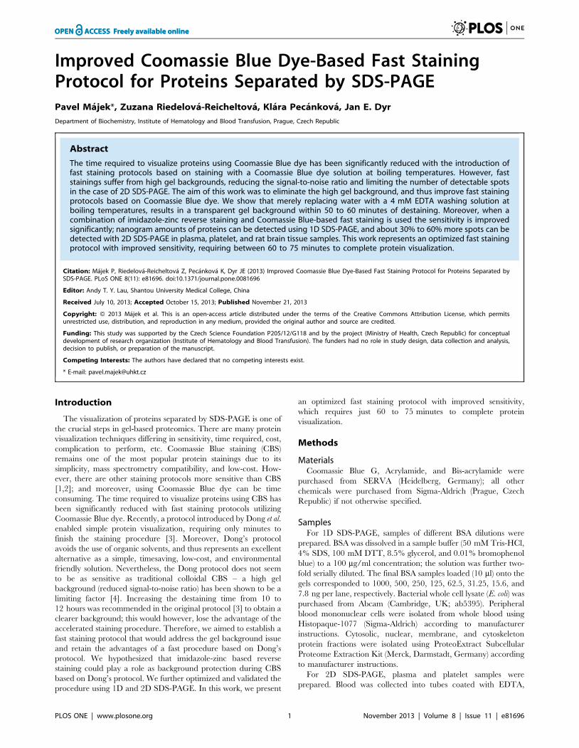
Figure 1. Comparing the effects of different protocols on thedestaining of the gel background and detection of BSA bands.The effects of different protocols on gel background destaining wascompared: gels were stained according to Dong et al. and destained sixtimes for 1 min in boiling water, according to the original protocol (A);gels were stained according to Dong et al. and destained for 1 hr inboiling water (B); gels were stained according to Dong et al. anddestained for 1 hr in a boiling EDTA solution (C); and gels were stainedaccording to our new proposed protocol with imidazole-zinc reversestaining followed by fast CBS, and destained for 1 hr in a boiling EDTAsolution (D). Gel background intensities were compared for all testedprotocols (E), the results are expressed as mean values 6 standarddeviations from three independent experiments ; a, b, c, and dcorrespond to the above mentioned protocols A, B, C, and D,respectively. To estimate the differences in detection of BSA bands ingels stained according to the protocols A (blue), B (red), C (green), andD (purple), corresponding band areas were measured (F); the insetshows more detail illustration for lanes 5–8. The results are expressed asmean values 6 standard deviations from three independent experi-ments. Two-fold serially diluted BSA samples were used for 1D SDS-PAGE and corresponded to 1000, 500, 250, 125, 62.5, 31.25, 15.6, and 7.8ng per lanes 1–8, respectively.doi:10.1371/journal.pone.0081696.g001
Figure 2. The influence of temperature on gel destaining. Gelswere destained with a boiling EDTA destaining solution for 1 hr (1); thecombination of six washes in a boiling EDTA solution for 1 min followedby a 1 hr room temperature EDTA washing solution (2); a roomtemperature EDTA washing solution for 1 hr (3) or overnight (4); and thecombination of six washes in a boiling EDTA solution for 1 min followedby a 6 hr room temperature washing (5). Illustrations of gelbackgrounds after destaining with appropriate procedures are shown(A). Average area intensity values and their standard deviations for gelsdestained using appropriate procedures are presented as estimatedusing ImageJ software (B).doi:10.1371/journal.pone.0081696.g002
Figure 3. The influence of EDTA concentration on geldestaining. Illustrations of gel backgrounds after 1 hr incubationusing different EDTA concentrations (0, 0.5, 1, 2, 4, 8, 16, 32, and 40 mMEDTA solutions, respectively) are shown (A). Average area intensityvalues and their standard deviations for gels destained with corre-sponding EDTA concentrations are presented as estimated usingImageJ software (B).doi:10.1371/journal.pone.0081696.g003
Improved Fast Staining Protocol for SDS-PAGE
PLOS ONE | www.plosone.org 2 November 2013 | Volume 8 | Issue 11 | e81696

solution containing 200 mM imidazole and 0.1% SDS, then
washed with water for 15 sec, and developed by incubation in
200 mM zinc sulfate for approximately 30 sec. Developing was
stopped by a quick wash in excess of water. Reverse stained gels
were then stained with CBS based on the protocol by Dong et al.
[3]. Gels were boiled for 2 min in the staining solution (0.05%
Coomassie Blue G in water) and then destained. The washing
solution was replaced when it turned light blue – that was
approximately every two to five minutes.
Image analysisGels were scanned (16-bit grayscale), and the digitized images
were processed with ImageJ [9] software (1D electropherograms).
Gels from the same batch were compared for the influence of the
temperature and concentration of the disodium EDTA washing
solution on gel destaining. Three technical replicates were used for
the estimation of sensitivity of different protocols using 1D SDS-
PAGE. For 2D SDS-PAGE, Progenesis SameSpots software v3.2
(Nonlinear Dynamics, Newcastle upon Tyne, UK) was used to
evaluate the number of protein spots (results are expressed as
means 6 standard deviations); three technical replicates were used
for the analyses.
Results and Discussion
Recently, it has been shown that a high gel background is the
limiting factor of fast CBS [4]. In an attempt to resolve this issue,
we hypothesized that imidazole-zinc based reverse staining could
play the role of background protection during fast CBS. In reverse
stained gels, the background is covered with the precipitate while
protein bands or spots are not; therefore, access of the Coomassie
Blue dye to the gel background during fast CBS might be limited.
To test this assumption, gels were reverse stained with imidazole-
zinc, followed by fast CBS, and then destained with an EDTA
(disodium salt) solution at a boiling temperature (EDTA was
chosen as a chelating agent removing imidazole-zinc precipitate,
to maintain acidic conditions, and as being odorless at boiling
temperatures). The background of gels was completely destained
(to a clear transparent background) in 50 to 60 minutes. To
maintain comparable conditions for the following protocol
evaluation, the 60 minute washing time was chosen as no more
destaining was observed following this interval. To test the
sensitivity of the protocol, serially diluted BSA samples were used
for 1D SDS-PAGE (from 1000 to 7.8 ng per lane), and the gels
were treated as follows: (A) gels were stained according to Dong
Figure 4. 2D SDS-PAGE of blood platelet, undepleted plasma, and rat brain tissue samples. The effect of an appropriate protocol on gelbackground destaining and spot detection is illustrated using blood platelet, undepleted plasma, and rat brain tissue samples analyzed by 2D SDS-PAGE. The gels were processed according to the original Dong (Protocol A) and the final (Protocol D) protocols. Magnified insets were exported fromthe Progenesis SameSpots software (brightness and contrast of the insets were adjusted by the software) and correspond to the gray areashighlighted in the gels. Spots that were detected in gels stained according to the final protocol D only are indicated with circles or arrows.doi:10.1371/journal.pone.0081696.g004
Improved Fast Staining Protocol for SDS-PAGE
PLOS ONE | www.plosone.org 3 November 2013 | Volume 8 | Issue 11 | e81696

et al. [3] and destained six times for 1 min in boiling water,
according to the original protocol; (B) gels were stained according
to Dong et al. and destained for 1 hr in boiling water; (C) gels were
stained according to Dong et al. and destained for 1 hr in a boiling
EDTA solution; and (D) gels were stained according to our new
proposed protocol – i.e., with imidazole-zinc reverse staining
followed by fast CBS – and destained for 1 hr in a boiling EDTA
solution.
The results are illustrated in Figure 1A-D; the dark gel
background is obvious with gels treated according to the original
Dong protocol (A), prolonging the washing step with boiling water
to 1 hr reduced the gel background (B). Using a combination of
imidazole-zinc and fast CB stainings (D), significant improvement
could be observed, and a clear gel background was obtained.
However, a clear gel background was also observed when the
boiling EDTA solution (C) was used instead of water (B);
moreover, nearly clear gel background can be achieved with
water (B) when increasing the frequency of washing solution
changes. Comparison of gel background intensity values achieved
with the tested protocols (using three replicates) is shown in Figure
1E. It is apparent that prolonging the washing resulted in
reduction of the gel background, particularly for protocols C
and D utilizing the EDTA washing solution. In order to compare
detection of BSA bands in gels stained according to appropriate
protocols, corresponding band areas from three independent
replicates were measured (Figure 1F). It is obvious that using the
EDTA washing solution (protocols C and D) improved the
detection when compared to both protocols A and B. Moreover,
our new proposed protocol D was more effective in detection of
bands, especially those containing low amounts of BSA. Thus,
using an EDTA solution instead of water at a boiling temperature,
and prolonging the washing to 50–60 min, considerably improved
the sensitivity of the Dong protocol [3]; however, the combination
of imidazole-zinc reverse and CB stainings followed by EDTA
destaining further improved detection sensitivity.
The influence of the temperature of the EDTA washing solution
on gel destaining was estimated (Figure 2). Gels were destained
with: (i) a boiling solution, (ii) the combination of six washes in a
boiling solution for 1 min followed by a room temperature
washing solution, or (iii) a room temperature washing solution.
While using the boiling EDTA solution (i) produces a transparent
gel background in 50 to 60 min, washing with room temperature
EDTA (iii) does not result in a clear background even after
overnight washing. With the gels that had been washed six times
for 1 min with a boiling EDTA solution and then followed by a
room temperature solution washing (ii), a transparent gel
background was achieved after six hours. Therefore, using the
EDTA solution at a boiling temperature is of critical importance
and significantly reduces the time required for the destaining step.
The influence of EDTA concentration on gel destaining was
estimated; 0.5, 1, 2, 4, 8, 16, 32, and 40 mM EDTA solutions were
used (Figure 3). After 1 hr incubation destaining was apparent
using 1 mM EDTA; however, complete destaining was achieved
using 4 mM EDTA. Higher EDTA concentrations seemed to
slightly reduce destaining time (maximal decrease of 5 min during
1 hr incubation); however, using concentrations of EDTA more
than 8 mM seemed to have no significant effect. Thus, using
2 mM EDTA provided clear gel backgrounds, but the time
required exceeded 1 hr; increasing the EDTA concentration to
8 mM or higher reduced the washing time by a maximum of only
5 min. Therefore, keeping the protocol effective and at the same
time low-cost, a 4 mM EDTA solution was consequently used as a
destaining solution.
The final protocol is summarized as follows: immediately after
electrophoresis, gels are incubated in a 200 mM imidazole/0.1%
SDS solution for 10 min, then briefly rinsed with water for 15 sec,
and developed by incubation in 200 mM of zinc sulfate for
approximately 30 sec. Developing is stopped by rinsing in an
excess of water. At this point the procedure can be interrupted,
and the gels stored in water in a refrigerator for later use. In the
next step, gels are boiled for 2 min in the staining solution (0.05%
Coomassie Blue G in water) and destained with a 4 mM EDTA
(disodium salt) solution at a boiling temperature until a transparent
gel background is achieved (approximately 50 to 60 min); it is of
critical importance to keep the washing solution at or just below
the boiling point. The washing solution should be changed
regularly as it turns light blue (approximately 3 to 5 min) – longer
durations between changing of the solution may prolong the
destaining process. The duration of gel destaining should not vary
too much among batches as different gel background levels may be
produced and thus hamper gel comparisons.
Using a 1D SDS-PAGE based approach together with BSA
dilution is however, not predictive enough and is not satisfactory
for 2D SDS-PAGE, capable of resolving hundreds to thousands of
proteins [4]. Therefore, 2D SDS-PAGE of blood platelets,
undepleted plasma samples, and rat brain tissue samples (Figure
4) was used to compare the number of protein spots; the original
Dong protocol [3] and the final protocol combining imidazole-zinc
and CB stainings were compared. 520640, 886683, and 497640
spots were detected using the Dong protocol in the plasma,
platelet, and rat brain tissue samples, respectively. Using the final
protocol combining imidazole-zinc and CB stainings, 667619,
1195648, and 785616 spots were detected in the plasma, platelet,
and rat brain tissue samples, respectively. Therefore, there were
28%, 35%, and 58% increases in the number of detected spots in
the plasma, platelet, and rat brain tissue samples, when the final
protocol combining imidazole-zinc and CB stainings was used.
The increase in the plasma sample corresponds to the previously
reported difference between the number of detected spots when
Figure 5. A comparison of bacterial (E. coli) proteins staining.Whole cell lysate (E. coli) was separated by 1D SDS-PAGE and gels werestained according to the original Dong protocol (lane 1) and accordingto our new proposed protocol with imidazole-zinc reverse stainingfollowed by fast CBS and EDTA destaining (lane 2) to illustrate the effectof both protocols. Brightness and contrast of the magnified insets wereadjusted for better illustrations.doi:10.1371/journal.pone.0081696.g005
Improved Fast Staining Protocol for SDS-PAGE
PLOS ONE | www.plosone.org 4 November 2013 | Volume 8 | Issue 11 | e81696
Dong’s protocol and colloidal CBS were compared [4]; a higher
gel background was found to be the reason for the lower number
of detected spots. Thus, these results suggest that using the
combined strategy directly solves the problem of a higher
background; and it is obviously suitable for 2D SDS-PAGE
analyses, as it considerably increases the number of detectable
spots. The results also point out that there may be another factor
contributing to the difference in the number of detected spots.
Several spots were found to disappear from the 2D spot pattern
when the original Dong protocol was used, nevertheless, those
spots were stained properly when our final protocol was used; an
example is illustrated in Figure 4 (panel 4). It is likely that the
missing spots/proteins were washed out from the gel during the
staining and destaining steps. The final protocol prevented this
phenomenon probably due to the previous imidazole-zinc staining
of the gels that eliminated protein elution. In addition to 2D SDS-
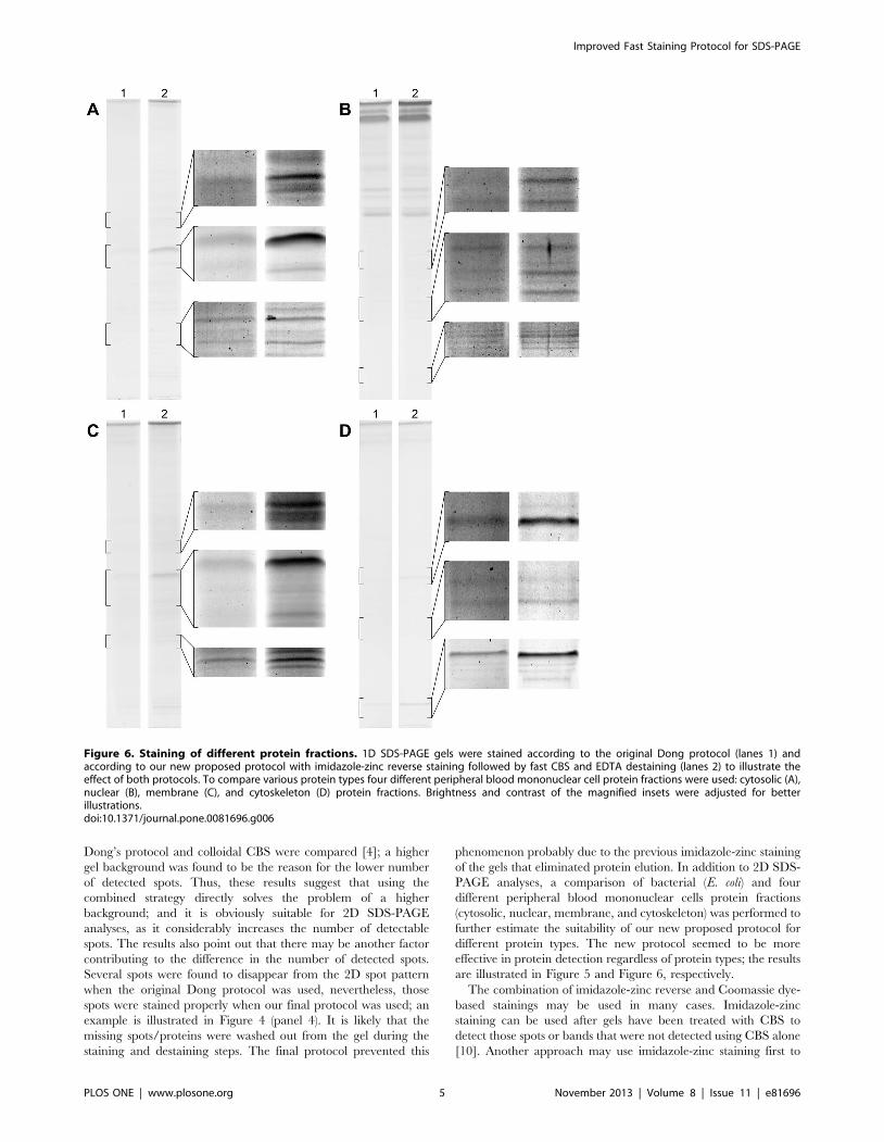
PAGE analyses, a comparison of bacterial (E. coli) and four
different peripheral blood mononuclear cells protein fractions
(cytosolic, nuclear, membrane, and cytoskeleton) was performed to
further estimate the suitability of our new proposed protocol for
different protein types. The new protocol seemed to be more
effective in protein detection regardless of protein types; the results
are illustrated in Figure 5 and Figure 6, respectively.
The combination of imidazole-zinc reverse and Coomassie dye-
based stainings may be used in many cases. Imidazole-zinc
staining can be used after gels have been treated with CBS to
detect those spots or bands that were not detected using CBS alone
[10]. Another approach may use imidazole-zinc staining first to
Figure 6. Staining of different protein fractions. 1D SDS-PAGE gels were stained according to the original Dong protocol (lanes 1) andaccording to our new proposed protocol with imidazole-zinc reverse staining followed by fast CBS and EDTA destaining (lanes 2) to illustrate theeffect of both protocols. To compare various protein types four different peripheral blood mononuclear cell protein fractions were used: cytosolic (A),nuclear (B), membrane (C), and cytoskeleton (D) protein fractions. Brightness and contrast of the magnified insets were adjusted for betterillustrations.doi:10.1371/journal.pone.0081696.g006
Improved Fast Staining Protocol for SDS-PAGE
PLOS ONE | www.plosone.org 5 November 2013 | Volume 8 | Issue 11 | e81696

detect as many spots as possible, followed by complete destaining
of the gel and then using CBS; such an approach would enable
maximal spot detection and quantification using Coomassie dye.
However, this is the first time imidazole-zinc reverse staining is
being used in combination with fast CBS; this protocol solves the
problem of the high gel background after fast CBS, and thus
increases the sensitivity of fast staining protocols based on
Coomassie Blue dye. Moreover, the above described procedure
can be discontinued after imidazole-zinc reverse staining; gels can
be scanned and images of both stainings of the gel can be
obtained. Reverse stained gels can also be stored in distilled water
in a refrigerator, and a batch of gels can be further processed
efficiently by treating them all with boiling CB at once.
Author Contributions
Conceived and designed the experiments: PM ZRR JED. Performed the
experiments: PM ZRR KP. Analyzed the data: PM ZRR KP. Contributed
reagents/materials/analysis tools: PM ZRR KP JED. Wrote the paper:
PM ZRR JED.
References
1. Westermeier R (2006) Sensitive, quantitative, and fast modifications for
Coomassie Blue staining of polyacrylamide gels. Proteomics 6: 61–64. doi:
10.1002/pmic.200690121.
2. Smejkal GB (2004) The Coomassie chronicles: past, present and future
perspectives in polyacrylamide gel staining. Expert Rev Proteomics 1: 381–
387. doi: 10.1586/14789450.1.4.381.
3. Dong WH, Wang TY, Wang F, Zhang JH (2011) Simple, time-saving dye
staining of proteins for sodium dodecyl sulfate-polyacrylamide gel electropho-
resis using Coomassie blue. PLoS One 6: e22394. doi: 10.1371/journal.
pone.0022394.
4. Majek P, Riedelova-Reicheltova Z, Suttnar J, Dyr JE (2013) Staining of proteins
for 2D SDS-PAGE using Coomassie Blue – speed versus sensitivity?
Electrophoresis 34: 1972–1975. doi: 10.1002/elps.201300087.
5. Reicheltova Z, Majek P, Riedel T, Suttnar J, Dyr JE (2012) Simplified platelet
sample preparation for SDS-PAGE-based proteomic studies. Proteomics Clin
Appl 6: 374–381. doi: 10.1002/prca.201100101.
6. Majek P, Reicheltova Z, Stikarova J, Suttnar J, Sobotkova A, et al. (2010)
Proteome changes in platelets activated by arachidonic acid, collagen, andthrombin. Proteome Sci 8: 56. doi: 10.1186/1477-5956-8-56.
7. Majek P, Reicheltova Z, Suttnar J, Maly M, Oravec M, et al. (2011) Plasma
proteome changes in cardiovascular disease patients: novel isoforms ofapolipoprotein A1. J Transl Med 9: 84. doi: 10.1186/1479-5876-9-84.
8. Fernandez-Patron C, Castellanos-Serra L, Hardy E, Guerra M, Estevez E, et al.(1998) Understanding the mechanism of the zinc-ion stains of biomacromole-
cules in electrophoresis gels: generalization of the reverse-staining technique.Electrophoresis 19: 2398–2406. doi: 10.1002/elps.1150191407.
9. Schneider CA, Rasband WS, Eliceiri KW (2012) NIH Image to ImageJ: 25
years of image analysis. Nat Methods 9: 671–675. doi: 10.1038/nmeth.2089.10. Fernandez-Patron C, Hardy E, Sosa A, Seoane J, Castellanos L (1995) Double
staining of coomassie blue-stained polyacrylamide gels by imidazole-sodiumdodecyl sulfate-zinc reverse staining: sensitive detection of coomassie blue-
undetected proteins. Anal Biochem 224: 263–269. doi: 10.1006/abio.
1995.1039.
Improved Fast Staining Protocol for SDS-PAGE
PLOS ONE | www.plosone.org 6 November 2013 | Volume 8 | Issue 11 | e81696


