Lionel Rohit Mathew et al JMSCR Volume 05 Issue 01 January 2017 Page 15884
JMSCR Vol||05||Issue||01||Page 15884-15901||January 2017
Immunohistochemistry in 169 Cases of Pituitary Adenoma
Authors
Lionel Rohit Mathew1, S. Annapurneswari
2, V. S. Mallikarjuna
3
1Dept. of Pathology, Sri Muthukumaran Medical College Hospital and Research Institute, Chikkarayapuram,
Mangadu, Chennai – 600069 2Senior Consultant, Histopathology, Apollo Speciality Hospital, Teynampet, Chennai - 600035
Email: [email protected] 3Chief of Laboratory Services and Senior Consultant, Histopathology, SRM Institute of Medical Sciences,
Vadapalani, Chennai - 600026.
Email: [email protected]
Corresponding Author
Dr Lionel Rohit Mathew
1C2 Happy Windows, A. R. Nagar, Kattupakkam, Chennai - 600056
Email: [email protected], Phone: (044) 49580418, Mobile: 9884192922
Aims and Objectives
To study the morphological and immunohisto-
chemical profile of Pituitary Adenomas in
relation to determination of cell differentiation
& classification, clinical features, radiological
features and biochemical data.
Introduction
The Human Pituitary Gland, despite being a small
bean-shaped organ, is the commander of the major
endocrine glands in the body. It exerts control
over virtually every system and is located
intracranially, close to areas of higher function of
the Central Nervous System.
It is therefore, not surprising, for a Pituitary
Adenoma, the most common tumour arising from
it, to present with a wide range of symptoms that
challenge clinicians of various specialties, who
must assemble all the information they can obtain,
with an open mind, to arrive at the diagnosis.
Materials and Methods
The study was performed in the Department
of Histopathology, Apollo Specialty Hospital,
Chennai. 172 cases of Pituitary Adenoma was
collected over a period of three years (1st
January 2007 to 31st December 2009). Of
these, 169 cases were included in our study.
Year Number of cases studied
2007 47
2008 56
2009 66
Total number of cases studied = 169
Clinical, biochemical and radiological data
were obtained for as many cases as possible.
Criteria for Exclusion
Biopsies showing extensive infarction.
www.jmscr.igmpublication.org
Impact Factor 5.84
Index Copernicus Value: 83.27
ISSN (e)-2347-176x ISSN (p) 2455-0450
DOI: https://dx.doi.org/10.18535/jmscr/v5i1.106
Lionel Rohit Mathew et al JMSCR Volume 05 Issue 01 January 2017 Page 15885
JMSCR Vol||05||Issue||01||Page 15884-15901||January 2017
Histology
Tissue sections from formalin-fixed Paraffin-
Embedded blocks were stained by
Haematoxylin and Eosin and observed by
Light Microscopy.
Immunohistochemistry
4 – micrometre thick paraffin sections were
collected on Triaminopropylethoxysilane-coated
glass slides and subjected to Immunohisto-
chemistry using the Streptavidin-Biotin
Complex Immunoperoxidase method, with the
following Antibodies:
CK - Dako - Mouse Monoclonal
(1/200 dilution)
HGH - Dako - Mouse Monoclonal
(Prediluted)
TSH - Biogenex - Mouse Monoclonal
(Prediluted)
ACTH - OSB - Rabbit Polyclonal
(Prediluted)
FSH - Biogenex - Mouse Monoclonal
(Prediluted)
LH - Biogenex - Mouse Monoclonal
(Prediluted)
PRL - Biogenex - Mouse monoclonal
(Prediluted)
Positive Controls were employed for the
Antibodies as below.
S. No. Antibody Positive Control
1. CK Colonic Mucosa
2. FSH Pituitary, K/c/o Gonadotroph
Adenoma
3. PRL Pituitary, K/c/o Prolactinoma
4. HGH Normal Pituitary
5. TSH Normal Pituitary
6. LH Normal Pituitary
7. ACTH Normal Pituitary
Immunostaining was evaluated in the fields
consisting of regions of the tumour having the
greatest number of immunoreactive cells, as
assessed qualitatively at low-power examination.
Cytoplasmic staining, in > 10% of cells was
regarded as positive.
Review of Literature
The Adenohypophysis, is composed of five
pituitary-specific cell lineages that produce six
trophic hormones1.
Immunohistochemistry of Pituitary Adenoma
identifies the cell-type and enables a functional
classification.2
Epidemiology
Pituitary Adenomas form 10 –15% of Intracranial
Neoplasms. They occur in adults, with 2% of
cases being children. PRL-producing adenomas
are the most common hormone-producing
tumours in both adults and children. ACTH-
producing adenomas are more common in
children. 3
Clinical Features
Clinically, they present with signs and
symptoms of Intracranial mass, visual field
disturbances, hormone excess, deficiency of
hormones from the normal portions of the
gland or failure of target-organ (thyroid, gonad
or adrenal). Mild Hyperprolactinemia (<200
ng/ml) may occur, due to pituitary stalk
compression, called ‘Stalk-Section Effect’ 3, 20
.
Visual disturbance progresses from Superior
Quadranopia, to Bitemporal Hemianopia and
eventually blindness. Sometimes, they may
show suprasellar extension, or extension into
paranasal sinuses and cavernous sinuses to
compress cranial nerves, causing ophthalmo-
plegias. 3 Pituitary Apoplexy may cause sudden
enlargement of an adenoma, resulting in visual
disturbance. 6
Imaging
With its multiplanar capability and dramatic
soft tissue contrast, M. R. I. is the radiological
investigation of choice. 3 mm T1-weighted
coronal images and sagittal images are studied,
before and after administration of Gadolinium
contrast. Computerised Tomography has less
sensitivity and specificity. M. R. I. helps to study
the size, location and extent of tumour. Most

Lionel Rohit Mathew et al JMSCR Volume 05 Issue 01 January 2017 Page 15886
JMSCR Vol||05||Issue||01||Page 15884-15901||January 2017
Pituitary Adenomas produce Suprasellar
Extension. Post-operative Radiological follow-
up is also of use to look for residual or
recurrent tumor.3, 19
Histopathology
Macroscopically, they have a soft, pudding-
like consistency, with calcification seen only
in Prolactinomas.
Microscopically, they display patternless sheets
of uniform cells, with intervening capillary
network. At perivascular areas, they display
columnar shape, regimentation, tapering ends
and polarization of nuclei. Pseudorosettes and
papillae may be seen. The cells stain uniformly.
Nuclei are round to oval and may show ‘salt
and pepper chromatin’.
Classification
A number of attempts have been made, to classify
pituitary Adenomas. The two main groups are
“Clinically Functioning Adenomas” and “Clinic-
ally Non-functioning Adenomas”, according to
whether or not an endocrine syndrome is present.
Two thirds are clinically functioning.5 Non-
functioning adenomas are known to produce
Gonadotropin subunits.¹
According to Tumour size and gross anatomic
features, pituitary adenomas are divided into
microadenomas (<1cm in diameter) and
macroadenomas (>1cm in diameter). Rarely Giant
Adenomas occur (>4cm in diameter)3.
Macroadenomas show an increased tendency
toward suprasellar extension, gross invasion and
recurrence.5
Rarely, Ectopic Adenomas may arise from the
sphenoid sinus, oral mucoperiosteum, nasal
cavity, suprasellar region, or third ventricle.
Pituitary Adenoma may also be a component of an
ovarian teratoma. 6
Morphological classification of Pituitary Aden-
omas depended on the staining characteristics of
the tumour cells. Thus they were designated as
Acidophilic, Basophilic or Chromophobic. But
this classification did not provide information
about the specific adenoma type.5
The
eosinophilic adenomas once considered solely
GH-producing may manufacture GH, PRL, or
both. Alternatively, they may lack hormone
immunoreactivity altogether. Although basophilic
adenomas are PAS-positive and they are usually
associated with Cushing disease, weakly PAS-
reactive tumors may also produce FSH, LH, or
TSH. Lastly, chromophobic adenomas may either
lack hormone staining or may contain the full
range of pituitary hormones. 7
Pituitary Adenomas have a variety of growth
patterns (diffuse, papillary, trabecular, etc) that
may be present in any adenoma type. Although
these histological features are of no prognostic
significance, they may be considered in the
differential diagnosis of Pituitary Adenomas.5
Reticulin stain helps to distinguish normal
Pituitary tissue from adenomatous tisse10
(Images 3,4)
Immunohistochemistry serves to assess the
hormone content of the various Pituitary
Adenomas, but does not discriminate specific
subtypes, which are of prognostic importance. In
such situations, ultrastructural information
becomes necessary.5
Based on Immunohistochemical and Ultrastru-
ctural features, the Pituitary Adenomas have been
classified in the W.H.O. CLASSIFICATION
(2004) ³.
Apart from the Pituitary Hormones, these tumours
may show staining for Synaptophysin and
Chromogranin15
. Proliferating rates may be
assessed using Ki-67 or Proliferating Cell Nuclear
Antigen.9 Matrix Metalloproteinase-9 is
significantly increased in Invasive Pituitary
Adenomas.¹¹ Immunostaining studies are not of
use in infarcted tissue. 6
Cell biology studies have provided abundant data
regarding the nature and behavior of pituitary
adenomas. For instance, X-chromosome
inactivation studies have shown that they are
clonal lesions. DNA ploidy analysis, despite
reports to the contrary, is of limited use in
identifying invasive and aggressive lesions.

Lionel Rohit Mathew et al JMSCR Volume 05 Issue 01 January 2017 Page 15887
JMSCR Vol||05||Issue||01||Page 15884-15901||January 2017
Silver-staining nucleolar organizing region
(AgNOR) analysis is not much more promising.
On the other hand, cell proliferation studies using
flow cytometry (S-phase analysis, as well as Ki67
and bromodeoxyuridine labeling, show a
correlation between elevated indices and the
capacity of a tumor to invade and to metastasize.
The same is true of mitotic indices and p53
protein immunoreactivity. In situ hybridization
permits the identification of hormone mRNA
expression, a modality useful in the study of so-
called silent adenomas. The degree of secretory
activity exhibited by pituitary adenomas shows
little correlation with indicators of cell
proliferation3. The hemolytic plaque assay has
provided useful information regarding rates of
hormone secretion, but it and the more
conventional tissue culture methods remain
experimental modalities. 7
Aetiology
Inherited Genetic abnormalities seen are:
MEN-1 with involvement of 11q13
Carney Complex with involvement of
2p16 and 17q
Familial acromegaly with involvement of
11q13 and other loci
McCune-Albright syndrome with
involvement of 20q13.2
Increased production of the hypothalamic
releasing hormones can give rise to pituitary
adenomas.
Abnormal hormone receptors in the pituitary
gland and abnormal signal-transduction can also
lead to pituitary adenomas. 3
Molecular Pathogenesis
Point mutations in the α-subunit of the stimulatory
G-protein (Gsα), leads to the oncogene gsp, which
encodes for a constitutively active form of Gsα,
leading to elevated levels of cyclicAMP. This
mutation is seen in 40% of somatotroph adenomas
and a few non-functional and corticotroph
adenomas. Hypermethylation of p16 in
gonadotroph and null-cell adenomas.
Mutations in the α-subunit of the inhibitory GTP-
binding protein, GI2α, resulting in the oncogene
gip2, is also seen in some tumours.8
Silencing of the RB1 promoter is seen, by means
of methylation at a CpG island.
There is reduced p27 expression in ACTH
producing adenomas.
A truncated kinase-containing variant of
Fibroblast Growth Factor Receptor, bearing an
alternative initiation site, (known as ‘Pituitary-
derived Kinase’ or ptd-FGFR4) is expressed in
most tumours.
Other facors involved are oncogenes PTTG and,
Cyclin D1.
The transcription factor PIT-1 is found within
cells of the acidophil cell line, which are
somatotrophs, lactotrophs and thyrotrophs. This
may explain the presence of these cells in
plurihormonal adenomas.
Therapy
Surgery is the main modality of the treatment, and
is necessary to relieve the pressure-effects of the
tumour. The usual approach is trans-sphenoidal
minimally-invasive procedure. Medical therapy is
used initially, for patients with GH-producing and
PRL-producing adenomas. Radiation therapy used
to be performed before the trans-sphenoidal route
was in vogue, but its side-effects are
hypopituitarism and radiation-induced glioma and
sarcoma.3, 18
Gamma knife stereotactic surgery
may have a role in recurrent or residual disease
and in pituitary carcinomas.3
Results, Observations & Analysis
Proportion of Each Tumour Type
The distribution of cases in each tumour
category as classified by Immunohistoc-
hemistry is as given in Figure 1.

Lionel Rohit Mathew et al JMSCR Volume 05 Issue 01 January 2017 Page 15888
JMSCR Vol||05||Issue||01||Page 15884-15901||January 2017
Figure 1 - Proportion of Each Tumour Type
Age
Despite a wide age range (16 - 77), the
maximum incidence was noted in the fourth to
sixth decade. The mean age at diagnosis of
each individual tumour type was studied . The
Kruskal-Wallis test showed a highly significant
statistical difference with a P value of
<0.00001. Cases of Prolactinoma, Somatotroph
Adenoma and Corticotroph Adenoma showed
a significant lower mean age compared to
Gonadotroph Adenoma, Plurihormonal Aden-
oma and Null Cell Adenoma.
Table 1 - Age Distribution
Diagnosis Age Range Mean Age Median Age
Somatotroph Adenoma 28-62 38.9 35.0
Prolactinoma 18-67 38.0 34.5
Mixed GH-PRL Adenoma 35-44 38.0 35.0
Thyrotroph Adenoma 48 48.0 48.0
Corticotoph Adenoma 16-53 35.8 36.5
Gonadotroph Adenoma 23-77 49.9 50.5
Plurihormonal Adenoma 16-72 49.5 54.0
Null-Cell Adenoma 21-65 43.2 43.0
ALL 16-77 44.5 43.0
Table 2 - Incidence in Age Groups Age Group Total Male Female L S SL T C G P N
≤19 3 1 2 1 - - - 1 - 1 -
20-29 21 11 10 8 1 - - 4 2 4 2
30-39 43 20 23 4 7 2 - 8 10 6 6
40-49 37 19 18 4 1 1 1 2 6 9 13
50-59 37 26 11 6 1 - - 3 9 13 5
60-69 22 14 8 1 1 - - - 6 13 1
≥70 6 5 1 - - - - - 5 1 -
L = Prolactinoma, S = Somatotroph, SL = Mixed GH/PRL, T = Thyrotroph, C = Corticotroph, G = Gonadotroph, P =
Plurihormonal, N = Null Cell
S 6%
L 14%
SL 2%
T 1%
C 11%
G 22%
P 28%
N 16%
S = Somatotroph
L = Prolactinoma
SL = Mixed GH/PRL
T = Thyrotroph
C = Corticotroph
G = Gonadotroph
P = Plurihormonal
N = Null Cell
Lionel Rohit Mathew et al JMSCR Volume 05 Issue 01 January 2017 Page 15889
JMSCR Vol||05||Issue||01||Page 15884-15901||January 2017
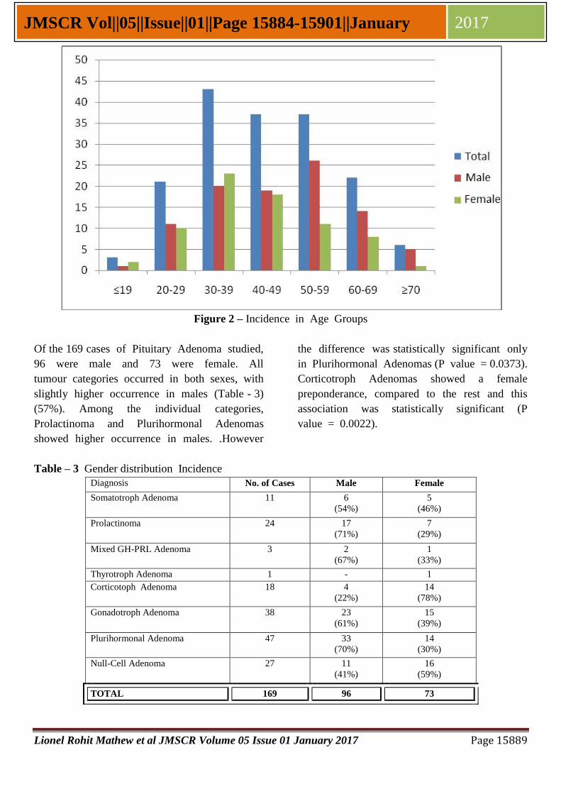
Figure 2 – Incidence in Age Groups
Of the 169 cases of Pituitary Adenoma studied,
96 were male and 73 were female. All
tumour categories occurred in both sexes, with
slightly higher occurrence in males (Table - 3)
(57%). Among the individual categories,
Prolactinoma and Plurihormonal Adenomas
showed higher occurrence in males. .However
the difference was statistically significant only
in Plurihormonal Adenomas (P value = 0.0373).
Corticotroph Adenomas showed a female
preponderance, compared to the rest and this
association was statistically significant (P
value = 0.0022).
Table – 3 Gender distribution Incidence
Diagnosis No. of Cases Male Female
Somatotroph Adenoma 11 6
(54%)
5
(46%)
Prolactinoma 24 17
(71%)
7
(29%)
Mixed GH-PRL Adenoma 3 2
(67%)
1
(33%)
Thyrotroph Adenoma 1 - 1
Corticotoph Adenoma 18 4
(22%)
14
(78%)
Gonadotroph Adenoma 38 23
(61%)
15
(39%)
Plurihormonal Adenoma 47 33
(70%)
14
(30%)
Null-Cell Adenoma 27 11
(41%)
16
(59%)
TOTAL 169 96 73

Lionel Rohit Mathew et al JMSCR Volume 05 Issue 01 January 2017 Page 15890
JMSCR Vol||05||Issue||01||Page 15884-15901||January 2017
Duration
Details on duration of symptoms was available
in 103 cases. It was highly variable, ranging
from days to years. Patients presented with
symptoms of either intracranial mass lesion,
hormonal symptoms, or both. Symptoms due
to intracranial mass lesion were Headache,
visual field defects notably Bitemporal
Hemianopia, Vomiting or Giddiness and
patients with these symptoms presented earlier
than those with hormonal symptoms. Patients
with acromegalic features presented with a
mean duration of 45.2 months, while patients
with hypothyroid, or lactational symptoms
presented within the first year of onset.
On studying the duration of symptoms for
each tumour type, the overall variation was
not statistically significant.
Figure 3 - Symptomatology
Table 4 – Duration of Symptoms
(Data Available = 84)
S. No. Tumour Type
No. of
Cases
Duration (months)
Range Mean Median
1. Prolactinoma 11 1 – 120 22.6 12
2. Somatotroph 4 2 – 60 33.5 36
3. Mixed GH/PRL 1 12 12.0 12
4. Corticotroph 6 1 – 144 31.7 8
5. Gonadotroph 21 1 – 48 9.8 6
6. Plurihormonal 26 1 – 120 30.9 9
7. Null Cell Adenoma 15 1 – 48 12.7 6
ALL 84 1 – 144 21.25 6
Radiology
Radiological information on tumour size was
available in 66 cases (Table - 5). Most of the
tumours were Macroadenomas. Of the 5 Giant
Adenomas seen, 4 were Plurihormonal
Adenomas and 1 was Prolactinoma.
Table 5 - Radiological Classification
(Data Available = 66)
S. No. Size No. of Cases
1. Microadenoma 1
2. Macroadenoma 60
3. Giant Adenoma 5

Lionel Rohit Mathew et al JMSCR Volume 05 Issue 01 January 2017 Page 15891
JMSCR Vol||05||Issue||01||Page 15884-15901||January 2017
The most common pattern of tumour extent
was Suprasellar (seen in 62% of cases with
available data). 31% showed involvement of
Optic Chiasma.
Details on Radiological follow-up was
available in 29 cases, of which 5 cases
showed residual/recurrent lesion. In this
category, 4 were Plurihormonal Adenomas,
and one was Gonadotroph Adenoma..
Biochemical Studies
Serum Prolactin levels were mildly elevated in
tumours other than Prolactinomas, in keeping
with the Stalk-Section Effect. Serum HGH or
IGF were elevated in 3 cases of Somatotroph
Adenoma and 3 cases of Plurihormonal
Adenoma.
Most of the patients underwent Endoscopic
Transnasal Transsphenoidal Surgery.
Microscopy
On H & E stained sections, the architectural
pattern of most tumours were predominantly
sinusoidal . Gonadotroph, Plurihormonal and
Null Cell Adenomas showed sinusoidal or
Papillary Pattern, with Pseudorosettes also.
Corticotroph Adenomas had arrangement of
cells in sinusoids as well as diffuse sheets.
Few Prolactinomas showed Amyloid and
Calcospherites also.
Cytoplasmic Staining was observed as below
(Table 6, Figure 4).
Table 6 - Cytoplasmic Staining Properties
Cytoplasm
Prolactinom
a Somatotroph
Mixed
GH/PRL Corticotroph Gonadotroph
Plurihormon
al Null Cell
Clear 1 0 0 0 0 0 1
Pale 4 1 0 3 29 29 17
Eosinophilic 19 10 3 7 9 17 8
Pale Amphophilic 0 0 0 1 0 1 1
Amphophilic 0 0 0 7 0 0 0
Figure 4 - Cytoplasmic Staining
0
5
10
15
20
25
30
35
L S SL C G P N
Clear Cytoplasm
Pale Cytoplasm
Eosinophilic Cytoplasm
Pale Amphophilic Cytoplasm
Amphophilic Cytoplasm

Lionel Rohit Mathew et al JMSCR Volume 05 Issue 01 January 2017 Page 15892
JMSCR Vol||05||Issue||01||Page 15884-15901||January 2017
Nuclei were mostly uniform, with mild
pleomorphism and prominent nucleoli being
noted in Prolactinomas and Somatotroph
Adenomas.
Of the 24 Prolactinomas, 8 had biochemical
correlation with serum PRL level. Seven of
these had elevated levels, with 6 of them, in
excess of 100 ng/dl and 5 above 200 ng/dl.
All these 7 cases showed strong immunopos-
itivity for PRL in >60% of tumour cells. Two
of these cases also showed significant drop in
PRL levels after surgery.
Of the 11 Somatotroph Adenomas, 4 showed
acromegalic features. Biochemical correlation
with serum levels of HGH was available in 4
cases, 3 of whom showed raised levels of
HGH, with one of them showing elevated
serum IGF level also.
Of the Plurihormonal Adenomas, the commo-
nest combination of hormone immunopositivity
was FSH, LH and TSH, which was seen in
26 cases (55%). 18 cases had FSH and PRL
positivity (38%).
Seven cases of our study were recurrent cases,
of which 4 were Pluihormonal Adenomas.
Image 1. The normal Pituitary Gland, H & E 10x
Image 2. The Normal Pituitary Gland.
Multiplicity of cell-types is appreciated. H & E,
40x
Image 3. Normal Pituitary Gland Reticulin Stain
10x
Image 4. Pituitary Adenoma Reticulin Stain 10x
Image 5. Prolactinoma H & E, 40x

Lionel Rohit Mathew et al JMSCR Volume 05 Issue 01 January 2017 Page 15893
JMSCR Vol||05||Issue||01||Page 15884-15901||January 2017
Image 6. Prolactinoma, PRL, 40x
Image 7. Prolactinoma - Amyloid H & E, 10x
Image 8. Prolactinoma, Amyloid, Congo Red,10
x
Image 9. Prolactinoma Congo Red, Polarized, 10x
Image 10. Somatotroph Adenoma H & E, 40x
Image 11. Somatotroph Adenoma Dot Positivity,
CK 40x
Image 12. Somatotroph Adenoma, HGH 40x
Image 13. Mixed GH/PRL Adenoma H & E, 10x

Lionel Rohit Mathew et al JMSCR Volume 05 Issue 01 January 2017 Page 15894
JMSCR Vol||05||Issue||01||Page 15884-15901||January 2017
Image 14. Mixed GH/PRL Adenoma HGH, 40x
Image 15. Gonadotroph Adenoma H & E 40x
Image 16. Gonadotroph Adenoma LH 40x
Image 17. Corticotroph Adenoma H & E 40x
Image 18. Corticotroph Adenoma ACTH 40x
Image 19. Plurihormonal Adenoma Sinusoidal
Pattern, H & E 40x
Image 20. Plurihormonal Adenoma HGH 10x
Image 21. Plurihormonal Adenoma FSH 40x

Lionel Rohit Mathew et al JMSCR Volume 05 Issue 01 January 2017 Page 15895
JMSCR Vol||05||Issue||01||Page 15884-15901||January 2017
Image 22. Plurihormonal Adenoma PRL 40x
Image 23. Plurihormonal Adenoma PRL 40x
Image 24. Null Cell Adenoma Papillary and Pseudorosette Pattern, H & E 40x

Lionel Rohit Mathew et al JMSCR Volume 05 Issue 01 January 2017 Page 15896
JMSCR Vol||05||Issue||01||Page 15884-15901||January 2017
2007

Lionel Rohit Mathew et al JMSCR Volume 05 Issue 01 January 2017 Page 15897
JMSCR Vol||05||Issue||01||Page 15884-15901||January 2017
2008

Lionel Rohit Mathew et al JMSCR Volume 05 Issue 01 January 2017 Page 15898
JMSCR Vol||05||Issue||01||Page 15884-15901||January 2017
2009

Lionel Rohit Mathew et al JMSCR Volume 05 Issue 01 January 2017 Page 15899
JMSCR Vol||05||Issue||01||Page 15884-15901||January 2017
Discussion
The ratio of the various tumour types, seen in
the study conducted by Osamura, Kajiya and
Takei1
showed a preponderance of Non-
Functioning Adenomas, which comprised Null
Cell Adenomas and Oncocytomas1, 19
, which
was followed, in order, by Somatotroph
Adenomas, Prolactinomas, Corticotroph Adeno-
mas and Gonadotroph Adenomas. Plurihorm-
onal Adenomas were very rare in Western
literature3, 9
. In our study, the commonest
tumour was Plurihormonal Adenoma (28%),
followed by Gonadotroph Adenoma (22%),
Null Cell Adenoma (16%), Prolactinoma
(14%) and Corticotroph Adenoma (11%).
Plurihormonal Adenomas had a frequency of
36.1% in the study conducted by Pawilowski
et al19
and 31% in the study of Ho et al21
.
71.5% of cases occurred during the fourth,
fifth and sixth decades. Corticotroph
Adenomas had a relatively younger mean age
of occurrence (35.8), which was in accordance
with 30 to 40, mentioned in literature
available3. The mean age of presentation of
Null Cell (43.2), Plurihormonal (49.5) and
Gonadotroph Adenomas (49.9) was in
accordance with the mean ages described in
literature 19, 21, 12
.
All tumours occurred in both sexes, with a
slight male preponderance. Plurihormonal
Adenomas showed a statistically significant
male preponderance. Corticotroph Adenomas
were commoner in females, which was in
accordance with literature3.
In patients for whom clinical history was
available, 77% presented with signs and
symptoms of symptoms of intracranial mass
lesion (Headache, Bitemporal Hemianopia) in
comparison with 74% of patients with only
visual field defects, in the study conducted by
Cury et al.19
. Patients with these symptoms
presented earlier than those with only
endocrine symptoms.
In patients with available radiological data,
91% presented with Macroadenoma. Most
cases showed Suprasellar Extension.
Involvement of Optic Chiasma, seen in 31%
of cases with available data, could account for
Visual Field Defects.
Serum PRL levels were available in 60 cases,
of which 27 showed elevated values. Values
below 200 ng/dl, in non-prolactinomas, could
be attributed to the stalk-section effect3. Of
the 5 cases in our study with values above
200 ng/dl, 4 were Prolactinomas and one was
Plurihormonal Adenoma.
Reduction in values of T4 were noted in non-
thyrotroph adenomas, explaining the deficiency
of hormones produced by the uninvolved
Pituitary gland3.
Most tumours showed predominantly Sinuso-
idal Pattern, with Corticotroph Adenomas
forming Diffuse sheets and Gonadotroph,
Plurihormonal and Null Cell Adenomas7
showing Papillary and Pseudorosette patterns.
Cytoplasmic Staining Properties on H & E
were consistent with the Histologic
Classification of the various Tumour types7.
Only one case of Thyrotroph Adenoma was
seen (48/F).
Most Plurihormonal Adenomas expressed FSH
hormone immunohistochemically. The comm.-
onest combination of hormone immunop-
ositivity seen was FSH, LH and TSH (55%).
Plurihormonal Adenomas were the commonest
tumour types known to recur, as seen in
literature1.
Conclusions
Pituitary Adenomas can be classified
immunohistochemically.
Patients with symptoms of Intracranial Mass
Lesion present earlier than those with
endocrine symptoms.
Plurihormonal Adenoma is the most common
type of tumour in our study Population. They
are most likely to recur.
The most common combination of hormone
immunopositivity in Plurihormonal Adenomas
seen in our study is FSH, LH and TSH.
Lionel Rohit Mathew et al JMSCR Volume 05 Issue 01 January 2017 Page 15900
JMSCR Vol||05||Issue||01||Page 15884-15901||January 2017
Corticotroph Adenomas are relatively
commoner in females and Plurihormonal
Adenomas in males.
Radiologic and Biochemical correlation and
correlation with H & E stained light
microscopy is necessary to explain
symptomatology
Acknowledgements
We are grateful to Dr. Moses Ambroise and
Dr. Anil Tarigopula for their valuable inputs.
The Departments of Neurosurgery, Histopat-
hology , Biochemistry, Radiology and Medical
Records were co-operative and helpful.
References
1. Robert Y. Osamura, Hanako Kajiya, Mao
Takei et al. Pathology Of The Human
Pituitary Adenomas Histochem Cell Biol
(2008) 130: 495-507
2. R. Yoshiyuki Osamura, Shigeyuki Tahara,
Reiko Kurotani, et al. Contributions of
Immunohistochemistry and In Situ
Hybridization to the Functional Analysis
of Pituitary Adenomas, J Histochem
Cytochem 2000; 48:445–458,
3. E. Horvath, K. Kovacs, R. V. Lloyd et al,
Tumours Of The Pituitary Gland In: World
Health Organization Classification of
Tumours – Pathology and Genetics of
Tumours Of Endocrine Organs 2004 pp
9-48.
4. J M Bilbao. Pituitary Gland. In: Juan
Rosai, editor. Rosai and Ackerman's
Surgical Pathology 2004 9th
ed. pp: 2683
- 2712
5. M. Beatrix S. Lopes. Tumors of the
Pituitary Gland. In : C D M Fletcher,
Diagnostic Histopathology Of Tumours 3rd
ed, 2007, Massachussets, Churchill
Livingstone, pp 971 – 996.
6. B. W. Scheithauer , P. C. Burger, F. S.
Vogel, Surgical Pathology Of The
Nervous System And Its Coverings, 4th
ed,
2004, Churchill Livingstone, PA, USA pp
437 -498.
7. B. W. Scheithauer, The Pituitary and
Sellar Region, In: Stacey E. Mills
Sternberg’s Diagnostic Surgical Pathology
4th
Edn, 2004, Lippincott Williams and
Wilkins, PA, USA, pp 521 – 556.
8. Disorders Of The Anterior Lobe of
Pituitary, In: David Lowe, James
Underwood, Recent Advances in
Histopathology 19; pp1-13.
9. Wolfgang Saeger, Dieter K Lüdecke,
Michael Buchfelder et al.
Pathohistological classification of pituitary
tumors: 10 years of experience with the
German Pituitary Tumor Registry.
European Journal of Endocrinology 156:
203–216.
10. Raymond V. Randall, Bernd W.
Scheithauer, Kalman Kovac. Pituitary
Adenomas: Historic Considerations In:
Kamal Thapar Diagnosis and Management
of Pituitary Tumours . New Jersey:
Humana Press 2001. pp 1-12.
11. Isa M. Hussaini, Christy Trotter, Yunge
Zhao et al. Matrix Metalloproteinase-9 Is
Differentially Expressed in Nonfunct-
ioning Invasive and Noninvasive Pituitary
Adenomas and Increases Invasion in
Human Pituitary Adenoma Cell Line. Am J
Pathol 2007, 170:356–365.
12. E. Horvath, K. Kovacs. Gonadotroph
Adenomas of the Human Pituitary: Sex-
Related Fine-Structural Dichotomy; A
Histologic, Immunocytochemical, and
Electron-Microscopic Study of 30
Tumors; Am J Pathol 1984, 117:429-440
13. Katsuya Umeoka, Naoko Sanno, R
Yoshiyuki Osamura,
et al. Expression of
GATA-2 in Human Pituitary Adenomas
Mod Pathol 2002;15(1):11–17
14. Zhi Rong Qian, Chiun Chei Li, Hiroyuki
Yamasaki,, et al, Role of E-Cadherin, α-,
β-, and γ-Catenins, and p120 (Cell
Adhesion Molecules) in Prolactinoma

Lionel Rohit Mathew et al JMSCR Volume 05 Issue 01 January 2017 Page 15901
JMSCR Vol||05||Issue||01||Page 15884-15901||January 2017
Behavior, Mod Pathol 2002;15(12):1357–
1365.
15. Marek Pawlikowski, Anna Gruszka,
Maciej Radek et al, Chromogranin A in
pituitary adenomas: immunohistochemical
detection and plasma Concentrations,
Folia Histchemica et Cytobiologica, Vol.
42, No. 4, 2004, pp. 245-247.
16. Oyama K., Sanno N., Teramoto A. et al.
Expression of Neuro D1 in Human Normal
Pituitaries and Pituitary Adenomas, Mod
Pathol 2001;14(9):892–899.
17. Heaney AP, Fernando M, Melmed S:
PPAR-γ receptor ligands: novel therapy
for pituitary adenomas, J. Clin. Invest.
2003 111:1381–1388.
18. Pawlikowski M, Kunert-Radek J, Radek
M.: Plurihormonality of Pituitary
Adenomas in light of Immunohistoc-
hemical Studies, Endokrynol Pol 2010; 61
(1): 63-66.
19. M. L. C. R. Cury, J. C. Fernandes, H. R.
Machado, L. L. Elias, et al: Non-
functioning pituitary adenomas: clinical
feature, laboratorial and imaging
assessment, therapeutic management and
outcome; Arq Bras Endocrinol Metab.
2009;53/1
20. K Kovacs, E Horvath, S Vidal.
Classification of pituitary adenomas
Journal of Neuro-Oncology , 2001 54:
121–127.
21. T Ho, Y Hsu, T Ting et al, Plurihormonal
Pituitary Adenomas: Immunostaining of
all Pituitary Hormones is mandatory for
Correct Classification, Histopathology
2001 Sep;39(3):310-319.


