www.my-ray.com
Plant - Via Bicocca, 14/c - 40026 Imola - Bo (Italy) tel. +39 0542 653441 - fax +39 0542 653555
Headquarters - Cefla s.c. Via Selice Provinciale, 23/a - 40026 Imola - Bo (Italy) tel. +39 0542 653111 - fax +39 0542 653344
Cefla North America, Inc. - 6125 Harris Technology Blvd. Charlotte, NC 28269 - U.S.A. Toll Free: (+1) 800.416.3078 Fax: (+1) 704.631.4609
Hyperion X5
3D/2D2D
Suspended imaging system
Dat
a su
bjec
t to
chan
ge w
ithou
t not
ice.
02/
2016
M
X53D
GB1
61S0
0
Hyperion X5, 3D/2D imaging.2 3
WALL-MOUNTED
MRT
(Patented)
10x103D HD-QuickSCAN
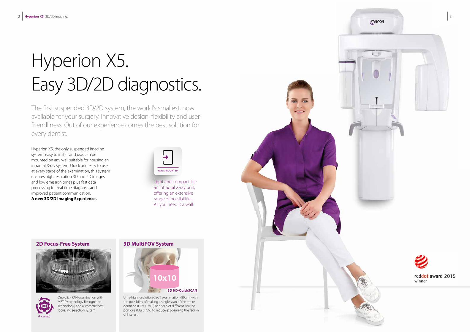
Hyperion X5, the only suspended imaging system, easy to install and use, can be mounted on any wall suitable for housing an intraoral X-ray system. Quick and easy to use at every stage of the examination, this system ensures high resolution 3D and 2D images and low emission times plus fast data processing for real time diagnosis and improved patient communication. A new 3D/2D Imaging Experience.
Hyperion X5. Easy 3D/2D diagnostics.The first suspended 3D/2D system, the world’s smallest, now available for your surgery. Innovative design, flexibility and user-friendliness. Out of our experience comes the best solution for every dentist.
Light and compact like an intraoral X-ray unit, offering an extensive range of possibilities. All you need is a wall.
2D Focus-Free System 3D MultiFOV System
One-click PAN examination with MRT (Morphology Recognition Technology) and automatic best focussing selection system.
Ultra-high resolution CBCT examination (80μm) with the possibility of making a single scan of the entire dentition (FOV 10x10) or a scan of different, limited portions (MultiFOV) to reduce exposure to the region of interest.
Hyperion X5, 3D/2D imaging.4 5
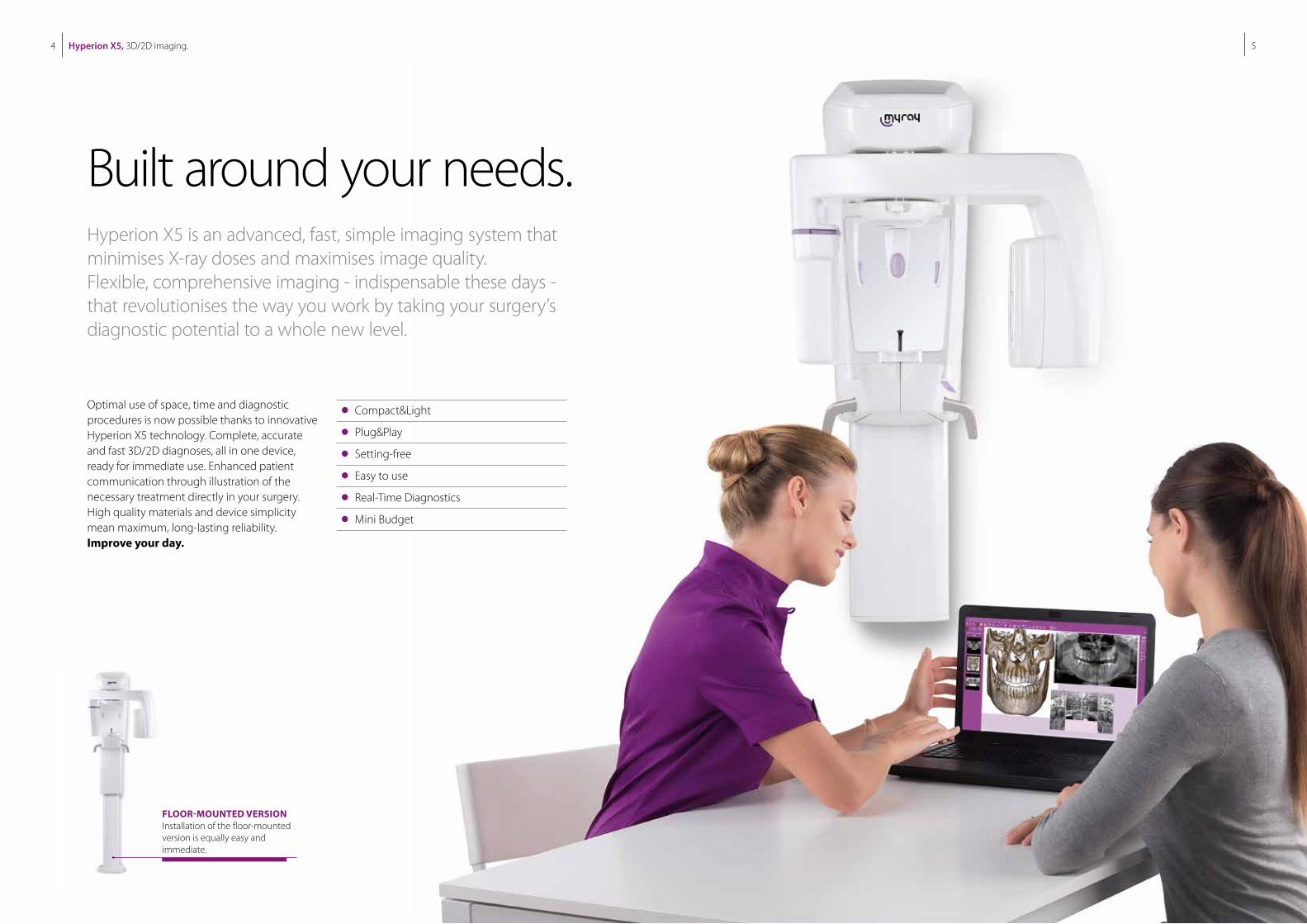
Optimal use of space, time and diagnostic procedures is now possible thanks to innovative Hyperion X5 technology. Complete, accurate and fast 3D/2D diagnoses, all in one device, ready for immediate use. Enhanced patient communication through illustration of the necessary treatment directly in your surgery. High quality materials and device simplicity mean maximum, long-lasting reliability. Improve your day.
Compact&Light
Plug&Play
Setting-free
Easy to use
Real-Time Diagnostics
Mini Budget
FLOOR-MOUNTED VERSIONInstallation of the floor-mounted version is equally easy and immediate.
Built around your needs.Hyperion X5 is an advanced, fast, simple imaging system that minimises X-ray doses and maximises image quality. Flexible, comprehensive imaging - indispensable these days - that revolutionises the way you work by taking your surgery’s diagnostic potential to a whole new level.
Hyperion X5, 3D/2D imaging.6 7
MAXI FLEX MULTI VISION
MULTI FOV MULTI PAN
Suitable for all diagnostic needs
Clever collimators
3D Cone Beam MultiFOV technology
MultiPAN system
Up to 22 2D programmes
A single 3D/2D-QuickScan sensor
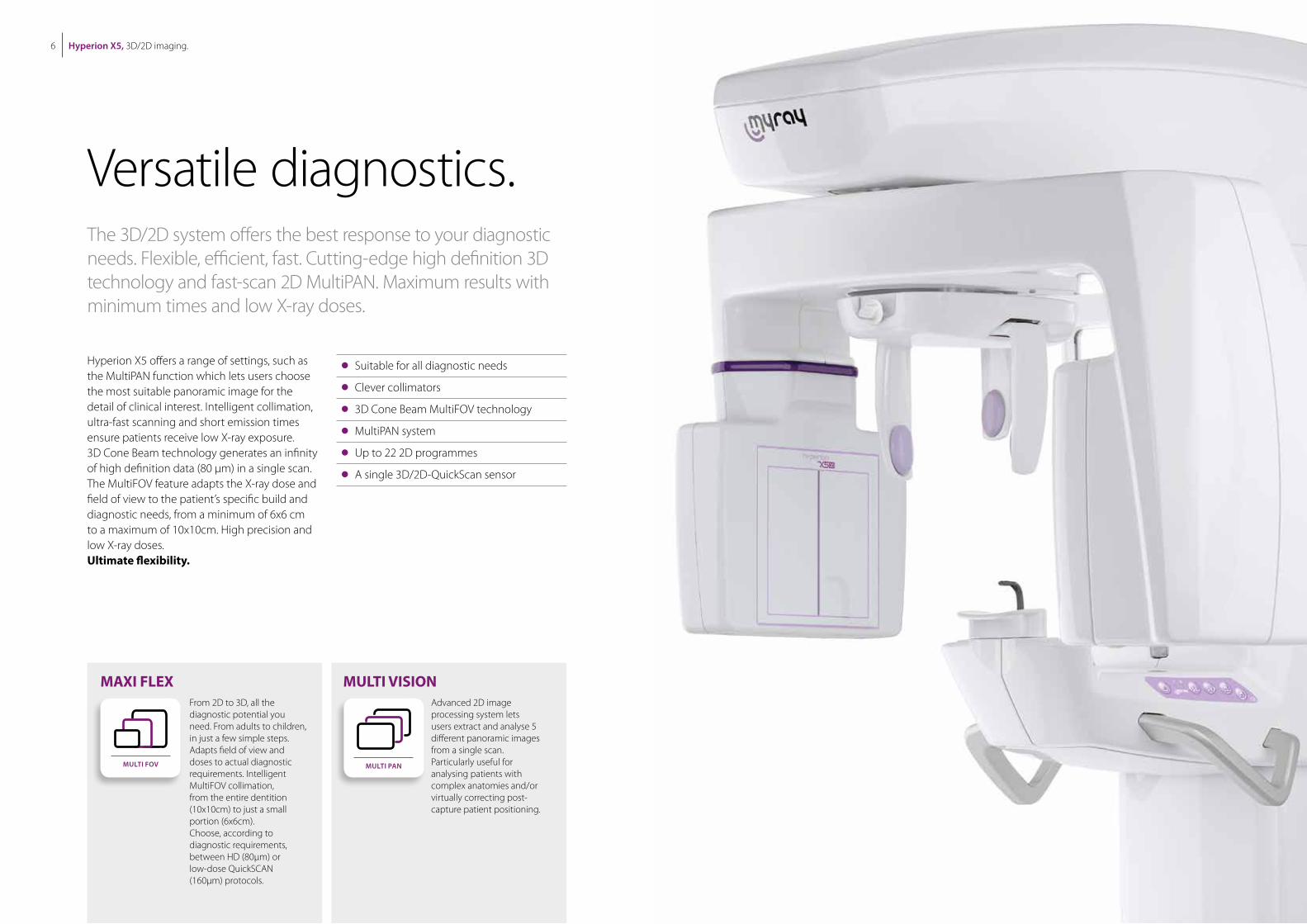
Hyperion X5 offers a range of settings, such as the MultiPAN function which lets users choose the most suitable panoramic image for the detail of clinical interest. Intelligent collimation, ultra-fast scanning and short emission times ensure patients receive low X-ray exposure. 3D Cone Beam technology generates an infinity of high definition data (80 μm) in a single scan. The MultiFOV feature adapts the X-ray dose and field of view to the patient’s specific build and diagnostic needs, from a minimum of 6x6 cm to a maximum of 10x10cm. High precision and low X-ray doses.Ultimate flexibility.
Versatile diagnostics.The 3D/2D system offers the best response to your diagnostic needs. Flexible, efficient, fast. Cutting-edge high definition 3D technology and fast-scan 2D MultiPAN. Maximum results with minimum times and low X-ray doses.
From 2D to 3D, all the diagnostic potential you need. From adults to children, in just a few simple steps. Adapts field of view and doses to actual diagnostic requirements. Intelligent MultiFOV collimation, from the entire dentition (10x10cm) to just a small portion (6x6cm). Choose, according to diagnostic requirements, between HD (80μm) or low-dose QuickSCAN (160μm) protocols.
Advanced 2D image processing system lets users extract and analyse 5 different panoramic images from a single scan. Particularly useful for analysing patients with complex anatomies and/or virtually correcting post-capture patient positioning.
3D Hyperion X5, 3D/2D imaging. 2D
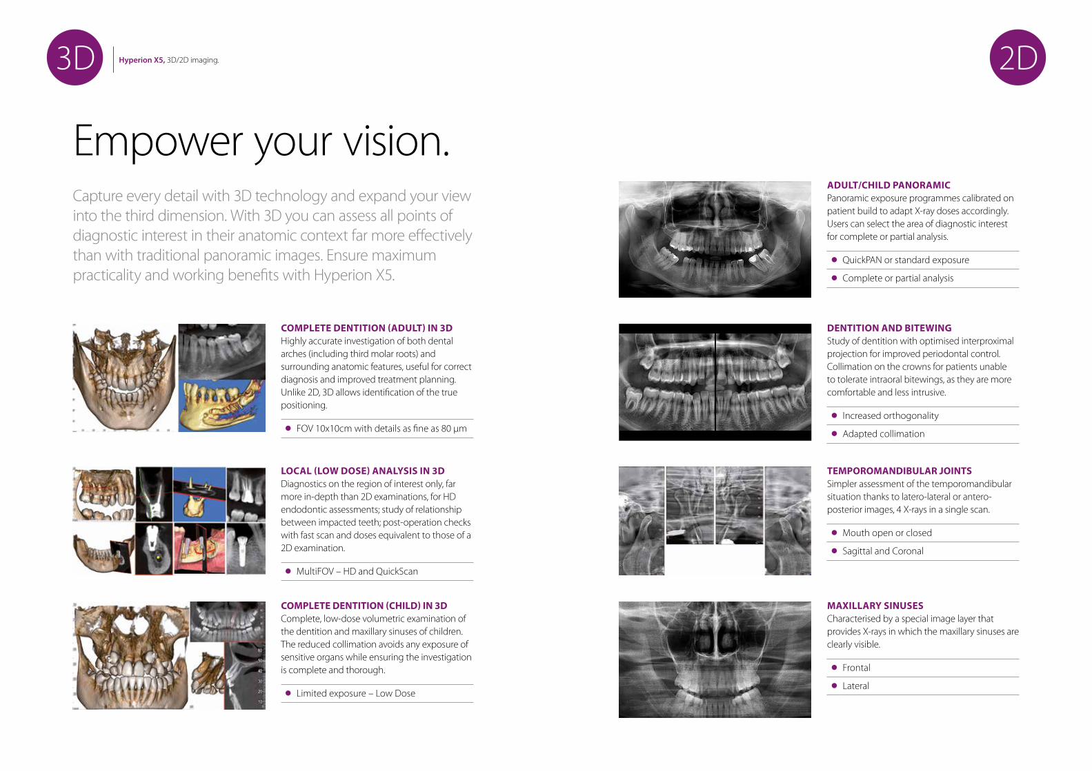
Empower your vision.Capture every detail with 3D technology and expand your view into the third dimension. With 3D you can assess all points of diagnostic interest in their anatomic context far more effectively than with traditional panoramic images. Ensure maximum practicality and working benefits with Hyperion X5.
ADULT/CHILD PANORAMICPanoramic exposure programmes calibrated on patient build to adapt X-ray doses accordingly. Users can select the area of diagnostic interest for complete or partial analysis.
QuickPAN or standard exposure
Complete or partial analysis
DENTITION AND BITEWINGStudy of dentition with optimised interproximal projection for improved periodontal control. Collimation on the crowns for patients unable to tolerate intraoral bitewings, as they are more comfortable and less intrusive.
Increased orthogonality
Adapted collimation
TEMPOROMANDIBULAR JOINTSSimpler assessment of the temporomandibular situation thanks to latero-lateral or antero-posterior images, 4 X-rays in a single scan.
Mouth open or closed
Sagittal and Coronal
MAXILLARY SINUSESCharacterised by a special image layer that provides X-rays in which the maxillary sinuses are clearly visible.
Frontal
Lateral
COMPLETE DENTITION (ADULT) IN 3DHighly accurate investigation of both dental arches (including third molar roots) and surrounding anatomic features, useful for correct diagnosis and improved treatment planning. Unlike 2D, 3D allows identification of the true positioning.
FOV 10x10cm with details as fine as 80 μm
LOCAL (LOW DOSE) ANALYSIS IN 3DDiagnostics on the region of interest only, far more in-depth than 2D examinations, for HD endodontic assessments; study of relationship between impacted teeth; post-operation checks with fast scan and doses equivalent to those of a 2D examination.
MultiFOV – HD and QuickScan
COMPLETE DENTITION (CHILD) IN 3DComplete, low-dose volumetric examination of the dentition and maxillary sinuses of children. The reduced collimation avoids any exposure of sensitive organs while ensuring the investigation is complete and thorough.
Limited exposure – Low Dose
Hyperion X5, 3D/2D imaging.10 11
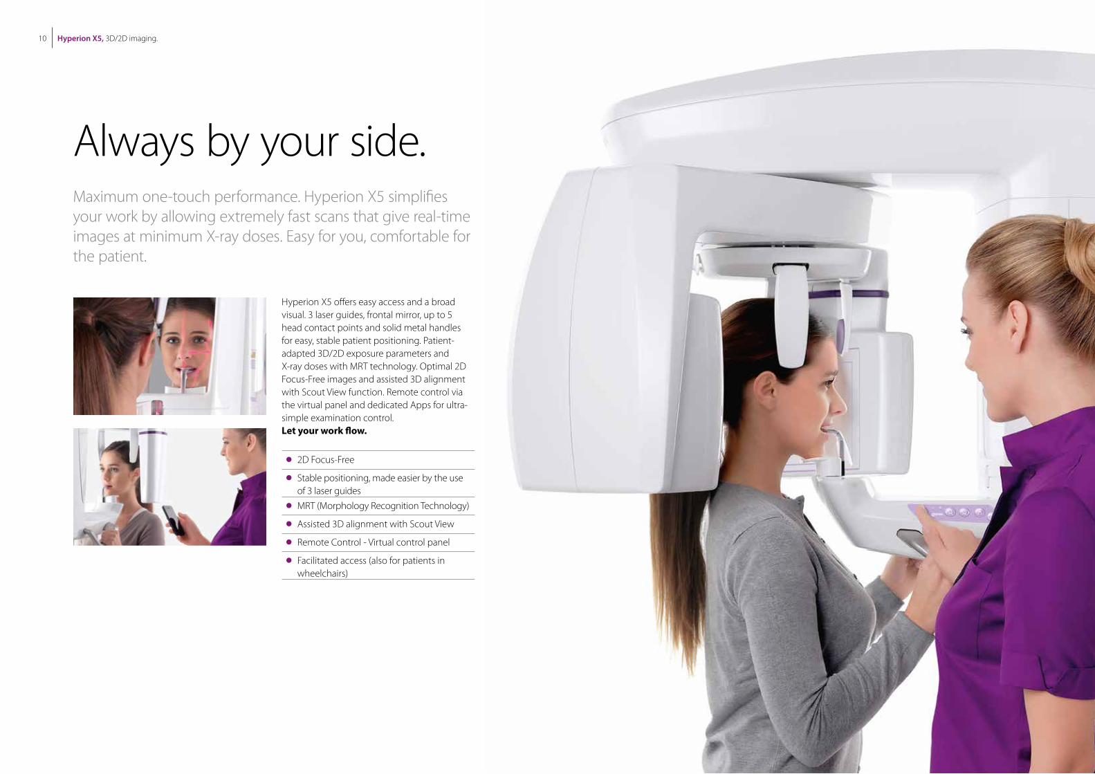
Hyperion X5 offers easy access and a broad visual. 3 laser guides, frontal mirror, up to 5 head contact points and solid metal handles for easy, stable patient positioning. Patient-adapted 3D/2D exposure parameters and X-ray doses with MRT technology. Optimal 2D Focus-Free images and assisted 3D alignment with Scout View function. Remote control via the virtual panel and dedicated Apps for ultra-simple examination control. Let your work flow.
2D Focus-Free
Stable positioning, made easier by the use of 3 laser guides
MRT (Morphology Recognition Technology)
Assisted 3D alignment with Scout View
Remote Control - Virtual control panel
Facilitated access (also for patients in wheelchairs)
Always by your side.Maximum one-touch performance. Hyperion X5 simplifies your work by allowing extremely fast scans that give real-time images at minimum X-ray doses. Easy for you, comfortable for the patient.
Hyperion X5, 3D/2D imaging.12 13
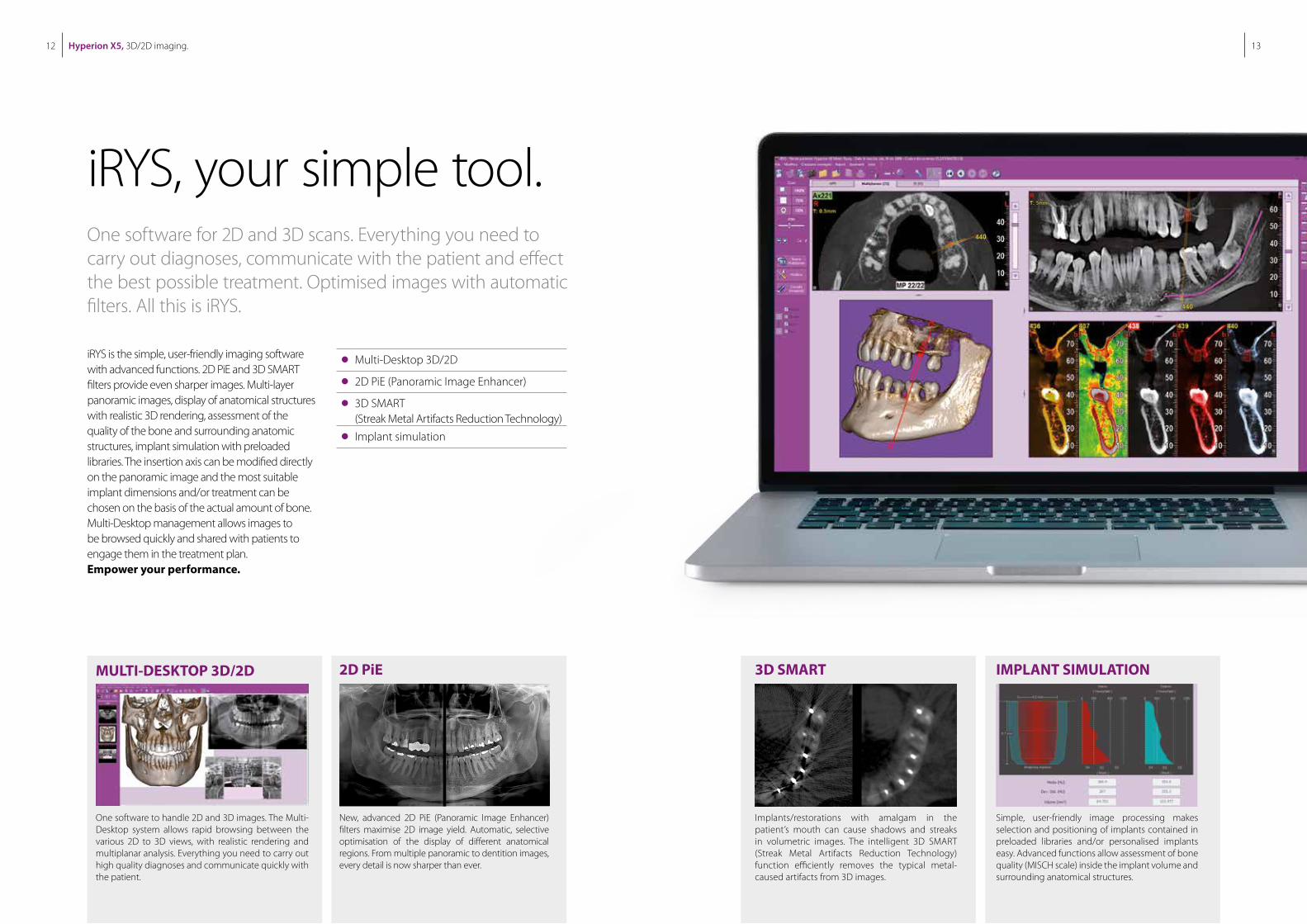
iRYS is the simple, user-friendly imaging software with advanced functions. 2D PiE and 3D SMART filters provide even sharper images. Multi-layer panoramic images, display of anatomical structures with realistic 3D rendering, assessment of the quality of the bone and surrounding anatomic structures, implant simulation with preloaded libraries. The insertion axis can be modified directly on the panoramic image and the most suitable implant dimensions and/or treatment can be chosen on the basis of the actual amount of bone. Multi-Desktop management allows images to be browsed quickly and shared with patients to engage them in the treatment plan.Empower your performance.
iRYS, your simple tool.One software for 2D and 3D scans. Everything you need to carry out diagnoses, communicate with the patient and effect the best possible treatment. Optimised images with automatic filters. All this is iRYS.
MULTI-DESKTOP 3D/2D 2D PiE
One software to handle 2D and 3D images. The Multi-Desktop system allows rapid browsing between the various 2D to 3D views, with realistic rendering and multiplanar analysis. Everything you need to carry out high quality diagnoses and communicate quickly with the patient.
New, advanced 2D PiE (Panoramic Image Enhancer) filters maximise 2D image yield. Automatic, selective optimisation of the display of different anatomical regions. From multiple panoramic to dentition images, every detail is now sharper than ever.
3D SMART IMPLANT SIMULATION
Implants/restorations with amalgam in the patient’s mouth can cause shadows and streaks in volumetric images. The intelligent 3D SMART (Streak Metal Artifacts Reduction Technology) function efficiently removes the typical metal-caused artifacts from 3D images.
Simple, user-friendly image processing makes selection and positioning of implants contained in preloaded libraries and/or personalised implants easy. Advanced functions allow assessment of bone quality (MISCH scale) inside the implant volume and surrounding anatomical structures.
Multi-Desktop 3D/2D
2D PiE (Panoramic Image Enhancer)
3D SMART (Streak Metal Artifacts Reduction Technology)
Implant simulation
Hyperion X5, 3D/2D imaging.14 15
REMOTE CONNECTION
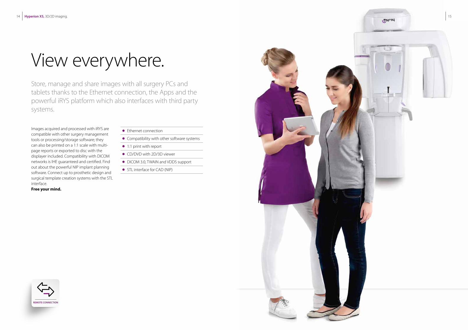
Images acquired and processed with iRYS are compatible with other surgery management tools or processing/storage software; they can also be printed on a 1:1 scale with multi-page reports or exported to disc with the displayer included. Compatibility with DICOM networks is IHE guaranteed and certified. Find out about the powerful NIP implant planning software. Connect up to prosthetic design and surgical template creation systems with the STL interface.Free your mind.
Ethernet connection
Compatibility with other software systems
1:1 print with report
CD/DVD with 2D/3D viewer
DICOM 3.0, TWAIN and VDDS support
STL interface for CAD (NIP)
View everywhere.Store, manage and share images with all surgery PCs and tablets thanks to the Ethernet connection, the Apps and the powerful iRYS platform which also interfaces with third party systems.
Hyperion X5, 3D/2D imaging.16 17
1101
(43.
3)
872(34.3)
664(26.1)
436(17.2)
868
(34.
2)23
4(9
.2)
790(31.1)
748
(29.
5)10
70M
IN.
/ 1
690
MA
X
(
42.1
/ 6
6.5)
657(25.9)
1661
MIN
./
228
1 M
AX
(65
.3 /
89
.8)
40 (1.6
)
1101
(43.
3)
872(34.3)
664(26.1)
436(17.2)
868
(34.
2)23
4(9
.2)
790(31.1)
748
(29.
5)10
70M
IN.
/ 1
690
MA
X
(
42.1
/ 6
6.5)
657(25.9)
1661
MIN
./
228
1 M
AX
(65
.3 /
89
.8)
40 (1.6
)
1636
MIN
/ 225
6 M
AX
(64.
4
/
88,
8)
104
(4.1
)86
8(3
4.2)
130(5.1)
276(10.9)
320(12.6)
436(17.2)
872(34.3)
1060
(41.
7)41
5(1
6.3)
405
MIN
/ 10
25 M
AX
(15.
9
/ 4
0.4)
657(25.9)
1045
MIN
/ 166
5 M
AX
( 41.
2
/
65.6
)
972
(38.
3)755
(29.
7)
664(26.1)
1636
MIN
/ 225
6 M
AX
(64.
4
/
88,
8)
104
(4.1
)86
8(3
4.2)
130(5.1)
276(10.9)
320(12.6)
436(17.2)
872(34.3)
1060
(41.
7)41
5(1
6.3)
405
MIN
/ 10
25 M
AX
(15.
9
/ 4
0.4)
657(25.9)
1045
MIN
/ 166
5 M
AX
( 41.
2
/
65.6
)
972
(38.
3)755
(29.
7)
664(26.1)
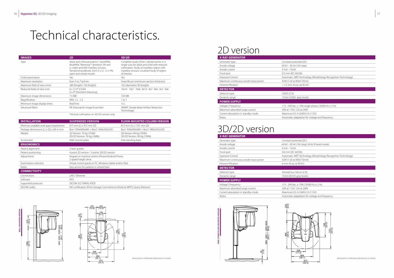
Technical characteristics.
FREE
-STA
ND
FLO
OR-
MO
UN
TED
VER
SIO
N
dimensions in millimetres (dimensions in inches)
X-RAY GENERATORGenerator type Constant potential (DC)
Anode voltage 60 kV – 85 kV (1kV step)
Anode current 4 mA - 15mA
Focal spot 0.5 mm (IEC 60336)
Exposure Control Automatic. MRT Technology (Morphology Recognition Technology)
Maximum continuous anode input power 42W (1:20 at 85kV/10mA)
Inherent filtration > 2.5 mm Al eq. (at 85 kV)
DETECTORDetector type CMOS (CSI)
Dynamic range 14 bit (16383 grey levels)
POWER SUPPLYVoltage | Frequency 115 - 240 Vac, ± 10% single-phase | 50/60 Hz ± 2 Hz
Maximum absorbed surge current 20A at 115V; 12A at 240V
Current absorption in standby mode Maximum 0.5 A (240V);1A (115V)
Notes Automatic adaptation for voltage and frequency
IMAGES 2D 3D/2DType Adult and child panoramic*, QuickPAN,
MultiPAN, “Bitewing”* dentition, PA and LL (right and left) maxillary sinuses, Temporomandibular Joint (2 x LL +2 x PA) open and closed mouth.
Complete study of the 2 dental arches in a single scan for adult and child with reduced collimation; Study of maxillary region with maxillary sinuses; Localised study of region of interest.
Child examination Yes Yes
Maximum resolution from 5 to 7 lp/mm Voxel 80 µm (minimum section thickness)
Maximum field of view (mm) 280 (length); 150 (height) 102 (diameter); 96 (height)
Reduced fields of view (cm) 6 x 12.5* (Child) 6 x 9* (Dentition bitewing)
10x10 - 10x7 - 10x6 - 8x10 - 8x7 - 8x6 - 6x7 - 6x6
Maximum image dimensions 7.5 MB 720 MB
Magnification PAN: 1.2 - 1.3 1 to 1
Minimum image display times RealTime 15 s
Advanced filters PiE (Panoramic image Enancher) SMART (Streak Metal Artifact Reduction Technology)
*Vertical collimation on 3D/2D version only
INSTALLATION SUSPENDED VERSION FLOOR-MOUNTED COLUMN VERSIONMinimum available work space requirement 872 mm (L) x 972 mm (D) 872 mm (L) x 1101 mm (D)
Package dimensions (L) x (D) x (H) in mm Box1 930x690x960 + Box2 1460x350x350 Box1 930x690x960 + Box2 1860x355x350
Weight 2D Version: 70 kg (154lb) 3D/2D Version: 76 Kg (168lb)
2D Version: 84 kg (185lb)3D/2D Version: 90 Kg (199lb)
Accessories Wall counter-plate Free standing base
ERGONOMICSPatient alignment 3 laser guides
Patient positioning 4 point 2D version - 5 points 3D/2D version
Adjustments Keypad on machine and/or iPhone/Android Phone 2-speed height drive
Examination selection Virtual control panel on PC, Windows tablet and/or iPad
Notes Easy access for patients in wheelchairs
CONNECTIVITYConnections LAN / Ethernet
Software iRYS
Supported protocols DICOM 3.0, TWAIN, VDDS
DICOM nodes IHE certification (Print; Storage Commitment; WorkList MPPS; Query Retrieve)
2D version
3D/2D versionX-RAY GENERATORGenerator type Constant potential (DC)
Anode voltage 60 kV – 85 kV (1kV step); 90 kV (Pulsed mode)
Anode current 4 mA - 15mA
Focal spot 0.6 mm (IEC 60336)
Exposure Control Automatic. MRT Technology (Morphology Recognition Technology)
Maximum continuous anode input power 42W (1:20 at 85kV/10mA)
Inherent filtration 6 mm Al eq. (a 90 kV)
DETECTORDetector type Amorphous Silicon (CSI)
Dynamic range 16 bit (65535 grey levels)
POWER SUPPLYVoltage | Frequency 115 - 240 Vac, ± 10% | 50/60 Hz ± 2 Hz
Maximum absorbed surge current 20A at 115V; 12A at 240V
Current absorption in standby mode Maximum 0.5 A (240V);1A (115V)
Notes Automatic adaptation for voltage and frequency
dimensions in millimetres (dimensions in inches)
WA
LL-M
OU
NTE
D V
ERSI
ON
Hyperion X5, 3D/2D imaging.18 19
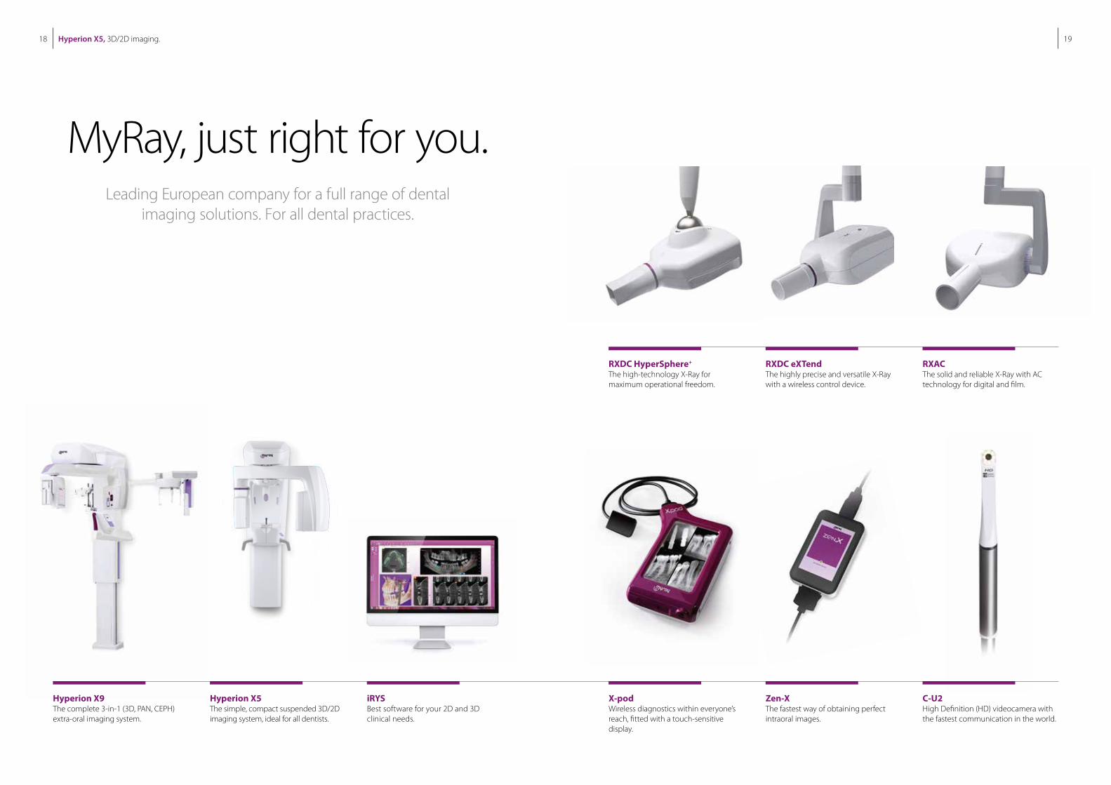
Hyperion X9The complete 3-in-1 (3D, PAN, CEPH) extra-oral imaging system.
Hyperion X5The simple, compact suspended 3D/2D imaging system, ideal for all dentists.
iRYSBest software for your 2D and 3D clinical needs.
X-podWireless diagnostics within everyone’s reach, fitted with a touch-sensitive display.
RXDC HyperSphere+
The high-technology X-Ray for maximum operational freedom.
Zen-XThe fastest way of obtaining perfect intraoral images.
RXDC eXTendThe highly precise and versatile X-Ray with a wireless control device.
C-U2High Definition (HD) videocamera with the fastest communication in the world.
RXACThe solid and reliable X-Ray with AC technology for digital and film.
MyRay, just right for you.Leading European company for a full range of dental
imaging solutions. For all dental practices.


