Hyaluronan Esters Drive Smad Gene Expression andSignaling Enhancing Cardiogenesis in Mouse Embryonicand Human Mesenchymal Stem CellsMargherita Maioli1,2, Sara Santaniello1, Andrea Montella1, Pasquale Bandiera1, Silvia Cantoni2,3, Claudia
Cavallini2,3, Francesca Bianchi2,3, Vincenzo Lionetti2,4, Flavio Rizzolio5, Irene Marchesi1,5, Luigi
Bagella1,5, Carlo Ventura2,3,6*
1 Department of Biomedical Sciences, University of Sassari, Sassari, Italy, 2 National Institute of Biostructures and Biosystems, University of Bologna, Bologna, Italy,
3 Laboratory of Molecular Biology and Stem Cell Engineering, Cardiovascular Department, National Institute of Biostructures and Biosystems, S. Orsola - Malpighi Hospital,
University of Bologna, Bologna, Italy, 4 Sector of Medicine, Scuola Superiore S. Anna, CNR Institute of Clinical Physiology, Pisa, Italy, 5 Sbarro Institute for Cancer Research
and Molecular Medicine, Center for Biotechnology, Temple University, Philadelphia, Pennsylvania, United States of America, 6 Bioscience Institute, Falciano, Republic of
San Marino
Abstract
Background: Development of molecules chemically modifying the expression of crucial orchestrator(s) of stem cellcommitment may have significant biomedical impact. We have recently developed hyaluronan mixed esters of butyric andretinoic acids (HBR), turning cardiovascular stem cell fate into a high-yield process. The HBR mechanism(s) remain stilllargely undefined.
Methodology/Principal Findings: We show that in both mouse embryonic stem (ES) cells and human mesenchymal stemcells from fetal membranes of term placenta (FMhMSCs), HBR differentially affected the patterning of Smad proteins, one ofthe major conductors of stem cell cardiogenesis. Real-time RT-PCR and Western blot analyses revealed that in both celltypes HBR enhanced gene and protein expression of Smad1,3, and 4, while down-regulating Smad7. HBR acted at thetranscriptional level, as shown by nuclear run-off experiments in isolated nuclei. Immunofluorescence analysis indicated thatHBR increased the fluorescent staining for Smad1,3, and 4, confirming that the transcriptional action of HBR encompassedthe upregulation of the encoded Smad proteins. Chromatin immune precipitation and transcriptional analyses showed thatHBR increased the transcription of the cardiogenic gene Nkx-2.5 through Smad4 binding to its own consensus Smad site.Treatment of mouse ES cells and FMhMSCs with HBR led to the concomitant overexpression of both Smad4 and a-sarcomeric actinin. Smad4 silencing by the aid of lentiviral-mediated Smad4 shRNA confirmed a dominant role of Smad4 inHBR-induced cardiogenesis.
Conclusions/Significance: The use of HBR may pave the way to novel combinatorial strategies of molecular and stem celltherapy based on fine tuning of targeted Smad transciption and signaling leading to a high-throughput of cardiogenesiswithout the needs of gene transfer technologies.
Citation: Maioli M, Santaniello S, Montella A, Bandiera P, Cantoni S, et al. (2010) Hyaluronan Esters Drive Smad Gene Expression and Signaling EnhancingCardiogenesis in Mouse Embryonic and Human Mesenchymal Stem Cells. PLoS ONE 5(11): e15151. doi:10.1371/journal.pone.0015151
Editor: Branden Nelson, Seattle Children’s Research Institute, United States of America
Received August 6, 2010; Accepted November 1, 2010; Published November 30, 2010
Copyright: � 2010 Maioli et al. This is an open-access article distributed under the terms of the Creative Commons Attribution License, which permitsunrestricted use, distribution, and reproduction in any medium, provided the original author and source are credited.
Funding: This work was supported by Regione Emilia Romagna, Programma di Ricerca Regione - Universita 2007/2009, Area 1b ‘‘Medicina rigenerativa’’, Italy;Fondazione Luisa Fanti Melloni, Bologna, Italy; Tavola Valdese, Rome, Italy; Sintofarm S.p.A. (Guastalla, Reggio Emilia), Italy; Fondazione Banco di Sardegna, andRegione Autonoma della Sardegna, Italy. The funders had no role in study design, data collection and analysis, decision to publish, or preparation of themanuscript.
Competing Interests: The authors have declared that no competing interests exist.
* E-mail: [email protected]
Introduction
Embryonic stem (ES) cells differentiate in vitro into various cell
types including cardiac myocytes [1,2], by tracing or mimicking
developmental processes occurring in vivo. Moreover, ES cells
besides being a tool to investigate the molecular mechanisms
underlying cardiogenesis, represent a source in regeneration
therapies for damaged tissues [3]. Although ES cells have
unlimited replication capacity, the cardiogenic process is ineffi-
cient, with only a limited number of cells becoming cardiac
myocytes [4]. Consequently, developing new molecules that
maximize the functional expression of crucial signaling orchestra-
tor(s) of targeted commitments within a population of precursor
cells may offer significant advantages in tissue repair and clinical
therapies. These molecules may also provide new insights into the
dynamic circuitry underlying developmental decisions.
We have developed a mixed ester of hyaluronan with butyric
and retinoic acids (HBR), acting as a differentiating agent,
remarkably increasing the yield of cardiomyocytes derived from
mouse ES cells [4]. This compound was also effective as a tool
implementing the cardiogenic process in human mesenchymal
stem cells (hMSCs) isolated from the bone marrow and alternative
PLoS ONE | www.plosone.org 1 November 2010 | Volume 5 | Issue 11 | e15151

sources, including the dental pulp and fetal membranes of term
placenta (FMhMSCs) [5]. The cardiogenic program induced by
HBR was due to the transcriptional induction of two key
regulators of cardiogenic commitment, GATA-4 and Nkx-2.5,
early expressed during heart formation [6,7]. HBR also enhanced
prodynorphin gene transcription and the expression of its
biologically active peptide, the k opioid receptor agonist dynorphin
B, both being directly implicated in cardiac lineage specification
[8–11].
The Smad proteins are a family of signal transducers which can
be divided into three distinct subfamilies: receptor-regulated
Smads (R-Smads), common-partner Smads (Co-Smads) and
inhibitory Smads (I-Smads) [12,13]. Compelling evidence suggests
that a Smad signaling pathway is important in stem cell
cardiovascular differentiation. In vertebrates and Drosophila, heart
formation is an early event occurring in the embryo, which
requires the activation of a bone morphogenetic protein (BMP)
signaling, representing a part of the transforming growth factor
beta (TGF-b) superfamily of growth factors [14–16]. Activated
BMP receptor kinases specifically and transiently interact with and
phosphorylate particular R-Smads. Subsequently, the activated R-
Smads recruit Co-Smads and heteromeric complexes accumulate
in the nucleus [12,13]. Nkx-2.5 is the earliest known marker of the
cardiac lineage, and is expressed together with the cardiogenic
commitment of cells from anterior-lateral mesoderm [17,18]. Its
expression is maintained in the heart throughout prenatal and
postnatal life. In Drosophila, Smad4 and R-Smads (Smad1, Smad5,
and Smad8), may regulate Nkx-2.5 gene expression via a Smad-
binding domain located in its promoter region [19]. Moreover,
mouse Smad4 mutant embryos [20] show significant reduction of
Nkx-2.5 expression in prospective cardiac mesoderm, confirming
previous findings, indicating a requirement for Smad1/4 in the
activation of Nkx-2.5 gene transcription [21].
In the current study, the gene and protein expression profile of
Smad1,3,4, and 7 were investigated for different periods of time in
both mouse ES cells and FMhMSCs exposed to HBR. Our data
provide evidence that the mixed ester is able to drive a complex
circuitry of Smad transcription and signaling that is essential in
stem cell cardiogenesis.
Results
HBR Modulates the Gene Expression of Smad1,3,4, and 7in GTR1 ES Cells and FMhMSCs
GTR1 cells [9,11], a derivative of R1 ES cells bearing the
puromycin-resistance gene driven by the cardiomyocyte-specific a-
myosin heavy chain promoter, were used as an in vitro model of
cardiogenesis to elucidate whether a recruitment of Smad
signaling may be a mechanism underlying the cardiogenic
commitment induced by HBR.
When cultured in the absence of LIF and in suspension, these
cells aggregate in EBs, evolving into spontaneously beating
cardiomyocytes throughout 7–8 days of culturing. After puromy-
cin selection, a virtually pure population of ES-derived cardiomy-
ocytes can be obtained.
The gene expression of Smad1,3,4, and 7 was investigated by
real-time RT-PCR at different times of GTR1 ES culture in the
absence or presence of HBR. To further investigate whether HBR
may have a role in Smad patterning, the hyaluronan mixed ester
was also applied to FMhMSCs, a hMSC population that due to
HBR treatment could be committed to the cardiovascular lineage
in vitro, affording remarkable myocardial repair in vivo after
transplantation in infarcted rat hearts [5]. In both cell types,
HBR was used at a concentration of 1.5 mg/ml that was
previously shown to afford a maximal cardiogenic response [4,5].
After 8 hours of culture, following LIF withdrawal (time zero),
EBs consistently exhibited Smad1,3 and 4 gene expression
(Figure 1A–1C). HBR-treated EBs revealed a remarkably higher
gene expression of each isoform, as compared to untreated cells.
At this time point, Smad7 mRNA declined compared to time zero,
and was further significantly downregulated in the presence of
HBR (Figure 1D).
After 24 hours, the gene expression of Smad1-4 increased
compared to time zero, retaining higher levels in HBR-treated
than untreated cells (Figure 1A–1C). The spontaneous gene
expression of these Smads declined after 72 hours of culture, with
Smad4, and to a lesser extent Smad3, still exhibiting higher
mRNA levels in HBR-treated than in unexposed cells (Figure 1B,
1C). On the contrary, at this time point, Smad1 was similarly
expressed in both groups of cells (Figure 1A).
Comparative analysis of Smad1,3, and 4 mRNA was also
performed at 10 days in ES-derived cardiomyocytes recovered
after exposure in the absence or presence of HBR from the time of
LIF removal throughout 4 days of puromycin selection. Under
these experimental conditions, HBR reversed its effect, downreg-
ulating the gene expression level, as compared to unexposed
controls (Figure 1A–1C).
The gene expression of Smad7 progressively increased in EBs
after 24 hours of culture, approaching the time zero level after 10
days in ES-derived cardiomyocytes, but remaining downregulated
in HBR-exposed cells, as compared with the corresponding
untreated time control (Figure 1D).
To further address whether the currently observed response to
HBR may represent a general feature of this molecule in stem cell
patterning, the effect of HBR on Smad gene expression was also
investigated in FMhMSCs. We have previously shown that after
exposure to HBR these cells exhibited a consistent increase in the
transcription of the cardiogenic genes GATA-4 and Nkx-2.5, and
differentiated into a high-yield of cardiac marker-expressing
elements [5]. As shown in figure 2, FMhMSCs spontaneously
expressed Smad1,3, and 4 mRNA. Interestingly, the gene
expression of these Smads was significantly enhanced following a
48-hour exposure to the mixed ester, remaining upregulated
during a subsequent period of 3 days, as compared with the
untreated group (Figure 2A–2C). Confirming the results in HBR-
treated GTR1 ES cells, FMhMSCs exposed to HBR displayed a
lower amount of Smad7 mRNA, compared to unexposed cells
(Figure 2D).
HBR Affects Smad Protein Expression During theCardiogenic Commitment in both GTR1 ES Cells andFMhMSCs
The cellular levels of Smad proteins result from a delicate
balance between the rates of transcription of the corresponding
genes and post-transcriptional regulatory mechanisms. Therefore,
it was important to examine the expression of targeted Smad
proteins in EBs and ES-derived cardiomyocytes exposed in the
absence or presence of HBR.
As shown by Western blot and densitometric analyses, the
spontaneous expression of Smad1 and 3 proteins, gradually
increased after 8 hours, following LIF withdrawal (time zero), up
to 10 days throughout ES cell commitment toward the cardiac
phenotype (Figure 3A, 3B). During the same time-course Smad4
expression was already increased at 8 hours, with less appreciable
changes in the subsequent time points (Figure 3C). HBR treatment
differentially affected over time the expression of these Smad
proteins, as compared with untreated cells, raising Smad1 at
Hyaluronan Esters, Smads, and Cardiogenesis
PLoS ONE | www.plosone.org 2 November 2010 | Volume 5 | Issue 11 | e15151
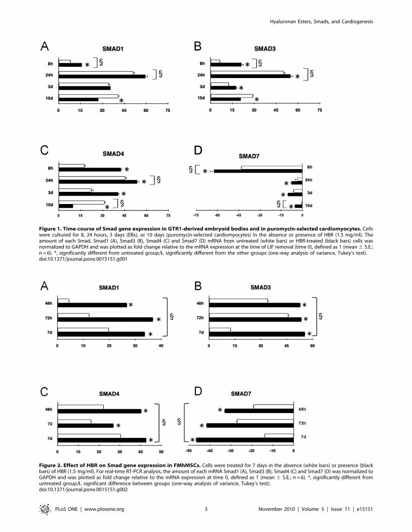
Figure 1. Time-course of Smad gene expression in GTR1-derived embryoid bodies and in puromycin-selected cardiomyocytes. Cellswere cultured for 8, 24 hours, 3 days (EBs), or 10 days (puromycin-selected cardiomyocytes) in the absence or presence of HBR (1.5 mg/ml). Theamount of each Smad, Smad1 (A), Smad3 (B), Smad4 (C) and Smad7 (D) mRNA from untreated (white bars) or HBR-treated (black bars) cells wasnormalized to GAPDH and was plotted as fold change relative to the mRNA expression at the time of LIF removal (time 0), defined as 1 (mean 6 S.E.;n = 6). *, significantly different from untreated group;1, significantly different from the other groups (one-way analysis of variance, Tukey’s test).doi:10.1371/journal.pone.0015151.g001
Figure 2. Effect of HBR on Smad gene expression in FMhMSCs. Cells were treated for 7 days in the absence (white bars) or presence (blackbars) of HBR (1.5 mg/ml). For real-time RT-PCR analysis, the amount of each mRNA Smad1 (A), Smad3 (B), Smad4 (C) and Smad7 (D) was normalized toGAPDH and was plotted as fold change relative to the mRNA expression at time 0, defined as 1 (mean 6 S.E.; n = 6). *, significantly different fromuntreated group;1, significant difference between groups (one-way analysis of variance, Tukey’s test).doi:10.1371/journal.pone.0015151.g002
Hyaluronan Esters, Smads, and Cardiogenesis
PLoS ONE | www.plosone.org 3 November 2010 | Volume 5 | Issue 11 | e15151
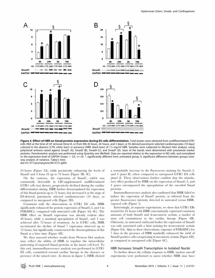
24 hours (Figure 3A), while persistently enhancing the levels of
Smad3 and 4 from 24 up to 72 hours (Figure 3B, 3C).
On the contrary, the expression of Smad7, which was
consistently detectable in LIF-supplemented undifferentiated
GTR1 cells (not shown), progressively declined during the cardiac
differentiation timing. HBR further downregulated the expression
of this Smad protein up to 72 hours, but increased it at the stage of
ES-derived, puromycin selected cardiomyocytes (10 days), as
compared to unexposed cells (Figure 3D).
Consistent with the observations in GTR1 ES cells, HBR
significantly enhanced the protein expression of Smad1,3, and 4 in
FMhMSCs, compared with untreated cells (Figure 4A–4C). The
HBR effect on Smad4 expression was already evident after
48 hours, while a maximal upregulation of Smad1, and 3 was
achieved after 72 hours of treatment. As in GTR1 cells, HBR
accentuated the decline of Smad 7 expression observed up to
72 hours, but significantly counteracted the downregulation of this
Smad at a later time (Figure 4D).
We then assessed whether results from Western blot analysis
may reflect the ability of HBR to regulate the intracellular
patterning of targeted Smad proteins at the intact cell level. To
this end, immunofluorescence analysis was performed in GTR1
ES cells, committed toward cardiac lineage in the absence or
presence of the mixed ester. As shown in figure 5, HBR elicited
a remarkable increase in the fluorescent staining for Smad1,3,
and 4 (panel B), when compared to unexposed GTR1 ES cells
(panel A). These observations further confirm that the stimula-
tory effect produced by HBR on the expression of Smad1,3, and
4 genes encompassed the upregulation of the encoded Smad
proteins.
Immunofluorescence analysis also confirmed that HBR failed to
induce the expression of Smad7 protein, as inferred from the
greater fluorescence intensity detected in untreated versus HBR-
exposed cells (Figure 5).
Interestingly, in separate experiments, we show that GTR1 EBs
treated for 24 hours with HBR concomitantly expressed consistent
amounts of both Smad4 and a-sarcomeric actinin, a marker of
stem cell commitment to the cardiac lineage (Figure 6B).
Otherwise, in untreated embryoid bodies the expression of Smad4
was only associated with a faint staining for a-sarcomeric actinin
(Figure 6A). Akin to these observations, exposure of FMhMSCs for
7 days in the presence of HBR markedly enhanced the yield of
Smad4 positive cells coexpressing a-sarcomeric actinin (Figure 6D),
as compared to unexposed cells (Figure 6C).
HBR Increases Smad4 Transcription in Isolated NucleiTo further dissect the cellular response to HBR, nuclear run-off
experiments were performed to assess whether HBR may have
Figure 3. Effect of HBR on Smad protein expression during ES cells differentiation. Total lysates were obtained from undifferentiated GTR1cells (ND) at the time of LIF removal (time 0), or from EBs (8 hours, 24 hours, and 3 days), or ES-derived puromycin selected cardiomyocytes (10 days)cultured in the absence (CTR, white bars) or presence (HBR, black bars) of 1.5 mg/ml HBR. Samples were subjected to Western blot analysis, usingpolyclonal antisera raised against Smad1 (A), Smad3 (B), Smad4 (C), and Smad7 (D). Sizes of the bands were determined with prestained markerproteins. Densitometric analysis was performed using Quantity one (BioRad). Data are reported relative to the expression in ND cells, and normalizedto the expression level of GAPDH (mean 6 S.E.; n = 6). *, significantly different from untreated group; 1, significant difference between groups (one-way analysis of variance, Tukey’s test).doi:10.1371/journal.pone.0015151.g003
Hyaluronan Esters, Smads, and Cardiogenesis
PLoS ONE | www.plosone.org 4 November 2010 | Volume 5 | Issue 11 | e15151

affected the rate of Smad4 gene transcription and whether, in the
affirmative, it may have acted as a unit or after hydrolysis of its
hyaluronan grafted moieties. Figure 7 shows that nuclei isolated
from HBR-treated GTR1 ES cells or FMhMSCs exhibited a
consistent increase in the transcription rate of Smad4, as
compared with nuclei isolated from untreated cells. In separate
experiments, nuclei were isolated from untreated cells and
subsequently incubated with HBR, or exposed to HA, BU, or
RA administered alone or in combination. While nuclear
incubation with HBR or HA was ineffective, direct nuclear
treatment with BU or RA enhanced Smad4 gene transcription
(Figure 7). The transcription rate was further enhanced when
nuclei were exposed to a combination of BU and RA (Figure 7).
Nkx-2.5 Gene Expression During HBR-inducedCardiogenesis is Smad4-mediated
To examine whether Smad4 was a transcription factor crucial
in the HBR-mediated molecular program of cardiogenesis we
performed chromatin immunoprecipitation (ChIP) analysis on ES
cells treated in the absence or presence of HBR. We found that
HBR-treated EBs exhibited a Smad4-binding on the promoter
region of Nkx-2.5 gene (Figure 8A). Immunoprecipitation with a
Smad4 antibody revealed that nuclear extracts from control and
HBR-treated EBs generated an amplified product for the SBE
binding site on the Nkx-2.5 promoter. Moreover, the amount of
amplified DNA was significantly higher in HBR-treated GTR1
EBs, as compared to untreated controls (Figure 8A). The input
control revealed that similar amounts of DNA were present in
both untreated and HBR-stimulated EBs. To establish whether
this mechanism may have a causal role in HBR-induced
cardiogenesis, we performed a comparative analysis of the effect
of HBR on the yield of ES derived cardiomyocytes in wild type
GTR1 cells and GTR1 cells subjected to Smad4 silencing using
lentiviral-mediated Smad4 shRNA, an approach that led to
consistent silencing of Smad4 protein and mRNA expression
(Figure 8B, 8C). Both groups of cells were cultured in the absence
or presence of HBR from the time of LIF removal throughout 4
days of puromycin selection. Confirming our previous studies [4],
GTR1 cell exposure to HBR resulted into a consistent increase in
the number of spontaneously beating colonies, as compared with
untreated controls (Figure 8D). Highlighting the important role of
Smad4 in cardiac differentiation, Smad4 silencing nearly abro-
Figure 4. Effect of HBR on Smad protein expression during the differentiation of FMhMSCs. Total lysates were collected at time 0 (ND),considered as the time at which cells from the 3rd passage reached 80% confluence, or from FMhMSCs cultured for different periods of time in theabsence (CTR, white bars) or presence of 1.5 mg/ml HBR (HBR, black bars). Samples were analyzed by Western blot, using polyclonal antisera againstSmad1 (A), Smad3 (B), Smad4 (C), and Smad7 (D). Sizes of the bands were determined with prestained marker proteins. Densitometric analysis wasperformed using Quantity one (BioRad). Data are reported relative to the expression in ND cells, and normalized to the expression level of GAPDH(mean 6 S.E.; n = 6). *, significantly different from untreated group; 1, significant difference between groups (one-way analysis of variance, Tukey’stest).doi:10.1371/journal.pone.0015151.g004
Hyaluronan Esters, Smads, and Cardiogenesis
PLoS ONE | www.plosone.org 5 November 2010 | Volume 5 | Issue 11 | e15151

Figure 5. HBR modulates Smad protein expression in intact GTR1 ES cells. Smad1,3,4, and 7 protein expression was assessed in cellsaggregated for 24 hours as EBs and cultured for additional 24 hours in the absence (A) or presence (B) of HBR (1.5 mg/ml). Scale bars are 40 mm.Nuclei are labeled with Propidium Iodide (red). Representative of five separate experiments.doi:10.1371/journal.pone.0015151.g005
Figure 6. Immunofluorescence analysis of the expression of a-sarcomeric actinin and Smad4 in GTR1 ES cells and FMhMSCs.Concomitant expression of a-sarcomeric actinin [(FITC) green immunofluorescence] and Smad4 [(rhodamine) orange immunofluorescence] wasassessed in GTR1-derived EBs or FMhMSCs cultured respectively for 3 or 7 days in the absence or presence of 1.5 mg/ml HBR. Scale bars are 40 mm.Nuclei are labeled with DAPI (blue). Representative of five separate experiments.doi:10.1371/journal.pone.0015151.g006
Hyaluronan Esters, Smads, and Cardiogenesis
PLoS ONE | www.plosone.org 6 November 2010 | Volume 5 | Issue 11 | e15151

gated ES cell cardiogenesis, leading to a remarkable decline in the
number of puromycin-resistant, spontaneously beating cells,
representing only 1 to 2% of the cell population yielded from
GTR1 wild type cells (Figure 8D). Interestingly, HBR failed to
affect the amount of the few contracting aggregates spontaneously
yielded in Smad4-silenced cells (Figure 8D).
Discussion
Stem cell fate is controlled by multiple intrinsic regulators and
by the extracellular matrix context. Under appropriate cell culture
conditions, stem cells can spontaneously differentiate into various
cell types, including cardiomyocytes. Nevertheless, spontaneous
Figure 7. Analysis of Smad4 gene transcription in isolated nuclei. Nuclei were isolated from GTR1-derived EBs or FMhMSCs cultured for24 hours in the absence (A) or presence (B) of 1.5 mg/ml HBR. From lanes C through H, nuclei were isolated from untreated cells and then directlyincubated for 12 hours without any drug (C) or in the presence of 2.0 mg/ml HBR (D), 1.5 mg/ml hyaluronic acid (HA) (E), 2.5 mM butyric acid (BU) (F),1028M retinoic acid (RA) (G), or with a combination of BU and RA (H). Autoradiographic exposure was for 2 days on Kodak X-Omat film with anintensifying screen. The right side of each panel reports the position of radiolabeled DNA markers showing that the single protected fragmentsmigrated with a molecular size comparable to Smad4 (268 bases), or GAPDH (597 bases) mRNA. Autoradiograms are representative of six separateexperiments.doi:10.1371/journal.pone.0015151.g007
Figure 8. Smad4 binding to the Nkx-2.5 gene is crucial for HBR-mediated cardiogenesis. (A) ChIP was performed with Smad4 antibody onchromatin obtained from GTR1 ES cells, cultured in the absence (Control) or presence of 1.5 mg/ml HBR (HBR), at several hours of differentiation. Theprecipitated DNA was amplified by real-time PCR using specific primers for Nkx-2.5 promoter. Input DNA was used as reference and normal IgGimmunoprecipitated DNA as calibrator. (B) Lentiviral-mediated Smad4 shRNA led to consistent silencing of Smad4 protein expression in GTR1 ES cells.A scrambled shRNA was used as a negative control. Samples were subjected to Western blot analysis, using polyclonal antisera raised against Smad4.Sizes of the bands were determined with prestained marker proteins. (C) Real-time RT-PCR analysis of Smad4 mRNA expression in GTR1 ES cellstransduced in the absence (control, wild type) or presence of Smad4 or scrambled shRNA. Data are reported relative to the expression in GTR1 wildtype cells (mean 6 S.E.; n = 6). *, significantly different from wild type. (D) Analysis of the yield of puromycin-selected beating colonies obtained fromwild type GTR1 ES cells or cells subjected to Smad4 silencing by the aid of lentiviral-mediated Smad4 shRNA. From the time of LIF removalthroughout 4 days of puromycin selection, both groups of cells were treated in the absence (open circles and diamonds, wild type and Smad4-silenced, respectively) or presence of 1.5 mg/ml HBR (filled circles and diamonds, wild type and Smad4-silenced, respectively) (mean 6 S.E.; n = 6).*, significantly different from untreated wild type cells.doi:10.1371/journal.pone.0015151.g008
Hyaluronan Esters, Smads, and Cardiogenesis
PLoS ONE | www.plosone.org 7 November 2010 | Volume 5 | Issue 11 | e15151
cardiogenesis is generally inefficient even in ES cells, occurring
within a context of differentiated and undifferentiated cells, which
are not suitable for cell therapy approaches and complicate efforts
for the molecular dissection of differentiating programs. The ex vivo
expansion and differentiation of both ES and adult multipotent
stem cells are usually handled by cocktails of growth factors and
genetic manipulation. Therefore, more efficient and selective
methods are needed to drive targeted signaling orchestrator(s) of
lineage commitment in stem cells, avoiding the cumbersome use of
viral-mediated gene transfer technologies that are not readily
envisionable in clinical practice.
There is compelling evidence that the Smad pathway is
necessary for both heart development in vivo and cardiomyocyte
differentation from pluripotent stem cells. Here, we sought
evidence that HBR, a cardiogenic agent in both mouse ES cells
and hMSCs [4,5] that has recently shown to afford myocardial
survival and repair even without stem cell transplantation [22], is
able to finely regulate the molecular circuitry related to the Smad
group of signal transducers. The finding that HBR concomitantly
overexpressed Smad1 and 4 at both gene and protein level in
GTR1 ES cells as well as in FMhMSCs is particularly rewarding.
In fact, cooverexpression of Smad1 and 4, but not the transfection
with either Smad1 or 4 alone, fully restored the process of
cardiogenesis in P19CL6noggin, a line which constitutively
overexpresses the BMP antagonist noggin loosing the ability to
differentiate into cardiomyocytes [23]. The current observations
indicate that the Smad1/4 system can be efficiently activated by
HBR without the needs of BMP stimulation. Moreover, the
finding that HBR-mediated Smad1/4 overexpression involved the
differentiation in cardiac marker expressing cells of both GTR1
ES cells and FMhMSCs indicates that the mixed ester was able to
couple the activation of Smad signaling with the execution of a
program of cardiac lineage specification.
The functional implications of HBR-induced Smad3 gene and
protein expression remain to be clarified. However, it has been
shown that upon R-Smad phosphorylation, Smad3 may have a
pivotal role in the continuation of the signaling cascade being
involved in the assembly of Smad4 into a heterotrimeric complex
with a stoichiometry of two Smad3 subunits to one Smad4 subunit
[24]. HBR-mediated increase in Smad1 protein expression was
short-lived compared to the mixed ester effect on Smad3 and 4.
Recently, it has become evident that Smad levels are subjected to
complex, differential regulation by selected players within the
ubiquitination-proteasome pathway [25]. We cannot exclude that
differences in time-course response of individual Smads to HBR
may reflect inherent changes in these still largely unexplored
patterning of post-transcriptional modifications. Additional work is
needed to clarify the possible implications of the HBR effect on the
carefully regulated interplay of Smad members.
The downregulation by HBR of gene and protein expression of
Smad7, an inhibitory Smad, may represent a relevant part of the
cardiogenic effect of the mixed ester. In fact, Smad7 gene
expression has been found to be potently increased by LIF, a
cytokine capable of maintaining ES cells in an undifferentiated
state [9]. It is also evident that Smad7 blocks the activation of R-
Smads, and/or competes with activated R-Smads for heteromeric
complex formation with Smad4 [26,27], abrogating cardiac
differentiation in pluripotent cells [23]. Hence, the cardiogenic
action of HBR may have been enhanced by its ability to suppress
an autoregulatory negative feedback loop exerted by inhibitory
Smads during the early period of Smad1/4 upregulation. On the
other hand, it has been recently shown that Smad7 deficient mice
died in utero due to multiple defects in cardiovascular development,
suggesting that Smad7 has an important role in development and
function of heart in vivo [28]. Moreover, long-term treatment with
B-type natriuretic peptide significantly attenuated cardiac hyper-
trophy via the Smad7 pathway in vivo and in vitro [29]. These
findings indicate that once the cardiac phenotype is established,
Smad7 may have an important role in preventing myocardial
remodeling at both tissue and cardiomyocyte level. Intriguingly,
while HBR decreased Smad7 gene and protein expression in EBs,
during the maximal induction of Smad1/4, it reversed its action
counteracting the spontaneous decline of the Smad7 protein
expression at a later period when cardiomyocytes were formed
from GTR1 ES cells or a cardiac-like phenotype was established
from FMhMSCs. These observations suggest that HBR, by finely
regulating the balance of different Smads, may also control cell
growth homeostasis throughout the initial cardiogenic commit-
ment and the terminal lineage specification.
Mechanisms underlying HBR-mediated changes in Smad gene
expression remain to be fully elucidated. We have previously
shown that the ability of HBR to increase the expression of a
number of genes enrolled in stem cell survival and/or cardiovas-
cular commitment involves a direct action of HBR-grafted
moieties, particularly BU and RA, on the nuclear transcriptional
machinery [5,22]. Akin to these observations, nuclear run-off
experiments revealed that the increase in Smad4 mRNA elicited
by HBR was mediated at the transcriptional level, and could be
reproduced with additive effects by a direct exposure of isolated
undifferentiated nuclei to BU and RA, but not to the intact mixed
ester. These findings prompt the hypothesis that, at least at nuclear
level, HBR may have acted following the hydrolysis of its grafted
moieties. Within this context, we have recently shown that HBR is
able to enhance histone acetylation in both infarcted rat hearts and
isolated rat cardiac myocytes and Stro-1 positive stem cells [22].
Consonant with the current observations, Smad transcription and
protein expression have been reported to be largely affected
through chromatin remodeling induced by BU and other HDAC
inhibitors [30–32]. An inference of the retinoid moiety of HBR in
Smad4 transcription is consistent with previous findings showing
that retinoic acid activation of BMPR/Smad transcription and
signaling is an important molecular trait in a number of
differentiating processes [33–36]. Moreover, RXR/RAR hetero-
dimer action is enhanced by histone deacetylase inhibitors,
promoting major developmental pathways in pluripotent cells
[37]. Studies are in progress to further elucidate the trascriptional
and nuclear signaling profile(s) coupled with the wide spectrum of
Smads in response to the mixed ester.
We have previously shown that both in mouse ES cells and
hMSCs isolated from different sources the cardiogenic action of
HBR encompassed an increase in the gene expression of Nkx-2.5
[4,5]. It is now evident that in early heart progenitor cells the
Smad patterning converges to the binding of Smad4 to a highly
conserved Smad site in the Nkx-2.5 cardiac enhancer and that this
mechanism is central for the entire cardiogenic process. The N-
terminal, Mad homology-1 domain of Smad4 binds DNA via a
Smad binding element (SBE) identified as AGAC [18]. The
presence of a Smad regulatory element in the 59flanking region of
the mouse Nkx-2.5 gene has also been described [38–42].
The current ChIP experiments show that nuclear extracts from
control and HBR-treated EBs generated an amplified product for
the SBE binding site on the Nkx-2.5 promoter and that the
amount of amplified DNA yielded from nuclei extracted from
HBR-treated ES cells was significantly higher than that derived
from control cell nuclei. These findings provide evidence that a
synthetic molecule can be used to efficiently enhance Smad4
binding on Nkx-2.5 gene. The observation that in GTR1 cells
subjected to Smad4 silencing HBR completely lost its ability to
Hyaluronan Esters, Smads, and Cardiogenesis
PLoS ONE | www.plosone.org 8 November 2010 | Volume 5 | Issue 11 | e15151

enhance the formation of spontaneously beating ES-derived
cardiomyocytes indicates that HBR-mediated recruitment of
Smad4 to the Nkx-2.5 gene is a mandatory step in the execution
of the cardiogenic program driven by the mixed ester. Assessing
the consequences of HBR-induced increase in Nkx-2.5 gene
expression and Smad patterning in hMSCs will require further
investigation to establish whether these cells may exhibit
electrophysiological features and contractile activity of fully
compliant cardiomyocytes. Nevertheless, we have shown that
HBR-treated FMhMSCs retained in vivo a cardiac-like phenotype
when transplanted in infarcted rat hearts, remarkable enhancing
cardiac repair, as compared with hearts transplanted with
untreated cells [5].
In conclusion, the clinical use of stem cells will be hampered in a
near future by a number of interrelated challenges, including: (i)
high-throughput bioprocess development and improved down-
stream processing problems; (ii) significant modification, improve-
ment and re-testing of current strategies of stem cell culturing and
lineage commitment complying with all standards of Good
Manufacturing Practice (GMP); (iii) the development of new
chemistry to maximize differentiation efficiencies. Within this
context, we have developed HBR, a synthetic compound affording
a chemical manipulation of one of the major family of signal
transducers, optimizing stem cell cardiogenesis through a fine
tuning of Smad transcription and signaling without the needs of
viral vector mediated gene transfer technologies.
Materials and Methods
ES CellsGTR1, a derivative of R1 ES cells bearing the puromycin-
resistance gene driven by the cardiomyocyte-specific a-myosin
heavy chain (MHC) promoter, were kindly provided by Dr. W.L.
Stanford (University of Toronto, Canada). GTR1 cells were
maintained in the undifferentiated state by culturing onto a layer
of mitotically inactivated mouse embryo fibroblasts in the presence
of Knockout DMEM containing 15% fetal bovine serum,
supplemented with a final concentration of 1000 U/ml Leukemia
Inhibitory Factor (LIF). Cell differentiation and puromycin-
mediated selection of ES-derived cardiomyocytes were performed
as previously described [4]. Briefly, cells were plated onto specialty
plates (Costar ultra low attachment clusters), containing the culture
medium lacking supplemental LIF. After 2 days of culture, the
resulting embryoid bodies (EBs) were plated onto tissue culture
dishes. When spontaneous contractile activity was noticed (7 days
after LIF removal), puromycin (2 mg/ml) was added to eliminate
non-cardiomyocytes, and puromycin-selected cells were cultured
for an additional period of 4 days.
Lentivirus Production and Infection of GTR1 ES CellsThe packaging cells (293FT cell line, Invitrogen, Cat. N. R700-
07) were seeded at 1.56105 cells/ml (6 ml per plate) in low-
antibiotic growth media (DMEM +10% FBS) in 6 cm tissue
culture plates. After 24 hours, the packaging cells were transfected
with 3 lentivirus plasmids. 1 mg of Hairpin- pLKO.1 vector
(Smad4 shRNA, Sigma), 0.9 mg of packaging plasmid psPAX2,
and 0.1 mg of envelope plasmid pMD2G were diluted in OPTI-
MEM to a total volume of 250 ml. 24 ml of FuGene HD were
added to the plasmids mix and incubated 20 minutes at room
temperature. The transfection mix was transferred to the
packaging cells. The cells were incubated at 37uC, 5% CO2.
18 hours post-transfection, the medium was replaced with fresh
high-serum medium. After 24 hours, the viruses in the medium
were harvested and then replaced with high-serum media.
Twenty-four hours after the first harvest, the virus was harvested
and the packaging cells were discarded. The media containing
virus was filtered with 0.45 mm filter. The eluate was transferred to
a sterile polypropylene storage tube. GTR1 cells were infected
with 1 ml (1 MOI) of virus solution. After 18–24 hours of
incubation, the media were replaced with growth media. Real-
time RT PCR was used to assess the downregulation of Smad4
mRNA expression with the following primers: Smad4 forward:
GGACGACTTCAGGTGGCTG; Smad4 reverse: CCTGAGA-
GATCAATTCCAGGTG. Smad4 protein expression was also
determined by Western blot analysis.
FMhMSCsFMhMSCs were isolated as described [5]. Briefly, term placenta
obtained from caesarian sections were rapidly rinsed in PBS
containing penicillin and streptomycin and used immediately.
Pieces from fetal membranes were minced and digested for 10
minutes in DMEM with 0.25% trypsin, 10 U/ml DNaseI and
0.1% collagenase. Tissues were pipetted vigorously up and down
avoiding foam for 5 minutes; larger pieces of tissue were allowed to
settle under gravity for 5 minutes. Each supernatant was
transferred to a fresh tube, neutralized with FBS, then spun at
10006 g for 10 minutes. Each pellet was resuspended in 5 ml of
DMEM containing 20% FBS, 10 U/ml penicillin and 100 mg/ml
streptomycin. FMhMSCs were seeded into 25-cm2 flasks and
grown at 37uC in 5% CO2. Non-adherent cells were removed
after 1 week and medium (with 10% FBS) was changed
subsequently every four days.
Gene ExpressionTotal RNA was isolated using Trizol reagent according to the
manufacturer’s instruction (Invitrogen). Total RNA was dissolved
in RNAase-free water and, for RT-PCR, cDNA was synthesized
in a 50-ml reaction volume with 1 mg of total RNA and MuMLV
reverse transcriptase (RT) according to the manufacturer’s
instruction (Invitrogen). Quantitative real-time PCR was per-
formed using an iCycler Thermal Cycler (Bio-Rad). Two ml cDNA
were amplified in 50-ml reactions using Platinum Supermix UDG
(Invitrogen), 200 nM of each primer, 10 nM fluorescein (BioRad),
and Sybr Green. After an initial denaturation step at 94uC for
10 min, temperature cycling was initiated. Each cycle consisted of
94uC for 15 s, 55–59uC for 30 s and 60uC for 30 s, the
fluorescence being read at the end of this step. All primers used
in this study were from Invitrogen and previously described by
other Authors [43,44]. To evaluate the quality of product of real-
time PCR assays, melting curve analysis was performed after each
assay. Relative expression was determined using the ‘‘delta-CT
method’’ with GAPDH as reference gene [45].
Nuclear Run-off Transcription AssayIsolation of nuclei and assessment of nuclear purity were
performed as detailed elsewhere [5,22]. Only freshly isolated
nuclei were used in each experiment. Nuclear run-off experiments
were carried out as previously described [5,22]. Nuclear RNA was
isolated by using guanidine thiocyanate and acid phenol
extraction, followed by purification on RNAMATRIXTM. Equal
counts of 32P-labeled RNA (about 5?106 cpm) were then subjected
to a solution hybridization RNase protection assay and were
hybridized for 12 hours at 55uC in the presence of unlabeled
antisense Smad4 mRNA. To generate these cRNA probes, cDNA
fragments of rat Smad4 (268 bp), or GAPDH (597 bp) genes were
inserted into a pCRII-TOPO vector. Transcription of plasmids
linearized with BamHI generated antisense strands of GAPDH
mRNA, whereas transcription of plasmids linearized with EcoRI
Hyaluronan Esters, Smads, and Cardiogenesis
PLoS ONE | www.plosone.org 9 November 2010 | Volume 5 | Issue 11 | e15151
produced an antisense strand of Smad4 mRNA. Samples were
then incubated with a combination of RNase A and T1 and
exposed to proteinase K. The protected fragments were recovered
after phenol chloroform extraction and electrophoretically sepa-
rated in a polyacrylamide non-denaturing gel. Autoradiographic
exposure was for 48 hours.
Immunoblotting analysisES cells and FMhMSCs were collected in PBS, than pellets were
lysates with cell extraction buffer (Invitrogen). Total cell lysates,
from GTR1 ES cells and from FMhMSCs were electrophoresed
on 10% Novex Tris-glycine polyacrylamide gels (Invitrogen, CA),
in MOPS SDS Running Buffer, using an XCell SureLockTM
Mini-Cell, according to the instruction provided by the manufac-
turer. After protein transfer to polyvinylidene difluoride (PVDF)
membranes (Invitrogen, CA), membrane saturation and washing,
the immunoreaction was carried out for 1 hour at room
temperature in the presence of the primary antibody (antisera
against Smad1, Smad3, Smad4, and Smad7) (AbCAM) diluted
1:1000. After additional washing, membranes were incubated with
anti-rabbit horseradish peroxidase (HRP) conjugated secondary
antibody (AbCAM). Targeted Smad expression was assessed by a
chemioluminescence detection system (ECL Western blotting
detection reagents were from Amersham Biosciences).
ChIP AnalysisCells were plated in ultralow attachment clusters plates (Costar)
in growth medium without LIF. After treatment with HBR, cells
were collected at 24, 48, and 72 hours. ChIP analysis was carried
out with the Upstate Biotechnology’s ChIP Assay Kit (Upstate
Group), following manufacturer’s instructions. After fixation with
1% formaldehyde (Sigma, St. Louis, MO) for 20 min at room
temperature, cells were collected and lysed in ice for 10 minutes.
Lysates were then sonicated at amplitude 10% in eleven cycles of
30 s, spaced out 15 s, using a Fisher Model 550 Sonic
Dismembrator (Fisher, Pittsburgh, PA). ChIP was performed
overnight using 2 mg of either normal rabbit IgG or anti-Smad4
(Santa Cruz). Following the elution, samples were phenol-
chloroform extracted and resuspended in nuclease-free water.
Real-time PCR was performed using the Applied Biosystem 7300
System with a FastStart Universal SYBR Green Master (ROX)
(Roche). Data from real-time PCR experiments were analyzed
with the ‘‘delta-CT method’’ [45]. Each data point was obtained
from three independent experiments. Input DNA (1% of total
chromatin used for the immunoprecipitation reactions) was used
as reference and the background signal (normal IgG immunopre-
cipitated DNA) as calibrator. The sequences of the primers used
are: Nkx2.5 forward ACAGAAACCCCCATCTGTTTCC;
Nkx2.5 reverse CTGCAATCAGCCGCGAAAAGTA. Final val-
ues shown here are fold differences relative to the background,
obtained with the formula:
2[(Ct background – Ct Input background) – Ct Ab – CT Input
Ab)]
where Ct is the threshold cycle, background is the normal rabbit
IgG immunoprecipitated sample, and Ab is the Smad4 immmu-
noprecipitated sample. To ensure specific PCR amplification,
every real-time PCR run was followed by a dissociation phase
analysis (denaturation curve).
ImmunostainingPuromycin-selected cells were treated with trypsin, and the
resulting suspension was cultured at low density to permit
visualization of individual cells. The cultures were fixed with 4%
paraformaldehyde. Cells were exposed for 1 hour at 37uC to
mouse monoclonal antibodies against a-sarcomeric actinin or
Smad1, or with rabbit polyclonal antibodies against Smad3,
Smad4, and Smad7, and stained at 37uC for 1 hour with
fluorescein-conjugated goat IgG.
For the double staining immunofluorescence we used the
parallel approach. Briefly, cells were simultaneously labeled for
1 hour at 37uC with anti a-sarcomeric actinin mouse monoclonal
antibody and with anti Smad4 rabbit polyclonal antibody (1:100).
Cells were then stained with fluorescein-conjugated goat IgG and
with rhodamine conjugated mouse anti-rabbit IgG. All microscopy
was performed with a Leica confocal microscope (Leica TCSSP5).
DNA was visualized with Propidium Iodide (1 ug/ml) or DAPI
(1 mg/ml).
Data analysisThe statistical analysis of the data was performed by using a
one-way analysis of variance followed by Tukey’s multiple
comparison test, and assuming a p value less than 0.005 as the
limit of significance.
Author Contributions
Conceived and designed the experiments: MM LB SC VL. Performed the
experiments: MM LB SC VL. Analyzed the data: MM LB SC VL. Wrote
the paper: CV. Interpretation of the experiments: MM LB SC VL.
Collection and assembly of data: AM PB CC FB. Conception and design,
data analysis and interpretation: CV. Final approval of manuscript: CV.
References
1. Smith AG (2001) Embryo-derived stem cells: of mice and men. Annu Rev Cell
Dev Biol 17: 435–462.
2. Chen L, Yang M, Dawes J, Khillan, JS (2007) Suppression of ES cell
differentiation by retinol (vitamin A) via the overexpression of Nanog.
Differentiation 75: 682–693.
3. Rajasingh J, Bord E, Hamada H, Lambers E, Qin G, et al. (2007) STAT3-
dependent mouse embryonic stem cell differentiation into cardiomyocytes:
analysis of molecular signaling and therapeutic efficacy of cardiomyocyte
precommitted mES transplantation in a mouse model of myocardial infarction.
Circ Res 101: 910–918.
4. Ventura C, Maioli M, Asara Y, Santoni D, Scarlata I, et al. (2004) Butyric and
retinoic mixed ester of hyaluronan. A novel differentiating glycoconjugate
affording a high throughput of cardiogenesis in embryonic stem cells. J Biol
Chem 279: 23574–23579.
5. Ventura C, Cantoni S, Bianchi F, Lionetti V, Cavallini C, et al. (2007)
Hyaluronan mixed esters of butyric and retinoic acid drive cardiac and
endothelial fate in term placenta human mesenchymal stem cells and enhance
cardiac repair in infarcted rat hearts. J Biol Chem 282: 14243–14252.
6. Lints TJ, Parsons LM, Hartley L, Lyons I, Harvey RP (1993) Nkx-2.5: a novel
murine homeobox gene expressed in early heart progenitor cells and their
myogenic descendants. Development 119: 419–431.
7. Arceci RJ, King AA, Simon MC, Orkin SH, Wilson DB (1993) Mouse GATA-4:
a retinoic acid-inducible GATA-binding transcription factor expressed in
endodermally derived tissues and heart. Mol Cell Biol 13: 2235–2246.
8. Ventura C, Maioli M (2000) Opioid peptide gene expression primes
cardiogenesis in embryonal pluripotent stem cells. Circ Res 87: 189–194.
9. Ventura C, Zinellu E, Maninchedda E, Maioli M (2003) Dynorphin B is an
agonist of nuclear opioid receptors coupling nuclear protein kinase c activation
to the transcription of cardiogenic genes in gtr1 embryonic stem cells. Circ Res
92: 623–629.
10. Ventura C, Zinellu E, Maninchedda E, Fadda M, Maioli M (2003) Protein
kinase C signaling transduces endorphin-primed cardiogenesis in gtr1 embryonic
stem cells. Circ Res 92: 617–622.
11. Maioli M, Asara Y, Pintus A, Ninniri S, Bettuzzi S, et al. (2007) Creating
prodynorphin-expressing stem cells alerted for a high-throughput of cardiogenic
commitment. Regen Med 2: 193–202.
12. Attisano L, Wrana JL (2000) Smads as transcriptional co-modulators. Curr Opin
Cell Biol 12: 235–243.
13. Massague J, Chen YG (2000) Controlling TGF-beta signaling. Genes Dev 14:
627–644.
14. Bodmer R (1993) The gene tinman is required for specification of the heart and
visceral muscles in Drosophila. Development 118: 719–729.
Hyaluronan Esters, Smads, and Cardiogenesis
PLoS ONE | www.plosone.org 10 November 2010 | Volume 5 | Issue 11 | e15151

15. Azpiazu N, Frasch M (1993) Tinman and bagpipe: Two homeo box genes that
determine cell fates in the dorsal mesoderm of Drosophila. Genes Dev 7:1325–1340.
16. Bodmer R, Jan LY, Jan YN (1990) A new homeoboxcontaining gene, msh-2, is
transiently expressed early during mesoderm formation of Drosophila.Development 110: 661–669.
17. Harvey RP (1996) NK-2 homeobox genes and heart development. Dev Biol 178:203–216.
18. Lien CL, McAnally J, Richardson JA, Olson EN (2002) Cardiac-specific activity
of an Nkx2–5 enhancer requires an evolutionarily conserved Smad binding site.Dev Biol 244: 257–266.
19. Xu X, Yin Z, Hudson JB, Ferguson EL, Frasch M (1998) Smad proteins act incombination with synergistic and antagonistic regulators to target Dpp responses
to the Drosophila mesoderm. Genes Dev 12: 2354–2370.20. Song L, Yan W, Chen X, Deng CX, Wang Q, et al. (2007) Myocardial Smad4 is
essential for cardiogenesis in mouse embryos. Circ Res 101: 277–285.
21. Brown CO, Chi X, Garcia-Gras E, Shirai M, Feng XH, et al. (2004) TheCardiac Determination Factor, Nkx2-5, Is Activated by Mutual Cofactors
GATA-4 and Smad1/4 via a Novel Upstream Enhancer. J Biol Chem 279:10659–10669.
22. Lionetti V, Cantoni S, Cavallini C, Bianchi F, Valente S, et al. (2010)
Hyaluronan mixed esters of butyric and retinoic acid affording myocardialsurvival and repair without stem cell transplantation. J Biol Chem 285:
9949–9961.23. Monzen K, Hiroi Y, Kudoh S, Akazawa H, Oka T, et al. (2001) Smads, TAK1,
and their common target ATF-2 play a critical role in cardiomyocytedifferentiation. J Cell Biol 153: 687–698.
24. Moustakas A, Souchelnytskyi S, Heldin CH (2001) Smad regulation in TGF-
beta signal transduction. J Cell Sci 114: 4359–4369.25. Liu T, Feng XH (2010) Regulation of TGF-beta signalling by protein
phosphatases. Biochem J 430: 191–198.26. Hata A, Lagna G, Massague J, Hemmati-Brivanlou A (1998) Smad6 inhibits
BMP/Smad1 signaling by specifically competing with the Smad4 tumor
suppressor. Genes Dev 12: 186–197.27. Itoh F, Asao H, Sugamura K, Heldin CH, ten Dijke P, et al. (2001) Promoting
bone morphogenetic protein signaling through negative regulation of inhibitorySmads. EMBO J 20: 4132–4142.
28. Chen Q, Chen H, Zheng D, Kuang C, Fang H, et al. (2009) Smad7 is requiredfor the development and function of the heart. J Biol Chem 284: 292–300.
29. He JG, Chen YL, Chen BL, Huang YY, Yao FJ, et al. (2010) B-type natriuretic
peptide attenuates cardiac hypertrophy via the transforming growth factor-beta1/smad7 pathway in vivo and in vitro. Clin Exp Pharmacol Physiol 37:
283–289.30. Guh JY, Chuang TD, Chen HC, Hung WC, Lai YH, et al. (2003) Beta-
hydroxybutyrate-induced growth inhibition and collagen production in HK-2
cells are dependent on TGF-beta and Smad3. Kidney Int 64: 2041–2051.
31. Daniel C, Schroder O, Zahn N, Gaschott T, Steinhilber D, et al. (2007) The
TGFbeta/Smad 3-signaling pathway is involved in butyrate-mediated vitamin D
receptor (VDR)-expression. J Cell Biochem 102: 1420–1431.
32. Pajak B, Orzechowski A, Gajkowska B (2007) Molecular basis of sodium
butyrate-dependent proapoptotic activity in cancer cells. Adv Med Sci 52:
83–88.
33. Li X, Schwarz EM, Zuscik MJ, Rosier RN, Ionescu AM, et al. (2003) Retinoic
acid stimulates chondrocyte differentiation and enhances bone morphogenetic
protein effects through induction of Smad1 and Smad5. Endocrinology 144:
2514–2523.
34. Zhong H, Chen FY, Wang HR, Lin JY, Xu R, et al. (2009) Modification of
TGF-beta1 signaling pathway during NB4 cells differentiation by all-trans
retinoid acid induction. Int J Hematol 89: 438–444.
35. Zhang J, Li R, He Q, Li WI, Niu B, et al. (2009) All-trans-retinoic acid alters
Smads expression in embryonic neural tissue of mice. J Appl Toxicol 29:
364–366.
36. Zhang W, Deng ZL, Chen L, Zuo GW, Luo Q, et al. (2010) Retinoic acids
potentiate BMP9-induced osteogenic differentiation of mesenchymal progenitor
cells. PLoS One 5: e11917.
37. Dilworth FJ, Fromental-Ramain C, Yamamoto K, Chambon P (2000) ATP-
driven chromatin remodeling activity and histone acetyltransferases act
sequentially during transactivation by RAR/RXR In vitro. Mol Cell 6:
1049–1058.
38. Schwartz RJ, Olson EN (1999) Building the heart piece by piece: Modularity of
cis-elements regulating Nkx2–5 transcription. Development 126: 4187–4192.
39. Searcy RD, Vincent EB, Liberatore CM, Yutzey KE (1998) A GATA-
dependent nkx-2.5 regulatory element activates early cardiac gene expression in
transgenic mice. Development 125: 4461–4470.
40. Lien CL, Wu C, Mercer B, Webb R, Richardson JA, et al. (1999) Control of
early cardiac-specific transcription of Nkx2–5 by a GATA- dependent enhancer.
Development 126: 75–84.
41. Reecy JM, Li X, Yamada M, DeMayo FJ, Newman CS, et al. (1999)
Identification of upstream regulatory regions in the heart-expressed homeobox
gene Nkx2–5. Development 126: 839–849.
42. Tanaka M, Wechsler SB, Lee IW, Yamasaki N, Lawitts JA, et al. (1999)
Complex modular cis-acting elements regulate expression of the cardiac
specifying homeobox gene Csx/Nkx2.5. Development 126: 1439–1450.
43. Shen H, Huang G, Hadi M, Choy P, Zhang M, et al. (2003) Transforming
growth factor-b1 downregulation of Smad1 gene expression in rat hepatic
stellate cells. Am J Physiol Gastrointest Liver Physiol 285: G539–G546.
44. Wang H, Yang GH, Bu H, Zhou Q, Guo LX, et al. (2003) Systematic analysis of
the TGF-b/Smad signaling pathway in the rhabdomyosarcoma cell line RD.
Int J Exp Path 84: 153–163.
45. Pfaffl MW (2001) A new mathematical model for relative quantification in Real
Time PCR. Nucleic Acids Res 29: e45.
Hyaluronan Esters, Smads, and Cardiogenesis
PLoS ONE | www.plosone.org 11 November 2010 | Volume 5 | Issue 11 | e15151


