
Human Respiratory System

Why Do we need a Respiratory System?
We need a respiratory system to
1.provide oxygen for cellular respiration
2.To release the waste product CO2 from cellular respiration
Know this equation:
C6H12O6 + 6O2 36 ATP + 6CO2 + 6H2O

What is in the air we breathe?The air we breathe is
composed of
• 78% - N2
• 21% - O2
• 1% - Argon, noble gases, CO2* 21% of the air is oxygen is only marginally reduced in the air we breathe out which explains why we can give mouth-to-mouth resuscitation as there is plenty of oxygen left even in air we breathe out - enough to keep someone else alive during resuscitation if necessary. Nitrogen is roughly 80% of the air you breathe, it simply acts as an inert gas, not being used by the body. Humans breathe air order to obtain oxygen (20% of air).
Look at Composition of Air: Imagine the volume of air in a typical classroom that is 30 feet by 30 feet with a 10 foot high ceiling. Also assume, we separated all the gases. Oxygen would cover the room to about 2 feet deep. Nitrogen would fill almost to the ceiling (another 8 feet minus a couple of inches). Argon gas would fill a one inch layer over the whole room. The remaining gases fill the last one inch. Carbon dioxide has about the same volume of one student. Neon is 1.5 gallons. Helium would fill a one liter bottle. Methane gas would fill someone's 1/2 liter bottle. Krypton would fill a 12 oz soda can. Hydrogen would fill about half of a 12 oz soda can. And xenon gas would have the volume of a pencil's eraser.

The primary function of the respiratory system is to:
1.Allow O2 to diffuse into the blood 2.Allow CO2 to diffuse out of the
blood

The respiratory system also works with the cardiovascular system to accomplish the following:•Breathing/Ventilation: entrance and exit of gases (exchange of gases) into and out of the lungs•External respiration: exchange of gases between air and blood•Transport of gases: to and from the lungs and tissues•Internal respiration: exchange of gases between blood and tissue fluid

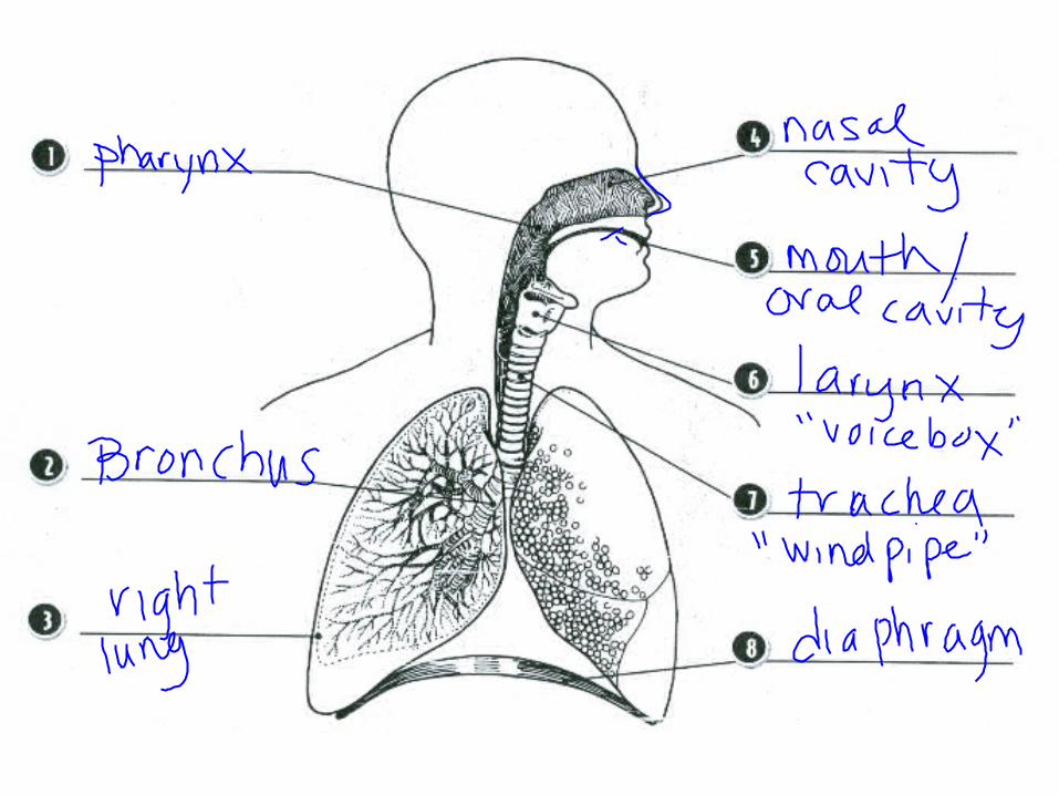
What are the organs of the Respiratory system?
• Nose
• Pharynx
• Larynx
• Trachea
• Bronchi
• Bronchioles
• Alveoli
• Lungs
Figure 13.1

Anatomy of the respiratory system
• respiratory basics – the 4 steps of respiration


Pleura/pleural cavity
Arterioles, capillaries, venules

Pleural membranes and pleural cavity

The Nose:• only externally visible
part of the respiratory system
• Air enters the nose through the nostrils
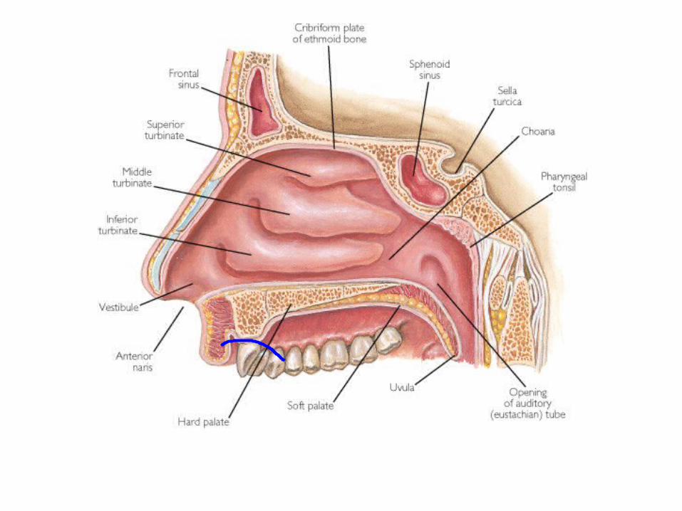
• The interior of the nose consists of a nasal cavity divided by a nasal septum

Anatomy of the Nasal CavityWhat is the Function of the nose?
1. Moistens air
2. Cleanses/filters -traps incoming foreign particles
3. Warms air
4. Sense of smell
How does the nose cleanse the air?
The cleansing is accomplished by the coarse hair in the nostrils. Also the cilia and mucus trap the particles

Why do we have a runny nose when we cry?
• The tear (lacrimal) glands drain into the nasal cavity by way of the tear ducts

Upper Respiratory Tract
Figure 13.2

Anatomy of the Nasal Cavity• Lateral walls have
projections called conchae– Increases surface area
for moistening and warming the air
– Odor receptors are on the cilia in the cells located here
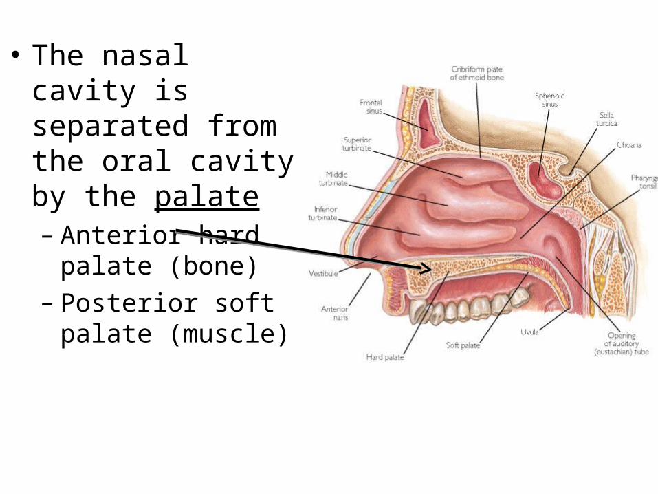
• The nasal cavity is separated from the oral cavity by the palate– Anterior hard palate
(bone)– Posterior soft palate
(muscle)

Paranasal SinusesSinuses are cavities
surrounding the nasal cavity
Sinusitis is an inflammatory disease that can be caused by allergy, bacteria, fungi or virus. The sinus tissue becomes inflamed, cannot drain, and causes pain

What are the function of the sinuses?1. Reduces the weight of the skull
2. Act as resonance chambers for speech
3. 1st line of defense of the immune system
4. Produce mucus that drains into the nasal cavity

Pharynx (aka:Throat)• Muscular passage
from nasal cavity to larynx
NOTE:There are two “tubes 1. Larynx 2.Esophagus (food
tube)

Three regions of the pharynx (throat)Nasopharynx – superior region behind nasal cavityOropharynx – middle region behind mouthLaryngopharynx – inferior region attached to larynx
The oropharynx and laryngopharynx are common passageways for air and food

What does the throat look like?
What are tonsils? Tonsils are a protective ring of lymphatic tissue that contain lymphocytes that help protect against pathogens

What does tonsillitis look like?
http://www.funscrape.com/Search/1/tonsillitis.html
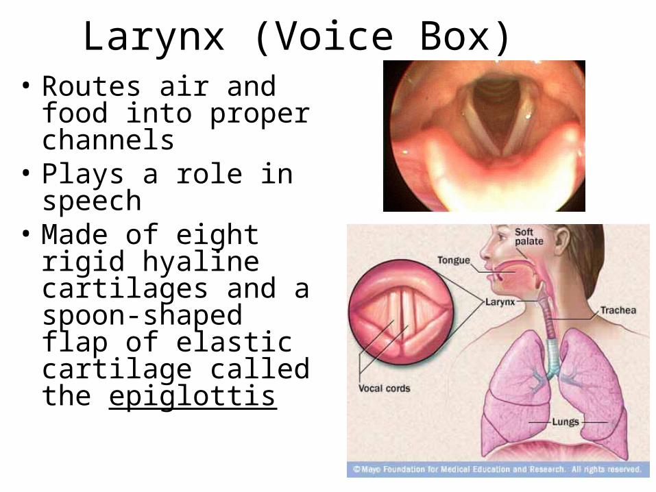
Larynx (Voice Box)• Routes air and food
into proper channels• Plays a role in
speech• Made of eight rigid
hyaline cartilages and a spoon-shaped flap of elastic cartilage called the epiglottis

Structures of the Larynx• Thyroid cartilage
– Largest hyaline cartilage– Protrudes anteriorly
(Adam’s apple)
• Epiglottis– Superior opening of the
larynx– Routes food to the
esophagus and air toward the trachea

Structures of the Larynx• Vocal cords (vocal folds)
– Vibrate with expelled air to create sound (speech)
• Glottis – opening between vocal cords
vocal cords


Trachea (Windpipe)• Connects larynx with bronchi• Has cartilage rings – keeps
trachea open while breathing
• Lined with ciliated mucosa cells– Cilia sweep debris away from
lungs– Goblet cells produce mucous
that trap dust and other particles
Video of cilia & goblet cells Super-detailed explanation of how goblet cells work

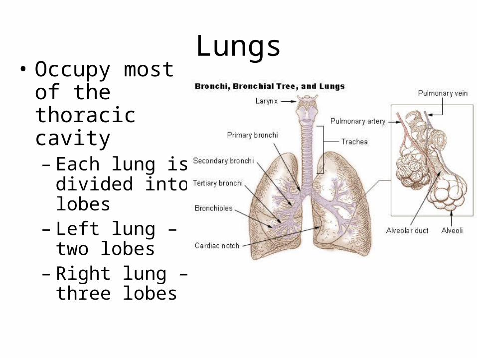
Lungs• Occupy most of
the thoracic cavity– Each lung is
divided into lobes
– Left lung – two lobes
– Right lung – three lobes


Bronchi and Bronchioles
The trachea divides into Bronchi
Bronchi - A pair of tubes that lead into the lungs that branch off into Bronchioles
Bronchioles – smaller tubes that end with air sacs called alveoli

Bronchioles
• Bronchi lead into tiny bronchioles
• Bronchioles terminate in alveoli
• Alveoli are tiny air sacs where diffusion of gasses occur

Alveoli• Gas exchange
takes place within the alveoli in the respiratory membrane
• Alveoli are covered in capillaries that allow for gas exchange

The pathway of air
Description Function
Nasal Cavity Hollow spaces in the nose Warm filter and moisten the air
Pharynx Chamber that lies between nasal cavity and larynx
Connects surrounding regions
Glottis Opening to the larynx Passage of air into the larynx
Larynx Cartilaginous organ that houses the vocal cords; voice box
Sound production
Trachea Flexible tube that connects larynx with the bronchi
Passage of air into the bronchi
Bronchi Paired tubes inferior to the trachea that enter the lungs
Passage of air to lungs
Bronchioles Branched tubes that lead from bronchi to alveoli
Passage of air to each alveoli
Alveoli Tiny air sacs that are covered in capillaries
Gas exchange

The respiratory system moves gases into
and out of the blood.• The lungs contain the
bronchi, bronchioles, and alveoli.
• Millions of alveoli give the lungs a huge surface area. (30-50 m2)
• The alveoli absorb oxygen from the air you inhale.


Gas exchange occurs in the alveoli of the
lungs. • Oxygen and carbon dioxide are carried by the blood to and
from the alveoli.– oxygen diffuses from alveoli into capillary – oxygen binds to hemoglobin (oxyhemoglobin) in red blood cells – carbon dioxide diffuses from capillary into alveoli
ALVEOLI GAS EXCHANGES
capillaries
alveolus
capillary
co2
o2
Co2 diffusesinto alveolus.
O2 diffusesinto blood.

Mechanics of Breathing (Pulmonary Ventilation)
• Two phases– Inspiration – flow of air into lung– Expiration – air leaving lung
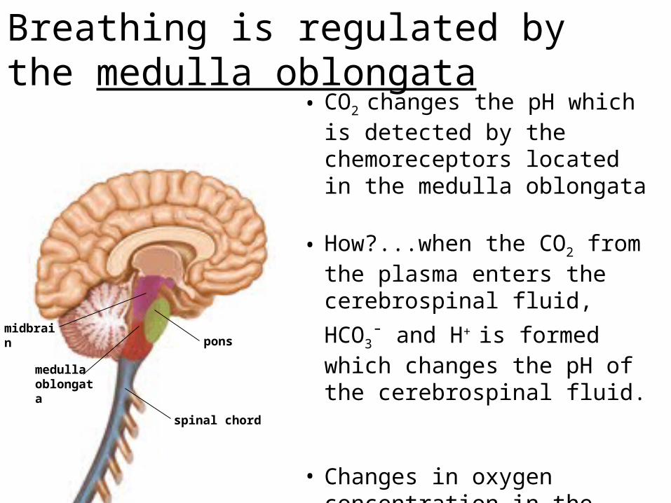
Breathing is regulated by the medulla oblongata
midbrainpons
medulla oblongata
spinal chord
• CO2 changes the pH which is detected by the chemoreceptors located in the medulla oblongata
• How?...when the CO2 from the plasma enters the cerebrospinal
fluid, HCO3- and H+ is formed
which changes the pH of the cerebrospinal fluid.
• Changes in oxygen concentration in the blood are detected by chemoreceptors in the aorta and carotid artery and the information is sent to the medulla oblongata
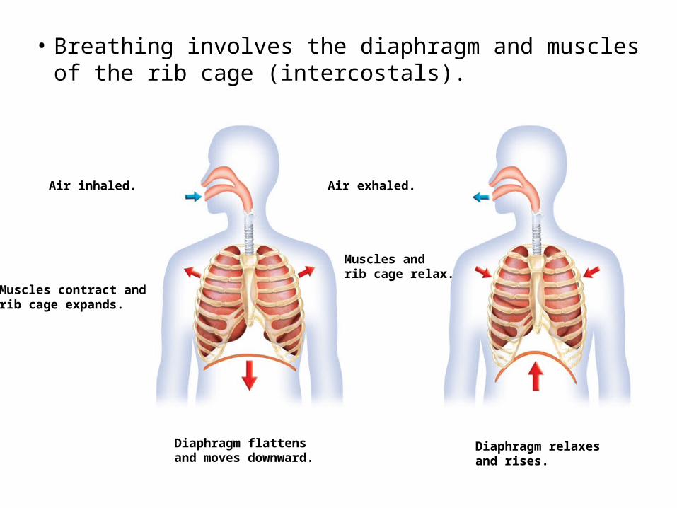
• Breathing involves the diaphragm and muscles of the rib cage (intercostals).
• Air flows from areas of high pressure to low pressure.
Air inhaled.
Muscles contract andrib cage expands.
Diaphragm flattensand moves downward.
Air exhaled.
Muscles andrib cage relax.
Diaphragm relaxesand rises.

Inhale:• Diaphragm and
intercostal muscles contract
• The size of the thoracic cavity increases
• External air is pulled into the lungs due to an increase in intrapulmonary volume
Exhale• Largely a passive
process which depends on natural lung elasticity
• As muscles relax, air is pushed out of the lungs
• Forced expiration can occur mostly by contracting internal intercostal muscles to depress the rib cage
Nonrespiratory Air Movements
• Can be caused by reflexes or voluntary actions
• Examples– Cough and sneeze – clears lungs of debris– Laughing– Crying– Yawn– Hiccup – spasm of the diaphragm

Respiratory Volumes and Capacities
• Normal breathing moves about 500 ml of air with each breath (tidal volume [TV])
(Tidal volume is the lung volume representing the normal volume of air displaced between normal inspiration and expiration when extra effort is not applied. )
• Many factors that affect respiratory capacity– A person’s size
– Sex
– Age
– Physical condition
• Residual volume of air – after exhalation, about 1200 ml of air remains in the lungs

Gas Transport in the BloodOxygen transport in the blood
– Inside red blood cells attached to hemoglobin (oxyhemoglobin [HbO2])
– 1 hemoglobin molecule + 4 oxygen molecules = Oxyhemoglobin
– A small amount is carried dissolved in the plasma
Carbon dioxide transport in the blood– Most is transported in the plasma as bicarbonate ion
(HCO3–)– A small amount is carried inside red blood cells on
hemoglobin, but at different binding sites than those of oxygen
– When bound to hemoglobin – HbCO2
Internal Respiration
• Exchange of gases between blood and body cells
• An opposite reaction to what occurs in the lungs– Carbon dioxide diffuses out of tissue to blood– Oxygen diffuses from blood into tissue

Internal Respiration
Figure 13.11
Oxyhemoglobin – HbO2 (oxygen binded to hemoglobin) Bicarbonate ion – HCO3
- (how carbon dioxide found in plasma)

Respiratory diseases interfere with gas exchange.
• Lung diseases reduce airflow and oxygen absorption.– Emphysema destroys alveoli.
– Pneumonia – fluid filled in alveoli
How will this affect gas exchange?
– Asthma constricts airways.– Cystic fibrosis is genetic and produces sticky
mucus.

What happens during pneumonia?Pneumonia – alveoli fill up with fluid, therefore gas exchange cannot occur.Pneumonia is caused by bacteria or virus. It is diagnosed by an x-ray

Emphysema
• Alveoli enlarge as adjacent chambers break through
• Chronic inflammation promotes lung fibrosis• Airways collapse during expiration• Patients use a large amount of energy to exhale• Overinflation of the lungs leads to a permanently
expanded barrel chest

Acute & Chronic Bronchitis
• The lower respiratory passages becomes severely inflamed and mucus production increases
• Pooled mucus impairs ventilation and gas exchange
• Risk of lung infection increases
• Pneumonia is common
• Hypoxia (low oxygen levels)and cyanosis (blueish color) occur early
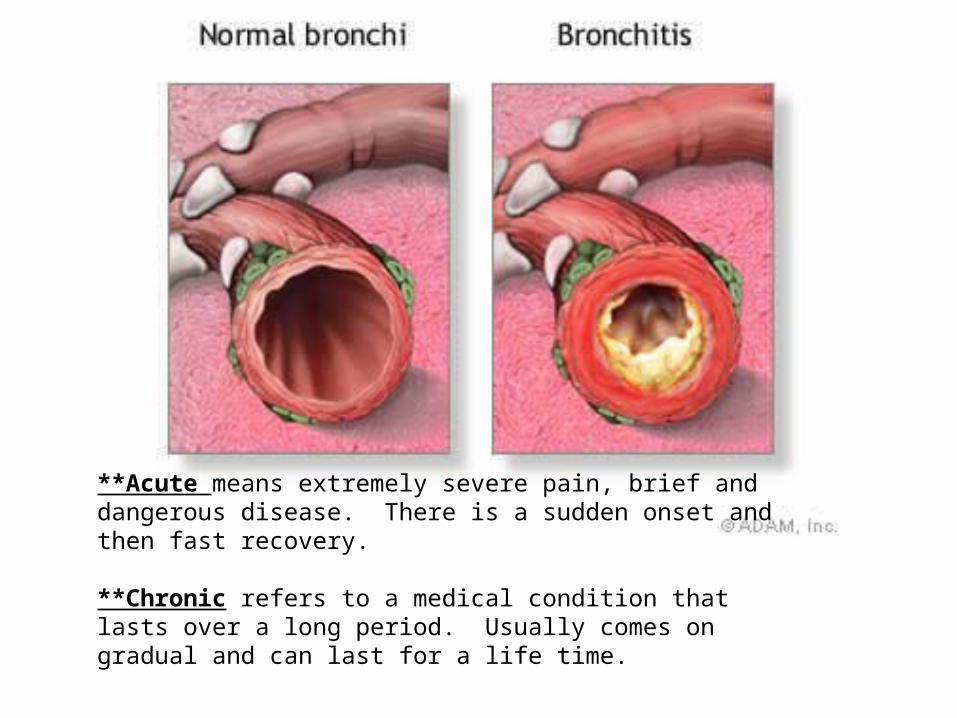
**Acute means extremely severe pain, brief and dangerous disease. There is a sudden onset and then fast recovery.
**Chronic refers to a medical condition that lasts over a long period. Usually comes on gradual and can last for a life time.

Chronic Obstructive Pulmonary Disease (COPD)
Figure 13.13
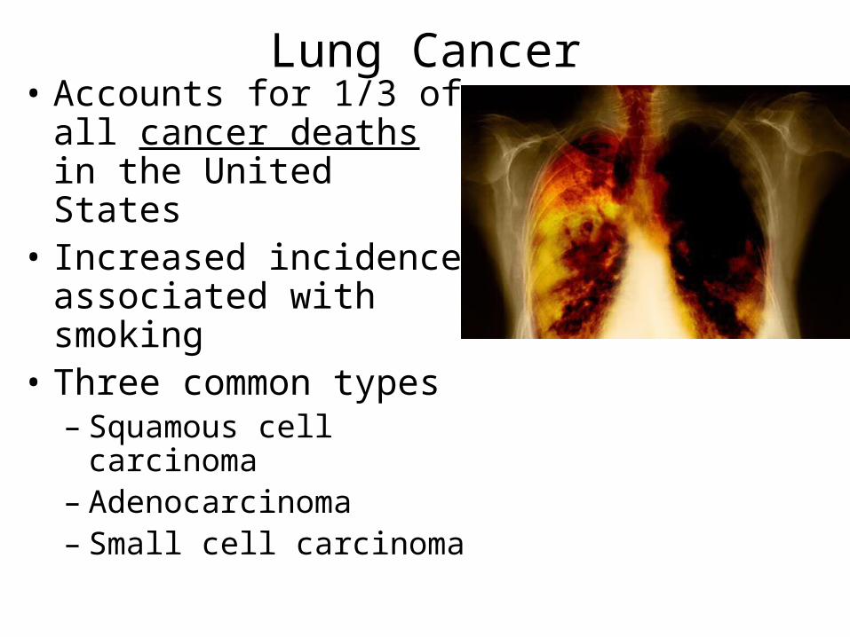
Lung Cancer• Accounts for 1/3 of all
cancer deaths in the United States
• Increased incidence associated with smoking
• Three common types– Squamous cell
carcinoma– Adenocarcinoma– Small cell carcinoma


• Smoking is the leading cause of lung diseases.

Sudden Infant Death Syndrome (SIDS)
• Apparently healthy infant stops breathing and dies during sleep
• Some cases are thought to be a problem of the neural respiratory control center
• One third of cases appear to be due to heart rhythm abnormalities

Asthma• Chronic inflamed hypersensitive
bronchiole passages
• Response to irritants with difficulty breathing (dyspnea), coughing, and wheezing

What happens during an asthma attack?
• During an asthma attack the smooth muscle of the bronchioles contracts
• Causing constriction, blocking airways and wheezing
Developmental Aspects of the Respiratory System
• Lungs are filled with fluid in the fetus
• Lungs are not fully inflated with air until two weeks after birth
• Surfactant that lowers alveolar surface tension is not present until late in fetal development and may not be present in premature babies

Important birth defects– Cystic fibrosis – oversecretion of thick mucus
clogs the respiratory system

What is a cleft palate?During development in the womb, tissue that forms the lip and the palate fail to connect. Children born with the condition can have problems with hearing, feeding and speech and teasing in the playground can lead to social isolation and misery.
Cleft palate is quite a common 1 in every 700 births.
Causes? Can be genetic, passed down from one or both parentsOr possibly drugs, viruses, or other toxins can all cause these birth defects.

Aging Effects
• Elasticity of lungs decreases
• Vital capacity decreases
• Blood oxygen levels decrease
• Stimulating effects of carbon dioxide decreases
• More risks of respiratory tract infection
Respiratory Rate Changes Throughout Life
• Newborns – 40 to 80 respirations per minute
• Infants – 30 respirations per minute
• Age 5 – 25 respirations per minute
• Adults – 12 to 18 respirations per minute
• Rate often increases somewhat with old age


