
Histology of Respiratory System
Respiratory System
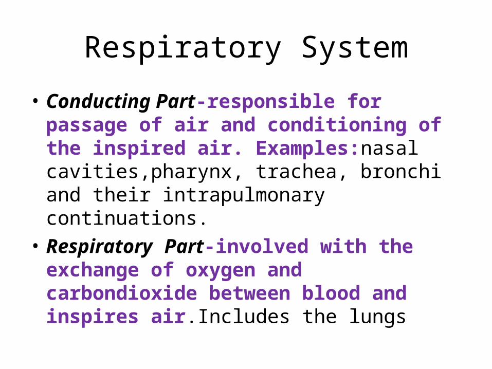
• Conducting Part-responsible for passage of air and conditioning of the inspired air. Examples:nasal cavities,pharynx, trachea, bronchi and their intrapulmonary continuations.
• Respiratory Part-involved with the exchange of oxygen and carbondioxide between blood and inspires air.Includes the lungs


RESPIRATORY SYSTEM HISTOLOGY
• Trachea • Bronchus
-Primary bronchus
-Secondary bronchus
-Tertiary bronchus• Bronchiole • Lung

Trachea (T.S. Low Power)

Trachea
• Mucosa
-Epithelium
-Lamina propria• Sub mucosa• Cartilage &muscle
layer• Adventitia

Trachea Mucosa• Epithelium -Pseudo stratified ciliated
columnar/ Respiratory epithelium
Cells-Ciliated columnar cells - Goblet cells -Brush cells - Basal cells -Granule (kulchitsky)
cells -Clara cells( bronchiolar
cells) surfactant secretion• Lamina propria - Elastic
fibre, Lymphocyte, Mast cells, Blood vessels


Respiratory Epithelium

Trachea( T.S. High Power)

Trachea• Sub mucosa-• Loose connective tissue• Tracheal glands-Mixed
(serous &mucus) glands• Blood vessels and ducts• Cartilage &smooth muscle
layer-• ”C” Shaped hyaline cartilage
having perichondrium and chondrocytes
• Ends of cartilage connected by smooth muscles
• Adventitia-fibro elastic tissue

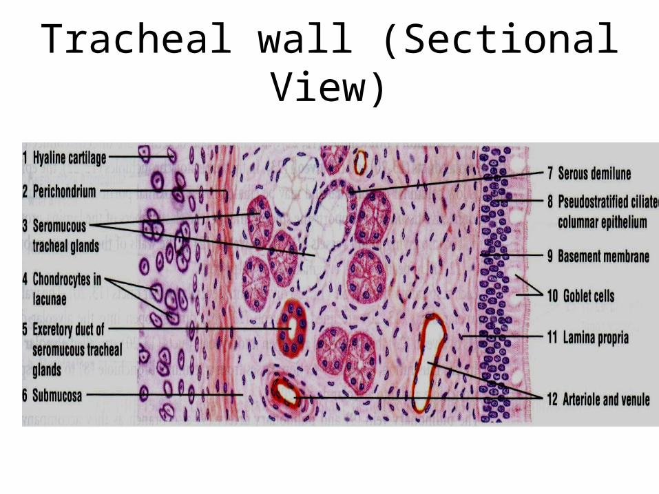
Tracheal wall (Sectional View)

Trachea and Oesophagus


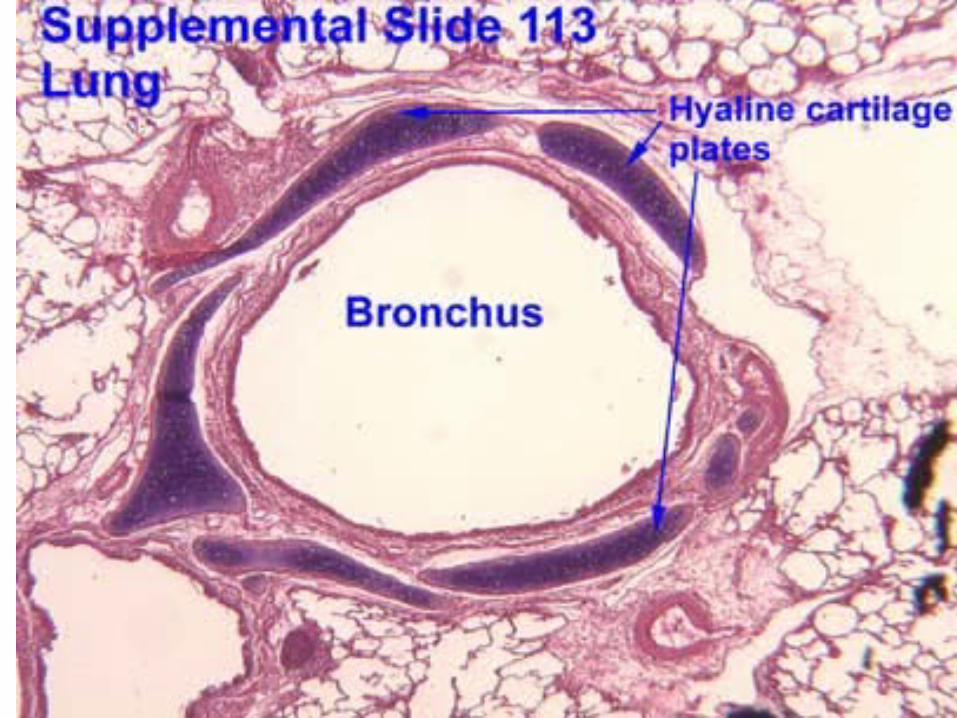
Bronchus
• Principal bronchus
-same as trachea
• Secondary /Lobar
• bronchus
-Irregular hyaline cartilage
-Pseudo stratified ciliated columnar
• Tertiary /Segmental bronchus
-Columnar epithelium
-Patches of cartilage

Changes as bronchi become smaller• Cartilage-irregular and smaller. Absent in
bronchioles.• Muscle- increases as bronchi becomes smaller.
(Spasm of these muscles bring difficulty in breathing in allergic conditions)
• Subepithelial Lymphoid Tissue-increases with decrease in the diameter of bronchi.
• Glands-few.Absent in the walls of capillaries.• Epithelium- pseudostratified ciliated columnar
epithelium in principal bronchi later simple ciliated columnar,non-ciliated columnar and later cuboidal in respiratory bronchioles

Bronchiole
• Terminal bronchiole
-Columnar epithelium
-No cartilage
- smooth muscle +
-Clara cells present
• Respiratory bronchiole
-Cuboidal epithelium
-No mucous gland

Bronchiole

Bronchus and Bronchiole


Bronchiole

Differences between Bronchi and Bronchioles
Bronchioles• No glands• No cartilage• No goblet cells• Thick smooth muscle
layer• Presence of Clara
cells• Many elastic fibres

Terminal Bronchiole

Terminal Bronchiole


Respiratory Bronchiole
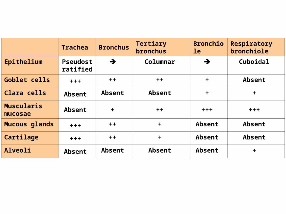
Trachea Bronchus Tertiary bronchus Bronchiole Respiratory bronchiole
Epithelium Pseudostratified
Columnar Cuboidal
Goblet cells +++ ++ ++ + Absent
Clara cells Absent Absent Absent + +
Muscularis mucosae
Absent + ++ +++ +++
Mucous glands +++ ++ + Absent Absent
Cartilage +++ ++ + Absent Absent
Alveoli Absent Absent Absent Absent +

Cells seen in the respiratory passages
• Goblet cells• Non-ciliated serous
cells• Basal cells• Cells of Clara• Brush cells • Argyrophil Cells similar
to diffuse endocrine cells of gut
• Lymphocytes

• Goblet cells: numerous and secrete mucous. Mucous traps the dust particles and is moved by ciliary action towards pharynx.
• Non-ciliated serous cells: secretes watery fluid that keeps the epithelium moist
• Cells of Clara: are non-ciliated cells predominantly seen in terminal bronchioles. Secrete a fluid that spreads over the alveolar surface forming a film that reduces surface tension. May function as stem cells

• Basal cells: Multiply and transform into other cell types replace the lost cells.
• Argyrophil cells: cells similar to diffuse endocrine cells of the gut containing granules, secrete hormones and active peptides including serotonin and bombesin.
• Lymphocytes and other leucoctes may be present in the epithelium.


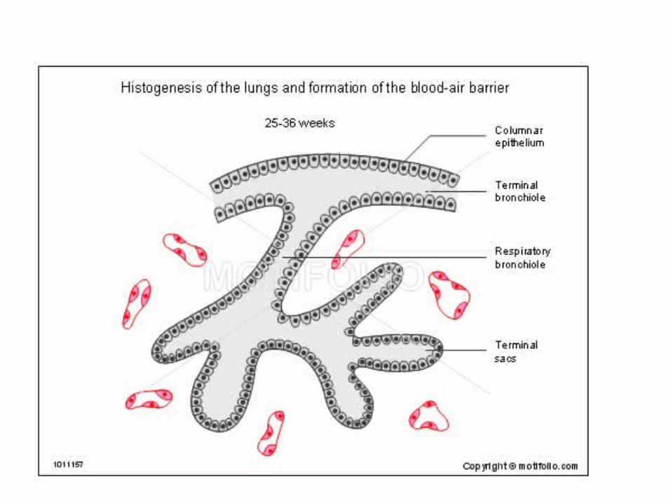
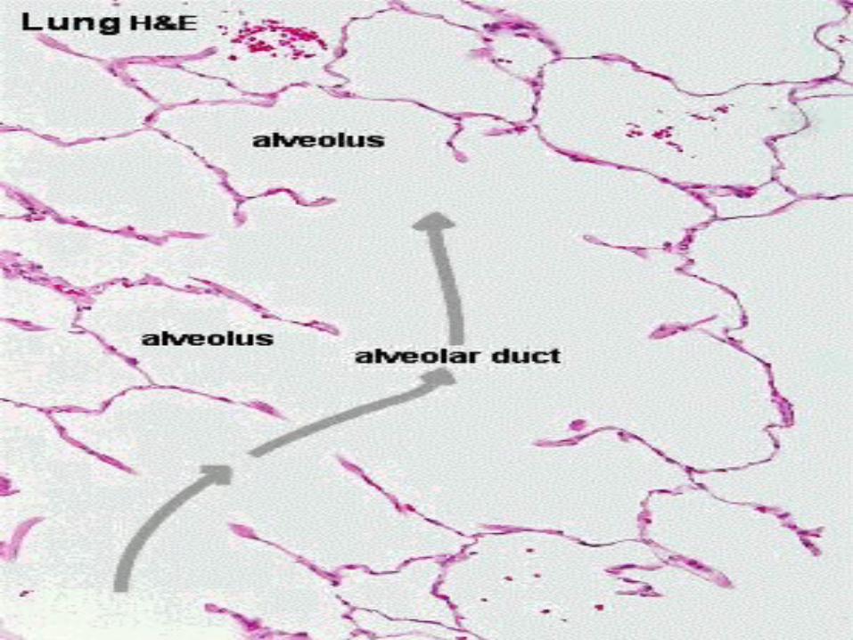
Alveolar Ducts and AlveolarSacs

Alveolar Sac

Alveoli
• 200 million in a normal lung
• Total area-75 square meters
• Total capillary surface area available for exchange-125square meters
• Are spongy and form the parenchyma of lung.
• Sac like evaginations present at the terminal end of the bronchial tree.

• In section, they resemble a honeycomb• Alveoli are separated by interalveolar septum
lying between thin epithelial lining of two neighbouring alveoli
• Interalveolar septum contains anetwork of capillaries supported by reticular and elastic fibres, occassionally fibroblasts, macrophages and mast cells.
• Septum containspores(ALVEOLAR PORES OF KOHN) help in passage of air from one alveolus to another, thus equalizing Pressure in the alveoli

• Elastic fibres-enable the alveoli to expand during inspiration and passively contract during expiration.
• Reticular fibres support and prevent overdistention of the alveoli

Cells in the Alveoli
• Type I Pneumocytes
• Type II Pneumocytes
• Macrophages or Dust cells

Pneumocytes
• Type I Alveolar or Type I Pneumocytes orSquamous Epithelial cells- Form the lining of 90% of the alveolar surface, numerous,squamous,thinness reduced to 0.05 to0.2 micron m, edges of the 2 cells overlap and are uniting by tight junctions- preventing leakage of blood from capillaries to the alveolar lumen
• Form Blood Air barrier

Type I Pneumocytes

Type II Alveolar or Type II pneumocytes
• Also known as Septal cells
• Rounded or cuboidal secretory cells with microvilli
• Secretory granules are made of several layers- Multilamellar bodies.
• These lamillar bodies are cytoplasmic inclusions made up of phospholipid which combines with other chemicals to form surfactant & then ooze out of the cell by exocytosis.
• Pulmonary Surfactant – is the fluid secreted that spreads over the alveolar surface
• These cells can multiply to replace damaged cells.
• Surfactant also has bactericidal properties
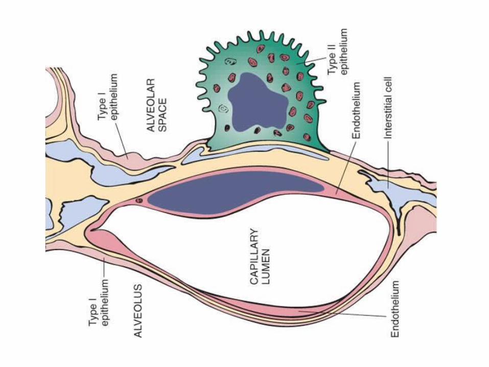

Type I and II Pneumocytes, capillaries and Dust cells

Pulmonary Surfactant
• Surfactant contains phospholipids, proteins and glycosaminoglycans, reduces the surface tension and prevents collapse of the alveolus during expiration.
• Is constantly renewed.• Removed from the
surface by Type I pneumocytes and macrophages
• The reduced surface tension in the alveoli decreases the force that is needed to inflate alveoli during inspiration.
• Therefore surfactant stabilizesthe alveolar diameters, facilitates their expansion and prevents their collapse by minimizing the collapsing forces

Blood Air Barrier• Consist of a thin layer of surfactant• Cytoplasm of Type I Pneumocytes• Basement membrane of Pneumocytes• Intervening Connective Tissue• Basement membrane of capillary endothelial cell• Cytoplasm of capillary Endothelial cells• Endothelial cells of alveolar capillaries are
extremely thin, have numerous projections increasing the surface area of the cell membrane exposed to blood for gaseous exchange. At places the 2 basement membranes are so fused reducing the thickness of Barrier.


Alveolar Macrophages or Dust cells
• Derived from Monocytes and are part mononuclear phagocytic system.
• Either seen in the septa or alveoli
• Cytoplasm contains phagocytosed inhaled carbon and dust particles
• Inhaled carbon and dust particles are passed on to them from pneumocyte I through pinocytic vesicles

Alveolar Macrophages or Dust cells
• Migrate from septum to alveolar surface and are carried to the pharynx through sputum
• Main function is to clean the alveoli of invading microorganisms and inhaled particulate matter by phagocytosis

Heart failure cells
• In congestive heart failure where pulmonary capillaries are overloaded with blood, the alveolar macrophages phagocytose erythrocytes that escape from capillaries
• These cells become red brick in color because of pigment Haemosiderin and are known as heart failure cells.


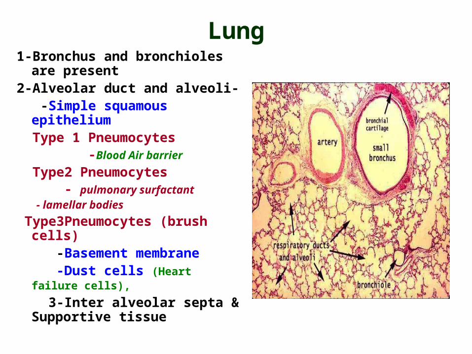
Lung1-Bronchus and bronchioles are
present2-Alveolar duct and alveoli- -Simple squamous epithelium Type 1 Pneumocytes -Blood Air barrier
Type2 Pneumocytes - pulmonary surfactant - lamellar bodies
Type3Pneumocytes (brush cells) -Basement membrane -Dust cells (Heart failure cells), 3-Inter alveolar septa &
Supportive tissue


Clinical• Bronchiectasis: Permanent dilatation of bronchi
and bronchioles full of mucous. This is caused by tissue destruction secondary to infection.
• Respiratory distress syndrome or Hyaline membrane disease: in premature new born babies there is deficiency of surfactant as it is produced in the last week of gestation. They have difficulty in expanding the already collapsed lungs. A fibrin rich eosinophilic material called hyaline membrane lines the respiratory bronchioles and alveolar ducts of babies.Synthesis of surfactant is induced by administration of corticosteroids.

MCQ
Heart failure cells are
• Type I Pneumocytes
• Type II Pneumocytes
• Macrophages
• Cells of Clara

MCQ
All of the following are true for Pulmonary Surfactant EXCEPT
• Lines the alveolar surface
• Secreted by Type I alveolar cells
• Secreted by Type II alveolar cells
• Prevents collapse of lungs

MCQ
Cartilage is seen in
• Bronchus
• Terminal bronchiole
• Respiratory bronchiole
• Alveolar duct

MCQ
Cells of Clara are predominantly seen in
• Trachea
• Primary Bronchus
• Secondary Bronchus
• Bronchioles
MCQ
Which of the following does not take part in the formation of Blood Air Barrier?
• Type I pneumocytes
• Type II Pneumocytes
• Capillary endothelium
• Basement membrane of Capillary Endothelium


