
GIS-K-26
INTESTINAL OBSTRUCTIONSyahbuddin Harahap
Division of Digestive Surgery
Department of Surgery
Faculty of Medicine University of North Sumatera Faculty of Medicine University of North Sumatera
Adam Malik Hospital

DEFINITION
• Bowel /Intestinal obstruction occurs when the normal
propulsion and passage of intestinal contents does not
occur
BO can involve:
– SBO � Small intestine– SBO � Small intestine
– LBO � Large intestine
– Generalized Ileus
via systemic alterations
involving both the small and large intestine

�Etiopathogenesis
- Mechanical obstruction
- Non mechanical (Functional ) obstruction
Mechanical obstruction (Dynamic ) ileus refers to a lack
of passage due to an “obstruction of the bowel”,
which can be located anywhere in the bowelwhich can be located anywhere in the bowel
Non mechanical Obstruction (Paralytic )(adynamic)
(Fungsional) ileus
Paralytic ileus refers to a lack of passage due to
“paralysis of the bowel”

Intestinal /Bowel Obstruction can also be classified according to :
�Time of presentation and duration of obstruction:- Acute- Chronic
�The extent of obstruction-Partial-Complete-Complete
�The type of obstruction-Simple-Closed-loop-Strangulation

Nonmechanical Obstruction�Paralytic (adynamic) (Fungsional) ileus due to :1. After abdominal operations 2. Inflammation �Peritonitis
3. Systemic disorders e.g. sepsis, hyponatremia, hypokalemia,
hypomagnesemia
4. Retroperitoneal disorders e.g. ureter, spine fractures ,
hematoma
5. Thoracic conditions e.g. pneumonia, rib fractures5. Thoracic conditions e.g. pneumonia, rib fractures
6. Drugs e.g opiates, psychotropics , General anesthesie
� Pseudo-ObstructionImbalance in the parasympathetic and sympathetic influences
on Colonic motility.
Acute colonic pseudo-obstruction, also known as Ogilvie
syndrome.

MECHANICAL OBSTRUCTION
at each age group
• Neonate
Congenital atresia
Volvulus neonatum
Meconeum ileus
Hirschsprung”s disease
Imperforate anus
• Middle ageAdhesesion and band
Strangulated Ing.hernia
Strangulated fem.hernia
Imperforate anus
• Infant
Stranggulated inguinal hernia
Intussuception
Complication of Meckel”s diverticulum
Hischsprung”s diseases
• Young adult
Adhesions and bands
Strangulated ing.hernia
Carcinoma colon
Volvulus
• ElderlyAdhesion and bandsStrangulated Ing.herniaStrangulated fem.herniaCarcinoma colonVolvulusImpacted faeces
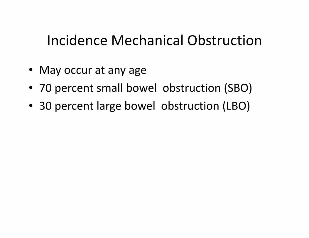
Incidence Mechanical Obstruction
• May occur at any age
• 70 percent small bowel obstruction (SBO)
• 30 percent large bowel obstruction (LBO)
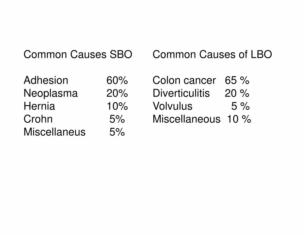
Common Causes SBO
Adhesion 60%
Neoplasma 20%
Hernia 10%
Crohn 5%
Common Causes of LBO
Colon cancer 65 %
Diverticulitis 20 %
Volvulus 5 %
Miscellaneous 10 %Crohn 5%
Miscellaneus 5%
Miscellaneous 10 %

Etiology?
• Extrinsic (Outside the wall )
• Intrinsic (Inside the wall )• Intrinsic (Inside the wall )
• Inside the lumen

Extrinsic (Outside the wall)
• Adhesions
• Hernia
-- inguinal, femoral, umbilical
• Neoplastic
– extraintestinal neoplasm
• Volvulus (sigmoid, cecal)

Intrinsic (Inside the wall )
• Congenital
– Malrotation
• Neoplastic
– Primary neoplasms
– Metastatic neoplasms– Metastatic neoplasms
• Inflammatory
– Crohn's disease
• Miscellaneous
– Intussusception
– Radiation

Intraluminal (Inside the lumen)
• Gallstone
• Enterolith
• Bezoar
• Foreign body
• Parasit �Bolus Ascaris

Clinical Picture
Mechanical obstruction
The classic quartet
1. Colicky abdominal pain
2. Abdominal distension2. Abdominal distension
3. Nausea and Vomiting
4. Decreased passage of stool or flatus

PathophysiologyDependent upon :
1. Degree of obstruction
2. Duration of obstruction
3. Presence and severity of ischaemia
Result in :
1. Accumulation of fluid and air(Sequestration within the dilated
loop)loop)
Fluid disturbances massive third space losses
8 – 10 L of fluid are secreted
Hypovolumic shock oliguria, hypotension,hemoconcentration
2. Electrolyte depletion3. Bacterial overgrowth �Rapid colonisation
-Maximal by 24 hrs after obstruction
-Bacterial translocation to node and portal system

4. Bowel distension
-Chest compression by pushing up diaghragma muscle
-Decreases the ability mucosa to absorb ,stasis intestinal content
of fluids and electrolytes-Increased intraluminal pressure � oedematous� cyanosis�
intraperitoneal exudation �necrosis �perforation�peritonitis
-ACS � impediment in venous return�arterial insufficiency
5. LBO5. LBO
Ileocaecal valve plays prominent role in pathophysiology of LBO.
If competent valve = Closed loop obstruction
In 10 – 20 % of individual ICV incompetentCaecal around 10 – 12 cm � the risk of perforation

Clinical Manifestations
Altered mental state
Vital Sign
Hypovolumic shock
TachicardiaTachicardia
Hypotension
Tachipnoe
Fever
Oliguria

Abdominal Examination
Patient �Supine position with the legs flexed at the hip
Abdominal Colicky pain The periodicity of pain:
3 to 4 minutes pain from proximal intestinal obstruction
15 to 20 minutes pain from distal small bowel or colon
On Inspection
Abdominal distensionAbdominal distension
Proximal obstructions may cause little or no distention
Distended small bowel loops usually occupy the central
abdomen Distended large bowel loops are typically seen
around the periphery .
Visible peristalsis which are indicative of acute small bowel
obstructionAbdominal Scars � Adhesion

On AuscultationPerformed for at least 3 to 4 minutes
�Metallic sound
�Borborygmi
�The absence of bowel tones :
Is typical of intestinal paralysis .
Late�Quiet abdomen (may also indicate Late�Quiet abdomen (may also indicate
intestinal fatigue from long-standing
obstruction).

On PalpationInguinal ,Femoral , Umbilical ,Incisional HerniasPalpable mass Abdominal asymmetry or a protruding mass
suggests an underlying malignancy, an abscess, or closed-loop
obstruction.
Peritoneal irritation
On PercussDull � Fluid or MassDull � Fluid or Mass
Tympanic � Air (Intraluminal or not )
Peritoneal irritation
DRE (Digital Rectal Examination )
For Mass , Impacted faeces

Vomiting – NG Aspirates
Consists� food and gastric chyme� bile �faeculent
�GOO � Clear , food and gastric chyme�Mid to distal SBO � Bilious/Bile�Distal SBO to LBO � Feculent

Mechanical Obstruction Nonmechanical Obstruction
Abdominal
Pain
colicky pain severity may decrease over time as a
result of bowel fatigue and atony.
3 to 4 minutes from proximal SBO
15 to 20 minutes distal SBO or LBO
Diffuse , usually mild
Inspection Abdominal distension
Visible peristalsis
Abdominal distension
Auscultation Metalic Sound Quiet abdomen
Borborygme
Late � Quiet Abdomen
Abd.X Ray
Erect
Supine
Large small intestinal loops
gas less in colon
Step ladder A/F levels
Gas diffusely through
intestine, incl. colon
May have large diffuse A/F
levels
Barium
Enema
Obvious transition point on contrast study No obvious transition point
on contrast study
Exudate No peritoneal exudate Peritoneal exudate if
peritonitis
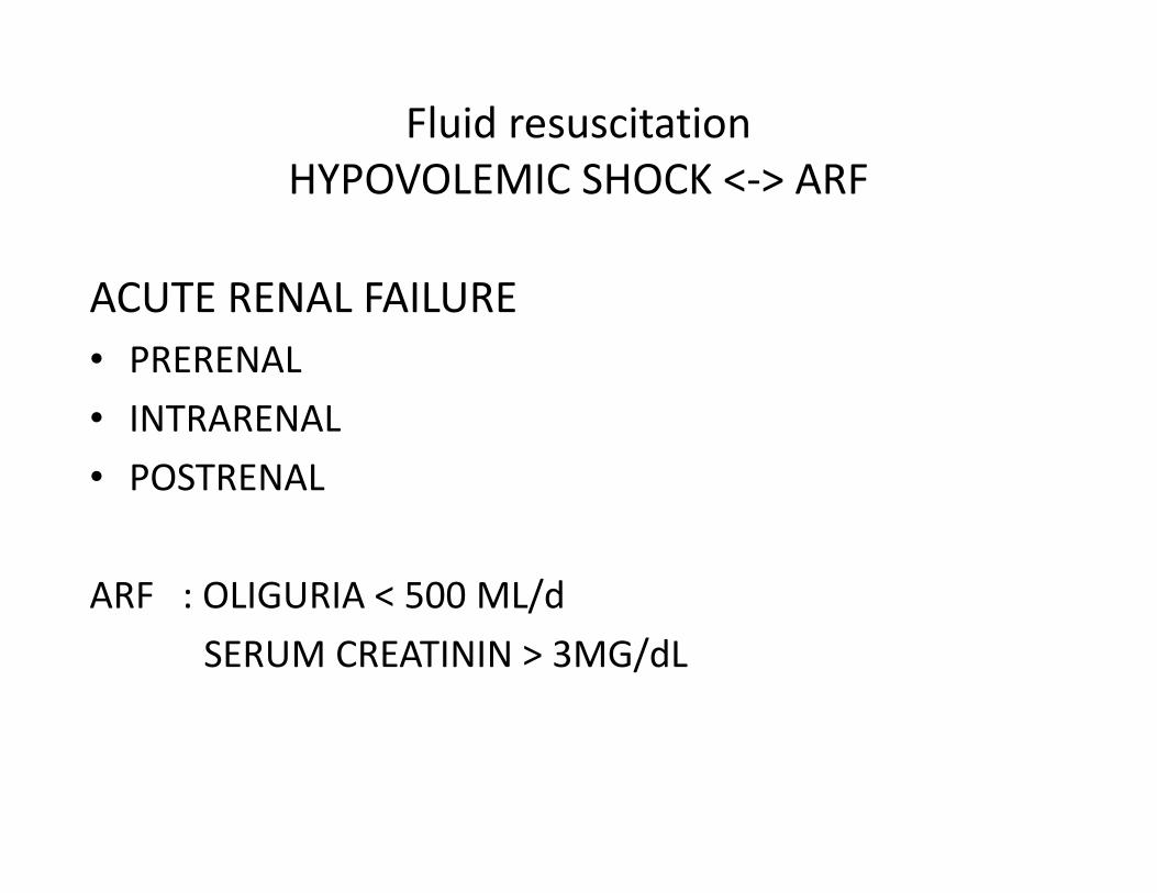
Fluid resuscitation
HYPOVOLEMIC SHOCK <-> ARF
ACUTE RENAL FAILURE
• PRERENAL
• INTRARENAL
• POSTRENAL
ARF : OLIGURIA < 500 ML/d
SERUM CREATININ > 3MG/dL
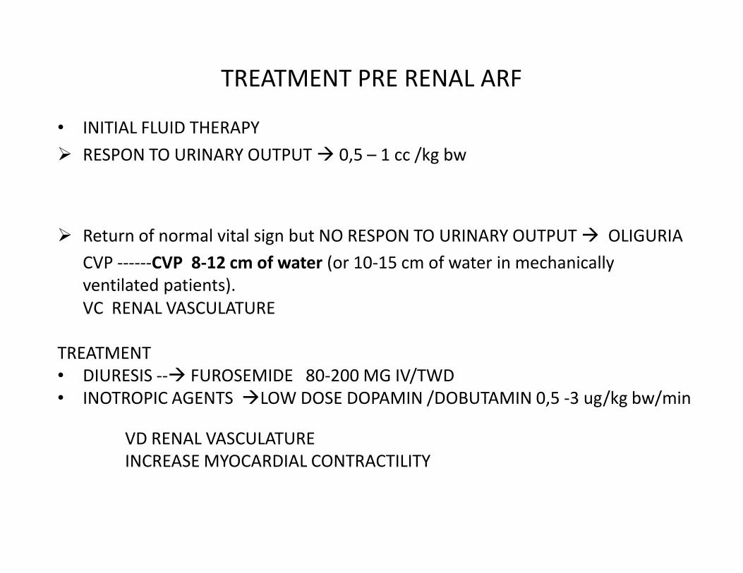
TREATMENT PRE RENAL ARF
• INITIAL FLUID THERAPY
� RESPON TO URINARY OUTPUT � 0,5 – 1 cc /kg bw
� Return of normal vital sign but NO RESPON TO URINARY OUTPUT � OLIGURIA
CVP ------CVP 8-12 cm of water (or 10-15 cm of water in mechanically
ventilated patients).ventilated patients).
VC RENAL VASCULATURE
TREATMENT
• DIURESIS --� FUROSEMIDE 80-200 MG IV/TWD
• INOTROPIC AGENTS �LOW DOSE DOPAMIN /DOBUTAMIN 0,5 -3 ug/kg bw/min
VD RENAL VASCULATURE
INCREASE MYOCARDIAL CONTRACTILITY
EVALUATION OF FLUID RESUSCITATION
• RETURN OF NORMAL VITAL SIGNS
• MENTAL STATUS
• URINARY OUTPUT
• ACID/BASE BALANCE• ACID/BASE BALANCE
• CVP

Diagnoctic Studies
�Laboratory test• Fecal Occult Blood Test
• CBC
• Serum electrolyte concentrations
• The serum creatinine concentration / BUN• The serum creatinine concentration / BUN
• The coagulation profile
• Urinalysis should be done to check for hematuria
• Liver function profile

�Sigmoidoscopy
Exclude a rectal or distal sigmoid obstruction.

�Imaging / X ray examination
�Chest x-ray Exclude a pneumonic process To look for subdiaphragmatic air.
�Plain abdominal X ray Erect and lying down � routinely
�Water soluble enema to exclude �Water soluble enema to exclude colonic obstruction.
Colonic pseudo obstuctionLBO + incompetent ileocecal thereby mimicking small bowel obstruction.

Barium enema X ray transition point on contrast study

bent inner tube = Coffe bean” appearance “Bird Beak “
SIGMOID VOLVULUS

Management of Bowel Obstruction
Principles• Fluid resuscitation
Requirements = Deficit + Maintenance + Ongoing losses
• Close monitoring hemodinamic
– Foley catheter� urine output
– CVP
• Electrolyte, acid-base correction
• NGT decompression
• Antibiotics
• Diagnostic study
• Informed concent
• Exploratory laporotomy


![GIS - K21 NECROTIZING ENTEROCOLITIS .ppt [Read-Only]ocw.usu.ac.id/course/download/1110000120... · •Affects 0.5 to 1 per 1000 live births •Incidence 3-10% in infants < 1500 g](https://cdn.vdocuments.us/doc/165x107/5e483a7dd7001478e66a51d5/gis-k21-necrotizing-enterocolitis-ppt-read-onlyocwusuacidcoursedownload1110000120.jpg)


