
INTRODUCTIONGerm cells are progenitors of the gamete found in the gonads. The ovaries and testes are paired organs. The ovaries are located on the sides of the uterus close to the lateral wall while the testes are suspended by the
1spermatic cordand are housed within the scrotum .
A substantial proportion of germ cell tumours have benign rather than malignant behaviour with diversity in
2biology and histologic heterogeneity . They originate in primordial germ cells within the yolk sac endoderm, which may subsequently undergo germinomatous or embryonic differentiation. At about four weeks gestation, the primordial germ cells are initially detectable in the yolk sac of the embryo, and their route of migration during embryogenesis from the yolk sac to either the testes or ovaries may account for the primarily midline location of most extragonadal germ
2cell tumours . Extragonadal germ cell neoplasms manifest when germ cells fail to reach their final destination and develop as tumours. In the International Classification of Childhood Cancer (ICCC), germ cell tumours are grouped together with
3trophoblastic and other gonadal neoplasms . Extragonadal germ cell neoplasms account for 2% to
43% of childhood cancers .
It is essential for a proper understanding of the incidence patterns for germ cell tumours of children to note that germ cell tumours of infancy and early childhood are biologically distinct from those that arise in older children. The ICCC provides the basis for understanding the incidence patterns and trends of germ cell tumours in children in their categorization of
3germ cell tumours .
Apart from case reports and sporadic reviews of teratomas, there is a significant lack of studies of germ cell neoplasms in the paediatric age group from Nigeria and other parts of Africa. However, in United States of America (USA) for example, each year, approximately 900 children and adolescents younger than 20 years of
2age are diagnosed with germ cell tumours , and germ cell tumours are known to represent 3.5% of cancer
2diagnoses for children younger than 15 years .
Considering the differences in the pattern of occurrence of germ cell tumours in childhood from Caucasian studies and the absence of any corresponding studies from Africa, therefore, the aim of this study is to determine the histological pattern of germ cell tumours among the paediatric age group in Ibadan, Nigeria.
Germ Cell Tumours in Children: A Twenty-year Retrospective Study (January 1988 - December 2007) in Ibadan, South-West Nigeria
Edegbe FO , Okani CO , Ogun GO , Akang EE*
ABSTRACT
Background: There is a significant lack of studies of germ cell neoplasms in the paediatric age group from Nigeria and other parts of Africa.
Objectives: The aim of this study was to determine the histological pattern of paediatric germ cell tumours in Ibadan, Nigeria.
Method: This is a retrospective study of cases of germ cell neoplasms histologically diagnosed at the Department of Pathology, University College Hospital (UCH), Ibadan between January 1988 and December 2007 among children aged 0-14 years. Haematoxylin and eosin stained slides were reviewed and where necessary, the paraffin blocks were recut and stained. The World Health Organization Histological Classification of paediatric germ cell tumours of 2004 was used in this study.
Result: Paediatric germ cell tumours comprised 0.05% of all surgical pathology specimens in the study period. There were 10 (25.6%) male and 29 (73.4%) female patients, giving a male to female gender ratio of 1:2.9. Extragonadal germ cell tumours constituted 25 (64.1%) and gonadal germ cell tumours 14 (35.9%) cases. Benign neoplasms accounted for 26 (66.7%) and malignant neoplasms accounted for 13 (33.3%) cases. Thirty-one (79.5%) of the 39 cases were teratomas, 7 (17.9%) were yolk sac tumours and one (2.6%) was a mixed germ cell tumour. The most common sites were the ovary (33.3%), sacrococcygeal region (20.5%) and neck (10.3%). There was a bimodal age distribution, with a first peak in infancy and a second peak between >5-10 years of age.
Conclusion: In view of the paucity of studies on germ cell tumours from Nigeria and other parts of Africa, further studies of this unique group of neoplasms are required in order to validate the findings of this study.
Keywords: Germ cell tumours, Children, Pathology, Nigeria.
1 2 3 3
1Department of Morbid Anatomy, Federal Teaching Hospital, Abakiliki, Ebonyi State, Nigeria.2Department of Anatomic Pathology, Anambra State University, Amaku-Awka, Anambra State, Nigeria.3Department of Pathology, University College Hospital, Ibadan, Oyo State, Nigeria.*Corresponding Author: Prof. EEU Akang, Department of Pathology, University College Hospital Ibadan, Oyo State, Nigeria.E-mail- [email protected]
Original Article_____________________________________________________________________________________________________

MATERIALS AND METHODSAll surgical specimens of paediatric germ cell tumours received in the Department of Pathology, University College Hospital Ibadan between January 1988 and December 2007 constituted the materials for the study. Only germ cell tumours from children aged 0-14 years were included in this study, in line with the recommendations of the World Health Organization (WHO) and International Agency for Research on
5Cancer .
The age, sex and original histopathological diagnosis were obtained from the surgical daybook and original request cards in the archives of the Pathology Department. The case notes of the patients were retrieved and possible associated factors such as cryptorchidism in the case of testicular neoplasms and other congenital malformations were sought.
Histopathological slides from the cases were reviewed and reclassified where necessary to determine the histological type. The 2004 WHO classification outlined
6by Eble et al. for testicular germ cell tumours was
adopted. If the slides were unavailable, freshly cut-paraffin embedded sections were prepared.
Cases excluded include those in which the demographic data were absent and in which both the histology slides and blocks could not be retrieved. The data obtained was subjected to statistical analysis using the SPSS software (version 17). Ethical clearance for the study was obtained from the joint University of Ibadan- University College Hospital Ethical review committee.
RESULTSGeneral Findings The total number of paediatric germ cell tumours received in the Department of Pathology, University College Hospital during the 20-year period of the study was 39 cases. During this period, a total number of 74,045 surgical pathology specimens were received in the department. Thus, paediatric germ cell tumours accounted for 0.05% of all the surgical pathology specimens. The paediatric germ cell tumours occurred in 10 male and 29 female patients, giving a male to
Table I- Sex Distribution of Histological Variants of Germ Cell Tumour
HISTOLOGICAL DIAGNOSIS MALE FEMALE TOTAL %
Teratoma 8 23 31 79.5
(Mature) (7) (19) (26)
(Immature) (1) (4) (5)
Yolk sac tumour 2 5 7 17.5
Mixed germ cell tumour 0 1 1 2.6
TOTAL 10 29 39 100
Table II- Age Group Distribution of Histological Variants of Germ Cell Tumour
HISTOLOGICAL DIAGNOSIS AGE (years) TOTAL (%)
0-1 >1-5 >5-10 >10-14
Teratoma 16 4 7 4 31 (79.5)
(Mature) (11) (4) (7) (4)
(Immature) (5) (0) (0) (0)
Yolk sac tumour 1 4 1 1 7 (17.9)
Mixed germ cell tumour 0 1 0 0 1 (2.6)
TOTAL (%) 17 (43.6) 9 (23.1) 8 (20.5) 5 (12.8) 39 (100)
AFRIMEDIC Journal Volume 5, No. 1, January - June, 201410
Original Article_____________________________________________________________________________________________________

Table III- Site Distribution of Germ Cell Tumours in Children
ANATOMICAL SITE TERATOMA YOLK SAC TUMOUR MIXED GERM CELL TUMOUR TOTAL (%)
Ovary 10 3 - 13 (33.3)
Sacrococcyx 4* 3 1 8 (20.5)
Neck 4* - - 4 (10.3)
Intracranial 2* - - 2 (5.1)
Lumbar 2 - - 2 (5.1)
Pelvis 2 - - 2 (5.1)
Tongue 2* - - 2 (5.1)
Anterior fontanelle 1 - - 1 (2.6)
Kidney 1* - - 1 (2.6)
Occiput 1 - - 1 (2.6)
Testis - 1 - 1 (2.6)
Thyroid 1 - - 1 (2.6)
Submandibular 1 - - 1 (2.6)
TOTAL 31 7 1 39 (100)
*One teratoma in each of these sites was immature
AFRIMEDIC Journal
____________________________________________________________________________________________________Original Article
Volume 5, No. 1, January - June, 2014 11
Figure I: Age and Sex distribution of cases.
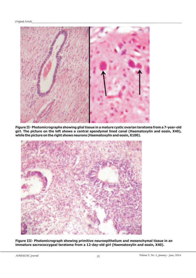
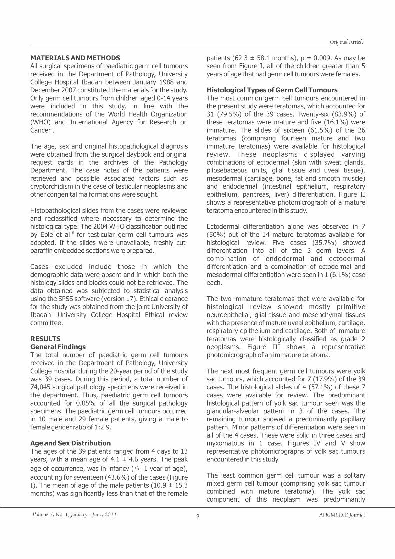
Figure II- Photomicrographs showing glial tissue in a mature cystic ovarian teratoma from a 7-year-old girl. The picture on the left shows a central ependymal lined canal (Haematoxylin and eosin, X40), while the picture on the right shows neurons (Haematoxylin and eosin, X100).
Figure III- Photomicrograph showing primitive neuroepithelium and mesenchymal tissue in an immature sacrococcygeal teratoma from a 12-day-old girl (Haematoxylin and eosin, X40).
AFRIMEDIC Journal Volume 5, No. 1, January - June, 201412
Original Article_____________________________________________________________________________________________________

Figure V- Photomicrograph showing solid pattern of yolk sac tumour from the ovary of a 7-year-old girl (Haematoxylin and eosin, X40).
Figure IV- Photomicrographs showing ovarian yolk sac tumour from a 13-year-old girl. The picture on the left shows a Schiller-Duval body, indicated by the arrow (Haematoxylin and eosin, X40). The picture on the right (from the same patient) shows multiple hyaline globules, indicated by the arrows (Haematoxylin and eosin, X100).
AFRIMEDIC Journal
____________________________________________________________________________________________________Original Article
Volume 5, No. 1, January - June, 2014 13

Figure VI- Photomicrographs showing sacrococcygeal mixed germ cell tumour from a 2½-year-old girl. The picture on the upper left shows bony tissue and squamous epithelium (Haematoxylin and eosin, X40). The picture on the lower left shows pancreatic tissue (Haematoxylin and eosin, X100). The picture on the right shows polyvesicular-vitelline pattern of yolk sac tumour in the same patient (Haematoxylin and eosin, X40).
DISCUSSIONIn the present study, only 0.05% of all surgical biopsy cases were paediatric germ cell tumours. This is in
7support of the observation of Cushing et al that germ cell tumours are infrequent in childhood. Paediatric germ cell tumours were more common overall in females than males in this study, with a female to male ratio of 2.9:1. The female predominance observed in this study is in agreement with the 2.2:1 female to male ratio reported by
8Marsden et al in the Manchester University Children's Tumour Registry. Teratomas demonstrated female gender predominance, with a female to male ratio of 2.9:1, which is consistent with the finding of female to male ratios ranging from 2:1 to 5:1 reported from
9-11 previous African series .
In the present study, paediatric germ cell tumours displayed a peak age of occurrence in the first year of life. This is in accordance with the report of germ cell
2tumours by Bernstein et al that there is high age specific incidence rate in the first year of life, followed
by a decline up until the age of eight years.
It was clearly shown that all of the paediatric germ cell tumours among subjects greater than five years of age in the present study were females. This can be explained by the fact that the majority of these neoplasms occurred in the ovaries and most of these cases were teratomas. In agreement with this finding a previous study from Ibadan showed that all of the childhood teratomas in patients aged 4 years and above
9were females . Similarly seventeen (89.5%) of the 19 children aged greater than 5 years in the South African teratoma study were females, which is also in
12agreement with our findings .
The most common germ cell tumours encountered in the present study were teratomas, which accounted for 79.5% of the cases. In a series of 137 childhood germ
8cell tumours, Marsden et al observed that teratomas were the most common germ cell tumours, comprising 63.5% of their cases, which is in agreement with the findings in the present study.
AFRIMEDIC Journal Volume 5, No. 1, January - June, 201414
Original Article_____________________________________________________________________________________________________

The majority (83.9%) of teratomas in the present study were mature (benign), teratomas while 16.1% were immature (malignant). In the series of Marsden
8et al from Manchester, England, 88.4% of cases were benign, 8.1% were malignant and 3.5% were of uncertain malignant potential, which closely corresponds to our findings. By contrast, in a study of 45 childhood teratomas from South Africa,
12 Bezuidenhout et al noted that although mature teratomas were most common (60%), immature teratomas accounted for 35% of cases.
In the present study, immature teratomas were restricted to the first year of life and all of them were extragonadal. In agreement with this finding, ten (90.9%) of the eleven extragonadal immature childhood teratomas in one South African series
12occurred in children less than one year of age .
The ovary, sacrococcyx and neck were the three most common locations of teratomas in this study. This is in agreement with previous studies of childhood teratomas from Ibadan, South Africa and England,
9, 8, which showed a predominance of ovarian teratomas 12. By contrast, some other African studies have revealed a predominance of sacrococcygeal over
11, 13ovarian teratomas in children .
All of the gonadal teratomas in the present series were mature and all of them occurred in the ovary. In contrast with our findings, in the series of ovarian
14 childhood tumours by Junaid 86.4% of teratomas in children less than 15 years of age were mature and 13.6% were immature. There was no case of testicular teratoma in the present study. This confirms the rarity
15of testicular germ cell neoplasms in blacks . In a series of 35 childhood and adolescent tumours from Ibadan,
16Junaid observed only one teratoma in the 0-5 years of age group and none in older children less than 15 years of age.
Yolk sac tumours were the second most common paediatric germ cell tumours encountered in the study, accounting for 17.9% of cases. This is in agreement
8with the findings of Marsden et al that yolk sac tumours were the second most frequent childhood germ cell tumours, overall, accounting for 25.5% of cases. In the present study, yolk sac tumours were the most frequent of all the malignant germ cell tumours encountered, which is also in agreement with the
8observation of Marsden et al .
The gonads were the most frequent location in the present study accounting for 57.1% of cases (three ovarian and one in the testis), followed by the sacrococcyx (3 cases) accounting for 42.9% of cases.
This is in agreement with the observation by Marsden et 8al that 54.3% of cases occurred in the gonads.
However, in our study, the ratio of ovarian to testicular 8yolk sac tumours was 3:1, whereas in Marsden et al's
study the ratio of ovarian to testicular yolk sac tumours was 1:2.3. This difference can be explained by the
relative rarity of testicular tumours in the Ibadan population.
CONCLUSIONThis study shows that paediatric germ cell tumours affect all age groups between 0 to 14 years. They are predominantly seen in females and the most common type is teratoma, which is most often seen in the first year of life. Extragonadal germ cell tumours are more common than the gonadal germ cell tumours in this study as compared to studies in Caucasian children, in which gonadal tumours are more than the extragonadal
8tumours . In view of the paucity of studies on germ cell tumours from Nigeria and other parts of Africa, it is recommended that further studies of this unique group of neoplasms should be carried out in other centres from Nigeria in order to confirm or refute the findings of this study.
REFERENCES
1. Rosai J, Cushing B, Perlman EJ. Male reproductive
system. In Rosai and Ackerman's Surgical th Pathology. 9 Edition. St. Louis, MO, USA: Elsevier
Mosby; 2004. Pp 412-482.
2. Bernstein L, Smith MA, Lui L, Deapen D. Friedman
DL. Germ cell, trophoblastic and other gonadal
neoplasms. In Cancer incidence and survival
among children and adolescents: United States
SEER Program 1975-1995. Bethesda, MD, USA:
National Cancer Institute; 1999. Pp125-137.
3. Marsden HB. The classification of childhood
tumours. In Parkin DM, Stiller CA, Draper GJ, Bieber
CA, Terracini B, Young JL (Eds.). International
incidence of childhood cancer. Lyon: IARC Scientific
Publications; 1988. Pp 9-16.
4. Streck CJ, Davidoff AM. Extragonadal germ cell
tumors. In Pediatric Surgery and Urology: Long ndTerm. Outcomes, 2 Edition. Cambridge:
Cambridge University Press; 2006. Pp 815-825.
5. Parkin DM. Materials and methods. In Parkin DM,
Stiller CA, Draper GJ, Bieber CA, Terracini B, Young
JL (Eds.). International incidence of childhood
cancer. No. 87. Lyon: IARC Scientific Publications;
1988. Pp 17-22.
6. Eble JN, Sauter G, Epstein JI, Sesterhenn IA. World
Health Organization Classification of Tumours.
AFRIMEDIC Journal
____________________________________________________________________________________________________Original Article
Volume 5, No. 1, January - June, 2014 15

Pathology and genetics of tumours of the urinary
system and male genital organs. Lyon: IARC Press;
2004. Pp 217-278.
7. Cushing B, Perlman EJ, Marina NM, Castleberry RP.
Germ cell tumours. In Pizzo P, Poplack D (Eds).
Principles and Practice of Paediatric Oncology, 4th
edition, Philadelphia: Hippincot Williams & Wilkins;
2001. Pp.1091-1113.
8. Marsden HB, Birch JM, Swindell R. Germ cell
tumours of childhood: a review of 137 cases. J Clin
Pathol. 1981; 34:879-883.
9. Akang EE, Odunfa AO, Aghadiuno PU, Childhood
teratomas in Ibadan, Nigeria. Hum Pathol. 1992;
23(4):449-453
10. Elesha SO, Aina A.O, Odunjo E.O. Sacrococcygeal
teratomas in Lagos, Nigeria: relationship of age,
sex, clinical type and histopathology to prognosis
in 30 cases. East Afr Med J. 1989; 66(10):685-692.
11. Mambo NC. Teratomas in children: Review of cases
in the 0-12 year age group seen at Harare Hospital
over ten year period, 1960-1969. Cent Afr J Med.
1974; 231-235
12. Bezuidenhout J, Schneider JW, Hugo F, Wessels G.
Teratomas in infancy and childhood at Tygerberg
Hospital, South Africa, 1973-1992. Arch Path Lab
Med. 1997; 121:499-502.
13. Nmadu PT. Childhood teratoma in Zaria, Nigeria.
East Afr Med J. 1995; 72(9):551-553.
14. Groeber WR. Ovarian tumours during infancy and
childhood. Am J Obstet Gynecol.1963: 1027-1035
15. Exelby PR. Testis cancer in children. Semin Oncol.
1979;6(1):116-120
16. Junaid TA. Testicular cancer in children and
adolescents in Ibadan, Nigeria. Urology.
1981;18:510-513.
AFRIMEDIC Journal Volume 5, No. 1, January - June, 201416
Original Article_____________________________________________________________________________________________________


