
1
Genetic diseases:
Hereditary disorders: Are derived from one parent, transmitted in
the gametes through the generation &therefore are familial.
Congenital diseases: simply present at birth. Note: not all genetic diseases are congenital ¬ all congenital
diseases are genetic e.g congenital syphilis.
Mutations: permanent changes in the DNA, those that affect germ cells are transmitted to the progeny &give rise to inherited disease,
while if in the somatic cells are not transmitted to the progeny but important in causation of cancers &some congenital malformations.
Types of mutations:
1- Point mutation: result from the substitution of a single
nucleotide base by a different base----replacement of one amino acid in the protein product e.g sickle cell anemia, this type is called
missense mutation.
While if the point mutation change an amino acid codon to a chain termination codon, or stop codon ---interrupt translation &the
resultant protein are rapidly degraded (nonsense mutation).
2- Frameshift mutation: occur when the insertion or deletion of one or two base pair alter the reading frame of the DNA strand (if
three or more this is not frame shift).
3- Trinucleotide repeat mutations characterized by the amplification of a sequence of three nucleotide e.g in fragile X
syndrome, there are 250-4000 repeats of the sequence CGG within
the gene called FMR-1. In the normal population, the number of repeats is small 29, so
amplification of FMR-1 giving rise to mental retardation.
We will discuss 3 types of genetic diseases: 1- Those related to the mutant genes (Mendelian disorders).
2- Dis. with multifocal inheritance (genetic &invironmental) e.g diabetes milletus.
3- Dis. arising from chromosomal aberration (number or structure of chromosome).
4- Heterogenous group that is single gene but do not follow Mendelian rules of inheritance.
1- Diseases caused by single gene defects.
Approximately 1% of all adult admittion to the hospital &6-8% of all
pediatric hospital.

2
Mutation involving single gene follow one of three patterns of
inheritance: * Autosomal dominant
Autosomal recessive * * X-linked disease.
A single gene mutation may lead to many phenotypic effects (pliotropy) &conversely several different mutations may produce the
same triat (genetic heterogeneity) e.g Marfan"s syndrome, the defect in the base of connective tissue---defect in skeleton, eyes,
cardiovascular, all of them from mutation in fibrillin.
While retinitis pigmentosa can be caused by several different types of mutations .
rders:diso ntAutosomal domina
1- At least one parent of an index case is affected 2- Both males &females are affected.
3- When an affected person marries an unaffected one, every child has 50% chance of having the dis.
N*N-------NN
N*N N*N NN NN
25% affected 25% affected normal
normal
50% 50%
4- With every autosomal dominant dis., some patients do
not have affected parents, so there is new mutation involving either the egg or the sperm from which they
derived. Their siblings are neither affected nor at high risk of the disease.
5- Clinical features can be modified by reduced penterance &variable expressivity
.
Reduced penterance: Some patients inherit the mutant
gene but are phenotypically normal.
Variable expressivity: If the trait is seen in all individuals carrying the mutant gene but is expressed differently
among individuals.

3
6- In many conditions, sign &symptoms do not appear until
adulthood.
7- 50% reduction in normal gene products is associated
with clinical symptoms.
8- Because a 50% loss of enzyme activity can be
compensated for, involved genes not encode enzyme proteins.
2 main types of nonenzyme proteins are affected:
a- Those involved in regulation of complex metabolic pathways e.g familial hypercholesterolemia.
b- Key structural proteins e.g collagen &cytoskeletal components of red cell membrane e.g spectrin----
hereditary spherocytosis. Examples of autosomal dominant disorders:
Marfan syndrome
Hypercholesterolemia
Polycystic kidney disease
Hereditary spherocytosis
Familial polyposis coli
Autosomal recessive disorders:
Occur when both of the alleles at a given locus are mutants, so characterized by the following features:
1- The trait does not usually affect the parents, but siblings
may show the disease.
2- Siblings have 25% risk for each birth (one chance in
four). N*N N*N
N*N* N*N N*N NN
affected carrier carrier normal
25% 25% 25% 25%

4
3- If the mutant gene occurs with a low frequency in the
population,there is a strong likelihood that the proband is the product of a consanguineous marriage.
4- The expression of the defect tends to be more uniform than autosomal dominant.
5- Complete penterance is common. 6- Onset of signs &symptoms early in life.
7- New mutation occur but rarely detected clinically
because the affected person is a symptomatic heterozygote, unless this heterozygous marry other
heterozygous &produce affected offsprings.
8- In heterozygotes equal amount of normal &defective enzymes are synthesized, cells with half of their
complement of enzyme function normally &50% reduction
not associated with clinical symptoms.
9- In many cases, enzyme proteins are affected by the mutation e.g phenylketonuria, thalassemia, sickle cell
anemia. Examples of autosomal recessive disorders:
Sickle cell anemia
Thalassemias
Cystic fibrosi
Phenylketonuria
Wilson disease
Glycogen storage diseases
Albinism
linked recessive):-(most are X linked disorders:-X
All sex-linked dis. are X-linked characterized by:
1- They are transmitted by heterozygous female only to sons who are hemizygous for the X chromosome.
2- Heterozygous female rarely express the full phenotypic
change, but if there is inactivation of one of the X

5
chromosome which is the normal X---full expression of the
disease in heterozygous female.
3- An affected male does not transmit the dis. to sons, but all daughters are carriers.
XX X*Y
X*X *XX XY XY
All daughters are carrier, sons not affected
4- Sons of heterozygous mothers are 50% chance of
affection.
XX* XY
XX XY XX* X*Y
Sons are 50% affected
5- There are few X-linked dominant &thier inherited pattern characterized by transmition of the dis. to 50% of
sons &daughters of an affected heterozygous female.
6- An affected male cannot transmit the dis. to his sons but all daughters are affected.
Examples of X-linked disorders:
Hemophilias A and B
Glucose-6-phosphate dehydrogenase deficiency (G6PD
deficiency)
Agammaglobulinemia
Duchenne muscular dystrophy
Diseases caused by mutation in structural protein
Marfan"s syndrome:
autosomal dominant dis. of connective tissue (fibrillin),
both quantitative &qualitative defects have been noted. 3 systems are mainly affected:
*Skeletal abnormalities: Slender patient with long legs,
arms &fingers, high arched palate, hyperextensibility of the

6
joints, kyphosis &the chest shows pectus excavatum or
pigeon breast deformity.
* Eyes: bilateral dislocation or subluxation of the lens due to weakness of suspensory ligament.
* Cardiovascular system: Fragmentation of the elastic
fibers in the tunica media of the aorta---aneurysmal dilatation &aortic dissection, dilatation of aortic valve ring--
-aortic incompetence, mitral &tricuspid valves regurgitation----congestive heart failure.
Death from aortic rupture may occur at any age.
*Variable expression of the features above between different patients.
Disease caused by mutation in receptor proteins:
Familial hypercholesterolemia:
1- Autosomal dominant dis., heterozygotes have 2-3 folds elevation of plasma cholesterol level, remain asymptomatic
until adulthood when develop xanthoma along tendon sheaths &premature coronary artery dis.
2- While homozygotes are much more severely affected,
cutaneous xanthoma in childhood &dying from myocardial infarction MI in the age of 15 years.
3- It is caused by mutation in the gene that specifies the
receptor for low density lipoproteins LDL.
4- Cholesetrol may be derived from diet or from
endogenous synthesis, endogenous synthesis of cholesterol &LDL begins in the liver.
Normally, there is LDL receptors in the hepatocytes, so LDL binds to the receptors &formation of very low density
lipoproteins VLDL which undergo lipolysis &converted into intermediate density lipopteins IDL, then to the liver (LDL
receptor) again.
Mutation in LDL receptor gene---accumulation of LDL cholesterol in the plasma, in addition the absence of LDL
receptors on the liver impair the transport of IDL to the liver---accumulation of IDL that converted to LDL .

7
Diseases caused by mutation in enzymes proteins:
Phenylketonuria (PKU)
1- Inborn error of metabolism, it is autosomal recessive disease.
2- homozygotes have severe lack of phenylalanine hydroxylase---hyperphenylalaninemia &PKU
3- The affected infants are normal at birth but within few
weeks to 6 months---increase phenylalanine in plasma with severe mental retardation, inability to walk, inability to
talk, seizures, decrease pigments of the skin &hair, eczema, musty odor of sweat.
4- Deficiency of the enzyme---inability to convert
phenylalanine into tyrosine, when this is blocked, minor
shunt to other pathways which lead to intermediates that excreted in sweat (odor), concomitant lack of tyrosine---
lack of melanin.
Treatment: restriction of phenylalanine intake early in life.
5- Many clinically normal PKU patients treated with diet
early in life &reach child bearing age, most of them have high serum phenylalanine because dietary treatment is
discontinued after reaching adulthood, children born to them are mentally retarded &have many congenital
abnormalities---maternal PKU.
Glycogen storage disorders (Glycogenoses):
An inherited deficiency of any of the enzymes involved in
glycogen synthesis or degradation, result in excessive accumulation of glycogen or abnormal form of glycogen in
various tissues .
Glycogen is most often stored within the cytoplasm or sometimes within the nuclei.
One variant called Pompe disease---lysosomal storage disease because the deficient enzyme localized to the
lysosomes.
Most glycogenoses are inherited as autosomal recessive
disease.
On the basis of pathophysiology they grouped into 3
categories:
8
1- Hepatic form: liver contains several enzymes that
synthesize or break down glycogen, so deficiency of an enzyme result in enlargement of the liver due to storage of
glycogen &hypoglycemia due to failure of glucose production e.g Glucose 6 phosphatase enzyme deficiency
called Von Gierke disease.
2- Myopathic form: In striated muscles glycogen derived by glycolysis, when enzyme are deficient so glycogen
storage disease of the muscle (muscle weakness), typically characterized by muscle cramps after exercise &failure of
the exercise to induce an evolution in blood lactate level due to block in glycolysis (McArdle disease) result from
deficiency of muscle phosphorylase enz.
3- Two other forms of glycolysis do not fit into either
of the above two
Pompe disease due to deficiency of lysosomal acid maltase so deposition of glycogen in every organ but
cardiomegaly is prominent .
Brancher glycogenoses is due to deposition of abnormal glycogen with effect on the liver, heart, muscles…
Diseases caused by mutation in protein that regulate
cell growth:
2 classes of genes that regulate cell growth, prooncogenes
&cancer suppressor genes, mutation affecting these genes----tumor.
5% of all CA, there is mutation affecting certain tumor suppressor genes are present in all cells of the body
including germ cells---transmitted to the offspring.
Neurofibromatoses type 1 &2: Neurofibromatoses type 1: accounts for 90% of the
cases &characterized by:
1- Multiple neurofibroma in the form of pedunculated nodules protruding from the skin , they are discrete,
unencapsulated, soft, sometimes the tumor form large
multilobar masses (plexiform NF).They are derived from

9
schwan cells, similar tumors may occur along nerve trunk,
cauda equine, cranial nerves, orbit, tongue &GIT.
2- Pigmented skin lesions (café-au-lait spots), sometimes overlie a NF.
3- Pigmented iris hamartomas (Lisch nodules), no clinical symptoms but helpful in the diagnosis.
* NF type1 gene on chromosome 17, it encodes a protein
that act as negative regulator of ras oncoprotein.
* NF type 2 on chromosome 22, rarer than type 1, in addition to NF, Café-au-lait spots +bilateral acaustic
neuroma.
Significance of NF:
1- Disfiguring condition.
2- Serious by its location e.g within the spinal cord.
3- In 3% of patient, NF leads to neurosarcoma. Usually malignant in the plexiform tumor attached to large
nerve trunk of the neck or extremities.
4- These patients are at greater risk of developing other tumors like optic glioma, menigioma, pheochromocytoma.
5- 30-50% of patients have associated skeletal
abnormalities like scoliosis, bone cysts.
Disorders with multifactorial inheritance:
Multifactorial trait may be defined as one governed by the additive effect of 2 or more genes of small effects but
conditioned by environmental influences.
There is some threshold effect so that the disorder becomes manifested.
The following features characterized multifactorial inheritance:
1- The risk of expressing a multifactorial disorder is
conditioned by the number of mutant genes inherited, so

11
the risk is greater in siblings of patient having severe
expression of the disease.
2- The rate of recurrence of the disorder is 2-7% is the same for all first degree relatives, so if parents have one
affected child, the risk that the next child will be affected is 2-7%.
3- Identical twins will be affected less than 100%(about
20-40%) but is much greater than the chance that both nonidentical twins will be affected.
4- The risk of recurrence of the phenotypic abnormality in
subsequent pregnancies depends on the outcome in previous pregnancies.
When one child affected---7% chance of the next child, while after 2 affected siblings---9% .
Multifactorial inheritance underlies * DM, hypertension
* Gout, schizophrenia * Manic depression
* Congenital heart dis. * Some skeletal abnormalities.
Cytogenetic disorders: chromosomal aberrations.
Karyotype is a paragraphic representation of a stained
metaphase spread in which the chromosomes are arranged in order of decreasing length.
Giemsa stain used, each chromosome have alternating light &dark bands of variable width.
1:200 newborn infants has some form of chromosomal
abnormalities, 50% of first trimester abortion is due to chromosomal abnormalities.
Chromosomal abnormalities either in: Number or structure &may affect:
Autosomes or sex chromosomes. What is Mitosis?
Mitosis produces two daughter cells that are identical to the parent cell.
If the parent cell is haploid (N), then the daughter cells will be haploid.
If the parent cell is diploid, the daughter cells will also be diploid.
This type of cell division allows multicellular organisms to grow and repair damaged tissue
Phases of Mitosis
Humans have a diploid number of 46.

11
Interphase
Chromosomes are not visible because they are uncoiled
Prophase
The chromosomes coil.
Ex. cell with 8 chromosomes. Each chromosome has 2
chromatids for a total of 16 chromatids.
Metaphase
The chromosomes become aligned.
Cell with 8 chromosomes. Each chromosome has 2 chromatids
for a total of 16 chromatids.
Anaphase
The chromatids separate; the number of chromosomes
doubles

12
The drawing shows a cell with 16 chromosomes. Each
chromosome has 1 chromatid for a total of 16 chromatids.
Telophase
The cell divides into two.
cell with 16 chromosomes. Each chromosome has 1 chromatid
for a total of 16 chromatids.
Interphase
The chromosomes have two chromatids each.
What is meiosis?
Meiosis produces daughter cells that have one half the number of
chromosomes as the parent cell.
2N → N
Meiosis enables organisms to reproduce sexually.
Gametes (sperm and eggs) are haploid.
Meiosis involves two divisions producing a total of four daughter
cells.
Prophase I Homologous chromosomes become paired.
Crossing-over occurs between homologous chromosome

13
.
Metaphase I
Homologous pairs become aligned in the center.
The random alignment pattern is called independent assortment.
Anaphase I
Homologous chromosomes separate
Phases of meiosis II

14
Mitosis&meiosis:
Numerical abnormalities
Normal chromosome count is 46 i.e 2n =46, any exact multiple of the haploid number is called euploid,
chromosomal number such as 3n & 4n called polypoid usually result in spontaneous abortion.
Any number which is not an exact multiple of n called
aneuploid .
The chief cause of aneuploidy is non disjunction of a
homologous pair of chromosome at first meiotic division or
failure of sister chromatides to separate during the second meiosis.

15
When non disjunction occurs at time of meiosis, the
gametes formed have either extrachromosome (n+1) or1 less chromosome (n-1), then fertilization lead to either
trisomy (2n+1) or monosomy (2n-1).
Autosomy involving an autosome is incompatible with life, while monosomy involving sex chromosomes is compatible
with life.
* Mosaicism: term used to describe the presence of 2 or
more populations of cells in the same individual, mosaicism affecting sex chromosomes is common while autosomal is
not.
Structural abnormalities:

16
Involve breakage of the chromosome &then
rearrangement, patterns of rearrangement as follows:
1- Translocation: transfer of a part of one chromosome to another chromosome, the process usually reciprocal ( i.e
fragment exchanged between chromosome)
A special pattern is called centric fusion type or
Robertsonian translocation involving 2 acrocentric chromosomes, typically the breaks occur close to the
centromere, transfer of the segment lead to one very large chromosome &one small, the short segments are lost &the
carrier has 45 chromosome, such loss is compatible with life, but difficulties arise during gametogenesis---formation
of unbalanced gametes---abnormal offspring.
2- Isochromosomes: result when the centromere divides
horizontally rather than vertically, one of the 2 chr. Is then lost.

17
3- Deletion: involve loss of a portion of a chromosome, single break may delete a terminal segment.
2 interstitial breaks with reunion pf the proximal &distal segment may result in loss of intermediate segment, the
isolated fragment almost never survive.
4- Inversion: occur when there are 2 interstitial breaks in a chromosome &the segment reunites after a complete
turnaround.
5- Ring chromosome: is a variant of deletion, after loss
of segments from each end of the chromosome, the arms uniting to form ring.
Notes:

18
* Chromosomal disorders may be associated with absence
(deletion or monosomy), excess (trisomy), or abnormal rearrangement (translocation).
* In general loss of chromosomal material produces more
severe defects than does gain of chromosomal material.
* Imbalances of sex chromosome (excess or loss) are tolerated much better than are similar imbalance of
autosomes, & often produce subtle manifestation, sometimes not detected at birth, usually it is infertility that
detected at adolescence.
Cytogenic disorders involving autosomes: Trisomy mainly (21, 18, 13).
Trisomy 21 (Down syndrome):
Is the most common, chromosomal count is 47, the most common cause is meiotic non disjunction in the ovum, the
parents are normal but maternal age is important, in women more than 45---1:25 birth.
In 4%, the extrachromosomal material is translocation of
long arm of chromosome 21 to 22 or 14. 1% is mosaicism with mixture of 46 &47 chromosome.
Clinical features: 1- Mental retardation.
2- Epicanthic folds &flat facial profile. 3- Abundant neck skin.
4- Simian creases. 5- Congenital heart defects &is the principle cause of death
in addition to serious infection. 6- Umbilical hernia.
7- Intestinal stenosis. 8- Hypotonia.
9- Gab between first &second toe. 10- Predisposition to leukemia.

19
Trisomy 13 (Patau syndrome):
1- Microcephaly &mental retardation.
2- Microphthalmia. 3- Cleft lips &palate.
4- Cardiac defects. 5- Umbilical hernia.
6- Renal defects. 7- Polydactyly.
8- Rocker-bottom feet.
disorders involving sex chromosomes
Are compatible with life due to:
1- Lyonization of X chromosome.
2- Scant amount of genetic information carried by Y chromosome.

21
One of the X chromosome is inactivated early in fetal life
&called Bar body.
Extra Y chromosome readily tolerated because the only information carried by it is related to male differentiation.
3 disorders:
1- Klinefilter syndrome:
Male hypogonadism develop when there are at least 2 X chromosome &one or more Y chromosome.
Karyotype: most patients are 47,XXY in 80% and mosaic in 20%.
Causes: * Advanced maternal age.
* History of irradiation of either parent. Clinical features:
Hypogonadism,
Marked testicular atrophy, Gynecomastia,
Reduced facial &body hair (failure of male secondary sexual characteristics development)
Increase length between the soles &the pubic bones (elongated body),
Serum testosterone decrease, Some with mental retardation
The principle clinical effect is sterility, only rare patient are fertile.
Histologically:
Hyalinization of tubules which appear as ghost like in
contrast lydig cells are prominent.

21
2- XYY males: Due to non disjunction at the second meiotic division, most
are phenotypically normal, but taller than usual also with antisocial behavior.
3- Turnner syndrome
Characterized by hypogonadism in phenotypic female
result from partial or complete monosomy of X chromosome.
Karyotype: 45 XO .
Clinical features:
* Short stature. * Low posterior hair line.
* Cubitus vulgus (increase in carrying angle of the arms). * Shield like chest with widely spaced nipples
* High arched palate. * Lymphoedema of the hands &feet.
* Variaty of congenital malformation e.g horseshoe kidney, coarctation of aorta.
* Failure of development of secondary sexual
characteristics.
* Genitalia remain infantile (little pubic hair, primary
amenorrhea.
* Ovaries fibrosed which is devoid of follicles.
* Ovarian estrogen decrease with no feed back inhibition so increase pituitary gonadotrophins.

22
Single gene disorders with atypical patterns of inheritance:
Three groups of diseases resulting from mutation affecting
single genes do not follow the mendelian rules of inheritance:
1- Diseases caused by triplet repeat mutation:
(Fragile X syndrome):
The mutation is characterized by long repeating sequence of three nucleotide, so disrupt the function of that gene
(FMR-1) .
It is characterized by mental retardation &an abnormality
of X chromosome.
It is one of the most common causes of familial mental retardation in males, the only distinctive physical
abnormality in 80% of postpubertal males is macroorchidism.
This disease is X-linked but unlike other X-linked diseases, 20% of males with the diseases are clinically
&cytogenically normal (carrier males).
Presence of mental retardation in 50% of the carrier females .
These unusual features have been related to the number of
triplet repeat, in normal population, the number of repeat is small about 29 whereas affected individuals have 250-
4000 repeat so called full mutation &the carrier male
&females are premutation.
Diagnosis: by cytogenetic demonstration of the fragile site or by DNA probe analysis.

23
2- Diseases caused by mutation in mitochondrial
genes:
Mitochondria contain several genes encodes for enzymes of
oxidative phosphorylation, usually the ovum contain the
large part of mitochondria, so the inheritance of mitochondrial gene is maternal.
Disease caused by mitochondrial genes are rare:
Leber"s optic neuropathy---progressive bilateral loss of
central vision.
3- Diseases associated with genomic imprinting: Prader Willi &Angleman syndromes:
All humans inherit two copies of each gene, carried on
homologous maternal &paternal chromosomes &there is no difference between normal homologous genes.
But now functional difference exists between maternal
&paternal genes is called genomic imprinting.
Prader Willi: mental retardation, short stature, hypotonia, hypogonadism, obesity, small hands &feet.
Causes: 50-60% deletion of band of long arm of
chromosome 15.
Angleman syndrome:

24
Mental retardation, ataxic gait, seizures, inappropriate
laughter called happy puppet syndrome.
Causes: Deletion of the same chromosomal region derived from
their mother.
Because DNA methylation affect gene expression, it is
strongly suspected that imprinting is associated with differential DNA methylation of paternal &maternal genes.
Diagnosis of genetic diseases:
About 4,000 human diseases are thought to be inherited
Scientists are making good progress figuring out where
genes are located on chromosomes and in understanding how the mutations in genes cause disease

25
Genetic map of chromosome 17
Genetic analysis:
In general, genetic testing can be divided into prenatal and postnatal analysis.
It may involve
Karyotype analysis
FISH,
Molecular diagnostics (PCR), or
a combination of these techniques. Karyotype analysis of human chromosomes:
Karyotype preparation &analysis:
Cells (from blood, amniotic fluid, etc) are grown in vitro (in a cell
culture dish) to increase their number
Cell division is then arrested in metaphase with colchicine
(prevents mitotic spindle from forming)
Cells are centrifuged and lysed to release chromosomes
Chromosomes are stained with Geimsa stain, photographed, and grouped by size and banding patterns
Photograph of the 46 chromosomes in a human somatic cell
arrested at metaphase

26
Limitations of karyotype analysis: * Resolution of this technique is fairly low
* It is applicable only to cells that are dividing or can be induced to divide in vitro.
FISH Flourescence insitu hybridization FISH utilizes DNA probes that recognize sequences specific to
chromosomal regions.
The probe binds to its complementary sequence on the
chromosome and thus labels the specific chromosomal region that can be visualized under a fluorescent microscope.
Many genetic diseases are caused by alterations at the nucleotide level (i.e., mutations) that cannot be detected by
FISH
Polymerase chain reaction PCR:
Many genetic diseases are caused by alterations at the
nucleotide level (i.e., mutations) that cannot be detected by FISH
Advantages over other techniques:
* It is remarkably sensitive.
* The use of polymerase chain reaction (PCR) allows several million-fold amplification of DNA or RNA, making it possible to
utilize as few as 1 or 100 cells for analysis. A few drops of blood or a piece of biopsy tissue can supply sufficient DNA for PCR
amplification.
Prenatal testing:
Is a testing for diseases or conditions in a fetus or embryo before it is born.
* The aim is to detect birth defect such as neural tube defect, Down"s syndrome, chromosome abnormalities, genetic diseases
&other conditions
,*
Diagnostic prenatal testing can be by
Invasive &non invasive methods
Amniocentesis:

27
* Used on pregnant women over 35 years of age
Performed during weeks 15-17 of pregnancy *
A needle is inserted into the amniotic sac *
* Amniotic fluid with fetal cells is removed
A karyotype of the fetal chromosomes is prepared *
Scientific breakthroughs:
Greater insights into disease will be achieved
Cures may be found Incurable diseases may be prevented
There will be new insights into the evolutionary origins of humans
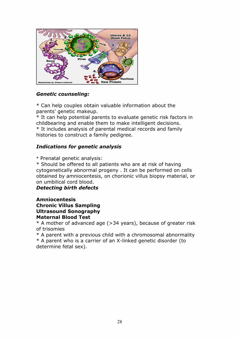
Gene therapy:
Genetic alteration of somatic cells to treat disease .
Researchers inject genes that are targeted to treat a particular
disease in to a patient’s blood stream.
When the genes arrive at the site of the defective genes, they
produce chemicals that can treat the problem.
Steps in gene therapy in utero
28
Genetic counseling:
* Can help couples obtain valuable information about the
parents’ genetic makeup.
* It can help potential parents to evaluate genetic risk factors in
childbearing and enable them to make intelligent decisions.
* It includes analysis of parental medical records and family
histories to construct a family pedigree.
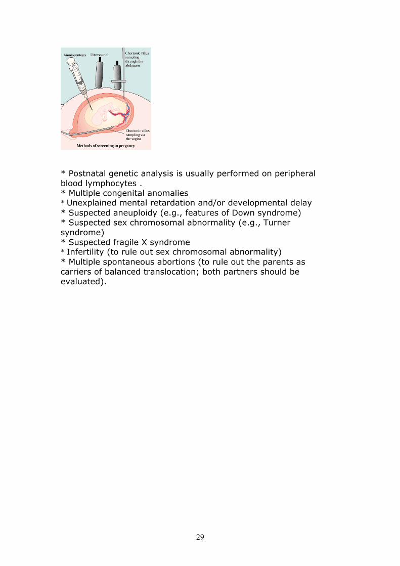
Indications for genetic analysis
Prenatal genetic analysis:*
* Should be offered to all patients who are at risk of having
cytogenetically abnormal progeny . It can be performed on cells obtained by amniocentesis, on chorionic villus biopsy material, or
on umbilical cord blood.
Detecting birth defects
Amniocentesis
Chronic Villus Sampling
Ultrasound Sonography Maternal Blood Test
* A mother of advanced age (>34 years), because of greater risk of trisomies
* A parent with a previous child with a chromosomal abnormality
* A parent who is a carrier of an X-linked genetic disorder (to
determine fetal sex).
29
* Postnatal genetic analysis is usually performed on peripheral
blood lymphocytes .
* Multiple congenital anomalies
Unexplained mental retardation and/or developmental delay *
* Suspected aneuploidy (e.g., features of Down syndrome)
* Suspected sex chromosomal abnormality (e.g., Turner
syndrome)
* Suspected fragile X syndrome
Infertility (to rule out sex chromosomal abnormality) *
* Multiple spontaneous abortions (to rule out the parents as
carriers of balanced translocation; both partners should be evaluated).

31


