
Flexible, Multi-Scale, Brain-Computer
Interfaces for Treating Epilepsy December 2, 2011
Brian Litt, MD
Associate Professor of Biology and Bioengineering
University of Pennsylvania
Philadelphia, PA
American Epilepsy Society | Annual Meeting

Disclosure
Name of Commercial
Interest
NeuroVista, Inc.
MC10
NeuroPace, Inc.
Type of Financial
Relationship
SAB, Consultant
SAB, Co-Founder (Medical Division)
Shareholder (from Licensed IP)
American Epilepsy Society | Annual Meeting

Learning Objectives
To understand:
• The need for high resolution recording and intervention in epilepsy
• Flexible, active electronic devices
• New high resolution findings in neocortical epilepsy, in-vivo
American Epilepsy Society | Annual Meeting

QuickTime™ and a decompressor
are needed to see t his pic ture.
QuickTime™ and a decompressor
are needed to see this picture.QuickTime™ and a
decompressorare needed to see this picture.

Courtesy of Gordon Baltuch, M.D., Ph.D., UPenn
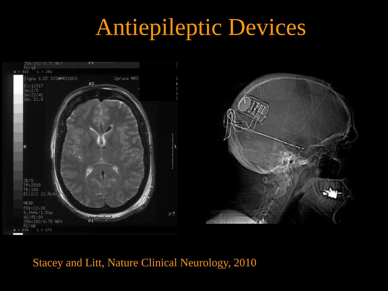
Stacey and Litt, Nature Clinical Neurology, 2010
Antiepileptic Devices

What is a seizure?
Basic epileptic unit?
Topology, architecture, scale?
How to intervene?
Fundamental Questions

Multiple Approaches
Hardware
Algorithms
Human studies
Animal electrophysiology
Computational Modeling
New Devices
The International Database
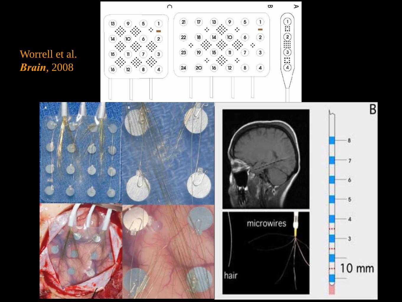
Worrell et al.
Brain, 2008

QuickTime™ and a decompressor
are needed to see this picture.
HFOs increased in SOZ vs. non-SOZ
Worrell et al. Brain, 2008

Justin Blanco

* * *
*
*

September, 2010
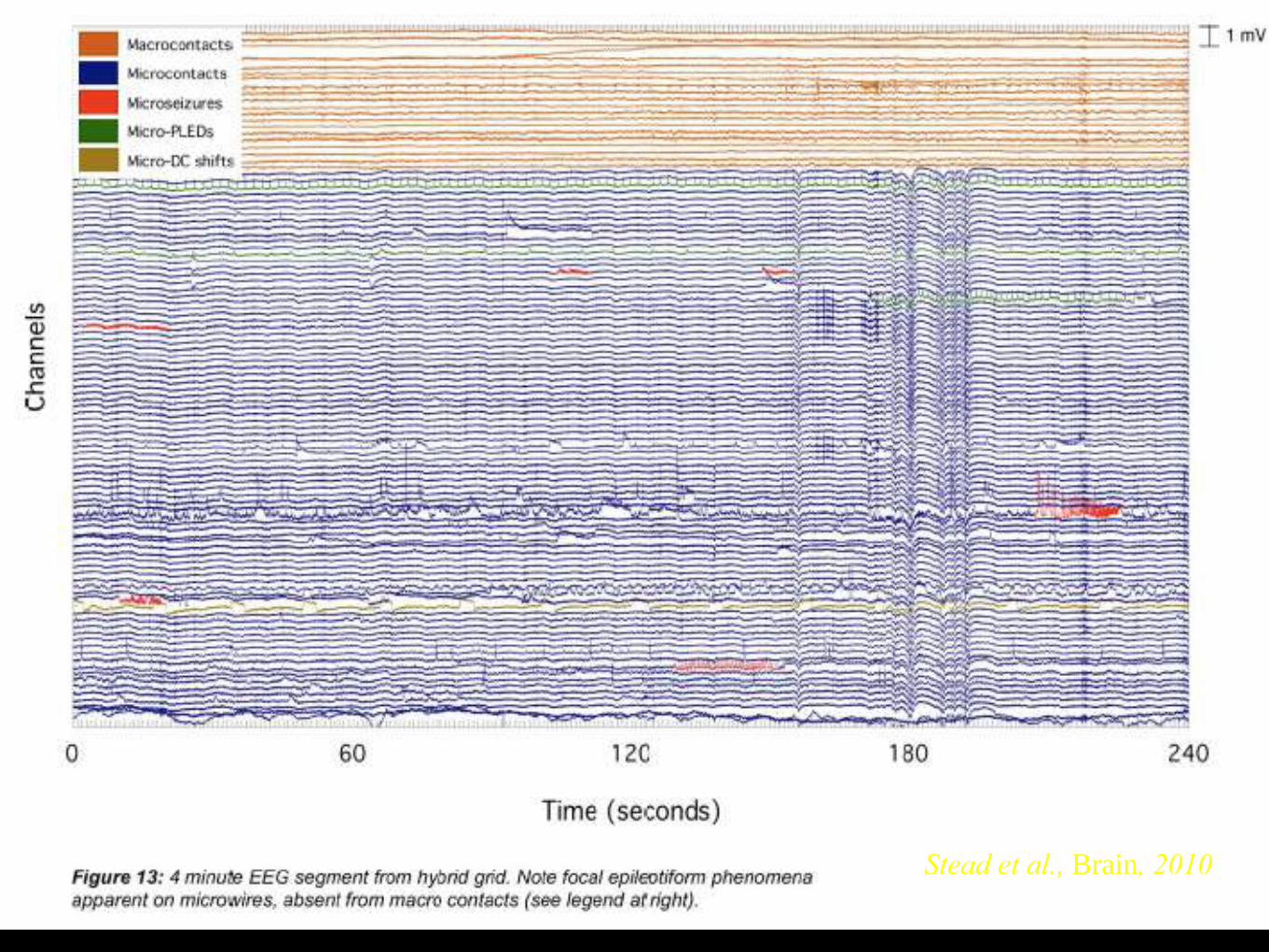
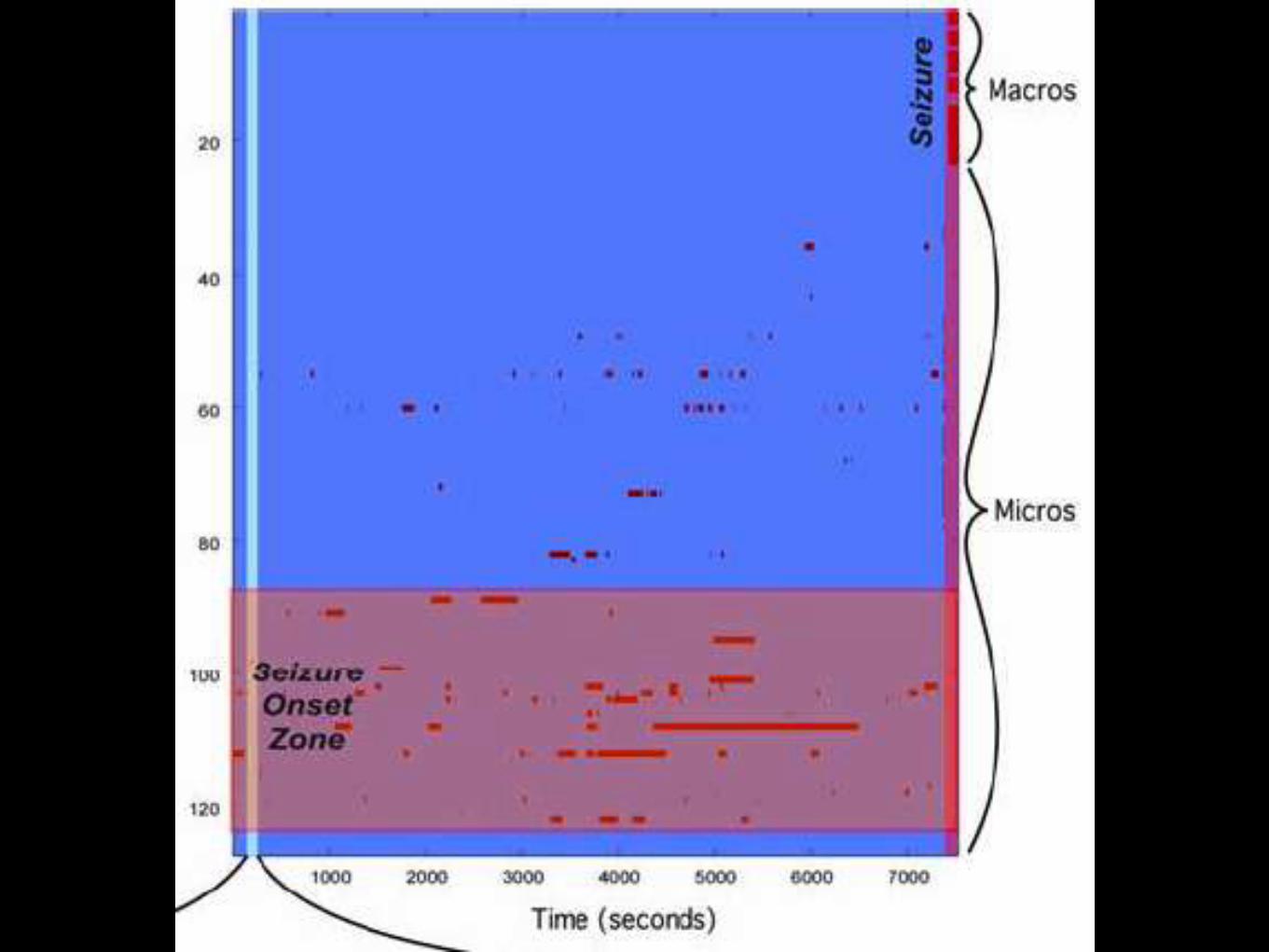
Stead et al., Brain, 2010


Lead to Sz

And Spatially…
•8 cm x 8 cm
•Individually wired
•Large volume increases morbidity
1 cm
12 Million Neurons 1 Electrode
1 cm

Collaborators
John A. Rogers Ph.D. Materials Science, UIUC
Cherie R. Kagan, Ph.D. ESE, UPenn
Diego Contreras M.D., Ph.D. Neuroscience, UPenn
David J. Callans, M.D. Cardiology, UPHS
Dae-Hyeong Kim, Ph.D. Materials Science, Soeul National Univ University
Jan Van der Spiegel Ph.D. ESE, UPenn
Jonathan Viventi Ph.D. ECE/Neuroscienc, NYU-Poly
Yale Cohen, Ph.D. Otorhinolaryngology, UPenn

Flexible and Stretchable Silicon Electronics
Kim, D-H. et al. Science 320, 507-11(2008).

Fabrication Steps
Viventi, J., Kim, D-H. et al. Science Translational Medicine 2, 24ra22 (2010).

Cross-Section

Flexible, Scalable BCI for Epilepsy

Flexible, Scalable BCI for Epilepsy

Catheter Delivery
1.5 mm
0.5 mm
hole
Science Translational Medicine, March 2010

720 Channel Array on Brain
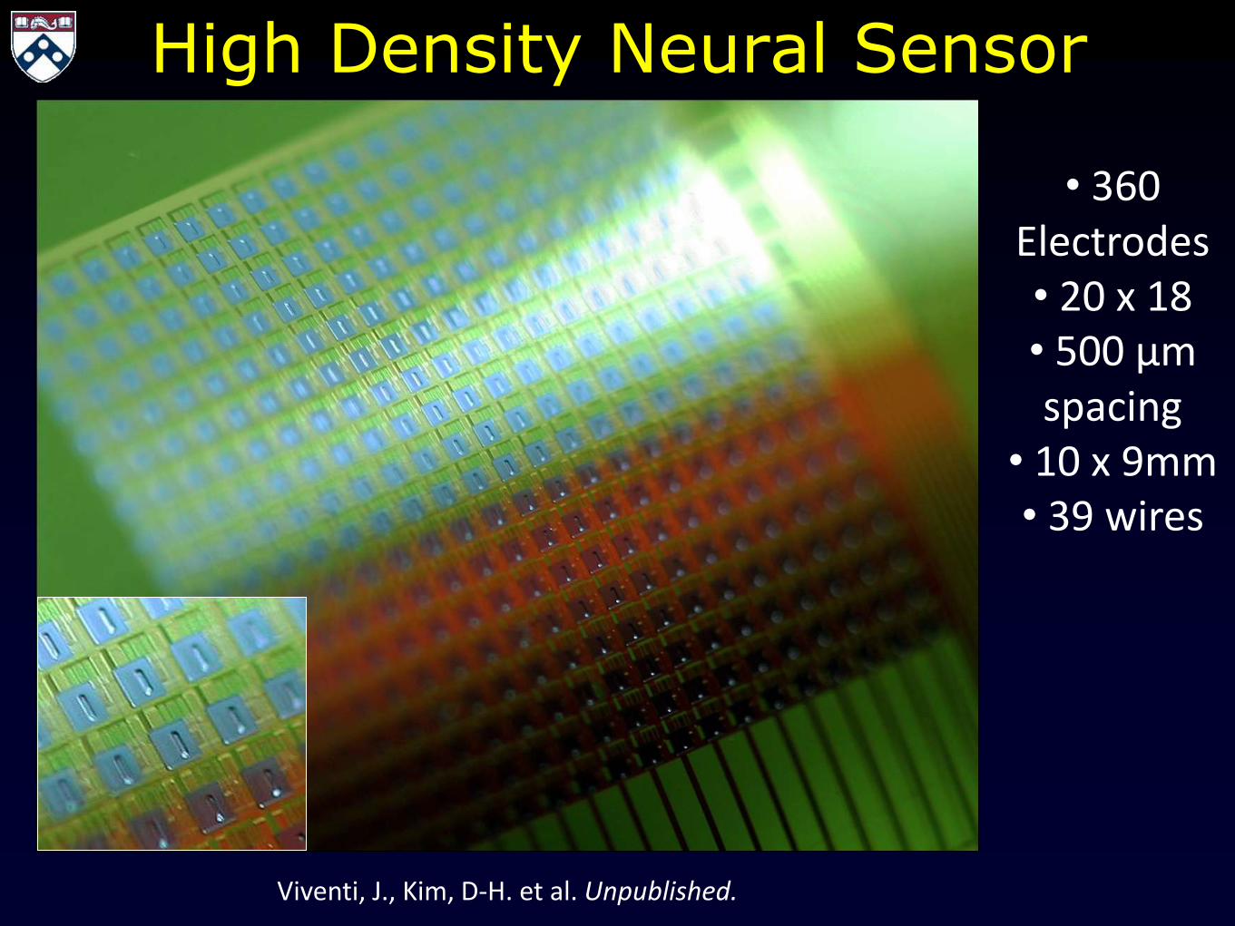
High Density Neural Sensor
Viventi, J., Kim, D-H. et al. Unpublished.
• 360 Electrodes • 20 x 18
• 500 µm spacing
• 10 x 9mm
• 39 wires
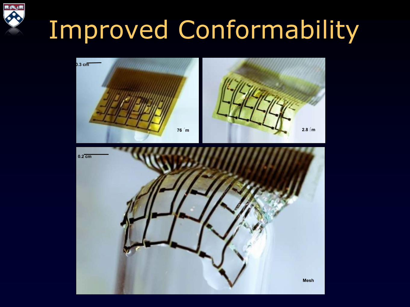
Improved Conformability
76 m 2.8 m
Mesh
0.3 cm
0.2 cm


Visual Cortex
QuickTime™ and a decompressor
are needed to see this picture.
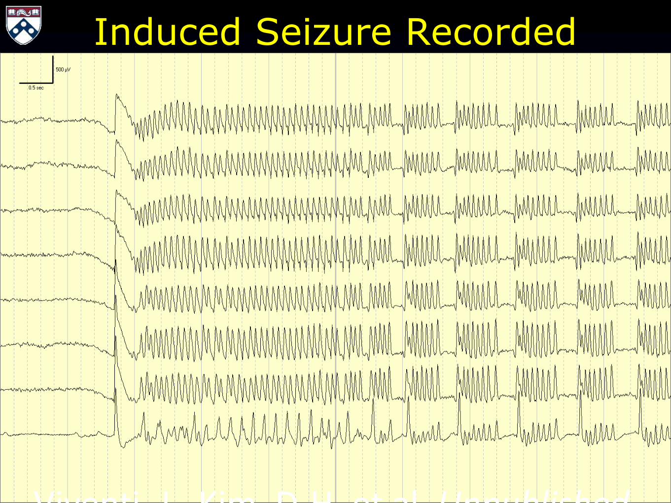
Induced Seizure Recorded
Viventi, J., Kim, D-H. et al. Unpublished.

Seizures
QuickTime™ and a decompressor
are needed to see this picture.
Viventi, J., Kim, D-H. et al. Nature Neuroscience, 12 2011.

Spike
500 ms
2 m
V
1. Spike Detection
QuickTime™ and a decompressor
are needed to see this picture.
25 ms
2 m
V
2. Delay Calculation
3 mV
-3 mV
0 mV
Movie of a Spike
38 ms
0 ms
19 ms
Resulting Delay Map

Automated Analysis Method II 4. PCA
5. Clustering
J.A. Blanco, M. Stead, A. Krieger, J. Viventi et al. Journal of neurophysiology 2900-2912 (2010).
3. Zero-Mean RMS
1.8 mV
0.4 mV
1.1 mV
http://www.cs.cornell.edu/courses/cs322/2008sp/images/thumb_PCA.
png
6. Gap Statistic
Tibshirani, R., Walther, G. & Hastie, T. Journal of the Royal Statistical Society: Series B 63, 411-423 (2001).

Cluster Homogeneity
0 ms 27 ms

Cluster Homogeneity
0 ms 22 ms

0 ms 24 ms
Plane
Wave
Clockwise
Spiral
0 ms 90 ms
Plane
Wave
0 ms 38 ms
Counter
Clockwise
Spiral
0 ms 165 ms
Plane
Wave
0 ms 39 ms
Plane
Wave
0 ms 38 ms
Waves within a seizure
500 ms
2 m
V
Viventi, J., Kim, D-H. et al. Nature Neuroscience, 12 2011.

Spike Clusters
0 ms 29 ms
0 ms 38 ms
0 ms 39 ms
0 ms 24 ms
0 ms 38 ms
40 ms
2 m
V
40 ms
2 m
V
40 ms
2 m
V
40 ms
2 m
V
40 ms
2 m
V
40 ms
2 m
V
0 ms 21 ms
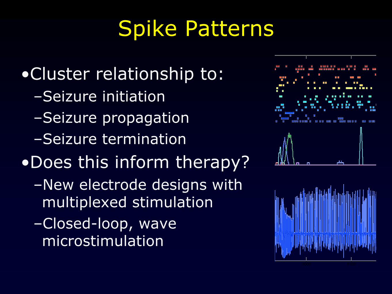
Spike Patterns
•Cluster relationship to:
–Seizure initiation
–Seizure propagation
–Seizure termination
•Does this inform therapy?
–New electrode designs with multiplexed stimulation
–Closed-loop, wave microstimulation

Support
NINDS (R01-NS041811-04, RO1-NS48598-01,
U24NS063930-01A1)
CURE
The Dr. Michel and Mrs. Anna Mirowski Discovery Fund

