
R E S I D E N T L E C T U R E 6 / 2 0 1 4
N I M A N A B A V I Z A D E H K R I S T I N A Y O U N G J O S E P H W A L L E R
Ewing’s Sarcoma

Epidemiology
Neuroectodermal origin Adolescents (40%), but 30% in <10 year olds 2nd most common bone tumor in children, after
osteosarcoma ~225 cases/yr M:F 1.5-2:1 white>>black /asian

Ewing Sarcoma Family of Tumors (ESFT)
ES of bone Extra-skeletal ES Askin’s tumor PNET

So Special They Named It
Askin’s tumor Primary lesion of rib Associated w/ direct pleural extension significant extraosseous soft tissue mass Female predominance Poor prognosis (median survival: 8 mos) RT delivered to hemithorax, 15-18 Gy

Presentation
Localized pain and swelling Constitutional symptoms 30%
fever, low appetite, weight loss Distribution
Axial skeleton 50% Skull 2% Chest wall 16% Spine 6% Pelvis 26%
Extremities Upper 9% Lower 41% (Femur 20%)
Metastatic disease (20-25%) Primary spread is hematogenous Most commonly to lungs, bones, BM, soft tissue, brain, spine Bilateral bone marrow biopsy part of staging, regardless of tumor size

Workup
H/P Lab: Nonspecific (increased ESR, LDH, WBC) Imaging studies: x-ray, CT (chest and primary site),
MRI, bone scan PET highly sensitive for detecting bone met (96%
sens, 92% spec) Ongoing study comparing whole body MRI and
conventional imaging for detecting distant mets Biopsy of mass (open preferred) and bone marrow

Imaging Studies
Bone scan, CXR, CT or MRI of primary, CT of chest Plain films show "onion skinning"
soft tissue mass growing out from the bone giving rise to multilamellated periosteal reaction vs "sunburst" pattern seen in osteosarcoma.
Diaphsysis rather than metaphysis (osteosarcoma) Periosteum displaced by underlying tumor
Codman triangle New bone formation beyond periosteal margin rare Associated soft tissue mass common

AJCC Staging (Bone Staging)
Primary Tumor: T1 - 8 cm or less in greatest
dimension T2 - >8 cm T3 - discontinuous tumors in the
primary bone site Regional Lymph Nodes: N0 - no N1 – yes Distant Metastases: M0 - no M1a - lung M1b - other distant sites
Stage Grouping: IA - T1 N0, Low grade IB - T2 N0, Low grade; or T3 N0, Low grade IIA - T1 N0, High grade IIB - T2 N0, High grade III - T3 N0, High grade IVA - M1a IVB - N1, M1b Note: Ewing's sarcoma is classified as grade 4

Simplified Staging
Stage Grade Size Node Metastasis 5y OS
IA Low Grade < 8cm None None
IB
Low Grade > 8cm None None
Low Grade
discontinuous (skip) lesion None None
IIA High Grade < 8cm none none 70%
IIB High Grade > 8cm none none 70%
III High Grade
discontinuous (skip) lesion none none 70%
IVA Any Any none lung 30%
IVB Any Any present
other than lung 15%

Pathology
Small round blue cell tumor likely arising in the bone marrow
Other small round blue cell tumors of childhood include Neuroblastoma Wilm's Tumor Rhabdomyosarcoma PNET Small cell lymphoma Desmoplastic small round cell tumor
Fusion between EWS gene and a partner gene which dysregulates cell growth t(11;22) EWS-FLI1 (85%) correlates with IHC expression of CD99 t(21;22) EWS-ERF (10-15%)

Prognostic Features
Disease site Favorable: non-pelvic
distal, ribs and other having the best prognosis Unfavorable: Pelvic Intermediate: Proximal
Age: younger is favorable Size: >8cm is unfavorable Labs Unfavorable: anemia, elevated ESR, leukocytosis, and elevated
LDH

Treatment Overview
Assume occult metastatic disease with chemotherapy as the backbone of treatment Radiation alone had cure rate ~10%, with majority failing distally
Chemotherapy is typically given for 12-15 weeks prior to local therapy
Local control is imperative (surgery or radiation therapy or both) No randomized studies comparing the two treatment approaches Surgery favored if complete resection is feasible without significant
morbidity and functional loss Radiation favored for central lesions

Surgical Technique
Limb-salvage preferred, if feasible Margins: >1cm bone, >0.5cm STS, >0.2cm fascia Preferred for accessible sites PORT offered to + margins, gross residual disease “Expendable sites” Proximal fibula, lateral 4/5th of clavicle, scapular body, ileum,
ischium, pubis, small bones of arms/feet – good functional results with surgery alone with no reconstruction (RT may be avoided in 75% of cases)

Local control: RT
Definitive RT: large tumors, location – vertebra, sacrum, periacetabular pelvis, soft tissue ESFTs
Post-op RT: + margins, poor histological responders, microscopic residual or tumor spill European data (EICSS) – local failure after WIDE RESECTION
<1% in good histologic responders (only 10% viable tumor in specimen) 12% for poor responders (>10% viable tumor) post-op RT brings down to 6%
Pre-op RT: used to downstage large tumors, increasingly used in European protocols
Radiation dose Doses >60 Gy result in unacceptable risk of secondary bone malignancies Doses <40 Gy have unacceptable local failures Currently, ~45 Gy are given for microscopic disease and ~55.8 Gy for gross
disease Whole lung radiation used for consolidation after chemotherapy (12-15 Gy)

Local control rates
Extremity lesions: 90-95% after RT, 70-80% for pelvic tumors
Tumors > 8cm diameter (80%) vs. 90% in < 8cm

CESS 86, Paulussen et al. JCO 2001
Does VAIA improve outcomes in high-risk (>100ml and/or central-sites) compared to VACA?
n=177, Nonrandomized, Chemo-sandwich Induction chemo x 3c: Standard risk: VACA High risk: VAIA
Surgery alone (23%), Surgery + RT (49%), RT alone (28%) RT alone: 60 Gy
QD vs BID Adj RT: 44.8 Gy
Proximal/distal margin: 5 cm Deep/lateral margin: 2 cm
Chemo x 9c (12 total)

CESS 86
5 yr OS: 69% No differences in OS/RFS for local tx LC: Surgery: 100% Surgery + RT: 95% RT alone: 86%
No difference for QD vs BID
DM: 24-52% Prognostic factors: Size (200 mL) Response to chemo VACA vs VAIA

INT 0091, Yock, JCO, 2006
75pts with pelvic tumors VACA vs. VACA-IE Local control modality chosen by physician Surgery alone – 16% RT alone – 56% Surgery +RT – 28%
5yr EFS : 49% No significant effect of local control modality

Combined results of CESS81, CESS86 and EICESS92 (Schuck, IJROBP, 2003)
1058 pts analyzed Again, local treatment modality up to physician
preference “wherever feasible, a surgical local therapy approach was used” EICESS 92 – pre-op RT introduced for pts with expected close
margins
Local failure significantly lower after surgery (with or without postop RT) than after definitive RT (7.5% vs 26.3%)
Local control rate with preop RT comparable to that of surgery (7.5% vs 5.3%)

RT for Ewing’s of Vertebrae (Ahrens, IJROBP, 2005)
Again, combined results of CESS 86, CESS 81 and EICESS 92
116 pts with primary tumors of C/T/L spine 65% had RT alone, 28% had RT + surgery, 3% had
surgery alone Definitive RT local control rate = 22.6% (comparable
to those of other tumor sites treated with definitive RT)
EFS and OS at 5 yrs, 47% and 58%
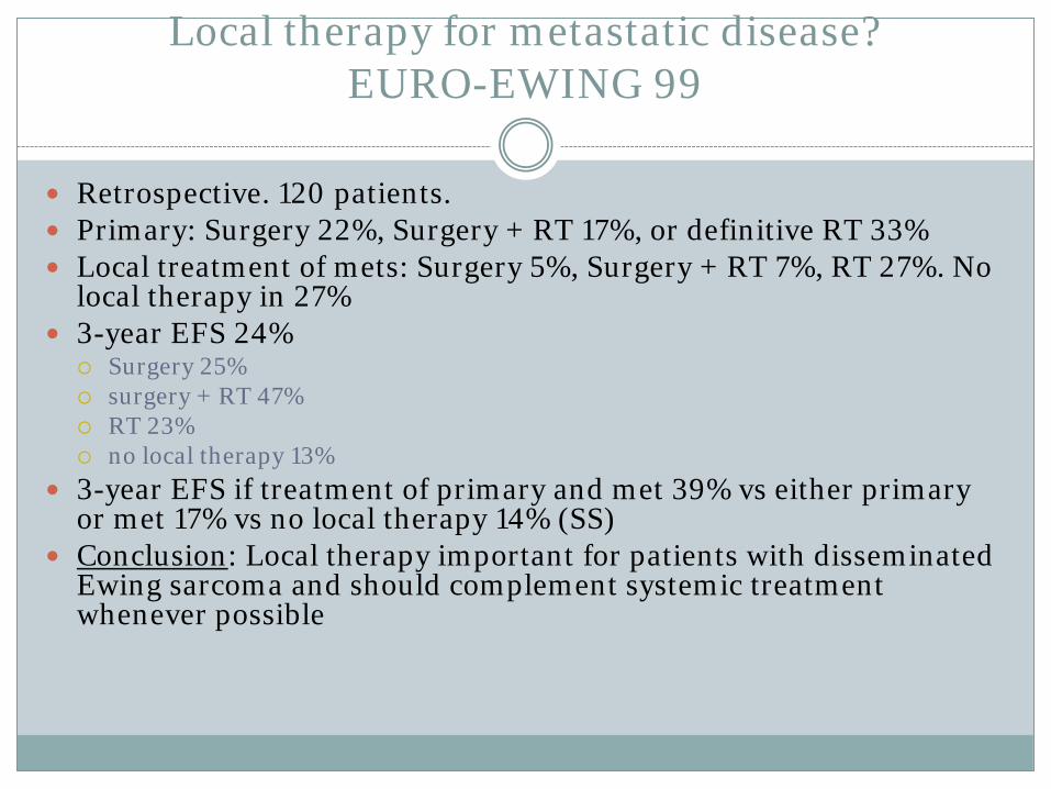
Local therapy for metastatic disease? EURO-EWING 99
Retrospective. 120 patients. Primary: Surgery 22%, Surgery + RT 17%, or definitive RT 33% Local treatment of mets: Surgery 5%, Surgery + RT 7%, RT 27%. No
local therapy in 27% 3-year EFS 24%
Surgery 25% surgery + RT 47% RT 23% no local therapy 13%
3-year EFS if treatment of primary and met 39% vs either primary or met 17% vs no local therapy 14% (SS)
Conclusion: Local therapy important for patients with disseminated Ewing sarcoma and should complement systemic treatment whenever possible

POG 8346: Donaldson et al. IJROBP 1998
IFRT equivalent to whole bone (SF) RT for LC? n=178, 1983-1988 Induction chemo: cyclophosphamide/doxorubicin x
12wks (5c) Local Tx based on response: PD RT + salvage chemo If CR/PR surgery (if feasible) + PORT if + margins/gross dz RT alone: randomized to IFRT vs SFRT
IF 55.8Gy SF 39.6 Gy + 16.2 Gy boost (GTV + 4cm)
VACA x 50 wks

POG 8346
EBM – POG 8346 No benefit to whole bone RT 5yr EFS: SF 37% vs. IF 39% 5yr LC: SF 53% vs IF 53%
Limitations: low accrual, high rate DM

Extracorporeal Irradiation
Pelvic tumors: poor prognosis Primary resection difficult, chemoRT mainstay Wide en-block resection ECI 50Gy @ 2Gy/min
debulking of tumor from bone re-implantation 13 patients, median age 16 yrs, no mets OS 69%, 9/13 NED at last followup, 4 died of
metastatic disease, no local relapse 7/13 with good/excellent functional outcomes
Krieg AH, J Bone Joint Surg 2009

RT Target Volume (AEWS1031)
RT to entire bone not necessary (POG 8346) GTV: pre-chemo bony disease and post-chemo soft
tissue disease CTV margin of 1-1.5cm Make sure scars and drain sites are wired and apply
bolus to ensure adequate coverage 45 Gy + 10.8 Gy (definitive RT or gross residual) 36 Gy (pre-op RT) 45-50.4 Gy (post-op RT)

RT Complications
Bone growth abnormalities > 20 Gy can prematurely close epiphysis > 20-30 Gy can cause permanent lymphedema Limb length discrepancy – 2-6 cm Permanent weakening of bone
High risk of fracture within 18 mos of RT
Dermatitis: recall-reaction w/ ADR and dactinomycin Decreased ROM 2/2 joint fibrosis Skin hyperpigmentation Cystitis (worse w/ cyclophosphamide/ifos) Second malignancies (5-10% @ 20yrs osteosarcoma)

Chemotherapy Regimens
For non-metastatic disease, standard 5-drug U.S.
regimen (VAC + IE) Vincristine Doxorubicin Cyclophosphamide Alternating with ifosfamide and etoposide x 48 weeks Actinomycin sometimes thrown in (VACA+IE)
For metastatic disease (VAC) Vincristine Doxorubicin Cyclophosphamide

IESS-I
342 pts. Localized Ewing's sarcoma of bone, previously untreated Group I Institutions: Randomized 3:2 to 1) RT to primary plus VAC + Adriamycin
or 2) RT plus VAC Group II Institutions: Randomized 3:2 to 3) RT to primary plus VAC and
bilateral pulmonary RT (BRP) or 2) RT plus VAC (same as above) Chemotherapy given x 6 weeks
Vincristine and cyclophosphamide q weekly and adriamycin given with the last dose.
After 6 weeks rest, pts had a 7 week course of continuation therapy that consisted of dactinomycin IV x 5 days followed 9 days later by VCR and cyclophosphamide weekly x 5 weeks. For treatment 1, adriamycin given with the last course in the 7th week of each course.
RT : entire involved bone to 45-55 Gy (based on age), followed by 10 Gy boost to gross radiographic tumor + soft tissue mass with margin. Lung RT: 15-18 Gy given at 150-180 cGy/day.

IESS-1
5-yr RFS treatment 1 - 60%, 2 - 24%, 3 - 44%. Similar trend for OS. Worse survival for pelvic sites. 15% LR overall. DM in 1-30%, 2-72%, and 3-42%. BPR was not effective in preventing
lung mets.
Conclusion: improved survival with addition of Adriamycin to VAC.

IESS-3
Non-metastatic pts 5-yr EFS 69% vs 54% for
VAC+ADR+IE vs VAC+ADR (RR=1.6)
5-yr OS 72% vs 61% (RR=1.6) Greater reduction in LR than in
distant mets. Greater benefit for large primary tumors or pelvic tumors.
For pts with mets, no difference between regimens: 5yr EFS 22% 5yr OS 34%
Conclusion: improved survival with addition of ifosfamide and etoposide (in non-metastatic pts)

WLI- EICESS 92 Bolling et al., Strahlenther Onkol, 2008
Any benefit to WLI? Toxicity? 99 with pulmonary mets, 70 received WLI, Local: VAIA +/- etop x14c WLI: wk 31, 12-21 Gy +/- boost to thoracic tumor to 54Gy
1.5 Gy QD vs 1.25 Gy BID AP/PA fields
5yr OS: 61% (WLI) vs 49% (none) p=0.36
5yr EFS 39% (WLI) vs 37% (none)

WLI- EICESS 92: Toxicity
PFT complications None Mild Moderate Severe
43 29 21 7

Treatment Overview
Chemotherapy is typically given for 12-15 weeks prior to local therapy VAC(A)+/- IE (no IE if metastatic)
Local Tx (surgery or radiation therapy or both) Surgery favored if complete resection is feasible without significant
morbidity and functional loss Radiation favored for central lesions (55.8Gy)
Radiation PORT if + margins: 45Gy Definitive RT or PORT w/ gross residual: 55.8Gy Whole lung radiation used for consolidation after chemotherapy
(15Gy/10fx), boost residual dz to 45Gy. Can consider resection if <=4 mets

Late (>5yr) recurrences in Ewing’s sarcoma)
>12k childhood cancer survivors Overall late relapse 4% and 6% at 10 and 20 years Two tumors stood out Ewing’s and CNS tumors
14% at 20 years
Importance of monitoring 15-20years from therapy
Wassilewski-Master, JNCI, 2009

Questions
What translocation is characteristic of Ewing’s sarcoma? A. t(11;22) B. t(12;16) C. t(9;22) D. t(x;18)
A

All of the following are true regarding Ewing’s sarcoma, except A. There is a predilection for whites B. It is more common among males than females C. Cytokeratin and neuron-specific enolase can be positive D. Half of patients present with localized disease at diagnosis
D
All of the following are true, except A. Ewing’s sarcoma exhibits chromosomal
translocation t(11;22) B. Codman’s triangle can be observed on radiography C. Presents more commonly with localized disease
than osteosarcoma D. Radiation plays a prominent role in therapy
C. Ewing’s presents with localized disease 75% of the time, osteosarcoma 90% of the time
In a patient with Ewing’s that has GRD after chemo and surgery, what is the correct RT dose and volume?
A. 45Gy to pre-chemo bone and post-chemo soft tissue tumor B. 45 Gy to post-chemo bone and post-chemo soft tissue tumor C. 55.8 Gy to the pre-chemo bone and pre-chemo soft tissue tumor D. 55.8 Gy to the pre-chemo bone and post-chemo soft tissue tumor
D

All of the following are true regarding IESS-1 in which adria was added to vincristine, actinomycin and cyclophosphamide, except:
A. The addition of adria improved OS B. The addition of adria improved DFS C. Pelvic disease sites fared no worse than nonpelvic disease sites D. Local recurrence did not differ by treatment
C. IESS-1: randomized 335pts to receive adria to VAC + RT (45-55 Gy + 10 Gy boost). Addition of VAC improved both DFS and OS. Pelvic disease sites had poorer survival than nonpelvic (34 vs 57 %). Local recurrence did not differ by treatment

All of the following are true regarding IESS-II in which intermittent high dose was compared to continuous moderate-dose chemo, except:
A. High dose chemo improved OS B. High dose chemo improved DFS C. High dose chemo arm had etoposide D. Cardiac toxicity was worse in high-dose arm
C. IESS-II randomized 214pt to receive VAC + adria by either moderate-dose continuous or high-dose intermittent regimen. High dose improved OS (77 vs 63%) but with greater cardiotoxicity

All of the following true regarding IESS-III in which ifosfamide and etoposide were added to VAC + adria, except:
A. The addition of IE improved OS in pts with both metastatic and non-metastatic disease
B. There was a greater reduction in local recurrence than in distant metastasis
C. A quarter of the enrolled patient had metastatic disease D. There was a greater benefit seen in pelvic tumors
A. IESS-III randomized 518pts to receive IE or not in addition to VAC + adr. 23% of pts had metastatic disease. In non-metastatic pts, addition of IE improved EFS and OS. Greater reduction in local recurrence than distant mets and a greater benefit for large or pelvic tumors. Patients with metastatic disease did not benefit from IE in terms of EFS or OS.

Q U E S T I O N S ?
THE END


